Chapter 4. RNA Secondary, Tertiary, and Quaternary Structure Principles
Scope Note
RNA structure is the physical arrangement of an RNA molecule, its intramolecular contacts, and the molecular partners that complete its biological state. This chapter introduces the structural vocabulary used throughout the book: primary sequence, secondary structure, tertiary structure, quaternary structure, motifs, domains, ribonucleoprotein assemblies, ion and ligand effects, and the limits of sequence-based inference. The chapter is a foundation chapter. It gives enough explanation for later chapters on folding thermodynamics, cellular folding, RNA-protein recognition, structural methods, prediction algorithms, viral RNA elements, riboswitches, ribosomes, CRISPR RNAs, long noncoding RNAs, and RNA-targeted therapeutics, but it does not replace those specialized treatments.
Executive Summary
RNA structure is commonly described in levels. Primary structure is the nucleotide sequence and polarity of the RNA chain. Secondary structure is the pattern of base pairing, usually represented as helices, hairpin loops, internal loops, bulges, multibranch junctions, and pseudoknots. Tertiary structure is the three-dimensional arrangement of secondary-structure elements and the long-range contacts that pack those elements into a fold. Quaternary structure is the assembly of multiple RNA molecules, proteins, ligands, ions, or larger ribonucleoprotein complexes. These levels are useful categories, but they are coupled rather than independent.
A secondary-structure diagram is a compact model of base pairing, not a complete physical description of an RNA molecule. A cloverleaf drawing of transfer RNA, or tRNA, captures major stems and loops, but the molecule functions as an L-shaped three-dimensional adaptor with modified nucleotides, an amino acid acceptor end, an anticodon loop, and binding surfaces for aminoacyl-tRNA synthetases, elongation factors, and the ribosome. A riboswitch diagram may show a ligand-binding aptamer and an expression platform, but ligand binding, ion conditions, transcription timing, and competing structures determine whether that diagram represents the state that controls gene expression.
Secondary-structure elements are the first reusable vocabulary. A stem is a run of base pairs. A hairpin is a stem capped by a loop. A bulge interrupts one side of a helix. An internal loop interrupts both sides. A junction connects three or more helical segments. A pseudoknot occurs when nucleotides in a loop pair with a region outside that loop, creating crossing base-pair dependencies that cannot be represented as a simple nested set of parentheses. Pseudoknots are biologically important in viral frameshifting elements, ribozymes, riboswitches, telomerase RNA, and ribosomal RNA, but many simple secondary-structure prediction frameworks exclude or approximate them.
Tertiary structure gives RNA its spatial architecture. Long-range contacts bring distant sequence regions together. Recurrent tertiary motifs include A-minor interactions, ribose zippers, tetraloop-receptor contacts, kissing-loop contacts, base triples, and coaxial stacking. These motifs help explain how RNAs form compact catalytic centers, ligand-binding pockets, ribosomal cores, and regulatory switches. They also explain why RNA structure is only partly modular: a motif can recur across RNAs, but its geometry and function often depend on neighboring helices, junctions, proteins, ions, and ligands.
Quaternary structure describes assemblies. Some assemblies are stoichiometric machines, such as ribosomes, spliceosomes, RNase P, signal recognition particles, telomerase, and CRISPR-Cas guide complexes. Other assemblies are transient, heterogeneous, or condition-dependent, such as RNA-RNA interaction networks and RNA-rich cellular bodies. Quaternary structure therefore includes both ordered RNA-protein machines and more dynamic ribonucleoprotein states.
RNA sequence constrains structure, but sequence alone does not uniquely determine the cellular structure or biological function of every RNA. Ion concentration, water, ligands, protein binding, molecular crowding, chemical modifications, transcription rate, folding history, ribosome occupancy, and cellular compartment can stabilize, destabilize, remodel, or select RNA structures. Strong structure-function claims require more than a predicted fold. They usually need structural evidence plus perturbation, compensatory rescue, interaction evidence, or a functional readout in a relevant biological context.
Concept Inventory
- RNA secondary structure: the base-pairing pattern within one RNA molecule or between RNA molecules. In diagrams, secondary structure is usually reduced to stems, loops, bulges, and junctions. The boundary case is important: a secondary-structure representation usually omits three-dimensional packing, dynamics, protein occupancy, ligand binding, and ion-specific interactions.
- Pseudoknot: a topology in which nucleotides in a loop pair with another region outside the loop. Pseudoknots create crossing pairing relationships. A pseudoknot can be essential for function, but it can also be difficult to infer because many algorithms are designed for nested structures.
- RNA tertiary structure: the three-dimensional arrangement of helices, loops, junctions, noncanonical pairs, base triples, backbone contacts, ion contacts, and long-range interactions. Tertiary structure determines whether a secondary-structure diagram becomes a compact fold, an extended scaffold, a ligand-binding pocket, or a protein-binding surface.
- Tertiary motif: a recurrent three-dimensional interaction pattern. Examples include A-minor interactions, ribose zippers, tetraloop-receptor contacts, kissing-loop contacts, coaxial stacks, and base triples. Motifs are useful because they recur, but motif transferability depends on context.
- Long-range contact: an interaction between sequence regions distant in primary sequence. The contact can be a base pair, a tertiary contact, a protein-mediated bridge, a ligand-mediated interaction, or an RNA-RNA contact between separate molecules.
- RNA junction: a structural element where multiple helices or single-stranded segments meet. Junctions control helix orientation and often determine whether domains pack into functional tertiary structures.
- RNA domain: a larger structural or functional unit within an RNA. A domain may fold partly independently, as in some riboswitch aptamer domains, but domain boundaries do not prove independent folding or autonomous function.
- RNA modularity: the reuse or recombination of structural elements, motifs, or domains. Modularity is real but conditional: an element that functions in one molecular environment can fail when transplanted into a different architecture.
- RNA quaternary structure: assembly involving multiple RNA molecules, RNA-protein complexes, RNA-ligand complexes, or higher-order ribonucleoprotein organization. Quaternary states range from stable machines to transient cellular assemblies.
- Ribonucleoprotein architecture: the spatial and compositional organization of RNA-protein assemblies. The ribosome is the canonical example, but spliceosomes, telomerase, RNase P, small nuclear ribonucleoproteins, small nucleolar ribonucleoproteins, signal recognition particles, and CRISPR-Cas complexes all show that RNA function often emerges from RNA-protein architecture.
- Metal-ion site: a position where a metal ion contributes to folding, catalysis, or stabilization. A specific metal-ion site differs from the diffuse ion atmosphere that screens the negatively charged phosphate backbone.
- RNA ligand-binding pocket: a structured RNA surface or cavity that binds a metabolite, drug-like molecule, ion, or other ligand. Binding in vitro does not by itself establish cellular target engagement.
- Context-dependent structure: structure whose population or function depends on conditions such as ions, proteins, ligands, modifications, transcription history, compartment, or cellular state.
- Structure-function relationship: a causal or mechanistic relationship between a structural feature and a biological or technological function. Prediction or correlation alone is not sufficient for a strong causal claim.
- Covariance: correlated evolutionary change that preserves a base pair or structural feature. Covariance can support structural inference, but alignment uncertainty, sparse sampling, lineage bias, and rapid turnover can weaken interpretation.
What to Know Before Reading This Chapter
The most important prerequisite is the difference between a molecule and a representation. A nucleotide sequence is a text-like representation of primary structure. A dot-bracket string or two-dimensional drawing is a representation of base pairing. A three-dimensional coordinate model is a representation of atomic or coarse-grained positions. A chemical-probing profile is an experimental readout of nucleotide reactivity or accessibility. A cryo-electron microscopy map is a density map interpreted through modeling. Each representation answers a different question.
The second prerequisite is that RNA is a polyanion. Every phosphodiester linkage contributes negative charge, so RNA folding is strongly affected by ions and electrostatics. The 2′-hydroxyl group of ribose gives RNA additional hydrogen-bonding and catalytic possibilities. Bases can form Watson-Crick pairs, wobble pairs, Hoogsteen-like contacts, base triples, stacking interactions, and noncanonical geometries. Chapter Chapter 2 explains nucleotide chemistry and base-pairing vocabulary, while Chapter Chapter 3 explains thermodynamics, kinetics, and ensemble concepts.
The third prerequisite is that RNA molecules often exist as ensembles. An ensemble is a population of conformations rather than a single static shape. One conformation may dominate under one condition, while another appears after ligand binding, protein binding, temperature change, transcriptional pausing, or mutation. An RNA structure statement should therefore specify the molecule, species, cellular or in vitro condition, method, and confidence level whenever those details matter.
Three running examples will anchor the chapter. tRNA shows the difference between a secondary-structure drawing and a tertiary fold. Riboswitches show how ligand binding, structure, and gene regulation can be linked. The ribosome shows how RNA secondary and tertiary architecture become part of a quaternary ribonucleoprotein machine. Viral RNA elements, guide RNAs, and long noncoding RNAs provide additional boundary cases.
4.1. Secondary-structure elements and pseudoknots
Secondary structure is the pattern of base pairing in RNA. The term often refers to intramolecular pairing within a single RNA molecule, but RNA-RNA duplexes between separate molecules also have secondary-structure features. Secondary structure is usually the first structural level taught because it is compact, sequence-linked, and visually intuitive. A reader can often see how a sequence folds into stems and loops without needing a full three-dimensional model.
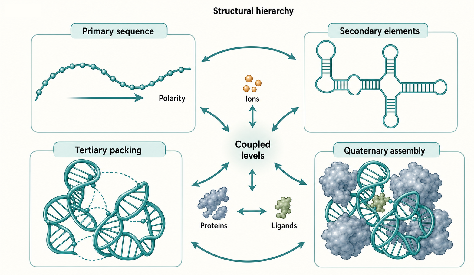
Figure 4.1. RNA Structural Hierarchy. RNA organization is described at four coupled levels: the nucleotide sequence and chain polarity that define primary structure; the base-pairing pattern of stems, hairpins, bulges, junctions, and pseudoknots that define secondary structure; the three-dimensional arrangement of long-range contacts and tertiary motifs that pack secondary elements into a compact fold; and the quaternary assembly of RNA with proteins, ligands, or other RNA molecules into ribonucleoprotein machines. The figure emphasizes that these levels are coupled rather than independent, because secondary elements require ions and proteins to adopt tertiary geometry, and quaternary assembly can remodel both secondary and tertiary architecture.
Table 4.1. Structural Elements and Evidence Standards. Summary of the main RNA secondary and tertiary structural elements, their definitions, common and stronger evidence types, and frequent sources of overstatement.
| Element | Definition | Common evidence | Stronger evidence | Common artifact or overreach |
|---|---|---|---|---|
| Helix or stem | Run of paired nucleotides. | Prediction, probing protection. | Covariation, compensatory rescue, high-resolution pair geometry. | Treating predicted pairing as cellular state. |
| Hairpin | Stem capped by loop. | Prediction, probing, processing pattern. | Mutational tests of loop and stem function. | Assuming any hairpin is regulatory. |
| Bulge | Unpaired segment on one side of a helix. | Probing reactivity, structural model. | Protein or ligand recognition plus perturbation. | Calling a bulge functional without a readout. |
| Internal loop | Unpaired segments on both sides of a helix. | Probing, comparative model. | Motif structure and rescue. | Ignoring alternative conformations. |
| Multibranch junction | Three or more helices meet. | Secondary model, probing. | Tertiary orientation, ligand or protein tests. | Assuming diagram angle reflects real geometry. |
| Pseudoknot | Crossing base-pair topology. | Specialized prediction, probing. | Covariation, high-resolution structure, compensatory rescue. | Missing it with nested algorithms or overpredicting unsupported knots. |
| Long-range contact | Distant regions interact. | Crosslinking, proximity, modeling. | Orthogonal contact and functional perturbation. | Interpreting proximity as direct pairing. |
| Tertiary motif | Recurrent 3D contact. | Structural model or analogy. | High-resolution motif plus functional tests. | Assuming transferability across contexts. |
| Domain | Larger structural or functional unit. | Conservation, deletion mapping. | Isolated and full-length evidence. | Treating boundaries as independent folding proof. |
| Quaternary assembly | Multi-molecule RNA, RNP, or RNA-ligand state. | Co-purification, imaging, structure. | Stoichiometry, architecture, dynamics, function. | Calling colocalization a defined assembly. |
Table 4.2. Context Factors That Alter RNA Structure. Each factor can shift RNA structural populations away from the state predicted from sequence alone; the table describes the mechanism, claim type, and evidence needed for each.
| Factor | Mechanism of effect | Example claim type | Evidence needed |
|---|---|---|---|
| Mg2+ | Screens charge, stabilizes specific sites, supports catalysis. | Mg2+-dependent folding. | Titration, structural site evidence, physiological comparison. |
| Monovalent ions | Screen charge and alter specific motifs. | Salt-dependent stability. | Controlled salt series and orthogonal readout. |
| Water | Mediates hydration and hydrogen bonding. | Hydration-supported pocket. | High-resolution structure or thermodynamic inference. |
| Ligand | Selects or induces a conformational state. | Riboswitch or drug-binding mechanism. | Binding, structure, mutation, expression or target-engagement readout. |
| RNA-binding protein | Stabilizes, blocks, recognizes, or remodels RNA. | RNP-dependent structure. | Binding evidence, structural or probing change, functional perturbation. |
| Helicase | Unwinds or remodels RNA. | ATP-dependent remodeling. | Enzyme assay, substrate specificity, in-cell relevance. |
| Modification | Changes pairing, stacking, recognition, or immune sensing. | Modification-dependent structure. | Stoichiometry, mapping, perturbation of writer, eraser, reader, or synthetic RNA. |
| Transcription speed | Changes folding path and available partners. | Cotranscriptional folding. | Nascent RNA assays, pause-site analysis, kinetic perturbation. |
| Ribosome occupancy | Unwinds or protects mRNA regions. | Translation-dependent mRNA structure. | Translation perturbation, ribosome profiling, probing comparison. |
| Crowding or compartment | Alters effective concentration and equilibria. | Compartment-specific structure. | In-cell and in vitro comparison, dynamics, composition. |
| Condensate environment | Concentrates RNA and proteins; can alter folding kinetics and accessibility. | Condensate-dependent structural state. | Imaging, fractionation, composition analysis, functional perturbation. |
A stem or helix is a run of paired nucleotides. Most stems contain Watson-Crick G-C and A-U pairs, but many RNA helices include G-U wobble pairs or noncanonical pairs. A stem is not just a ladder of hydrogen bonds. Base stacking, helix geometry, loop constraints, and ion conditions also influence stability. A short stem may be stable in one context and transient in another.
A hairpin loop forms when a stem is capped by unpaired nucleotides. Hairpins can be simple structural caps, recognition elements, or parts of more complex motifs. A bacterial transcription terminator hairpin can help terminate transcription. A microRNA precursor hairpin can be recognized by processing factors. A viral RNA hairpin can participate in replication, packaging, immune evasion, or translation. These functions depend on more than the presence of a hairpin; loop sequence, stem length, flanking regions, protein binding, and cellular context all matter.
A bulge is an unpaired nucleotide or stretch of nucleotides on one side of a helix. An internal loop contains unpaired nucleotides on both sides of a helix. Bulges and internal loops can bend helices, create protein-binding surfaces, expose bases for recognition, or participate in tertiary motifs. They are often more than defects in pairing. In structured RNAs, interruptions in helices can be deliberate architectural features.
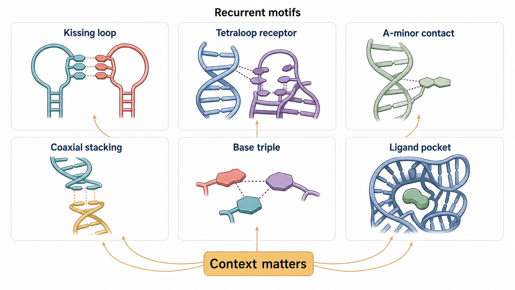
Figure 4.2. Recurrent Tertiary Motifs and Long-Range Contacts. Six recurrent tertiary interaction types are illustrated to build a shared visual vocabulary for structural chapters: kissing-loop contacts, in which bases in two loops pair with one another; tetraloop-receptor contacts, in which a four-nucleotide loop docks into a structured receptor elsewhere in the RNA; A-minor-like interactions, in which an adenosine reads the minor groove of a helix; coaxial stacking, in which two helices align across a junction to form a continuous stack; base triples; and a ligand-binding pocket, in which a folded cavity recognizes a small molecule. Together these motifs show that RNA tertiary architecture is built from recurrent but context-dependent modules.
A multibranch junction is where three or more helical segments meet. Junctions are common in tRNA, ribozymes, riboswitches, rRNA, viral RNA elements, and engineered RNA scaffolds. A junction can behave as a flexible hinge, a rigid architectural hub, or a ligand-dependent switch. Because junctions define helix orientation, they are a bridge between secondary and tertiary structure.
A pseudoknot is a base-pairing topology in which nucleotides within a loop pair with a region outside that loop. A simple way to imagine a pseudoknot is to draw a hairpin and then let the loop form a second stem with a downstream sequence. The resulting base-pair pattern crosses the original nested pairing pattern. Standard nested diagrams and dot-bracket strings can represent ordinary hairpins easily, but pseudoknots require additional notation or specialized modeling. This is why pseudoknots are a conceptual and computational boundary.
Pseudoknots matter because topology can become function. Some viral RNAs use pseudoknot-containing signals to alter translation, including programmed ribosomal frameshifting. Some ribozymes and riboswitches use pseudoknots to organize catalytic or ligand-binding architectures. Telomerase RNA and rRNA contain structured regions where pseudoknot-like topologies contribute to assembly or function. A pseudoknot review supports the general definition and functional range here; viral frameshifting, telomerase, ribozyme, and riboswitch examples receive class-specific treatment in later chapters.
Secondary-structure evidence varies in strength. A computational minimum-free-energy model is a hypothesis based on sequence and an energy model. Comparative covariation can support conserved base pairs. Chemical probing can support paired and unpaired regions under specific conditions. Mutations that disrupt a stem and compensatory mutations that restore function provide stronger evidence that base pairing matters. High-resolution structure can show base-pair geometry, but it is still condition-specific.
The main caution is that secondary-structure diagrams are summaries. A diagram may show a base pair that is present in one conformation but absent in another. It may omit a protein that protects a nucleotide from probing. It may omit a ligand that stabilizes a pocket. It may show a pseudoknot as though it is static even when the molecule samples multiple states. The diagram is useful precisely because it simplifies; the danger is forgetting what was simplified.
4.2. Long-range contacts and tertiary motifs
Tertiary structure begins when secondary-structure elements are placed in three-dimensional space. Two helices in a secondary-structure diagram may look separate, but in the folded RNA they can stack coaxially, dock through loop-receptor interactions, or be brought together by a protein. Long-range contacts are interactions between regions distant in the primary sequence. They can join the ends of an RNA, pack distant domains together, or create a pocket for a metabolite or small molecule.
A coaxial stack forms when two helices align so that their base pairs stack across a junction or loop, resembling a continuous helix. Coaxial stacking can stabilize a fold and reduce the number of flexible orientations available to a junction. In a riboswitch aptamer, coaxial stacking can help build a ligand-binding architecture. In a ribozyme, stacking can align catalytic regions. In an engineered RNA nanostructure, stacking can help define global shape.
An A-minor interaction is a common RNA tertiary contact in which an adenosine contacts the minor groove of an RNA helix. The concept is important because it shows how a single nucleotide can read helical shape without forming an ordinary Watson-Crick pair. A-minor-like interactions are prominent in large structured RNAs, including ribosomal RNA, and are one example within broader motif taxonomies that also include tetraloop receptors, ribose zippers, kissing loops, coaxial stacking, and base triples.
A ribose zipper is a tertiary interaction involving hydrogen-bonding networks between ribose 2′-hydroxyl groups. The motif highlights the special importance of the RNA ribose. DNA lacks the same 2′-hydroxyl chemistry, so RNA can build tertiary hydrogen-bond networks that are not available to ordinary DNA duplexes.
A tetraloop-receptor contact occurs when a small loop, often a stable four-nucleotide loop, docks into a receptor motif elsewhere in the RNA. This kind of interaction helped establish the idea that RNA folds through recurrent modules. A kissing-loop contact occurs when bases in two loops pair with one another. Kissing-loop interactions can be intramolecular or intermolecular. They are relevant to RNA-RNA recognition, viral genome dimerization, regulatory small RNAs, and synthetic RNA design.
Base triples and noncanonical pairs also help build tertiary folds. A base triple contains three bases interacting in one local arrangement. Noncanonical pairs do not follow ordinary Watson-Crick geometry but can be highly specific. These contacts expand the alphabet of RNA recognition. An RNA fold is therefore not merely a set of helices connected by passive linkers; it is a network of stacked bases, paired bases, backbone contacts, hydration, ions, and sometimes ligands or proteins.
Several named noncanonical conformers extend these principles. An RNA G-quadruplex stacks planar guanine quartets around monovalent cations; an RNA triple helix adds a third strand to a duplex through Hoogsteen-like contacts; and Z-RNA is a left-handed double-helical state that can arise transiently in suitable duplex sequences or protein-bound contexts. These labels describe different physical architectures, not one generic class of “unusual RNA.” The MALAT1 3′-end triple helix provides a concrete example: mutational, biochemical, and cellular reporter evidence connects base-triple formation to protection of a non-polyadenylated RNA end. Chapter 53 owns the motif-by-motif structures, proteins, ligands, mapping methods, and biological cases; this chapter owns the shared thermodynamic lesson that sequence potential, folding conditions, kinetic history, and molecular partners determine which conformer is populated.
Motifs make RNA partly modular. A motif can be recognized in unrelated RNAs because the local geometry recurs. The idea of modularity is powerful for interpreting natural RNAs and for designing synthetic RNAs. However, a motif is not a plug-and-play part in all contexts. Surrounding helices can impose incompatible angles. Ion conditions can change stability. A protein can occlude a motif. A ligand-binding pocket can require the rest of the domain to preorganize the local geometry.
Small molecules often recognize folded RNA surfaces rather than sequence alone. A ligand may bind a groove, bulge, junction, triple helix, or pocket that exists only in a subset of conformations. This is one reason RNA-targeted small-molecule discovery is both attractive and difficult. Folded RNA offers shapes and pockets, but those shapes can be dynamic, condition-dependent, and shared among RNAs. Cellular target engagement requires evidence that the molecule binds the relevant RNA in the relevant cellular state, not just that it binds a purified RNA fragment.
4.3. Junctions, domains, modularity, and quaternary assemblies
Junctions are local architectural decision points. In a three-way junction, three helices meet. In a four-way junction, four helices meet. Larger multibranch junctions occur in rRNA, ribozymes, riboswitches, and viral RNA elements. The sequence and geometry of the junction determine whether helices stack, bend, rotate, or remain flexible. Because helix orientation determines whether distant motifs can contact one another, junctions are central to tertiary folding.
Junctions also explain why secondary structure and tertiary structure are coupled. A secondary-structure diagram may show three stems connected at one point, but the biological question is how those stems are oriented. In one conformation, two stems may stack and the third may project outward. In another, ligand binding may stabilize a different stacking register. In another, protein binding may clamp the junction. The same local secondary structure can therefore support different tertiary outcomes.
A domain is a larger structural or functional unit within an RNA. Domains can be identified by conserved structure, function, folding behavior, or boundaries in a high-resolution structure. A riboswitch often has an aptamer domain that binds ligand and an expression platform that changes transcription, translation, splicing, or RNA stability. A group II intron contains domains that cooperate to position catalytic elements and exon substrates. rRNA contains large domains that assemble with ribosomal proteins and other rRNA regions.
The word domain can mislead if it is interpreted too rigidly. A domain boundary in a drawing does not prove that the domain folds independently. An isolated domain may fold differently from the same sequence in the full RNA. Conversely, some domains are experimentally separable under particular conditions. A careful statement distinguishes structural domain, functional domain, evolutionary domain, and experimentally isolated construct.
RNA modularity is the observation that motifs, junctions, domains, or folds can recur across RNAs or be recombined in engineering. Modularity is central to riboswitch comparison, ribozyme architecture, RNA nanotechnology, and some RNA design strategies. Yet natural RNA modularity is constrained by context. A tetraloop receptor, aptamer, or guide-RNA scaffold can fail when sequence flanking, helix length, spacing, ion conditions, or protein partners change. Chapter Chapter 65 treats programmable RNA architecture in more detail.
Quaternary structure is the structural organization of multiple molecules. In protein biochemistry, quaternary structure usually means assembly of multiple folded protein subunits. In RNA biology, quaternary structure includes RNA-RNA assemblies, RNA-protein assemblies, RNA-ligand assemblies, and larger ribonucleoprotein states. The definition must be broad because RNA function so often depends on partners.
The ribosome is the standard example of an ordered RNA-protein quaternary assembly. rRNA forms the catalytic and architectural core, while ribosomal proteins stabilize, extend, and tune the machine. The spliceosome is a more dynamic example. Small nuclear RNAs and proteins assemble, rearrange, catalyze splicing, and disassemble. RNase P, signal recognition particle, telomerase, small nucleolar RNPs, and CRISPR-Cas guide complexes provide additional examples in which RNA sequence, RNA structure, and protein assembly are inseparable.
RNA-RNA quaternary structure also matters. Some viral genomes dimerize. Bacterial small RNAs pair with target mRNAs, often with assistance from proteins such as Hfq or ProQ. Long-range intramolecular and intermolecular contacts can bring distant regulatory elements together. Methods such as proximity ligation, crosslinking, mutational profiling, and compensatory genetics can support these contacts, but each method has artifacts. Chapter Chapter 134 covers RNA-RNA interaction mapping.
Dynamic RNA-rich cellular bodies require especially careful language. A ribosome has defined subunits and a relatively well specified architecture. A stress granule, processing body, paraspeckle, nucleolus, or mitochondrial transcriptional condensate can contain many RNAs and proteins with variable stoichiometry, exchange rates, and local organization. Calling such a body a quaternary structure is possible in a broad sense, but the evidence standards differ from those for a crystallographic or cryo-EM structure. Material properties, composition, dynamics, and perturbation must be evaluated separately.
4.4. Metal ions, water, ligands, proteins, and crowding
RNA folding occurs in an environment. The phosphate backbone is negatively charged, so electrostatic repulsion opposes compact folding. Ions, especially monovalent cations and divalent cations such as Mg2+, reduce this repulsion. Water hydrates bases, ribose, and phosphate groups. Ligands can stabilize pockets or shift conformational populations. Proteins can bind, remodel, protect, destabilize, or expose structural elements. Molecular crowding and compartmentalization can change effective concentrations and folding pathways.
Mg2+ is often mentioned as though it has one simple role, but several mechanisms are possible. Diffuse Mg2+ can screen charge without occupying a specific site. Site-bound Mg2+ can stabilize a particular fold. Catalytic Mg2+ can participate directly in chemistry, as in some ribozyme active sites. Bridging Mg2+ can coordinate ligands and RNA atoms. An experiment that shows Mg2+-dependent folding does not automatically identify a specific metal-ion site. An experiment that resolves a metal ion in a structure still must address whether the site is occupied under physiological conditions. Water and crowding add further environmental variables: hydration shapes local contacts, and crowded solutions can shift nucleic-acid folding and interaction equilibria.
Monovalent ions also matter. K+, Na+, and other cations can screen charge, alter duplex stability, and influence G-quadruplexes or other specific structures. RNA structures measured under one salt condition may not transfer cleanly to another. This is a common source of discrepancy between in vitro probing, in-cell probing, biochemical assays, and computational predictions.
RNA G-quadruplex studies make that discrepancy unusually clear. rG4-seq can identify transcript regions that form potassium-dependent reverse-transcriptase stops under controlled folding conditions, establishing folding potential rather than automatic cellular occupancy. Chemical probing in living eukaryotic cells found that many such regions were predominantly unfolded, consistent with active remodeling and competition from proteins or alternative structures. The two results are complementary: one maps sequences capable of adopting an RNA G4, while the other tests whether those sequences occupy that state in a specified cell and condition. Stress, compartment, translation, ligand exposure, or helicase activity can shift occupancy, so neither a motif prediction nor an in vitro fold should be reported as a constitutive in-cell structure. Chapter 53 develops the corresponding evidence ladder and current controversies.
Water is not passive background. Hydration can stabilize grooves, mediate hydrogen bonds, fill pockets, and shape ligand recognition. A high-resolution structure may show ordered waters that are part of the architecture. Lower-resolution methods may not resolve water, but hydration still affects folding and binding. Because water is difficult to represent in simplified diagrams, students often underestimate its role.
Ligands can act through several mechanisms. A ligand may bind a pre-existing conformation and enrich that state. It may induce local ordering after initial contact. It may stabilize a junction, displace water, recruit ions, or alter protein binding. Riboswitch aptamers are the cleanest pedagogical example: metabolite binding to the aptamer changes the probability of downstream structures in the expression platform. Small-molecule drugs that target RNA use related principles, but target selectivity and cellular engagement are harder to establish.
Proteins can recognize sequence, shape, structure, modification, or combinations of these features. A double-stranded RNA-binding domain recognizes helical shape more than a unique sequence. A sequence-specific RNA-binding protein may prefer a short motif only when it is single-stranded. A ribosomal protein may stabilize an rRNA fold. A helicase may unwind or remodel RNA. A translating ribosome can disrupt coding-region structure as it moves along an mRNA. A protein can therefore make a predicted structure more likely, less likely, or irrelevant in vivo.
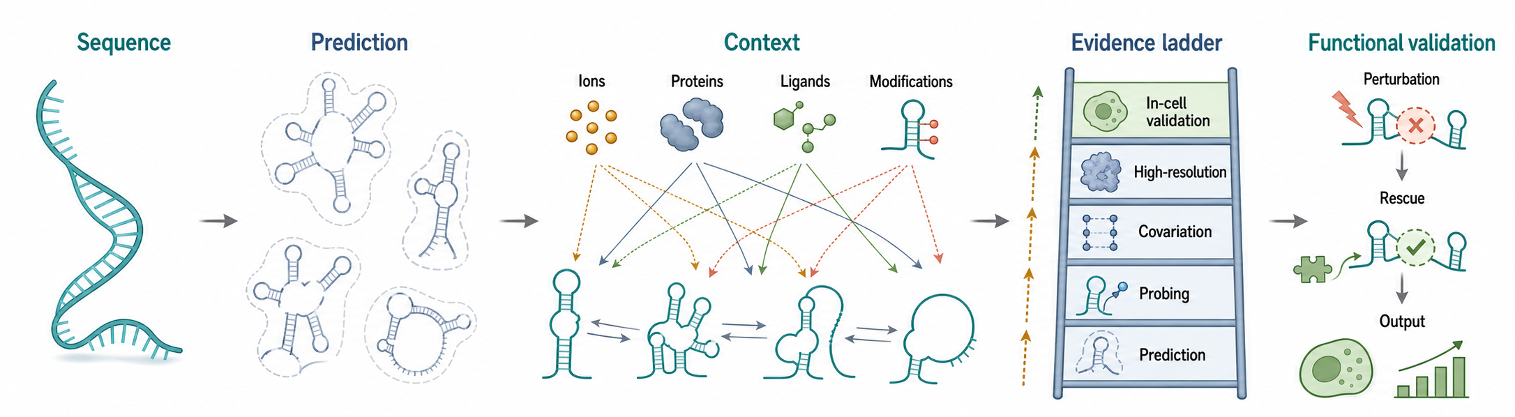
Figure 4.3. Sequence, Context, and Structure-Function Evidence. The figure presents a four-panel framework for evaluating RNA structure-function claims: sequence-derived hypotheses generated by computational prediction; environmental and RNP modifiers such as metal ions, ligands, proteins, and modifications that shift structural populations; a structural evidence ladder from prediction through in-cell validation; and functional validation by perturbation and compensatory rescue. The layout reinforces that sequence constrains possible structures, while context and orthogonal evidence determine which structural states are biologically relevant.
Chemical modifications are another context variable. A modified nucleotide can alter base-pairing potential, stacking, local flexibility, protein recognition, or immune sensing. The chapter on RNA modifications develops these mechanisms in detail, but the structural principle belongs here: a sequence written with A, U, G, and C is an incomplete chemical description if modified nucleotides are present.
Crowding and compartmentalization create additional boundaries. The inside of a cell is not dilute buffer. Macromolecular crowding can favor compact states, alter association equilibria, and change folding kinetics. Local compartments and RNP bodies can concentrate RNAs and proteins, but they can also introduce heterogeneity. A structure observed in dilute purified RNA may be a valid molecular state and still not be the dominant cellular state.
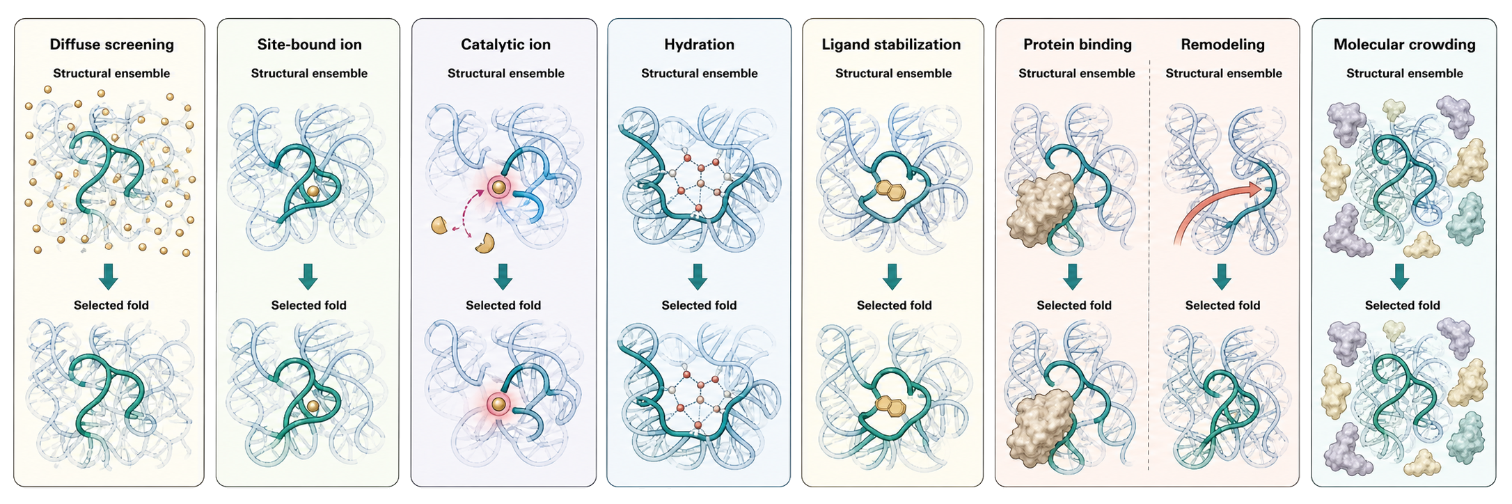
Figure 4.4. How the Environment Selects and Stabilizes an RNA Fold. Seven aligned mechanism modules compare environmental influences on the same RNA structural ensemble: diffuse ionic screening reduces phosphate-backbone repulsion; site-bound ions stabilize local geometry; catalytic ions participate directly in chemistry; hydration networks support grooves and pockets; ligand binding shifts conformational populations; proteins stabilize, destabilize, or actively remodel folds; and molecular crowding changes effective concentrations, kinetics, and equilibria. Population-weight graphics emphasize that context shifts an ensemble rather than assigning one deterministic fold to a sequence.
4.5. Structure-function relationships across RNA classes
RNA structure can support function by positioning chemical groups, creating binding pockets, exposing or hiding regulatory sequences, forming scaffolds, recruiting proteins, controlling translation, promoting decay, organizing chromatin, or packaging genomes. The same structural term can support different functions in different RNA classes, so structure-function reasoning must specify the RNA class and biological context.
tRNA is a compact adaptor. Its secondary structure is classically drawn as a cloverleaf with acceptor, D, anticodon, variable, and T arms. Its tertiary structure is an L-shaped molecule that places the amino acid acceptor end and anticodon at functionally distinct positions. Its modifications and identity elements help aminoacyl-tRNA synthetases attach the correct amino acid. The cloverleaf is a useful map, but tRNA function requires tertiary shape, modifications, protein recognition, ribosome binding, and codon-anticodon pairing; classic crystallographic work on yeast phenylalanine tRNA established the shape distinction.
rRNA shows structure at the scale of a molecular machine. Ribosomal RNAs contain secondary-structure domains, extensive tertiary packing, and binding surfaces for ribosomal proteins. In the ribosome, RNA is not a decorative scaffold; rRNA forms core functional centers. The ribosome also illustrates quaternary structure because rRNA folds in the presence of many proteins and interacts with mRNA, tRNAs, translation factors, and nascent peptide.
Riboswitches illustrate ligand-controlled structure-function relationships. A riboswitch aptamer binds a metabolite, and an expression platform changes gene expression. The mechanism can involve transcription termination, translation initiation, splicing, RNA stability, or another output depending on the system. A strong riboswitch claim typically needs ligand binding, structural or probing evidence, mutational tests of the aptamer, and a gene-expression readout. Chapter Chapter 79 covers riboswitch classes and mechanisms in depth.
Ribozymes illustrate catalytic structure. A ribozyme must position reactive groups, substrates, metal ions or general acid-base participants, and stabilizing contacts. A secondary-structure diagram can identify stems and loops, but catalytic explanation requires tertiary organization and chemistry. Some ribozymes are small and self-cleaving; others are large and embedded in RNP contexts. Chapter Chapter 8 and later enzymology chapters develop ribozyme evolution and mechanism.
mRNA structures can regulate translation, stability, localization, and decay. A 5′ untranslated region hairpin can affect scanning or initiation. Coding-region structure can slow ribosome movement or influence cotranslational events. A 3′ untranslated region structure can affect protein binding, microRNA targeting, localization, or decay. However, mRNAs are frequently remodeled by ribosomes, RNA-binding proteins, helicases, and decay machinery. A predicted mRNA structure should therefore be interpreted as a context-dependent regulatory hypothesis unless supported by cellular evidence.
Viral RNAs often use compact structural elements because viral genomes must encode many functions in limited sequence space. Viral RNA structures can regulate replication, translation initiation, ribosomal frameshifting, genome circularization, packaging, immune evasion, or host-factor recruitment. Some viral RNA elements are highly conserved and structurally constrained; others evolve rapidly. SARS-CoV-2 RNA elements have been modeled using experimentally supported secondary structures, illustrating how secondary-structure information can seed three-dimensional hypotheses. Chapter Chapter 117 treats viral RNA structures in detail.
Small regulatory RNAs often function through base pairing and RNP assembly. Bacterial small RNAs can pair with target mRNAs and recruit or require RNA chaperones. microRNAs and siRNAs guide Argonaute proteins through short sequence complementarity, where guide-target pairing, Argonaute conformation, target accessibility, and cellular concentration all matter. piRNAs depend on PIWI proteins, processing signatures, and pathway context. For these RNAs, secondary structure may affect biogenesis, loading, targeting, or stability, but the functional unit is usually an RNP.
Long noncoding RNAs require careful evidence standards. A long noncoding RNA may contain local structures, repeated elements, protein-binding domains, or modular regions. Some long noncoding RNAs have strong structure-function evidence. Many others have predicted folds or chemical-probing signals without a clear causal mechanism. The safe statement is not that long noncoding RNAs are unstructured, nor that every predicted structure is functional. The safe statement is that each proposed structural mechanism needs evidence appropriate to the claim. Chapter Chapter 91 develops lncRNA causality standards.
Therapeutic and engineered RNAs show why structure matters outside natural biology. Synthetic mRNAs require untranslated regions, coding sequence, poly(A) tail, cap structure, modified nucleotides, and formulation context that influence stability and translation. siRNAs and antisense oligonucleotides depend on duplex geometry, chemical modification, protein loading, target accessibility, and delivery. Aptamers and ribozymes exploit folded structures directly. RNA nanotechnology uses designed junctions, kissing loops, and modular motifs to build assemblies. These applications use the same structural principles but require additional pharmacological, manufacturing, and safety evidence.
Experimental Foundations and Evidence
Structural claims are strongest when multiple evidence types converge. The first evidence type is computation. A secondary-structure algorithm can produce a minimum-free-energy model, suboptimal models, base-pair probabilities, or structures constrained by experimental data. Computational models are useful, but their assumptions matter. Many simple models emphasize nested base pairing, nearest-neighbor thermodynamics, and purified RNA conditions. Pseudoknots, tertiary contacts, protein effects, co-transcriptional folding, and cellular remodeling may be absent or simplified.
Comparative sequence analysis asks whether evolution preserved a structure. If one species has a G-C pair at a position and another has an A-U pair at the aligned position, the compensatory change can support a conserved base pair. More complex covariance patterns can support conserved helices or motifs. However, covariance depends on correct alignment, adequate sampling, and appropriate evolutionary models. Lack of detected covariance is not definitive evidence against structure when the RNA family is poorly sampled, rapidly evolving, or hard to align.
Chemical probing measures how nucleotides react with chemicals under particular conditions. SHAPE reagents often report backbone flexibility or local nucleotide dynamics. Dimethyl sulfate and related chemistries can report accessibility or pairing-related protection for specific bases. Mutational profiling methods convert chemical modification into sequence changes that can be read by sequencing. These methods can reveal transcriptome-scale structure patterns and structure-dependent functions. Their limitation is that reactivity is not a direct photograph of base pairing. Protein binding, modification, solvent accessibility, RNA abundance, reverse-transcription bias, and library preparation can affect signal.
Crosslinking and proximity methods can identify contacts between RNA regions, between RNA and proteins, or between RNA and chromatin. These methods are powerful because they can operate in cells, but crosslinking efficiency, ligation bias, indirect proximity, and mapping ambiguity can complicate interpretation. A proximity signal may indicate direct base pairing, a protein-mediated contact, spatial colocalization, or an experimental artifact. Orthogonal validation is essential for strong mechanistic claims.
High-resolution structural methods define architecture more directly. X-ray crystallography can reveal atomic interactions in well ordered crystals. Nuclear magnetic resonance spectroscopy can describe solution structures and dynamics for suitable RNAs. Cryo-electron microscopy can resolve large RNP machines and multiple conformational states. These methods are indispensable, but they also have context limits. Crystallization can favor one state. NMR often favors smaller systems. Cryo-EM model quality varies by local resolution and particle heterogeneity. Purified constructs may omit cellular partners or sequence contexts.
Biochemical reconstitution tests whether purified components can reproduce a structural or functional state. Reconstitution can establish sufficiency under defined conditions. For example, if an RNA and protein assemble into a complex that binds ligand or catalyzes a reaction in vitro, the experiment supports a mechanistic model. But reconstitution does not automatically prove that the same state dominates in vivo. Cellular abundance, compartment, competing proteins, modifications, and RNA processing can change the outcome.
Genetic and functional evidence tests causality. A disruptive mutation can break a stem, motif, or binding pocket. A compensatory mutation can restore base pairing. A rescue experiment can show that structure rather than a particular sequence is important. A ligand-binding mutation can separate binding from expression output. A protein-binding mutation can separate RNP assembly from RNA abundance. Strong structure-function claims usually combine structural evidence with such perturbation and rescue logic.
Box 4.1. Structure-Claim Evidence Ladder
- Predicted structure: useful hypothesis under explicit model assumptions.
- Probing-supported model: experimental reactivity supports local pairing or accessibility under specified conditions.
- Comparative model: covariation or conservation supports a structural feature across species.
- Mutationally rescued model: disruptive and compensatory changes support causal base pairing or motif function.
- High-resolution structure: atomic or near-atomic architecture under defined construct and condition.
- In-cell validated mechanism: structural feature is connected to biological function in the relevant cellular context.
The ladder is not a rigid rule, but it prevents overstatement. A predicted structure is a hypothesis. A probing-supported model is an experimentally supported model. A comparative model has evolutionary support. A mutationally rescued model has causal support for a structural feature. A high-resolution structure provides molecular architecture under defined conditions. An in-cell validated mechanism connects structure to function in the relevant biological setting.
Biological Contexts Across Systems
In bacteria and archaea, RNA structures frequently couple transcription, translation, and RNA decay. Bacterial riboswitches can fold co-transcriptionally, meaning that the 5′ region of the RNA begins folding before the downstream region is synthesized. This timing can matter because early structures may trap the RNA in one path before all possible base-pairing partners exist. Bacterial small RNAs often pair with target mRNAs in ways influenced by RNA chaperones, target accessibility, and degradation machinery. Archaeal and bacterial CRISPR RNAs assemble with Cas proteins, where guide RNA structure is inseparable from RNP function.
In eukaryotes, RNA structure is shaped by processing and compartment. Pre-mRNAs are capped, spliced, cleaved, polyadenylated, exported, localized, translated, and degraded while bound by proteins. A structure present in a nascent transcript can differ from the structure of a mature cytoplasmic mRNA. Splicing can remove sequences that would otherwise pair. RNA-binding proteins can package transcripts into messenger RNPs. Ribosomes can unwind coding regions during translation. Subcellular localization can expose an RNA to different protein concentrations and modification states.
In organelles, RNA structure interacts with specialized processing and translation systems. Mitochondrial and chloroplast RNAs can have unusual processing, editing, stability, and translation mechanisms. Organellar ribosomes and RNA-processing RNPs often differ from cytosolic counterparts. A structure-function claim for organellar RNA should state the organism and organelle because plant chloroplasts, mammalian mitochondria, fungal mitochondria, and protist organelles can differ sharply.
In viruses, RNA structure is often under intense multifunctional constraint. The same segment of viral RNA may encode protein, regulate translation, bind host proteins, evade immune sensing, package the genome, and serve as a replication signal. Sequence changes can therefore affect codons, RNA structure, protein sequence, innate immune recognition, and RNA-RNA contacts at the same time. This overlap makes viral RNA a strong example of why sequence-to-function interpretation is difficult.
In disease contexts, RNA structure can create or modify pathology. Repeat-expansion RNAs can form structures that bind proteins or trigger toxicity. Splicing variants can disrupt structural elements or RNP assembly. Mutations in structured noncoding RNAs can affect processing, stability, localization, or interaction. Therapeutic oligonucleotides can bind structured targets or remodel splicing. These topics require caution because disease association alone does not reveal structural mechanism.
Technology, Computational, Clinical, and Engineering Links
RNA structure prediction connects sequence to hypotheses. Secondary-structure prediction can propose stems, loops, and alternative structures. Partition-function methods can estimate ensembles and base-pair probabilities. Comparative methods can use evolutionary information. Probing-constrained methods can integrate experimental reactivity. Tertiary modeling and molecular simulation can generate three-dimensional hypotheses. Machine-learning approaches can learn patterns from known structures, high-throughput experiments, or sequence-function maps. Chapters Chapter 60 through Chapter 65 develop these methods in depth.
The practical rule is that prediction should be matched to the question. If the question is whether a short bacterial leader might form a terminator hairpin, a secondary-structure model plus genetic tests may be appropriate. If the question is whether a long noncoding RNA has a conserved functional domain, comparative analysis, domain mapping, probing, and perturbation may be needed. If the question is whether a small molecule binds a folded pocket, structural, biochemical, and cellular target-engagement assays are needed.
High-throughput biochemistry maps sequence space. Large libraries can test how many sequence variants preserve folding, binding, or function. Such experiments reveal that many sequences can share a function, that some positions are constrained for structure or activity, and that epistasis can be strong: the effect of one mutation depends on other positions. These maps help connect sequence, structure, and function, but they are shaped by the assay design, library coverage, expression system, and readout.
RNA-targeted small-molecule discovery depends on folded structures and dynamic states. Ribosomal RNA antibiotics, splicing modifiers, riboswitch ligands, repeat-RNA binders, viral RNA ligands, and direct RNA-binding chemical probes all depend on some form of RNA recognition. A molecule may bind a conserved pocket, stabilize a rare conformation, alter RNP assembly, or recruit a degradation pathway. The central caveat is target validation: binding to a purified RNA or a short fragment is not the same as selective engagement of the intended RNA in cells.
RNA engineering uses structural principles deliberately. Aptamer engineering selects or designs ligand-binding pockets. Riboswitch engineering couples aptamers to expression platforms. RNA nanotechnology uses junctions, kissing loops, and modular motifs to assemble shapes. Guide-RNA engineering optimizes scaffold folding and protein recognition. Therapeutic mRNA design adjusts untranslated regions, coding sequence, modified nucleotides, and formulation to balance expression, stability, and immune recognition. In each case, sequence is a design variable, but the design target is a structure, ensemble, interaction, or function.
Clinical interpretation of RNA structure is still developing. Some variants in structured RNAs clearly disrupt known functions. Many predicted structural effects remain uncertain. A clinically useful structural claim needs a defined RNA, variant, disease context, mechanism, evidence level, and ideally a rescue or perturbation experiment. Prediction alone is rarely enough for clinical action.
4.6. What sequence can and cannot determine
RNA sequence determines the chemical identities and order of nucleotides. It constrains which base pairs, motifs, protein-binding sites, modification sites, and ligand-binding pockets are possible. In this sense, sequence is the starting condition for RNA structure. A sequence that lacks complementarity cannot form a particular Watson-Crick helix. A sequence that lacks required nucleotides cannot form a particular conserved motif. A guide RNA with changed spacer sequence will target a different nucleic acid.
Sequence does not, by itself, identify the dominant cellular structure in every context. The same sequence can fold differently during transcription than after full-length synthesis. The same RNA can adopt different structures at different Mg2+ concentrations. A protein can stabilize one conformation. A ligand can shift an ensemble. A chemical modification can change pairing or recognition. A ribosome can unwind an mRNA region. A mutation can have different effects depending on other sequence positions. This is the core of sequence-structure constraint.
Two errors are common. The first error is sequence determinism: assuming that one sequence has one biologically relevant structure that can be read directly from a prediction. This error is especially tempting when a diagram looks precise. The second error is sequence nihilism: assuming that because RNA is context-dependent, sequence tells us little. That is also wrong. Sequence is deeply informative. It constrains possible folds, encodes conserved motifs, creates or destroys binding sites, and records evolutionary selection. The mature view is conditional: sequence defines a space of possibilities, and context plus selection shapes which possibilities matter.
Evolutionary conservation can strengthen structural inference. If a base pair is preserved through compensatory changes, the structure may be under selection. If a motif is conserved while surrounding sequence diverges, the motif may be functional. If a structured RNA family preserves covariation across many species, comparative methods can infer a consensus structure. But conservation is not simple proof. Alignments can be wrong. Species sampling can be biased. Some structures are lineage-specific. Some functional RNAs evolve rapidly. Some conserved sequence reflects protein coding, protein binding, processing, modification, replication, or other constraints rather than RNA structure.
High-throughput sequence-function experiments add another layer. They can show which mutations disrupt folding, binding, expression, or activity. They can reveal redundant sequence solutions and unexpected constraints. They can also reveal epistasis, where a mutation is harmful in one background and neutral in another. Such results support the idea that RNA structure-function landscapes are shaped by networks of interacting positions rather than independent nucleotides.
The practical conclusion is that sequence-based claims should state their level. A sequence predicted to form a hairpin is a model. A hairpin conserved by compensatory mutations is stronger. A hairpin whose disruption reduces function and whose compensatory restoration rescues function is a causal structure-function statement. A hairpin resolved in a purified construct is a structural statement under defined conditions. A structure that controls gene expression in cells through a defined mechanism requires cellular functional evidence.
Box 4.2. Sequence Determinism Versus Sequence Nihilism
- RNA sequence constrains possible folds but does not uniquely determine the dominant cellular structure.
- Context — including ions, ligands, proteins, modifications, and cellular compartment — selects among structural populations.
- Conservation can support functional inference, but conservation alone is not proof of function.
- Lack of conservation can reflect poor sampling, lineage-specific function, or rapid evolutionary turnover rather than absence of structure.
- Functional claims require perturbation, compensatory rescue, or another causal test.
- Sound reasoning avoids both extremes: sequence is deeply informative, and structure is environment-dependent.
Recent Consensus
Current RNA structural biology treats secondary, tertiary, and quaternary structure as interdependent levels rather than isolated categories. Secondary-structure analysis remains essential because base pairing is often the most accessible and interpretable layer. Tertiary motifs explain how secondary elements form compact and specific folds. Quaternary assemblies explain why many RNAs are best understood as parts of RNP machines, RNA-RNA networks, or dynamic cellular bodies.
There is strong consensus that cellular RNA structure is context-dependent. Ions, water, ligands, proteins, modifications, transcription and processing history, ribosomes, and cellular compartments all alter structural populations. There is also strong consensus that computational prediction is valuable but insufficient as sole evidence for function. High-throughput probing, comparative analysis, high-resolution structure, and functional genetics are complementary rather than interchangeable.
There is growing consensus that folded RNA surfaces can be chemically targeted, especially when ligands recognize tertiary or quaternary features. However, the field remains cautious about selectivity, cellular target engagement, and whether in vitro binding events translate into mechanism. RNA-targeted drug discovery is therefore promising but evidence-intensive.
There is also consensus that evolutionary conservation and high-throughput sequence-function maps are powerful ways to understand structural constraint. These approaches help distinguish positions constrained by pairing, motif geometry, binding, or function. Their limits are equally important: conservation can be missed, alignment can mislead, and assay design can bias sequence-function maps.
Open Questions, Controversies, Deprecated Models, and Common Misconceptions
Open questions:
- How much of transcriptome-wide structure is functionally selected? Many RNAs show chemical-probing patterns consistent with structure. Some structures are clearly functional. Others may reflect physical folding without selected biological function. The difficult task is to separate causal structural elements from neutral or incidental folding.
- How should dynamic ensembles be represented? A single diagram can be misleading when an RNA samples several conformations. Ensembles can be represented by alternative structures, base-pair probabilities, kinetic paths, or structural states, but each representation loses some information. This is especially important for riboswitches, viral RNAs, long mRNAs, and RNP remodeling cycles.
- How transferable are RNA motifs? Recurrent motifs support modular thinking, and engineered systems can exploit this modularity. Natural contexts often impose constraints that make transfer incomplete. Predicting when a motif remains functional after transplantation is still difficult.
- How do condensate-like environments alter RNA structure? RNA-rich bodies can concentrate molecules and alter reaction environments, but the structural state of specific RNAs inside such bodies is often hard to define. Evidence from imaging, proximity mapping, biochemical fractionation, and functional perturbation must be interpreted carefully.
Common misconceptions:
- “A predicted minimum-free-energy structure is the cellular structure.” A predicted structure is a model under assumptions. It may be useful and even correct for some purposes, but cellular structure can be altered by proteins, ligands, modifications, ribosomes, transcription history, and compartment.
- “An unstructured RNA has no function.” Some RNAs function through flexible regions, short motifs, induced folding, protein-mediated scaffolding, or transient interactions. Lack of a stable global fold is not lack of function.
- “Every detected structure is functional.” RNA can fold because nucleotides obey physical chemistry. A structure-function claim requires evidence that the structural feature affects a biological or technological output.
- “Conserved sequence always means conserved structure.” Conserved sequence may reflect protein coding, protein binding, processing, modification, replication, or other constraints. Conserved structure is strongest when supported by covariation, probing, structural data, and functional tests.
Deprecated or weakened claims:
- Deprecated simplification: RNA structure is a static hierarchy. The hierarchy of primary, secondary, tertiary, and quaternary structure remains useful, but modern RNA biology treats these levels as coupled, dynamic, and environment-dependent.