Chapter 153. Engineering Translation and Stability in mRNA Therapeutics, Vaccines, saRNA, and circRNA
Scope Note
This chapter treats messenger RNA (mRNA) medicines as engineered RNA expression systems. The central object is not only a nucleotide sequence encoding a protein, but a designed molecular product whose cap, untranslated regions, open reading frame, codon pattern, poly(A) tail, nucleoside chemistry, impurity profile, formulation, dose, route, and manufacturing process jointly determine translation, stability, innate immune activation, tissue distribution, safety, and clinical effect. The chapter owns product-level engineering across conventional non-replicating mRNA, vaccines, protein-expression therapeutics, self-amplifying RNA (saRNA), trans-amplifying RNA (taRNA), and circular RNA (circRNA). Chapter 66 supplies decoding and tRNA biology, Chapter 36 supplies causal native codon-mediated decay mechanisms, and Chapter 72 supplies endogenous mRNA grammar; this chapter tests how those mechanisms constrain a manufactured therapeutic product.
Executive Summary
mRNA therapeutics use RNA as a transient expression cassette. A conventional non-replicating mRNA contains a 5′ cap, a 5′ untranslated region (5′ UTR), an open reading frame (ORF), a 3′ untranslated region (3′ UTR), and a poly(A) tail. Each feature can be engineered, but each feature is also interpreted by endogenous translation, RNA decay, and immune-surveillance machinery. A productive mRNA drug must reach a cell, escape from endosomes or another delivery compartment, enter the cytosol, recruit translation initiation factors, produce the intended protein at an appropriate amount and duration, and disappear without unacceptable toxicity.
The most visible mRNA products are prophylactic vaccines, where the encoded protein is an antigen and the intended pharmacology includes both antigen expression and immune education. Other mRNA therapeutics seek to replace a missing protein, express a genome-editing enzyme, deliver an immunomodulatory cytokine, encode a cancer antigen, or transiently reprogram a cell state. The same RNA design rules do not optimize all applications. A vaccine may benefit from inflammatory cues that recruit and mature antigen-presenting cells, whereas a protein-replacement therapy often tries to minimize innate immune activation so repeated dosing remains tolerable.
Translation tuning is a multi-constraint product problem. Cap structure, UTR architecture, codon choice, RNA secondary structure, upstream ORFs, poly(A) tail length, nucleoside modification, and impurities all influence ribosome loading, elongation, RNA stability, and immune sensing. Codon optimization can increase protein output, but synonymous ORF changes also alter GC content, structure, dinucleotide frequencies, pausing, co-translational folding, antigen processing, secretion, and susceptibility to native decay pathways. The mechanistic basis of decoding and decay belongs to Chapter 66 and Chapter 36; the engineering task here is to select and validate a sequence within the chemistry, formulation, route, dose, and desired expression window of the final product.
Lipid nanoparticles (LNPs) provide the dominant delivery system for many current mRNA products. LNPs condense and protect RNA, promote cellular uptake, and enable a small fraction of internalized RNA to escape into the cytosol. Biodistribution depends on lipid composition, particle properties, route, dose, serum protein interactions, tissue anatomy, and inflammatory state. For vaccines injected intramuscularly, local cells, draining lymph nodes, and antigen-presenting cells are central to the immune response. For systemic protein-expression therapies, liver exposure is often prominent. Reactogenicity arises from a mixture of desired immune activation, RNA sensing, LNP effects, tissue injury, complement or cytokine pathways, and product impurities.
Self-amplifying RNA, trans-amplifying RNA, and circular RNA platforms try to decouple dose from protein output. saRNA encodes viral replicase functions and a target antigen or protein in one RNA; taRNA separates replicase and cargo into distinct RNAs; circRNA aims for nuclease-resistant and sometimes cap-independent translation from a covalently closed transcript. These platforms can extend expression or reduce input dose, but they introduce their own constraints: larger RNA size, replicase immunogenicity, dsRNA intermediates, manufacturing complexity, translation-initiation engineering, and regulatory comparability.
The current consensus is that mRNA medicines are modular but not plug-and-play. The platform can be reused, yet every product needs empirical optimization of sequence, chemistry, purification, delivery, dose, route, potency, and safety. The major open questions concern tissue targeting beyond liver and injection-site immune tissues, quantitative endosomal escape, repeated-dosing immunology, circRNA and amplifying-platform durability, product-specific innate immune thresholds, and how to predict translation-stability tradeoffs from sequence before manufacturing and animal studies.
Concept Inventory
- mRNA therapeutic: a drug product in which an RNA molecule is delivered so cells make an encoded protein. The protein can be an antigen, missing enzyme, secreted factor, intracellular effector, gene-editing protein, receptor, or immune modulator.
- mRNA vaccine: an mRNA therapeutic whose encoded product is intended to elicit protective or therapeutic immunity. The RNA and formulation may also act as immunostimulatory components.
- Non-replicating mRNA: a linear mRNA whose protein output depends on the delivered copies and their translation lifetime, not on intracellular RNA amplification.
- Self-amplifying RNA: an RNA, usually derived from positive-strand RNA virus replicon logic, that encodes replicase machinery able to copy the RNA and amplify expression of the cargo.
- Trans-amplifying RNA: a split amplifying system in which replicase functions and target cargo are supplied on separate RNA molecules.
- Circular RNA platform: an engineered covalently closed RNA intended to express protein without free 5′ or 3′ ends, typically using internal ribosome entry, rolling-circle-derived design, or other cap-independent initiation strategies.
- Translation tuning: deliberate adjustment of RNA sequence, structure, chemistry, and delivery context to control protein amount, onset, duration, cell type, and immune consequences.
- Therapeutic codon design: selection of synonymous ORF sequence to meet product-specific targets for protein amount, duration, folding, localization, antigen processing, innate immune exposure, manufacturability, and safety.
- dsRNA impurity: double-stranded RNA species generated during in vitro transcription or amplification that can strongly activate innate immune sensors and suppress translation.
- Reactogenicity: expected short-term inflammatory symptoms after vaccination or RNA delivery, such as fever, pain, fatigue, or myalgia. Reactogenicity is not identical to dangerous toxicity, but it must be controlled and monitored.
What to Know Before Reading This Chapter
Readers should know that eukaryotic translation usually begins when cap-binding factors recognize a 5′ cap, recruit the small ribosomal subunit, scan the 5′ UTR, and initiate at a start codon in a favorable sequence context. Readers should also know that mRNA decay is not a single timer. Deadenylation, decapping, endonucleolytic cleavage, nonsense-mediated decay, codon-mediated decay, RNA-binding proteins, miRNAs, and stress responses all influence transcript lifetime. Minimal definitions are provided here, while Chapter 66, Chapter 36, and Chapter 72 provide the decoding, causal-decay, and endogenous-grammar foundations.
The chapter uses two running examples. The first is a prophylactic vaccine encoding a viral surface antigen, where success requires antigen expression, antigen processing and presentation, B-cell and T-cell activation, and acceptable reactogenicity. The second is a protein-replacement mRNA delivered repeatedly to express a missing or deficient human protein, where success requires enough protein in the correct tissue with limited innate immune activation across many doses. These examples share the same RNA grammar but place different weights on antigen-presenting cells, inflammatory signaling, duration, and tolerability.
153.1. mRNA Platform Architectures and Design Variables
An mRNA platform is an engineered expression architecture built around the logic of cellular messenger RNA. The simplest version is a purified linear RNA that resembles a mature eukaryotic mRNA: a 5′ cap, a 5′ UTR, an ORF, a 3′ UTR, and a poly(A) tail. This architecture is called non-replicating mRNA because each delivered molecule can be translated many times but is not copied inside the cell. The delivered RNA is therefore consumed by ordinary decay processes. Protein output is determined by the number of molecules reaching the cytosol, the rate of translation per molecule, and the lifetime of the RNA and encoded protein.
The therapeutic design problem begins by defining the intended protein product. In a vaccine, the encoded protein is usually an antigen or antigenic domain. A respiratory virus vaccine may encode a prefusion-stabilized surface glycoprotein so host cells make an antigen that resembles the infectious virion target. In a protein-replacement therapy, the ORF may encode a secreted enzyme, a clotting factor, or a missing metabolic protein. In a cell-engineering or oncology therapy, the ORF may encode an immune receptor, cytokine, antibody fragment, genome-editing nuclease, base editor, or tumor antigen. These examples differ in desired location, duration, dose, and immune context, so a sequence that is ideal for one product may be inappropriate for another.
Figure 153.1 frames the platform as a chain of linked design variables rather than a single RNA sequence.
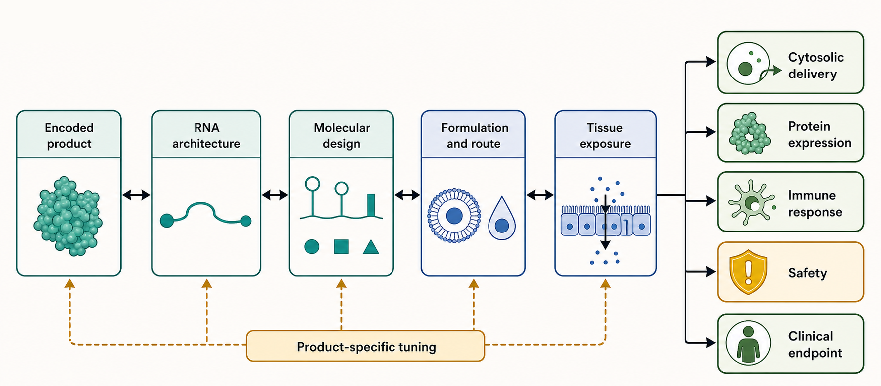
Figure 153.1. mRNA Medicine Architecture as a Linked Design System. “mRNA medicines are expression systems, not only sequences. Each design variable changes the next biological step, so platform reuse must still be product-specific.”
The first architectural distinction is whether the RNA is meant to be translated directly, amplified, or maintained in another molecular form. Non-replicating mRNA is translated directly. Self-amplifying RNA carries replicase functions that generate additional RNA templates in the cytosol. Trans-amplifying RNA separates the replicase and cargo RNAs so the cargo can be smaller or more modular. Circular RNA lacks free ends and therefore does not use the same cap and poly(A)-tail architecture as linear mRNA. Each architecture changes the relationship between administered dose and protein output. Direct linear mRNA is comparatively simple to analyze and manufacture. Amplifying and circular platforms can extend expression or reduce input dose, but they add replicase biology, noncanonical translation, or circularization chemistry to the product definition.
The second distinction is between immunological and non-immunological pharmacology. A vaccine usually wants antigen expression in a setting that activates innate and adaptive immunity. Antigen-presenting cells such as dendritic cells are especially important because they process antigen, display peptide-major histocompatibility complex molecules, provide costimulatory signals, and shape T-cell help for antibody responses. A protein-replacement mRNA usually wants expression with minimal inflammatory signaling because the same patient may require repeated dosing. Inflammation can reduce translation through interferon-stimulated pathways, increase adverse reactions, change biodistribution, and promote anti-drug immunity against the encoded protein or formulation components.
Table 153.1 summarizes the primary design variables that must be specified before an mRNA product can be interpreted biologically.
Table 153.1. Product-Level Design Variables for mRNA Medicines. Provide a structured way to evaluate an mRNA product before interpreting biological results.
| Variable | Design question | Typical evidence | Failure mode if ignored |
|---|---|---|---|
| Encoded protein and intended mechanism | Is the payload an antigen, secreted protein, intracellular effector, editor, cytokine, or cell-state regulator? | Protein expression, activity, antigen conformation, immune presentation, or pharmacodynamic endpoint. | Correct RNA can produce the wrong protein form, duration, location, or toxicity profile. |
| RNA architecture | Should the product be non-replicating mRNA, saRNA, taRNA, or circRNA? | Matched dose-route expression kinetics, RNA-species assays, potency, and innate-sensing readouts. | Dose-output assumptions fail; amplification, circularization, or byproducts dominate biology. |
| Cap and termini | Do the ends support translation, stability, and self-like discrimination? | Cap analysis, terminal integrity assays, translation potency, and innate immune readouts. | Poor initiation, exonuclease sensitivity, or sensing of defective 5′ ends. |
| UTRs | Which 5′ and 3′ UTRs tune initiation, stability, and cell-type regulation? | Reporter and payload assays, RNA half-life, motif review, and cell-type expression tests. | A high-expression cassette in one assay can be repressed or unstable in the target cell. |
| ORF and codon design | Do protein engineering and synonymous choices fit expression, folding, localization, and immune goals? | Synonymous variant panels, ribosome profiling, protein activity, secretion, or antigen-binding assays. | High output may yield misfolded protein, excessive innate motifs, decay, or toxic persistence. |
| Nucleoside chemistry | Does modified or unmodified RNA match the desired immune window? | Cytokine and interferon assays, translation assays, sensor-dependent tests, and tolerability data. | Excess inflammation can suppress translation; over-silencing may weaken vaccine adjuvanticity. |
| Impurity profile | Are dsRNA, truncated RNA, uncapped RNA, residual DNA, proteins, and endotoxin controlled? | Purity analytics, dsRNA assays, cap-tail characterization, and release testing. | Impurities can drive sensor activation, potency loss, lot variability, or safety signals. |
| Formulation | What carrier protects RNA and enables cytosolic delivery in the intended tissue? | Particle size, encapsulation, lipid composition, stability, expression, and safety assays. | Uptake may not become endosomal escape; formulation effects may drive toxicity or reactogenicity. |
| Route and dose | Which route, schedule, and dose give target exposure with acceptable innate activation? | Biodistribution, expression time course, dose-response, and clinical tolerability data. | Wrong tissue exposure, underdosing, or a dose ceiling from reactogenicity. |
| Intended cell type | Which cells must translate the RNA or present the encoded antigen? | Cell sorting, imaging, reporter or payload expression, and antigen-presenting-cell assays. | Expression in irrelevant or vulnerable cells can miss efficacy or create toxicity. |
| Clinical endpoint | What patient-relevant effect defines success for this product? | Immunogenicity, efficacy, pharmacodynamic biomarkers, safety, and comparability evidence. | Analytical success may not translate into protection, protein replacement, or benefit-risk support. |
Route and tissue exposure are part of the platform architecture, not afterthoughts. Intramuscular injection, intradermal injection, intravenous infusion, intratumoral injection, inhaled delivery, and local tissue administration expose different cell types and immune compartments. Intramuscular vaccination can use local inflammation and draining lymph nodes to support immune priming. Intravenous LNP delivery often produces strong liver exposure because serum proteins and hepatic uptake pathways favor liver accumulation. Inhaled delivery must negotiate mucus, airway epithelium, macrophages, and local inflammation. A platform cannot be evaluated by RNA sequence alone without specifying route, formulation, and intended cells.
The evidence basis for platform architecture comes from multiple layers. Cell-culture transfection assays measure whether an RNA can be translated, but they often overestimate productive delivery because transfection reagents and immortalized cells do not reproduce human tissue uptake. Animal biodistribution and expression studies add route and tissue context, but species differences in innate immune sensors, lipid metabolism, and antigen presentation limit direct extrapolation. Clinical vaccine trials provide strong evidence for immunogenicity and protection in defined populations, but they do not automatically validate the same RNA design for chronic protein replacement. The evidence standard therefore asks a narrow question: did the exact product, route, dose, and schedule produce the intended biological effect with acceptable safety in the relevant population?
Do not overgeneralize the term “platform.” A platform can simplify repeated development by reusing manufacturing methods, analytical assays, formulation families, and regulatory knowledge. A platform does not remove the need to optimize each ORF, UTR pair, purification process, LNP composition, route, dose, and indication. Sequence-dependent structure, protein toxicity, antigen biology, tissue distribution, immune memory, and patient state can all override generic expectations.
153.2. Cap, UTR, ORF, codon, and poly(A)-tail engineering tradeoffs
The cap is a modified nucleotide structure attached to the 5′ end of eukaryotic mRNA. In natural cytoplasmic mRNAs, the cap helps recruit translation initiation machinery, protects against exonucleases, and marks RNA as processed by the cell. Therapeutic mRNAs usually use enzymatic capping after transcription or co-transcriptional cap analog incorporation. Cap 0 refers to an N7-methylguanosine cap without ribose 2′-O-methylation on the first transcribed nucleotide. Cap 1 includes 2′-O-methylation on the first nucleotide and is common in many self RNAs. Cap state can influence translation and innate immune discrimination because some sensors and restriction factors respond differently to cap-deficient or improperly capped RNA.
The 5′ UTR is the untranslated region between the cap and start codon. It is not protein-coding, but it controls translation initiation. A strong therapeutic 5′ UTR usually avoids stable secondary structures close to the cap, upstream AUG codons, inhibitory upstream ORFs, cryptic splice-like motifs that matter during production or analysis, and sequence motifs that recruit inhibitory RNA-binding proteins. It also provides a start-codon context that supports efficient initiation. The best 5′ UTR is not always the shortest or least structured sequence. Some structure can protect RNA or tune initiation, and some natural UTRs contain motifs that improve expression in particular cell types.
The ORF encodes the protein. ORF engineering includes codon choice, GC content, avoidance of problematic repeats, removal of cryptic regulatory motifs, insertion of signal peptides or membrane anchors, domain stabilization, antigen prefusion stabilization, subcellular localization tags, and changes that alter protein half-life. For a secreted protein therapeutic, the ORF must include or preserve a signal peptide compatible with the target cell’s endoplasmic reticulum entry and secretion machinery. For a vaccine antigen, the ORF may encode a membrane-bound form, secreted form, stabilized trimer, or receptor-binding domain. Those protein-design choices change the immunological output even when RNA delivery is identical.
The 3′ UTR sits downstream of the stop codon and upstream of the poly(A) tail. Natural 3′ UTRs carry binding sites for RNA-binding proteins and miRNAs that regulate localization, translation, and decay. Therapeutic 3′ UTRs are often selected from stable human transcripts or assembled from elements that improve expression. However, a 3′ UTR sequence can behave differently across cell types because the relevant RNA-binding proteins and miRNAs differ. A motif that stabilizes RNA in one tissue may have little effect or an opposing effect in another tissue.
The poly(A) tail is a stretch of adenosines at the 3′ end of most eukaryotic mRNAs. It binds poly(A)-binding proteins, helps translation initiation communicate with the cap-binding complex, and influences deadenylation-dependent decay. In vitro transcribed mRNAs can receive the poly(A) tail from an encoded DNA template or from enzymatic polyadenylation. Template-encoded tails offer length precision but can create plasmid-stability and homopolymer issues. Enzymatic tailing can be efficient but may create length heterogeneity. Both approaches must be characterized because tail length and uniformity affect potency and product consistency.
Figure 153.2 shows the linear mRNA expression cassette as an integrated regulatory grammar.
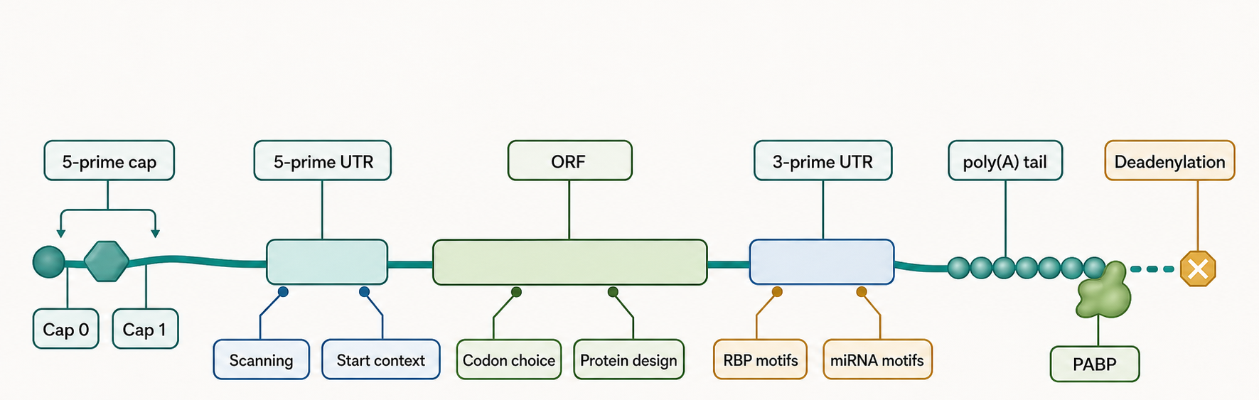
Figure 153.2. Linear mRNA Expression Cassette and Regulatory Grammar. “The therapeutic mRNA cassette is interpreted by normal cellular translation and decay machinery. Optimization of one component can change the effect of the others.”
Codon choice is often described as codon optimization, but the phrase can be misleading. Multiple synonymous codons encode the same amino acid, yet synonymous codons are not biologically equivalent. Codon choice changes decoding speed, tRNA demand, mRNA GC content, CpG and UpA frequencies, local RNA structure, ribosome pausing, protein co-translational folding, and the likelihood that decay pathways will interpret the transcript as stable or unstable. In an mRNA vaccine, codon changes may increase antigen output while also changing innate immune motifs or antigen conformation through altered translation kinetics. In an enzyme-replacement therapeutic, codon changes may improve yield but also alter folding, secretion, or post-translational modification if translation kinetics shape nascent-chain processing.
Table 153.2 distinguishes the main expression-cassette components and the failure modes that occur when each component is optimized in isolation.
Table 153.2. Linear mRNA Cassette Components and Coupled Failure Modes. Show that cap, UTR, ORF, and tail engineering cannot be optimized independently.
| Component | Primary function | Engineering choices | Coupled risk | Assay evidence |
|---|---|---|---|---|
| Cap structure | Recruits cap-dependent initiation factors and protects the 5′ end. | Enzymatic capping, co-transcriptional cap analogs, Cap 0 or Cap 1 target, and cap-quality limits. | Cap defects reduce initiation and expose RNA to immune discrimination. | Cap chemistry assays, translation potency, and innate immune readouts. |
| 5′ UTR | Controls scanning, start-codon access, and initiation efficiency. | Avoid upstream AUGs and inhibitory uORFs, tune structure, and select context-appropriate UTRs. | Generic high-expression UTRs may repress the payload or behave differently across cell types. | Reporter and payload expression, structure probing or prediction, and RNA half-life. |
| Start-codon context | Sets initiation-site fidelity and N-terminal protein identity. | Kozak-like context, removal of near-cognate starts, and management of start-proximal structure. | Leaky scanning, upstream initiation, or altered protein N terminus. | Ribosome profiling, reporter variants, and N-terminal protein checks when needed. |
| ORF protein design | Defines antigen or effector sequence, localization, and protein half-life. | Signal peptide, membrane anchor, prefusion stabilization, localization tags, and domain mutations. | Wrong conformation, secretory stress, abnormal localization, or encoded-protein toxicity. | Functional activity, antigen binding, secretion, surface display, or localization assays. |
| Synonymous codon pattern | Tunes elongation, RNA stability, structure, and motif content. | Codon adaptation, GC and dinucleotide balance, rare-codon retention, and motif removal. | Misfolding, ribosome collisions, innate motifs, or unexpected decay. | Synonymous variant panels, ribosome profiling, and RNA-protein time courses. |
| 3′ UTR | Regulates stability, localization, translation, and decay factor recruitment. | Stable-transcript UTR elements, motif edits, and miRNA detargeting sites where appropriate. | RNA-binding protein or miRNA differences can reverse expected effects in target tissues. | RNA half-life, payload duration, motif perturbation, and cell-type comparison. |
| Poly(A) tail | Supports poly(A)-binding protein, translation coupling, and deadenylation timing. | Template-encoded or enzymatic tailing with defined length and uniformity. | Tail heterogeneity can alter potency, decay, lot consistency, or template stability. | Tail-length analysis, RNA integrity, potency, and stability testing. |
| Terminal and residual sequences | Prevent process remnants from perturbing translation or sensing. | Remove vector scars, manage adapter or promoter remnants, and control residual DNA and proteins. | Cryptic motifs, immune sensing, release-test failures, or inconsistent expression. | Sequence and length identity, residual impurity assays, and biological potency tests. |
The evidence basis for expression-cassette engineering includes reporter assays, protein quantification, ribosome profiling, RNA stability measurements, structure probing, innate immune readouts, and product-specific animal or clinical data. A luciferase reporter can rank UTRs or tail designs, but a luciferase ranking may not transfer to a membrane antigen, secreted enzyme, or toxic intracellular effector. Ribosome profiling can show ribosome occupancy and pausing but does not by itself prove correct protein folding or immune presentation. RNA abundance measurements show stability, but high RNA abundance without translation can indicate blocked initiation, stress responses, or sequestration in nonproductive compartments.
Boundary cases matter. Some therapeutic goals intentionally reduce translation duration. A genome-editing nuclease or immune activator may require a short pulse to limit off-target activity or systemic toxicity. Some vaccines may accept lower expression if antigen quality, localization, or immune context improves. Some UTRs contain cell-type-specific regulatory motifs that are useful for targeting or safety, even if they reduce expression in generic reporter assays. Translation tuning is therefore not a race toward maximum protein output; it is a process of matching amount, timing, location, and immune context to the mechanism of action.
153.3. Therapeutic codon design: translation, stability, and immunity tradeoffs
Therapeutic codon design begins with a product objective, not with a universal codon score. A prophylactic vaccine may need enough correctly folded antigen for presentation during a limited inflammatory window. A secreted enzyme may need durable, repeated expression with efficient signal-peptide use and low innate activation. A cytokine or genome editor may require a short pulse because prolonged exposure increases toxicity. Synonymous ORF variants should therefore be ranked against amount, duration, protein quality, localization, immune response, dose, and safety rather than against peak reporter output alone.
The product-level variables are coupled. A codon substitution can redirect demand among tRNAs, change elongation and co-translational folding, alter local or global RNA structure, change GC content and CpG or UpA frequency, create or remove regulatory motifs, and modify susceptibility to translation-coupled decay. Chapter 66 owns the decoding-system explanation and Chapter 36 owns the causal decay mechanism. Here, those variables become engineering constraints: the same synonymous sequence must be synthesized reproducibly, remain sufficiently intact during storage and delivery, express the desired protein in the target cell, and avoid unacceptable immune or stress responses.
At least five design dimensions should be tested together. First, GC-rich recoding can improve expression in some mammalian systems but can also strengthen structure, alter in vitro transcription behavior, and complicate amplification or analytical interpretation. Second, CpG or UpA depletion can change innate or antiviral responses, but dinucleotide edits necessarily change codons and may change antigen processing or structure. Third, selected slower-decoded codons may be retained near domain boundaries, signal peptides, transmembrane segments, or folding-sensitive regions, yet excessive pausing can generate queues and quality-control responses. Fourth, start-proximal codon choices can change initiation-region structure, making an apparent elongation improvement irrelevant if ribosome loading falls. Fifth, synonymous changes can create or remove RNA-binding-protein, miRNA, endonuclease, or manufacturing-risk motifs. Endogenous integration of these features is developed in Chapter 72; therapeutic design must validate them in the final product context.
Figure 153.3 presents translation tuning as a tradeoff surface rather than a one-dimensional optimization score.
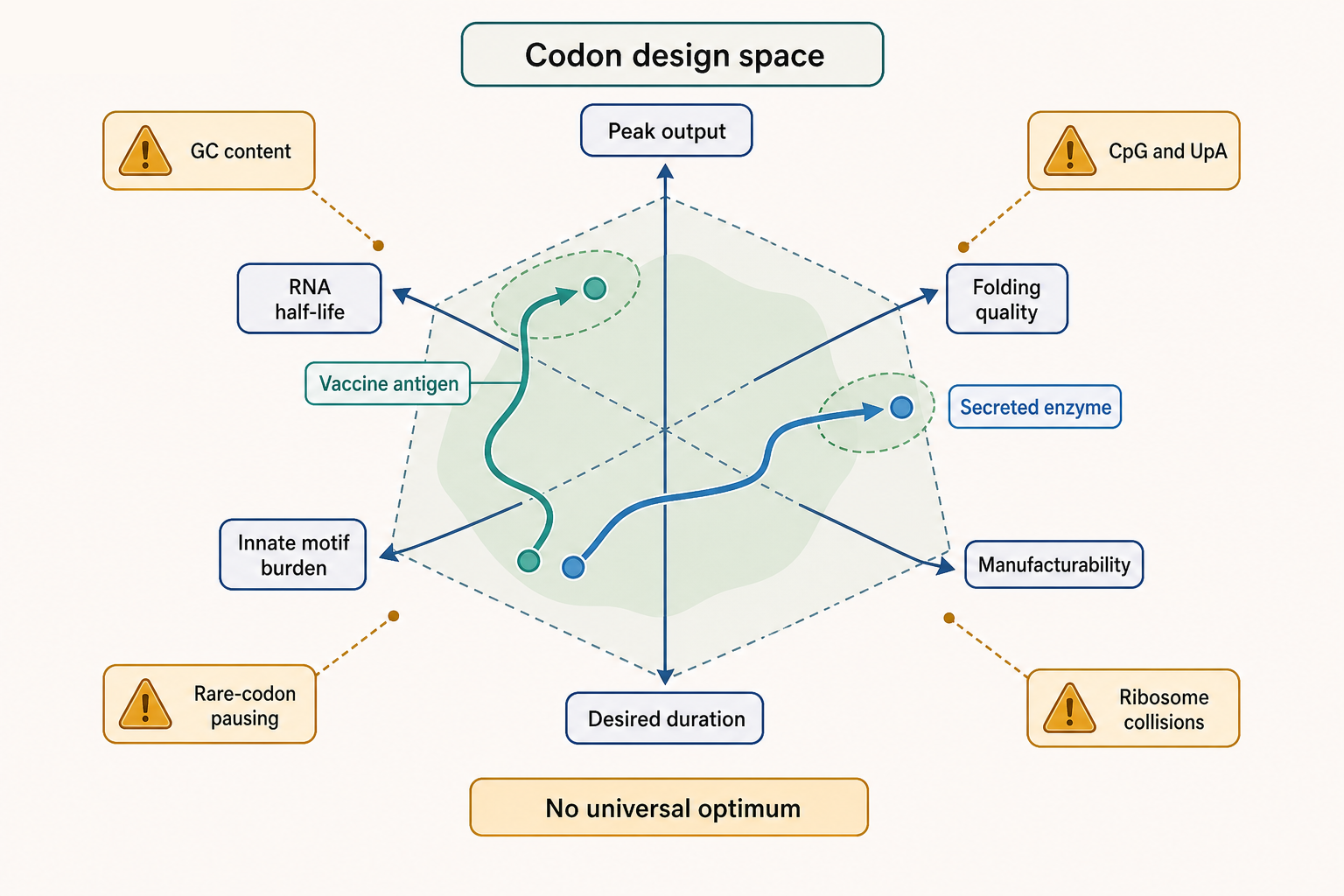
Figure 153.3. Codon Choice as a Translation-Stability Tradeoff Surface. “Synonymous codons tune more than translation speed. A useful codon design balances expression, stability, folding, immune sensing, and manufacturing constraints for a defined product.”
A concrete vaccine example is a viral spike or envelope glycoprotein antigen. Designers may alter the protein sequence to stabilize a prefusion conformation, add proline substitutions, change cleavage sites, or use a transmembrane anchor. They may also alter codons to improve mammalian expression. These protein and RNA changes are coupled. Higher expression of a misfolded antigen is not necessarily better than moderate expression of an antigen with the right conformation. Conversely, a perfectly designed antigen that is translated poorly may not drive enough immune response. Assays must therefore measure antigen amount, antigen conformation, cell-surface display or secretion, immune recognition, and innate immune activation.
A concrete protein-replacement example is a secreted enzyme. The ORF must produce a folded protein that enters the secretory pathway, receives appropriate post-translational modifications, and remains active in plasma or target tissue. Codon choices that maximize total intracellular protein could overload the endoplasmic reticulum, trigger stress, or reduce secretion efficiency. The desired output is functional enzyme exposure, not just total protein.
Table 153.3 gives a design checklist for codon and sequence tuning.
Table 153.3. Codon and Sequence Tuning Evidence Checklist. Help readers distinguish computational codon scores from product-relevant evidence.
| Design feature | Intended benefit | Possible liability | Product-relevant assay |
|---|---|---|---|
| Codon adaptation | Match efficient decoding and increase protein output or RNA stability. | Over-fast elongation can impair folding or create context-specific tRNA stress. | Matched mRNA variants measuring protein yield, activity, and RNA half-life. |
| GC content | Improve stability or expression in many mammalian design contexts. | Excess structure can block initiation and complicate synthesis or formulation. | Expression time course, structure probing or prediction, and manufacturability metrics. |
| CpG and UpA content | Reduce some innate-sensing or antiviral-restriction motifs. | Dinucleotide edits change codons and may alter antigen processing or RNA structure. | Dinucleotide-controlled variants with cytokine, interferon, and expression readouts. |
| Start-proximal structure | Keep scanning and early initiation efficient. | Hidden structure or upstream initiation can dominate total ORF optimization. | 5′ structure probing, reporter variants, and ribosome profiling near the start. |
| Rare-codon retention | Preserve pausing for folding, secretion, domain boundaries, or membrane insertion. | Excess pausing can create ribosome queues, quality control, decay, and low output. | Ribosome profiling plus folding, activity, secretion, or antigen-conformation assays. |
| Signal peptide and secretion context | Route the nascent protein into the secretory pathway when needed. | High intracellular synthesis can overload ER processing or reduce mature secretion. | Secreted protein quantification, maturation or glycosylation checks, and ER-stress markers. |
| Ribosome pausing | Tune co-translational folding, membrane insertion, or antigen processing. | Collisions can activate ribosome quality control and reduce productive protein. | Ribosome or disome profiling paired with protein function and abundance assays. |
| RNA-binding protein or miRNA motifs | Stabilize, detarget, localize, or restrict expression by cell type. | Unintended motifs can repress the target cell or destabilize the RNA. | Motif perturbation, cell-type expression panels, and RNA-protein half-life tests. |
The evidence basis for codon tuning should include direct comparisons of synonymous ORF variants in the relevant RNA chemistry and delivery system. Plasmid expression, stable cell lines, or viral vectors can be useful for protein engineering but do not reproduce the same RNA stability and innate immune constraints as delivered mRNA. Reporter libraries can reveal broad rules, but they may miss protein-specific folding and secretion requirements. Ribosome profiling can identify pauses or collisions, but interpretation requires careful normalization for RNA abundance, initiation rate, and nuclease biases. Proteomics, activity assays, and antigen-binding assays are often needed to decide whether increased translation improves the product.
Do not overgeneralize codon optimization as a universal good. The term can hide incompatible objectives: maximum peak expression, longer duration, lower innate stimulation, preserved folding, reduced sequence motifs, manufacturability, or conserved antigen processing. A mature design process names the objective, compares multiple synonymous ORFs in the intended RNA chemistry and delivery system, and tests whether a gain in one endpoint creates liabilities elsewhere.
Box 153.1. Define the Optimization Target Before Optimizing Codons
A useful codon-design claim starts with the endpoint. For a vaccine antigen, “better” may mean enough antigen in the right conformation, appropriate presentation, and innate activation that supports adaptive immunity. For a secreted enzyme, “better” may mean durable secretion of active protein with little inflammatory signaling. For a genome editor or cytokine, the desired expression window may be short. A codon panel should therefore compare matched RNAs that differ only in defined sequence features and should measure RNA abundance, protein amount, protein quality or activity, and innate immune readouts. A single codon-adaptation score is not evidence that the design is product optimal. The strongest inference comes when the same objective is improved in the delivery formulation, cell type, route, and dose that the product will use.
153.4. Modified Nucleotides, dsRNA Impurities, and Innate Immune Tuning
Innate immune tuning is the deliberate management of RNA-triggered immune pathways. Mammalian cells contain sensors that detect RNA features associated with infection, damage, or mislocalization. Endosomal Toll-like receptors can detect single-stranded RNA or RNA degradation products, especially in immune cells that sample extracellular material. Cytosolic RIG-I-like receptors detect particular double-stranded or structured RNA features. Protein kinase R (PKR) responds to double-stranded RNA and can inhibit translation through phosphorylation of eukaryotic initiation factor 2 alpha. OAS enzymes can activate RNase L in response to dsRNA, leading to RNA cleavage. These pathways are essential for antiviral defense, but they can suppress mRNA drug expression and cause inflammatory toxicity when activated inappropriately.
Modified nucleotides are nucleotides with chemical changes to the base or sugar. Natural RNAs contain many modifications, and therapeutic mRNAs can incorporate modified nucleoside triphosphates during in vitro transcription. Pseudouridine and N1-methylpseudouridine are prominent examples used to alter RNA immunogenicity and translation. The practical effect of a modification depends on the RNA sequence, modification fraction, purification, cell type, sensor expression, formulation, dose, and assay. A modified mRNA is not automatically non-immunogenic. It may still contain dsRNA impurities, uncapped RNA, 5′ triphosphate ends, damaged RNA, contaminating DNA, residual proteins, or formulation-associated triggers.
dsRNA impurities deserve separate attention because they can dominate biological response. In vitro transcription can generate double-stranded byproducts through template-independent extension, antisense transcripts, abortive products, self-complementary regions, or other polymerase-associated mechanisms. Even a small amount of dsRNA can activate sensors more strongly than the intended single-stranded mRNA. Purification methods such as chromatographic separation or cellulose-based enrichment can reduce dsRNA content and improve translation. The specification is not merely analytical neatness; it is part of the mechanism because impurity profile changes innate signaling and potency.
Box 153.2. Modified Nucleotides Do Not Replace Purification
Modified nucleosides alter how the intended RNA is interpreted by translation and immune-surveillance systems, but they do not remove all immunostimulatory material from a preparation. A batch can contain N1-methylpseudouridine or pseudouridine and still carry dsRNA byproducts, uncapped transcripts, 5′ triphosphate ends, nicked RNA, residual DNA, protein, solvent, or endotoxin. Purification reduces unwanted species; capping controls 5′-end identity; formulation and dose determine which cells see the RNA. These controls are complementary, not interchangeable. When an experiment reports lower cytokines or higher expression after a process change, the interpretation should ask which feature changed: nucleoside chemistry, cap quality, dsRNA burden, RNA integrity, LNP properties, or exposure level. Without analytical characterization, a biological improvement cannot be assigned confidently to chemical modification alone.
Figure 153.4 separates intended mRNA expression from impurity-driven sensing.
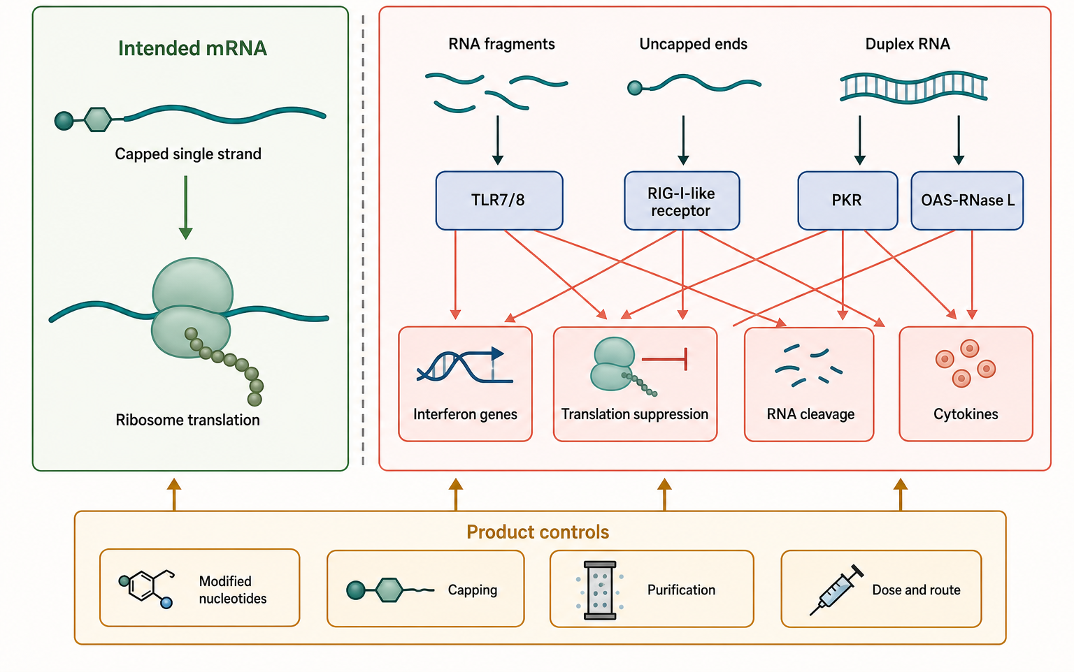
Figure 153.4. Intended mRNA Translation Versus Innate Immune Sensing of RNA Features and Impurities. “Innate immune sensing is feature- and context-dependent. Modified nucleotides, cap quality, and purification each address different parts of the sensing problem.”
The cap and ends are also immunological features. Cap-deficient RNA, improperly capped RNA, and 5′ triphosphate RNA can be interpreted as non-self or defective. Cap 1 methylation can help distinguish host-like RNA from foreign RNA in some sensor systems. However, immune response is combinatorial. A well-capped RNA delivered in a highly inflammatory particle can still cause strong cytokine induction. A modified RNA with low dsRNA content can still be immunogenic if it encodes an inflammatory protein or is delivered to immune cells at high dose.
For vaccines, innate immune activation is not purely harmful. Some inflammatory signaling promotes dendritic-cell maturation, antigen presentation, cytokine production, and adaptive immunity. The problem is dose and quality. Too little innate activation can reduce immunogenicity, especially if no adjuvant effect is supplied by the formulation. Too much activation can suppress translation, increase systemic symptoms, skew immune responses, or raise safety concerns. For repeated protein replacement, the acceptable inflammatory window is usually narrower because patients may receive many doses and because immunity against the encoded protein can undermine therapy.
The evidence basis for innate immune tuning includes cytokine assays, interferon-stimulated gene expression, translation measurements, sensor-knockout or inhibitor studies, dsRNA analytical assays, cap analysis, impurity profiling, and in vivo reactogenicity. Bulk cytokine release is useful but incomplete. It does not identify the sensor, cell type, RNA species, or causal impurity. A sensor knockout can show pathway involvement but may not translate across species or primary human cells. Analytical release tests can quantify product features, but the biological threshold for a feature depends on route, dose, and patient population.
Common misconceptions are especially risky here. Modified nucleotides do not make an mRNA invisible to the immune system. Unmodified mRNA is not automatically unacceptable; some vaccine and self-amplifying approaches may use unmodified RNA with formulation and dose choices that produce a desired immune profile. dsRNA depletion is not the same as complete immune silencing. Reactogenicity after a vaccine is not proof of better protection, and absence of reactogenicity is not proof of weak immunity. The correct question is whether the product produces the intended protein and immune or pharmacologic effect with a safety profile appropriate to the indication.
153.5. LNP Formulation, Biodistribution, and Reactogenicity Links
A lipid nanoparticle is a multicomponent particle that packages RNA with ionizable lipids and helper lipids. The common design includes an ionizable lipid that becomes positively charged in acidic environments, a phospholipid, cholesterol, and a polyethylene glycol (PEG)-lipid or other steric-stabilizing component. During formulation, the ionizable lipid helps complex the negatively charged RNA. After injection, the particle protects RNA from nucleases, circulates or drains according to route and composition, binds proteins, is taken up by cells, and traffics through endosomal compartments. Only a small fraction of internalized RNA must reach the cytosol to produce protein, but that fraction is often the limiting step.
Endosomal escape is the transition from endosomal confinement to cytosolic availability. It is central because ribosomes translate mRNA in the cytosol or on the endoplasmic reticulum, not inside endosomes. Ionizable lipids are designed to be relatively neutral at physiological pH and positively charged in acidic endosomes. This pH-responsive behavior can promote interactions with endosomal membranes, lipid mixing, membrane destabilization, and RNA release. The precise molecular events remain difficult to quantify in living tissues. Uptake and expression can diverge sharply: many cells may contain particle-associated RNA, while only a subset release enough RNA into the cytosol for translation.
Route controls the first biodistribution filter. Intramuscular injection places LNPs in muscle and interstitial tissue, where particles can be taken up locally or drain to lymph nodes. Dendritic cells, macrophages, monocytes, muscle cells, endothelial cells, and stromal cells can contribute to antigen production or immune activation, depending on formulation and dose. Intravenous dosing exposes serum proteins and hepatic uptake pathways, often producing liver expression. Intratumoral injection exposes tumor cells, stromal cells, vascular cells, and immune infiltrates. Inhaled or intranasal delivery must pass airway barriers and avoid excessive local inflammation.
Figure 153.5 connects formulation, tissue distribution, endosomal trafficking, and immune outcome.
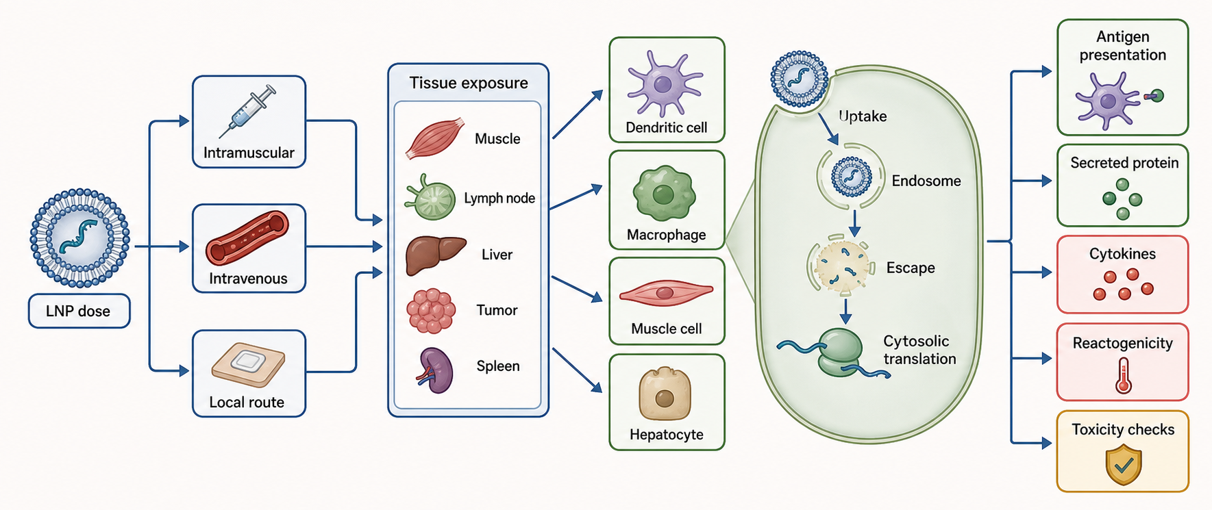
Figure 153.5. LNP Delivery Path from Injection to Antigen-Presenting Cell and Reactogenicity. “LNP biodistribution and endosomal escape determine which cells express the payload. The same delivery events can support efficacy and contribute to reactogenicity.”
Antigen-presenting cells are important for vaccine mechanisms, but they are not the only cells that matter. A cell that expresses antigen can be killed, secrete antigen, display antigen fragments, or transfer antigen to professional antigen-presenting cells. Dendritic cells can prime T cells when they receive antigen plus maturation signals. B cells can recognize conformational antigen and receive T-cell help in germinal centers. Innate immune cells can produce cytokines that shape the response. LNPs can affect each step by determining where antigen is made, how long it persists, and which inflammatory pathways are activated.
Reactogenicity is the clinical expression of acute inflammation. Pain at the injection site, fever, fatigue, headache, chills, myalgia, and lymph-node swelling can reflect cytokines, innate immune activation, local tissue injury, immune-cell recruitment, and antigen-specific immune responses. Reactogenicity is common for vaccines and can be acceptable when transient and balanced against protection. For other mRNA therapeutics, similar inflammatory symptoms may be dose-limiting or unacceptable, especially with repeated dosing. Safety assessment must distinguish common reactogenicity from rare serious adverse events, immune-mediated pathology, allergy-like responses, complement activation, myocarditis-like syndromes, liver enzyme elevations, and encoded-protein toxicity.
Table 153.4 organizes delivery and reactogenicity evidence into interpretable layers.
Table 153.4. Delivery, Biodistribution, Reactogenicity, and Safety Evidence Layers. Separate analytical, preclinical, and clinical evidence for delivery and safety.
| Evidence layer | What it measures | What it does not prove | Typical use in development |
|---|---|---|---|
| Particle characterization | Size, polydispersity, encapsulation, lipid ratio, pH, osmolality, and stability. | Cytosolic delivery, target-cell expression, or clinical potency by itself. | Release specification, lot comparability, and formulation triage. |
| Tissue RNA quantification | Total payload RNA in organs or tissues over time. | Intact RNA, endosomal escape, or productive translation. | Biodistribution, clearance, and route-dose comparison. |
| Reporter expression | Functional translation of a model payload after delivery. | Therapeutic cargo folding, secretion, antigen conformation, or immune quality. | Delivery screening and expression-kinetics benchmarking. |
| Cell-type sorting | Which cell populations contain payload, lipid label, or expressed reporter. | Full tissue context or causal immune priming without functional follow-up. | Identify antigen-presenting-cell, hepatocyte, muscle, stromal, or tumor exposure. |
| Cytokine and interferon assays | Acute innate activation, interferon-stimulated genes, and inflammatory window. | Protective immunity or the exact molecular trigger without perturbation tests. | Tune nucleoside chemistry, impurity burden, formulation, route, and dose. |
| Histology and clinical chemistry | Tissue injury, inflammation, liver enzymes, and organ-level toxicity signals. | The initiating RNA, lipid, or protein mechanism without supporting assays. | Preclinical safety, organ-liability monitoring, and dose selection. |
| Solicited reactogenicity | Expected short-term local and systemic symptoms after dosing. | Protective efficacy, serious toxicity, or mechanism of symptoms by itself. | Vaccine tolerability, schedule comparison, and dose-level selection. |
| Serious adverse-event monitoring | Medically significant uncommon events during trials or follow-up. | Complete rare-risk characterization when sample size or follow-up is limited. | Benefit-risk assessment, stopping rules, and labeling signal evaluation. |
| Pharmacovigilance | Real-world safety patterns across larger and more diverse populations. | Causality without epidemiology, case review, and mechanistic follow-up. | Detect rare, delayed, or population-specific safety signals after authorization. |
The evidence basis for LNP delivery includes particle characterization, encapsulation and size analysis, in vitro transfection, animal imaging, tissue RNA quantification, reporter expression, cell-type sorting, histology, cytokine profiling, and clinical safety data. Each method has a limitation. Fluorescent lipid labels can track the lipid rather than intact RNA payload. Total tissue RNA can reflect trapped or degraded RNA rather than productive cytosolic delivery. Reporter expression may not match a therapeutic protein with different folding or secretion. Mouse biodistribution can misrepresent primate or human distribution. Clinical reactogenicity is meaningful but does not identify the causal molecular trigger without supporting assays.
Boundary cases include extrahepatic targeting, repeated administration, pre-existing immunity to formulation components, and patient populations with inflammatory disease. Extrahepatic delivery often requires new lipid chemistries, local routes, targeting ligands, or changes in particle surface properties. Repeated dosing can be affected by anti-PEG antibodies, anti-drug antibodies, accelerated clearance, tissue inflammation, and cumulative toxicity. In cancer, intratumoral or systemic mRNA immunotherapy may deliberately induce inflammation, but the same inflammation can create systemic toxicity. Chapter 156 treats LNP chemistry and trafficking in greater depth; this chapter focuses on how delivery constraints feed back into mRNA expression and vaccine or therapeutic design.
153.6. Self-Amplifying RNA, Trans-Amplifying RNA, Circular RNA, and Next-Generation Platforms
Self-amplifying RNA is an RNA expression platform derived from replicon logic. A typical saRNA encodes nonstructural replicase proteins from a positive-strand RNA virus backbone and places the cargo protein under a viral subgenomic promoter or equivalent expression arrangement. After delivery to the cytosol, host ribosomes translate the replicase. The replicase then copies RNA through negative-strand intermediates and generates additional positive-strand templates or subgenomic RNAs that encode the cargo. The intended advantage is more protein output per input RNA molecule and longer expression at lower dose.
The advantage comes with biological costs. saRNA molecules are large, often many kilobases longer than conventional mRNA. Larger RNA can be harder to synthesize, purify, encapsulate, and deliver. Replication intermediates include double-stranded or structured RNA species that can activate innate immune sensors. Replicase proteins are foreign viral proteins and may be immunogenic or toxic in some contexts. Amplification makes dose-response less direct because small changes in delivery, replication, or sensing can produce nonlinear effects. The platform is attractive, but it is not simply “more mRNA.”
Trans-amplifying RNA splits the system. One RNA encodes replicase functions, and another RNA carries the cargo sequence arranged so the replicase can amplify or express it. This split can improve modularity and reduce the cargo RNA size, but it introduces stoichiometry problems. The same cell must receive enough replicase RNA and cargo RNA, and both RNAs must reach the cytosol in functional form. If the replicase RNA is delivered without cargo, it creates replicase exposure without intended product. If cargo is delivered without replicase, expression may be weak or absent. Formulation, co-encapsulation, dose ratio, and intracellular co-delivery become core product variables.
Circular RNA expression platforms use covalently closed RNA. Because circRNA lacks free 5′ and 3′ ends, it can resist some exonucleases and may persist longer than linear RNA. However, a circular RNA also lacks the canonical cap-dependent initiation structure and poly(A) tail. Protein expression therefore requires an internal ribosome entry site, engineered translation initiation element, or another cap-independent strategy. CircRNA manufacturing requires efficient circularization, removal of linear precursors, control of concatemer or nicked species, and proof that the circular product rather than contaminants drives expression.
Figure 153.6 compares direct, amplifying, split-amplifying, and circular platforms.
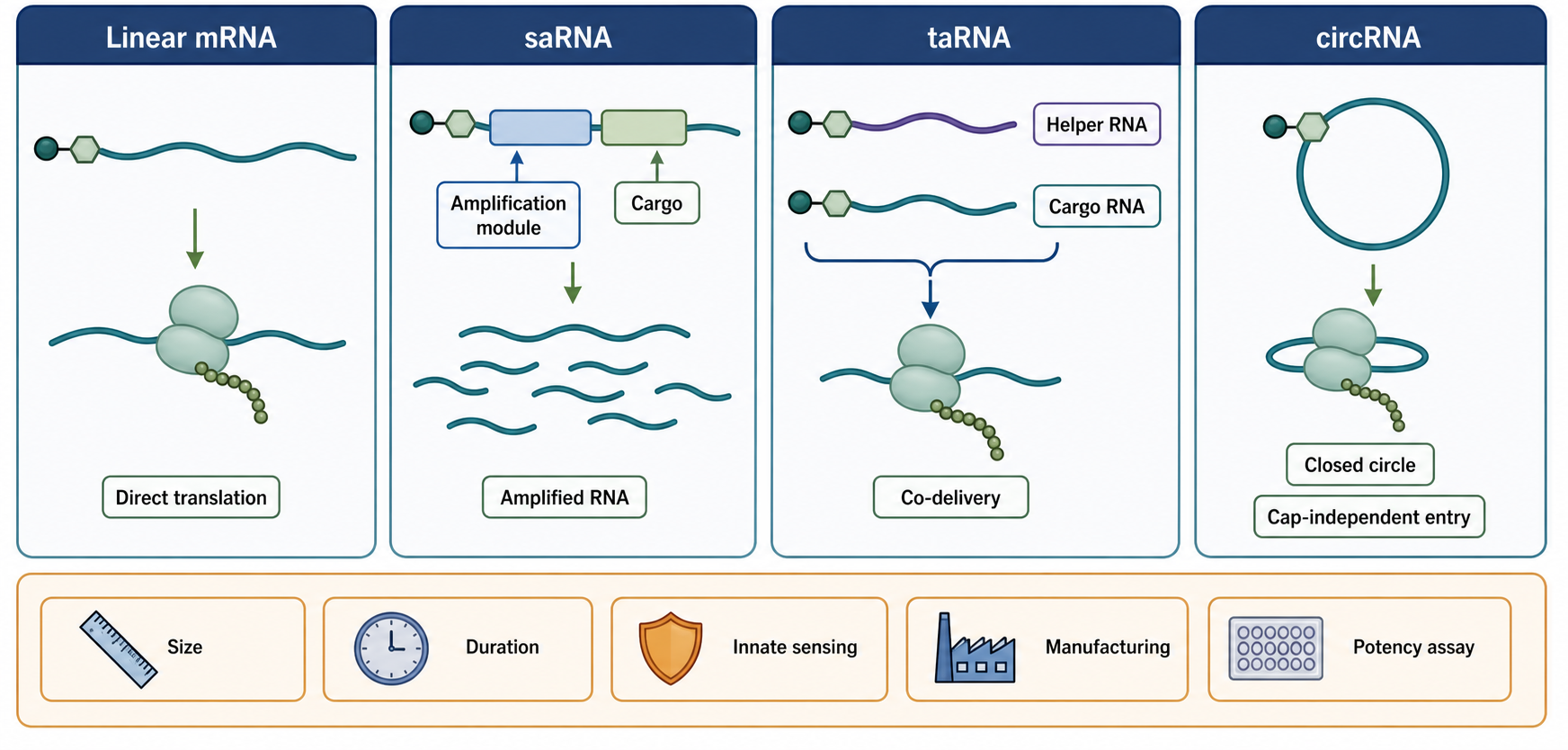
Figure 153.6. Non-Replicating mRNA, saRNA, taRNA, and circRNA Platform Comparison. “Next-generation RNA platforms change the relationship between administered RNA and protein output. Each architecture introduces distinct delivery, sensing, manufacturing, and regulatory questions.”
Circular RNA also changes innate immune questions. Natural and engineered circRNAs can be interpreted differently depending on their sequence, modifications, structure, protein partners, and purity. Some circularization methods produce byproducts that may be immunostimulatory. Some internal ribosome entry elements are viral or highly structured and can affect sensing or cell-type specificity. A long-lived RNA may improve protein duration but can be undesirable if the encoded protein is toxic, immunogenic, or needs tight temporal control.
Box 153.3. Lower Input Dose Is Not Automatically Lower Biological Exposure
Amplifying and circular platforms can reduce the mass of RNA administered or lengthen protein expression, but dose is not only the number of micrograms injected. Biological exposure includes the amount of input RNA delivered to cytosol, replicated or persistent RNA species, dsRNA intermediates, replicase protein, cargo protein, duration of expression, tissue distribution, and innate immune activation. A lower administered saRNA dose can still create strong local sensor activation if replication generates abundant structured RNA. A long-lived circRNA can be useful for a missing enzyme but problematic for a toxic cytokine or editor. Product claims should therefore report exposure in matched terms: input dose, RNA-species kinetics, protein output, duration, cell type, immune readouts, and reversibility. Lower dose is a design hypothesis, not a safety conclusion.
Next-generation mRNA platforms also include tissue-selective UTRs, miRNA target sites for detargeting, ligand-targeted particles, polymer or peptide formulations, thermostable formulations, personalized cancer vaccine workflows, multiplex antigen encoding, and combinations with genome editing or cell therapy. The unifying design question is whether the new feature solves a defined limitation. A longer expression window is useful for a secreted protein but may be risky for a cytokine. Lower dose is useful only if manufacturing, potency, and safety remain controllable. Tissue targeting is useful only if the payload is active in the target tissue and inactive or tolerable elsewhere.
The evidence basis for next-generation platforms should include side-by-side comparison with conventional mRNA under matched dose, route, cargo, and assay conditions. Claims of greater potency must specify whether potency means lower RNA dose, higher peak protein, longer duration, stronger immune response, better protection, lower reactogenicity, better thermostability, or easier manufacturing. For amplifying RNA, assays should distinguish input RNA, replicated RNA, subgenomic RNA, dsRNA intermediates, replicase protein, cargo protein, and innate immune activation. For circular RNA, assays should distinguish circular product, linear precursor, nicked circles, concatemeric products, and cap-independent translation from cryptic linear contaminants.
153.7. Clinical Evidence, Manufacturing, Safety, and Regulatory Issues
Clinical evidence for mRNA medicines is strongest for prophylactic vaccines where large trials and real-world use have shown that mRNA-LNP products can induce protective immunity at population scale. That evidence establishes the feasibility of the modality, not automatic success for every mRNA product. A vaccine against a rapidly spreading virus, a personalized cancer vaccine, an mRNA encoding a secreted enzyme, and an mRNA encoding a genome editor have different endpoints, schedules, safety tolerances, and risk-benefit thresholds.
Manufacturing starts upstream with DNA template design and in vitro transcription. The process must control template identity, promoter sequence, nucleoside triphosphate quality, polymerase reaction conditions, cap incorporation or enzymatic capping, poly(A) tail strategy, residual DNA, residual proteins, abortive transcripts, truncated RNA, dsRNA impurities, and RNA integrity. Purification and formulation are not separate from biology. A product with the correct nominal sequence but high dsRNA contamination can behave differently from a product with the same sequence and lower impurity burden. A formulation with the same lipid names but different ratios, particle size, mixing conditions, or storage history can show different potency or reactogenicity.
Analytical release testing must connect chemistry to function. Identity assays confirm the RNA sequence or expected length. Purity assays measure full-length RNA, truncated species, dsRNA, residual DNA, solvents, proteins, endotoxin, and other process-related impurities. Cap and tail assays characterize features required for translation and stability. Particle assays measure size, polydispersity, encapsulation, lipid composition, pH, osmolality, and stability. Potency assays test whether the product expresses the intended protein or produces a relevant biological activity. No single assay is sufficient. A potency assay that uses easy-to-transfect cells may miss delivery limits. A structural antigen assay may miss T-cell epitopes. A cytokine assay may miss rare immune risks.
Comparability becomes difficult when a platform changes after early studies. A new cap analog, altered UTR, improved purification step, different ionizable lipid, changed mixing device, larger manufacturing scale, or revised storage condition can improve a product while also changing the evidence package. Developers therefore need bridging assays that show whether the post-change product remains meaningfully comparable to the pre-change product. For an antigen vaccine, comparability may require RNA integrity, particle properties, antigen expression, antigen conformation, immunogenicity, and safety bridging. For a protein-replacement therapy, comparability may require functional protein activity, pharmacodynamic biomarkers, exposure duration, and repeated-dose tolerability. For saRNA, taRNA, or circRNA, comparability is even more platform-specific because amplification, circularization, or cap-independent translation can magnify small changes in RNA quality.
Safety evaluation spans RNA, encoded protein, delivery vehicle, route, schedule, and patient population. RNA-related risks include excessive innate immune activation, translation suppression, persistence outside the desired window, off-target expression, and impurities. Encoded-protein risks include antigen-driven autoimmunity, allergenicity, excessive cytokine activity, genome-editing off-target effects, abnormal localization, and immune responses against a therapeutic protein. Delivery risks include lipid toxicity, complement activation, allergy-like reactions, organ accumulation, local inflammation, and interactions with pre-existing antibodies. Schedule risks include repeated dosing, boosting, immune memory, anti-drug antibodies, and changes in exposure after inflammation.
Regulatory assessment asks whether the product is sufficiently defined, consistently manufactured, potent, stable, and clinically justified. For vaccines, regulators assess immunogenicity, efficacy, safety, lot consistency, pharmacovigilance, and variant or strain updates when relevant. For protein therapeutics, regulators assess pharmacokinetics, pharmacodynamics, toxicology, immunogenicity, dose selection, and clinical endpoints. For personalized cancer vaccines, regulators also consider rapid manufacturing, individualized sequence selection, release testing under time pressure, and comparability across patient-specific lots. For saRNA, taRNA, and circRNA products, additional questions concern replication competence, byproduct control, durability, reversibility, and platform-specific potency assays.
Clinical interpretation must avoid two opposite errors. The first error is platform exceptionalism: assuming that success of one vaccine proves safety and efficacy for unrelated mRNA therapeutics. The second error is platform dismissal: treating a failure in one indication as evidence that mRNA expression is generally unsuited to therapy. A failed program can reflect poor delivery, inadequate protein design, wrong tissue, excessive innate activation, weak clinical endpoint, manufacturing inconsistency, strategic competition, or true target failure. The evidence standard is product-specific and mechanism-specific.
Recent Consensus
Recent consensus can be stated cautiously. Non-replicating modified mRNA-LNP vaccines are a validated clinical modality for some infectious disease applications. mRNA can be manufactured rapidly relative to many protein or viral-vector platforms once sequence and process are established, but rapid sequence exchange does not eliminate process development, analytical release testing, or product-specific clinical evidence. The expression cassette is now understood as an integrated system: cap state, UTRs, ORF design, codon pattern, poly(A) tail, modified nucleotides, and impurity profile interact rather than acting as independent knobs [Chaudhary et al. 2021; Szabó et al. 2022; Jin et al. 2025].
The field also broadly agrees that delivery is the dominant constraint for many future mRNA therapeutics. LNP delivery is powerful but still limited by tissue targeting, endosomal escape, reactogenicity, and repeated-dosing constraints. Sequence engineering can tune translation and stability, but prediction remains imperfect because synonymous codons also affect RNA structure, dinucleotide content, ribosome kinetics, protein folding, innate sensing, and manufacturing behavior. Modified nucleotides and dsRNA depletion are complementary controls, not substitutes for one another. Amplifying and circular platforms are scientifically plausible and actively developing, but they need platform-specific evidence for manufacturing control, innate immune profile, expression duration, and clinical benefit.
Open Questions, Controversies, Deprecated Models, and Common Misconceptions
Open questions:
- How can developers predict protein output from RNA sequence, chemistry, and formulation before animal studies?
- Which endosomal escape events dominate in human tissues?
- How should dsRNA and other impurities be specified for different routes and doses?
- How much innate activation is optimal for each vaccine class?
- Can mRNA delivery be directed reliably to immune cells, lung, spleen, muscle, heart, tumor, or central nervous system without unacceptable exposure elsewhere?
- How will repeated dosing interact with formulation immunity and anti-protein immunity?
- Which circRNA translation elements are potent, safe, and predictable in primary human cells?
- Which saRNA and taRNA designs separate useful amplification from excessive innate sensing?
Common misconceptions:
- “Therapeutic mRNA must enter the nucleus or integrate into DNA before it can work.” Conventional mRNA products act after cytosolic delivery and ribosomal translation; durable downstream effects can arise from the encoded protein without DNA integration.
- “Maximizing protein output is always the correct design objective.” Vaccines, replacement proteins, cytokines, and genome editors require different amounts, durations, locations, and tolerability windows.
- “LNP uptake proves productive delivery.” Functional expression requires release of intact RNA into the cytosol; particle or lipid uptake can occur without endosomal escape.
- “Modified nucleotides make purification unnecessary.” Cap defects, dsRNA, truncated transcripts, residual template or proteins, and formulation-associated signals remain biologically important.
- “Reactogenicity is a surrogate for protective immunity.” Acute inflammatory symptoms and adaptive protection are distinct outcomes and require separate measurements.
Deprecated or weakened claims:
- Several older or overgeneralized models should be deprecated. mRNA medicines should not be described as simple sequence files that become drugs once encapsulated; the drug product includes RNA architecture, chemistry, impurities, formulation, route, dose, storage, and release specifications. Codon optimization should not be treated as a single monotonic algorithm for maximum expression, because vaccine antigens, secreted enzymes, cytokines, and genome editors have different desired expression windows and toxicity limits. Innate immune tuning should not be reduced to “modified is silent” and “unmodified is inflammatory”; cap quality, dsRNA impurities, RNA structure, dose, route, cell type, and formulation all change sensor exposure. Circular RNA and saRNA should not be assumed to be safer merely because they may require lower input doses or last longer, because amplification intermediates, circularization byproducts, and persistent expression can create different risks.