Chapter 37. Organellar, Lysosomal, and Autophagic RNA Turnover and Quality Control
Scope Note
This chapter explains two connected but nonidentical turnover landscapes. First, it covers how mitochondria, chloroplasts, and related endosymbiotic organelles degrade, trim, stabilize, and surveil their own RNAs. Second, it covers how lysosomes or vacuoles receive and digest cellular RNA through direct lysosomal import, ribosome-selective macroautophagy, and bulk autophagic delivery. It explicitly prevents four terms from collapsing into one: direct lysosomal RNA import called RNautophagy in a specific mammalian literature; macroautophagic ribosome turnover called ribophagy; intralysosomal nuclease digestion and nucleotide salvage; and loss of RNA because cytoplasm or whole organelles are engulfed. Chapter 32 owns comparative nuclease chemistry, Chapter 38 owns stress-dependent RNA-stability programs, and Chapter 89 owns the biology of tRNA- and rRNA-derived regulatory fragments. When organellar substrates require unusual end processing or ligation, this chapter explains the biological setting and hands comparative enzyme chemistry to Chapter 33. Chapter 17 covers organellar RNA genes and primary processing; Chapter 43 covers organellar ribosome assembly; and Chapter 51 covers organellar RNA editing.
Executive Summary
Organellar RNA turnover is the controlled destruction or remodeling of RNAs inside mitochondria and plastids. A mitochondrion or chloroplast does not simply inherit a bacterial RNA decay program unchanged. Endosymbiotic ancestry supplied bacterial-like genomes, ribosomes, and some RNA enzymes, but modern organelles depend heavily on nuclear-encoded proteins that are imported after translation in the cytosol. The result is a hybrid regulatory system: transcripts are made inside the organelle, many processing and decay factors are encoded in the nucleus, and RNA quality control must remain coordinated with organellar translation, energy metabolism, photosynthesis, stress signaling, and development.
The first organizing principle is that organellar transcripts often require processing before they become functional RNAs. Mammalian mitochondrial DNA is transcribed as long polycistronic precursor RNAs, and many mature mRNA and rRNA boundaries are generated by removal of flanking tRNAs. Plant mitochondrial and chloroplast genomes produce complex transcript populations with many overlapping transcription units, RNA editing sites, introns, intercistronic regions, and stable termini defined by RNA-binding proteins. Kinetoplastid mitochondria in trypanosomatids go even further: many transcripts require guide RNA-directed uridine insertion and deletion before they encode functional proteins. In all of these systems, turnover is inseparable from maturation because a molecule may be a precursor, an intermediate, a surveillance substrate, or a productive RNA depending on which processing steps have occurred.
The second organizing principle is that organellar polyadenylation does not mean the same thing in every compartment. In the cytoplasm of most eukaryotes, a long poly(A) tail commonly supports mRNA stability and translation. In bacteria, mitochondria, and chloroplasts, short poly(A)-rich or mixed tails often help exonucleases degrade structured RNA fragments by providing an accessible single-stranded entry point. This contrast is a common source of overgeneralization. Organellar tailing can stabilize some transcripts in some lineages or in protein-bound contexts, but many organellar tailing events function as surveillance marks or decay adaptors rather than as a cytoplasmic-style stability signal. Chapter 29 treats nuclear and cytoplasmic polyadenylation; this chapter focuses on organellar tailing as a quality-control and decay-coupled process.
The third organizing principle is that organellar quality control is constrained by RNA-end chemistry. A 5′ monophosphate, 5′ hydroxyl, 2′,3′ cyclic phosphate, 3′ phosphate, or 3′ hydroxyl can determine whether an organellar intermediate is trimmed, joined, stabilized, or eliminated. The general enzymology of end healing, RtcB- and ATP-dependent ligases, and repair-versus-turnover decisions belongs to Chapter 33. Here the central question is narrower: which of those reactions is localized to an organelle, on which endogenous substrate, and with what consequence for mitochondrial or plastid gene expression? Blocked termini can also evade ligation-dependent sequencing, so negative data require chemistry-aware controls (Scacchetti et al. 2024).
The fourth organizing principle is nuclear-organellar coordination. Most organellar RNA-binding proteins, ribonucleases, helicases, polymerases, aminoacyl-tRNA synthetases, modification enzymes, editing factors, and ribosome assembly factors are nuclear encoded. A defect in a nuclear gene can therefore appear as a mitochondrial or chloroplast RNA phenotype. Conversely, organellar dysfunction can alter nuclear transcriptional programs through retrograde signaling. In plants, pentatricopeptide repeat proteins and related RNA-binding factors define many organellar RNA termini, editing events, splicing steps, and stability states; recent review synthesis treats plant organellar RNA maturation as a strongly post-transcriptional and protein-guided system (Small et al. 2023). In metazoans, human mitochondrial decay reviews and disease genetics illustrate how broadly expressed RNA-remodeling factors can nevertheless produce tissue-specific disease (Santonoceto et al. 2024; van Esveld et al. 2022).
The fifth organizing principle is that delivery to a lysosome and digestion inside a lysosome are separate mechanistic questions. Direct uptake of purified RNA into isolated mammalian lysosomes supported the LAMP2C/SIDT2 pathway named RNautophagy. By contrast, ribophagy packages intact or partly disassembled ribosomes into autophagosomes before lysosomal or vacuolar delivery. RNASET2-family enzymes then participate in acidic-compartment RNA digestion, and the products can replenish nucleotide pools. Bulk macroautophagy, microautophagy, mitophagy, or chlorophagy can also make RNA disappear simply because RNA-containing cytoplasm or organelles are consumed. Lysosomal localization alone cannot distinguish these routes.
The final organizing principle is diversity and evidential restraint. Mammalian lysosomes, yeast vacuoles, animal developmental systems, mitochondria, and plastids do not use one universal RNA-autophagy program. A strong turnover claim specifies the organism, compartment, RNA class, delivery route, nuclease, perturbation, assay, and flux control. “RNautophagy” is therefore a named pathway supported in particular experimental systems, not a default label for every lysosomal RNA molecule or every autophagy-dependent loss of RNA.
Concept Inventory
- Organellar RNA turnover: the sum of pathways that remove, trim, remodel, or recycle RNAs inside mitochondria and plastids. Turnover includes bulk degradation, maturation-associated trimming, surveillance of defective RNAs, removal of antisense or noncoding transcripts, and clearance of processing fragments. A turnover event is not automatically a quality-control event. Quality control means that the cell preferentially recognizes a molecule as abnormal, incomplete, damaged, misprocessed, mistranslated, or surplus.
- Polycistronic precursor RNA: a transcript that contains multiple genetic units in one RNA molecule. In mammalian mitochondria, long transcripts are processed into mRNAs, rRNAs, and tRNAs. In plant organelles and chloroplasts, precursor transcript architecture is more complex because transcription, splicing, editing, intercistronic processing, and protein-defined termini all contribute to mature RNA populations.
- Organellar polyadenylation: post-transcriptional addition of adenosine-rich tails to organellar RNAs. Unlike the long stabilizing poly(A) tails on many nuclear-encoded cytoplasmic mRNAs, organellar poly(A)-rich tails often promote degradation by helping exonucleases engage structured RNA ends. The phrase should always be interpreted with the compartment and organism specified.
- Tailing: a broader term than polyadenylation. A terminal nucleotidyltransferase can add adenines, uridines, or mixed sequences to RNA ends. Tails can help decay enzymes, mark defective molecules, complete functional ends, or alter translation and stability. The same tail chemistry can have different consequences depending on protein occupancy and RNA class.
- Organellar RNA repair context: a demonstrated end-healing or joining reaction on an endogenous mitochondrial or plastid substrate. General ligase activity, a blocked terminus, or organellar colocalization alone is insufficient; localization, substrate chemistry, catalytic dependence, and product rescue are needed. Comparative repair enzymology is developed in Chapter 33.
- Organellar RNA ligation: joining of RNA ends during an organellar processing, editing, or rescue pathway. Kinetoplastid editosome ligation and organellar intron contexts are examples, but their biological ownership remains with the relevant processing system rather than implying one universal organellar repair pathway.
- Blocked RNA termini: RNA ends with chemical groups that interfere with standard enzymatic steps in sequencing or metabolism. Examples include 3′ phosphate or 2′,3′ cyclic phosphate ends. Standard RNA-seq often under-detects such molecules unless the library method removes the block or avoids ligation dependence (Scacchetti et al. 2024).
- Nuclear-organellar coordination: that organellar RNA metabolism is governed by both organellar transcription and nuclear-encoded factors imported into organelles. This coordination includes anterograde control, in which the nucleus supplies organellar gene-expression factors, and retrograde signaling, in which organellar state changes nuclear gene expression.
- RNautophagy: a pathway name introduced for ATP-dependent direct uptake of RNA across the lysosomal membrane in mammalian experimental systems, initially involving LAMP2C and later SIDT2. The name does not establish universality, endogenous substrate selectivity, or independence from other lysosomal delivery routes.
- Ribophagy: selective or enriched autophagic turnover of ribosomes. In budding yeast the starvation model requires Ubp3–Bre5 for the selective component; in mammalian cells NUFIP1–ZNHIT3 links starved ribosomes to LC3B. Ribophagy delivers an RNA–protein assembly and must not be equated with direct transport of naked RNA.
- Lysosomal RNA degradation: hydrolysis of RNA after it reaches a lysosome or vacuole. Delivery and digestion are distinct; RNASET2-family nucleases can be required for intralysosomal rRNA clearance even when autophagy supplies the cargo.
- Bulk autophagic RNA turnover: RNA loss caused by engulfment of cytoplasm or an RNA-containing organelle. Bulk delivery can coexist with selective delivery, and enrichment of one RNA in lysosomes does not by itself prove a receptor-mediated selection step.
What to Know Before Reading This Chapter
Mitochondria and chloroplasts originated from bacterial endosymbionts, but modern organelles are not free-living bacteria. Their genomes are reduced, their RNA processing systems are specialized, and most of the proteins required for organellar RNA metabolism are encoded in the nucleus. Therefore Chapter 34 is useful background for bacterial RNA decay, but bacterial rules cannot be transferred without checking lineage-specific organellar evidence.
An RNA molecule has polarity and chemically distinct ends. The 5′ end may carry a triphosphate, monophosphate, hydroxyl, cap-like structure, or other chemical state. The 3′ end may carry a hydroxyl, phosphate, cyclic phosphate, CCA sequence, poly(A)-rich tail, uridine-rich tail, or protein-protected structure. Nucleases and ligases read these chemical states. A decay enzyme that works well on a single-stranded 3′ hydroxyl end may fail on a structured end or a blocked terminus.
Organellar RNAs are often translated on organellar ribosomes. Mitochondrial and chloroplast translation systems are covered in later chapters, but the essential point for this chapter is that defective RNA can create defective protein synthesis. A mitochondrial mRNA that is incorrectly processed or incompletely edited can produce a faulty oxidative phosphorylation subunit. A chloroplast mRNA that is unstable or mistranslated can disturb photosynthetic complexes. Quality control therefore protects both RNA pools and membrane-associated energy-conversion machinery.
Many organellar RNA-binding proteins act as landmarks. A protein bound near an RNA end can block exonucleases, define a mature terminus, recruit processing enzymes, or protect a transcript until translation begins. In plants, pentatricopeptide repeat proteins are a major example (Small et al. 2023). In mammals, the SUV3–PNPase decay machinery, GRSF1, and transcript-specific factors such as LRPPRC–SLIRP illustrate distinct remodeling, surveillance, and stabilization roles rather than one interchangeable mitochondrial RNP system (Santonoceto et al. 2024; Rubalcava-Gracia et al. 2024).
Finally, assay interpretation is difficult. Steady-state abundance of an organellar RNA reflects transcription, processing, editing, tailing, degradation, and cell or tissue composition. A decrease in a mature mRNA may mean faster decay, failed processing, impaired transcription, defective organelle biogenesis, or secondary loss of organellar translation. Strong claims use RNA-end mapping, metabolic or inducible kinetics where possible, factor perturbation, rescue, organellar localization controls, and orthogonal readouts of translation and respiratory or photosynthetic function.
Autophagy experiments add a second layer of interpretation. An autophagosome is a double-membrane carrier; a lysosome or yeast vacuole is the acidic degradative compartment with hydrolases. Blocking lysosomal acidification can increase cargo because digestion stops, whereas stimulating autophagosome formation can either increase delivery or expose a later bottleneck. Autophagic flux means the rate at which material traverses the pathway, not the number of LC3-positive puncta at one time. A credible RNA-turnover study therefore pairs cargo abundance with inhibitor-aware flux measurements, genetics, and rescue.
37.1. Mitochondrial and chloroplast RNA decay pathways
Mitochondrial and chloroplast RNA decay pathways are the enzymatic routes by which organelles remove RNAs or trim them to mature ends. The main substrates are messenger RNAs, ribosomal RNAs, transfer RNAs, precursor transcript fragments, antisense RNAs, and damaged or misprocessed molecules. The starting point is different from a typical nuclear mRNA chapter. Many organellar RNAs are not born as individually capped and polyadenylated monocistronic mRNAs; they are generated from larger transcription units and become functional only after processing.
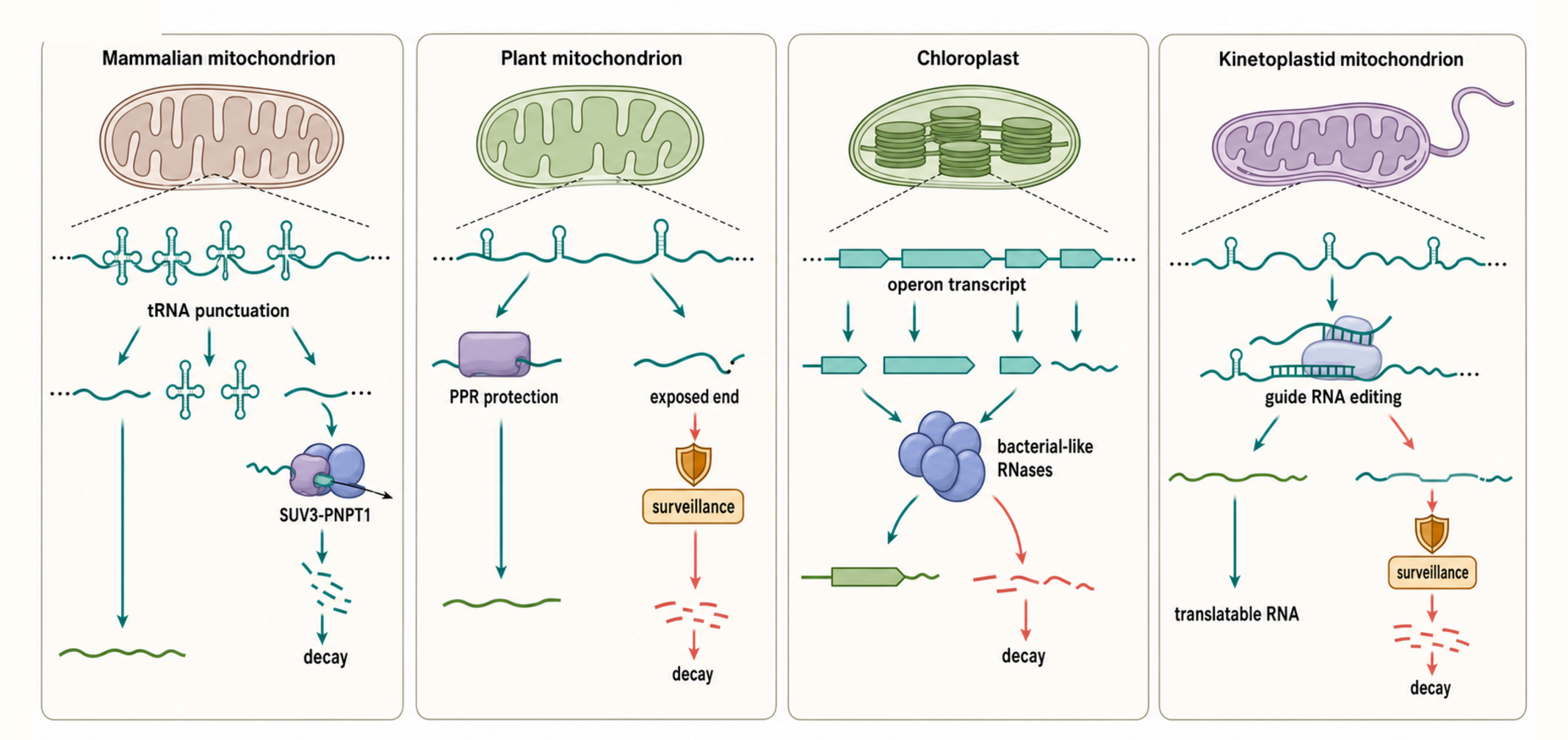
Figure 37.1. Comparative Organellar RNA Decay Map. Organelles share the need to remove defective RNAs, but each lineage defines productive and defective transcripts differently. Mammalian mitochondria process polycistronic precursors through tRNA punctuation and degrade unwanted RNAs via the SUV3–PNPT1 degradosome; a distinct consequence branch shows double-stranded RNA accumulation, mitochondrial escape, cytosolic MDA5 sensing, and type I interferon signaling as separable steps. Plant mitochondria rely on PPR protein protection and exposed-end decay tied to respiration and fertility. Chloroplasts use bacterial-like RNases and protein-defined termini within operon-like transcription units, and kinetoplastid mitochondria require guide RNA-directed uridine insertion and deletion before a transcript becomes translatable. Processing, editing, tailing, protein protection, decay, compartment escape, and downstream sensing must each be interpreted within the organism-specific pathway.
Table 37.1. Organellar RNA Decay Factors and Evidence Classes. Representative decay factors across organelles and lineages, with their proposed activities and the types of evidence supporting their roles.
| Organism | Organelle | Factor or complex | Proposed activity | Main RNA substrate class | Key evidence |
|---|---|---|---|---|---|
| Human | Mitochondria | SUV3–PNPT1 degradosome | Helicase-coupled 3′ to 5′ exonuclease | mRNAs, precursor RNAs, double-stranded RNA | Reconstitution; genetic depletion; double-stranded RNA accumulation with MDA5–interferon consequences |
| Yeast (S. cerevisiae) | Mitochondria | Suv3–Dss1 degradosome | Helicase-coupled 3′ to 5′ exonuclease | Mitochondrial mRNAs, precursor fragments | Temperature-sensitive mutants and RNA steady-state analysis |
| Plant (Arabidopsis) | Chloroplast | RNase J | 5′ to 3′ exonuclease and endonuclease | Chloroplast mRNAs, intercistronic fragments | RNA phenotypes in knockout lines |
| Plant (Arabidopsis) | Chloroplast and plastid | PNPase | 3′ to 5′ phosphorolytic exonuclease | mRNAs, poly(A)-rich tails, processing fragments | Biochemical activity and plastid RNA-end changes |
| Plants | Mitochondria and chloroplast | PPR proteins | Sequence-specific RNA-end protection and stabilization | Transcript-specific mRNAs and processing intermediates | RNA stability and editing changes in loss-of-function mutants |
| Trypanosomatids | Kinetoplast | Editosome-associated ligases and nucleases | RNA cleavage, uridine addition or deletion, ligation | Edited mRNAs and editing intermediates | Biochemical reconstitution and genetic depletion studies |
Table 37.2. Assay Interpretation for Organellar RNA Turnover. Each method illuminates one aspect of organellar RNA metabolism but requires complementary assays to distinguish decay, processing, and transcription defects.
| Assay | What it measures | What it cannot prove alone | Common artifact | Stronger follow-up |
|---|---|---|---|---|
| Steady-state organellar RNA-seq | Relative abundance of organellar RNA species | Whether change reflects decay, transcription, or processing | Cytosolic contamination; nuclear mitochondrial DNA reads | Organelle fractionation with marker controls; metabolic labeling |
| RNA gel blot or RT-qPCR | Abundance and apparent size of specific RNA | Mechanism of the abundance change | Probe or primer cross-reactivity | RNA-end mapping; pulse-chase kinetics |
| RNA-end mapping | 5′ and 3′ termini of transcripts | Whether a terminus is a mature end or a degradation intermediate | Ligation bias against blocked ends | Ligation-independent or end-healing protocols |
| Poly(A) or tail profiling | Tail composition and length on specific RNAs | Whether tail promotes decay or maturation | Oligo-dT enrichment selects preferentially for decay intermediates | Tail removal and RNA stability comparison |
| Ligation-independent small RNA sequencing | RNAs including those with blocked 3′ termini | Identity of factor generating the blocked ends | Amplification bias for short sequences | Direct comparison with ligation-dependent libraries |
| Organelle fractionation plus RNA analysis | RNA content of isolated organellar fraction | Absence of cytosolic or nuclear RNA contamination | Incomplete organelle purification | Marker protein and organellar RNA purity controls |
| Factor knockdown or knockout | RNA changes upon loss of specific factor | Whether the effect is direct or indirect organellar stress | Secondary organellar dysfunction from global stress | Rescue with wild-type and catalytic-dead alleles |
| In vitro nuclease or ligase assay | Enzymatic activity on defined substrates | Activity on authentic protein-bound organellar RNPs | Simplified substrate misses modification or protein context | Structured or protein-bound in vitro substrates |
| Organellar translation assay | Synthesis of organellar protein products | Which RNA processing step is defective | Secondary effects of organellar stress | Combine with RNA-end mapping and ribosome profiling |
| Respiratory or photosynthetic function assay | Organellar metabolic output | Which RNA or protein step is rate-limiting | Multistep distance from the primary RNA defect | Integrate with RNA abundance and translation measurements |
Table 37.3. Poly(A) and Tailing Outcomes in Different RNA Systems. Tail type, compartment, and transcript context together determine whether a poly(A)-rich or mixed tail promotes stability, maturation, or decay.
| Compartment or lineage | Tail type | Common consequence | Mechanistic explanation | Boundary case |
|---|---|---|---|---|
| Cytoplasmic mRNA | Long poly(A) | mRNA stabilization and translation support | Poly(A)-binding proteins protect from nucleases and stimulate translation initiation | Deadenylation triggers cytoplasmic mRNA decay |
| Bacteria | Short poly(A)-rich tail | Decay stimulation | Tail provides single-stranded 3′ entry for exonucleases on structured RNAs | Some transcripts can be transiently stabilized by tail-binding proteins |
| Mammalian mitochondria | Poly(A) tail | Stop-codon completion and transcript-specific maturation | Adenosines added post-transcriptionally complete incomplete UAA or UA stop codons | Aberrant or excessive tailing can intersect with decay and surveillance |
| Plant chloroplasts and mitochondria | Poly(A)-rich or mixed tail | Decay-associated surveillance marking | Exposed ends after protein protection fails are tailed and degraded by exonucleases | Some tailed molecules may be stable protected intermediates |
| Kinetoplastids | Uridine addition during editing | Sequence maturation of protein-coding mRNAs | Guide RNA-directed uridine insertion creates the correct open reading frame | Distinct from conventional tailing; editing intermediates must not be treated as defective RNAs |
Table 37.4. Disease and Phenotype Evidence Ladder for Organellar RNA Defects. Connecting a clinical or agronomic phenotype to an organellar RNA mechanism requires progressing from observation through increasingly direct mechanistic tests.
| Observation | Weak interpretation | Stronger mechanistic test | Example class |
|---|---|---|---|
| Patient variant in nuclear RNA helicase or nuclease | Variant causes mitochondrial disease | Confirm factor localizes to mitochondria and directly affects a specific RNA class | SUPV3L1 or PNPT1 variant |
| Lower mitochondrial mRNA abundance | Increased RNA decay | Distinguish failed processing from faster decay using RNA-end mapping and pulse-chase kinetics | Respiratory-chain mRNA reduction |
| Accumulation of precursor or tailed RNA | Surveillance failure | Map end chemistry and show that depletion of the specific factor reproduces accumulation | Mitochondrial tRNA or mRNA precursor buildup |
| Loss of mitochondrial translation product | mRNA-level defect | Distinguish RNA loss from ribosome assembly or aminoacylation defect | Oxidative phosphorylation subunit reduction |
| Respiratory-chain defect | General mitochondrial dysfunction | Identify which complex and which subunit are specifically affected | Complex I or Complex IV subunit loss |
| Tissue-specific clinical phenotype | Random distribution of disease | Connect high energy demand, factor expression level, and organellar load in affected tissue | Encephalomyopathy or cardiomyopathy |
| Rescue by wild-type but not catalytic mutant | Factor enzymatic activity is required | Confirm absence of dominant-negative effect from mutant allele | RNA helicase or nuclease active-site mutant |
| One patient with biallelic SIDT2 variants plus lysosomal phenotypes | Failed direct RNA import is the complete disease mechanism | Test variant RNA binding or transport, separate macroautophagy and other endolysosomal functions, and seek independent cases | SIDT2-associated cerebellar ataxia report |
Mammalian mitochondria provide a compact teaching example. The mitochondrial genome is transcribed into long RNAs from which most mature RNA boundaries are generated by excision of tRNAs. This “tRNA punctuation” model is not a decay pathway by itself, but it creates many processing intermediates and makes turnover dependent on precise cleavage and trimming. If tRNA excision, end maturation, or mRNA stabilization fails, abnormal precursor or partially processed RNAs accumulate. Current synthesis places RNase P, ELAC2, mitochondrial RNA granules, transcript-stabilizing RNPs, and RNA-degradation foci in a coupled maturation-surveillance network rather than a linear conveyor belt (Santonoceto et al. 2024).
The mammalian mitochondrial RNA degradosome is commonly described as a complex containing the helicase SUV3 and the 3′ to 5′ exonuclease polynucleotide phosphorylase, often abbreviated PNPase or PNPT1. A helicase uses nucleotide hydrolysis to unwind or remodel nucleic acids; an exonuclease removes nucleotides progressively from an end. Reconstitution showed cooperative degradation of double-stranded RNA by a SUV3–PNPase assembly, and cell studies placed decay in discrete mitochondrial foci (Wang et al. 2009; Borowski et al. 2013). This supports a remodeling-plus-digestion model, but abundance changes after factor loss can also reflect altered precursor processing or mitochondrial stress.
Failure of this surveillance system can expose mitochondrial RNA to a different compartment. Depletion of SUV3 or PNPase in mammalian models caused mitochondrial double-stranded RNA to accumulate, and cytosolic exposure of this RNA engaged the melanoma differentiation-associated protein 5 (MDA5) pathway and a type I interferon response. Fibroblasts from individuals with hypomorphic PNPT1 variants also showed mitochondrial double-stranded RNA accumulation with immune activation (Dhir et al. 2018). These experiments connect matrix RNA surveillance to innate sensing, but they do not make MDA5 a mitochondrial decay factor or identify a lysosomal turnover route. Accumulation inside mitochondria, release across mitochondrial membranes, cytosolic sensing, and downstream interferon signaling remain distinct causal steps.
Yeast mitochondria illustrate that decay machines vary even among eukaryotes. Budding-yeast mitochondrial turnover uses the DExH-box helicase Suv3 with the exoribonuclease Dss1. Genetic identification and biochemical reconstitution established that the two activities are tightly interdependent (Dziembowski et al. 1998, 2003; Malecki et al. 2007). Human PNPase and yeast Dss1 are distinct proteins, and their substrates differ; the reusable principle is that structured organellar RNA often requires remodeling before processive digestion.
Plant mitochondria and chloroplasts complicate the picture because transcript ends are frequently stabilized by bound proteins. A plant organellar transcript can be produced from a long precursor, edited at specific cytidines, spliced if it contains introns, cleaved at intercistronic sites, and protected at its 5′ or 3′ end by RNA-binding proteins. Pentatricopeptide repeat proteins and related factors can define RNA termini and prevent nucleases from chewing further into the molecule. In this context, a nuclease is not merely destructive; it can trim RNA up to a protein roadblock and thereby create a mature stable end. Small et al. review plant organellar RNA maturation as a network of processing, editing, splicing, and stabilization steps dominated by nucleus-encoded specificity factors (Small et al. 2023).
Chloroplast RNA decay retains clearer bacterial parallels than mammalian mitochondrial decay. Arabidopsis RNase E is chloroplast-localized and cleaves RNA with bacterial-like biochemical preferences, but its physiological role cannot be inferred from homology alone. RNase E knockout plants overaccumulate polycistronic precursors, lose several mature monocistronic mRNAs, and become deficient in chloroplast ribosomes, placing the enzyme at an intercistronic-processing step with downstream translation consequences (Schein et al. 2008; Walter et al. 2010). Chloroplast RNase J has both endo- and exonucleolytic activities and helps remove antisense RNA. Chloroplast PNPase is still more multifunctional: genetic and biochemical studies connect it to 3′-end maturation, intron-fragment and tRNA degradation, and polyadenylation, while partial loss and residual activities reveal compensation and substrate-specific effects (Walter et al. 2002; Germain et al. 2011; Sharwood et al. 2011; Halpert et al. 2019). Plant-specific protein barriers can nevertheless define stable ends. A factor may therefore be required for maturation and degradation in the same compartment, and neither sequence homology nor one steady-state RNA phenotype assigns its complete substrate range.
The evidence basis for organellar decay pathways includes genetics, RNA gel blots, quantitative reverse transcription PCR, RNA-seq, circularized RNA end sequencing, poly(A)-tail profiling, organelle fractionation, immunoprecipitation of RNA-binding proteins, in vitro nuclease assays, and measurements of organellar translation or complex assembly. Each method has limits. RNA-seq can confuse processing fragments with decay intermediates. Organelle purification can carry cytosolic contamination. Steady-state RNA abundance cannot distinguish failed synthesis from increased decay. In vitro nuclease assays may use simplified substrates that do not reproduce protein-bound organellar RNPs. A convincing pathway model connects a factor to organellar localization, substrate binding or processing, RNA-end changes, kinetic or genetic evidence, and physiological consequences.
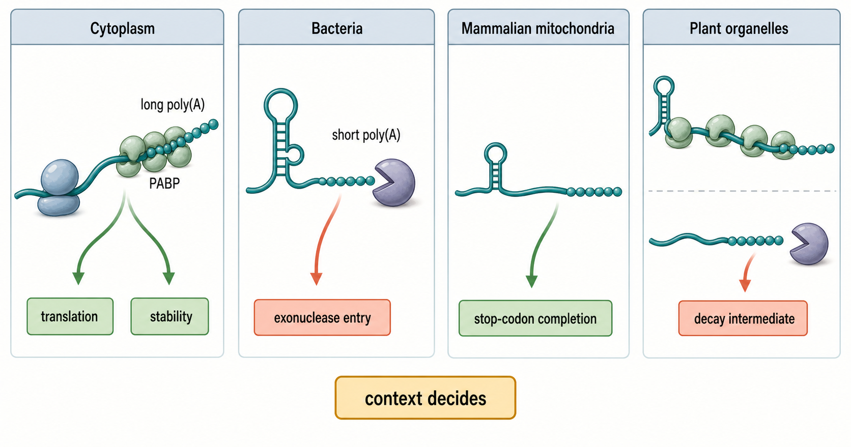
Figure 37.2. Poly(A) Tail Meanings by Compartment. Polyadenylation is a chemical event, not a universal functional signal. In the cytoplasm, long poly(A) tails recruit poly(A)-binding proteins to support translation and stability, whereas in bacteria and many organelles, short poly(A)-rich tails instead provide a single-stranded entry point for 3′ exonucleases, promoting decay of structured RNAs. In mammalian mitochondria, poly(A) addition can complete incomplete stop codons for specific protein-coding transcripts, and in plant organelles, poly(A)-rich or mixed tails often mark decay intermediates. Tail length, protein binding, compartment, organism, and transcript class together determine whether a tail stabilizes, matures, or destabilizes an RNA.
Do not overgeneralize from one organelle. Mammalian mitochondria have compact genomes and limited RNA classes. Plant mitochondria have large, recombinogenic genomes and extensive RNA editing. Chloroplasts have photosynthetic gene-expression demands and bacterial-like operon logic. Kinetoplastid mitochondria have guide RNA-directed editing systems that make “mature RNA” dependent on sequence remodeling. These systems all perform RNA turnover, but their substrates and specificity factors differ.
37.2. Organellar polyadenylation, tailing, and surveillance
Organellar polyadenylation is addition of adenosine-rich sequence to an RNA end inside an organelle. The term is deceptively familiar because cytoplasmic mRNAs often use long poly(A) tails to recruit poly(A)-binding proteins, support translation, and resist decay. In many bacteria, mitochondria, and chloroplasts, short poly(A)-rich tails instead promote degradation. The tail creates a single-stranded landing pad for 3′ to 5′ exonucleases, especially when the adjacent RNA is structured. This decay-assisting role is one of the cleanest examples of why compartment-specific definitions matter.
The mechanistic model can be described in steps. First, an organellar transcript or processing fragment exposes a 3′ end. Second, a terminal nucleotidyltransferase or poly(A) polymerase adds a short tail, often adenosine-rich and sometimes mixed with other nucleotides. Third, a 3′ exonuclease engages the tail more easily than it would engage a folded RNA end. Fourth, helicases and RNA-binding proteins remodel secondary structures or RNP barriers. Fifth, the transcript is degraded, or trimming stops when a protective protein, stable structure, or functional end is reached. This model resembles bacterial polyadenylation-assisted decay and contrasts with cytoplasmic tail-stabilized mRNA regulation discussed in Chapter 29.
Mammalian mitochondrial mRNAs introduce a boundary case because their poly(A) tails can be required to complete stop codons. Several protein-coding sequences end in an incomplete U or UA, and post-transcriptional adenosines complete UAA. Mitochondrial poly(A) polymerase perturbation also affects mRNA integrity and tRNA maturation, showing that tailing is embedded in processing rather than being a single decay mark (Chang and Tong 2012; Bratic et al. 2016). Tail function depends on transcript identity, tail length, protein binding, translation state, and organism.
Box 37.1. Poly(A) Is Not One Signal
- Cytoplasmic mRNA poly(A) tails often support translation and stability through poly(A)-binding proteins.
- Bacterial and organellar short poly(A)-rich tails can promote decay by providing exonuclease entry on structured RNA ends.
- Mammalian mitochondrial poly(A) tails can complete stop codons for specific transcripts rather than marking them for decay.
- The correct question is not whether an RNA is polyadenylated, but what the tail does in that compartment and transcript context.
Plant organelles use tailing as part of a broader surveillance landscape. Chloroplast and plant mitochondrial RNAs can acquire poly(A)-rich tails associated with degradation intermediates. Chloroplast PNPase is mechanistically unusual because it can catalyze phosphorolysis and polymerization; genetic and biochemical studies connect one factor to 3′-end maturation, decay, and tailing (Yehudai-Resheff et al. 2001; Walter et al. 2002; Zimmer et al. 2009). Because many plant organellar ends are protein-defined, tailing can expose where protection failed or processing generated an accessible terminus. A tail-enriched library is therefore not a direct inventory of productive organellar mRNAs.
Surveillance is the selective removal or containment of RNAs that are unlikely to produce useful products. In organelles, surveillance substrates can include unprocessed precursors, improperly edited RNAs, unspliced intron-containing RNAs, antisense RNAs, noncoding transcripts, truncated RNAs, RNAs with damaged bases, RNAs with blocked termini, and transcripts not assembled into translation-competent RNPs. Surveillance may be triggered by exposed ends, missing protective proteins, abnormal tailing, failure to bind translational activators, or persistence of an intermediate longer than expected.
Tailing can also create interpretation traps in high-throughput data. Oligo(dT)-based RNA-seq enriches RNAs with adenosine-rich tails, but in organelles those RNAs may be decay intermediates rather than stable mRNAs. A dataset enriched for polyadenylated mitochondrial or chloroplast RNA does not automatically represent the productive transcriptome. Conversely, protocols optimized for cytoplasmic mRNAs may miss nonpolyadenylated organellar RNAs, tRNAs, rRNAs, guide RNAs, and processed fragments with blocked ends. Tail-aware and ligation-independent methods are often needed to see the full substrate space.
The evidence for tailing functions should be graded carefully. Detection of a tail on an RNA end is an observation. Enrichment of tailed molecules after depletion of a nuclease suggests a decay relationship. Loss of mature RNA after perturbing a tailing enzyme suggests a maturation or stability role. Direct proof that a tail stimulates decay requires showing that tail addition changes degradation kinetics or nuclease access, ideally with substrate-specific assays and rescue. Many organellar studies combine genetics with RNA-end profiling rather than direct in organello decay kinetics, so claims should preserve the difference between strong mechanism and plausible pathway inference.
37.3. Organelle-specific end processing, ligation, repair, and rescue contexts
Organelles provide biological settings in which end processing and ligation can determine whether an RNA matures, is rescued, or is destroyed. The presence of structured tRNAs, rRNAs, introns, editing intermediates, and polycistronic cleavage products creates many candidate substrates, but a candidate is not a demonstrated repair substrate. An organelle-specific claim must identify the organelle, endogenous RNA, initial and product end states, responsible enzyme, and effect on gene expression.
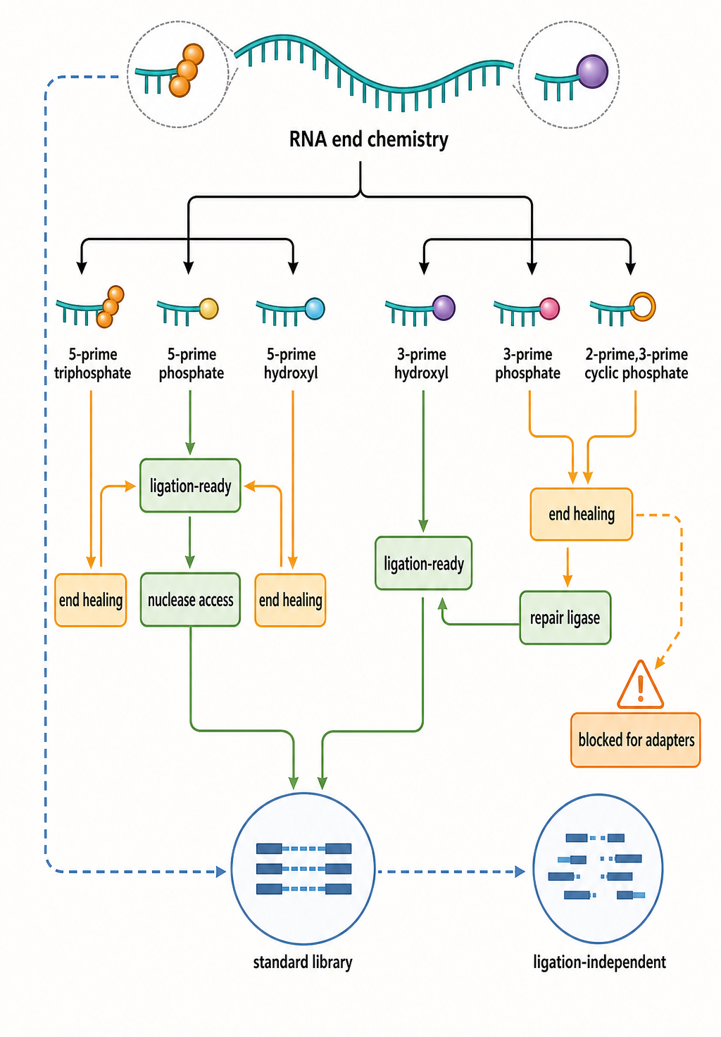
Figure 37.3. Organellar End-State and Fate Decision Tree. The figure begins with a mitochondrial or plastid substrate and asks whether its observed end is a programmed processing product, editing intermediate, damaged molecule, or decay fragment. End chemistry constrains which local reaction is possible, but localization, catalytic dependence, product recovery, and restoration of organellar translation are required before calling the route repair. A handoff points to Chapter 33 for comparative end-healing and ligase chemistry. A parallel assay lane shows how 3′ phosphate and 2′,3′ cyclic phosphate termini create blind spots in conventional ligation-dependent libraries.
The chemistry provides the eligibility rules. A 3′ phosphate can block many polymerases and ligases, a 5′ hydroxyl may require phosphorylation before joining, and a cyclic phosphate can require a different reaction route. Chapter 33 compares the relevant kinases, phosphatases, cyclic-phosphodiesterases, RtcB-family enzymes, and ATP-dependent ligases. This chapter asks which of those activities operates inside a given organelle rather than repeating their catalytic cycles.
RtcB is consequently a boundary example, not proof of a universal organellar pathway. Its unusual joining chemistry is well established in several cellular systems (Moncan 2023), yet that fact does not establish mitochondrial or plastid localization, substrate ownership, or physiological necessity in every lineage. Organelle-specific RtcB-like claims require import or localization evidence, loss of a defined ligated product after catalytic perturbation, accumulation of the predicted ends, and rescue by an active enzyme.
Organellar tRNAs make the context concrete. Mammalian mitochondria encode structurally unusual tRNAs, plant organelles use lineage-specific mixtures of encoded and imported tRNAs, and kinetoplastids import most or all mitochondrial tRNAs in many species. Cleavage, end loss, or failed CCA completion can impair translation, but observed fragments may represent regulated cleavage, failed processing, surveillance, or extraction damage. Chemistry-aware sequencing can reveal blocked fragments missed by standard adapter ligation (Scacchetti et al. 2024), while acid-preserving charging assays and targeted end mapping test whether a full-length functional tRNA is restored.
Ligation also occurs in organellar processing without being “repair” in the narrow sense. Plant organellar group II introns are removed through RNA-catalyzed chemistry assisted by proteins; kinetoplastid editosomes repeatedly cleave, add or remove uridines, and ligate mitochondrial mRNAs. These reactions are normal maturation or sequence editing, not evidence that damaged organellar RNAs are generally repaired. Chapter 51 owns guide-directed editing, and Chapter 9 compares catalytic RNAs and group introns.
RNA-mediated DNA repair is outside this ownership boundary. RNA can template or regulate DNA double-strand-break repair, but those observations do not establish restoration of a damaged RNA molecule. Conversely, RNA editing changes sequence information and may be required for organellar gene expression, yet editing is not synonymous with restoring a broken end. Explicit terminology prevents three distinct literatures from being merged.
The strongest evidence ladder begins with substrate end chemistry and compartment purity, then establishes direct enzyme-substrate activity, catalytic dependence, accumulation of predicted intermediates, recovery of the joined or healed product, restoration of organellar translation, and rescue by a properly localized active enzyme. Precursor accumulation alone can reflect failed cleavage, ligation, decay, transcription, import, or organelle stress. This is why the organellar chapter retains the physiological context while Chapter 33 owns comparative enzyme mechanism.
37.4. Organelle-nuclear coordination of RNA quality control
Organelle-nuclear coordination is the coupling between organellar gene expression and nuclear-encoded regulatory systems. The term is necessary because most organellar RNA quality-control factors are not encoded by organellar genomes. They are transcribed in the nucleus, translated in the cytosol, imported into mitochondria or chloroplasts, and assembled into organellar RNPs or enzyme complexes. As a result, organellar RNA phenotypes often originate from nuclear genes.
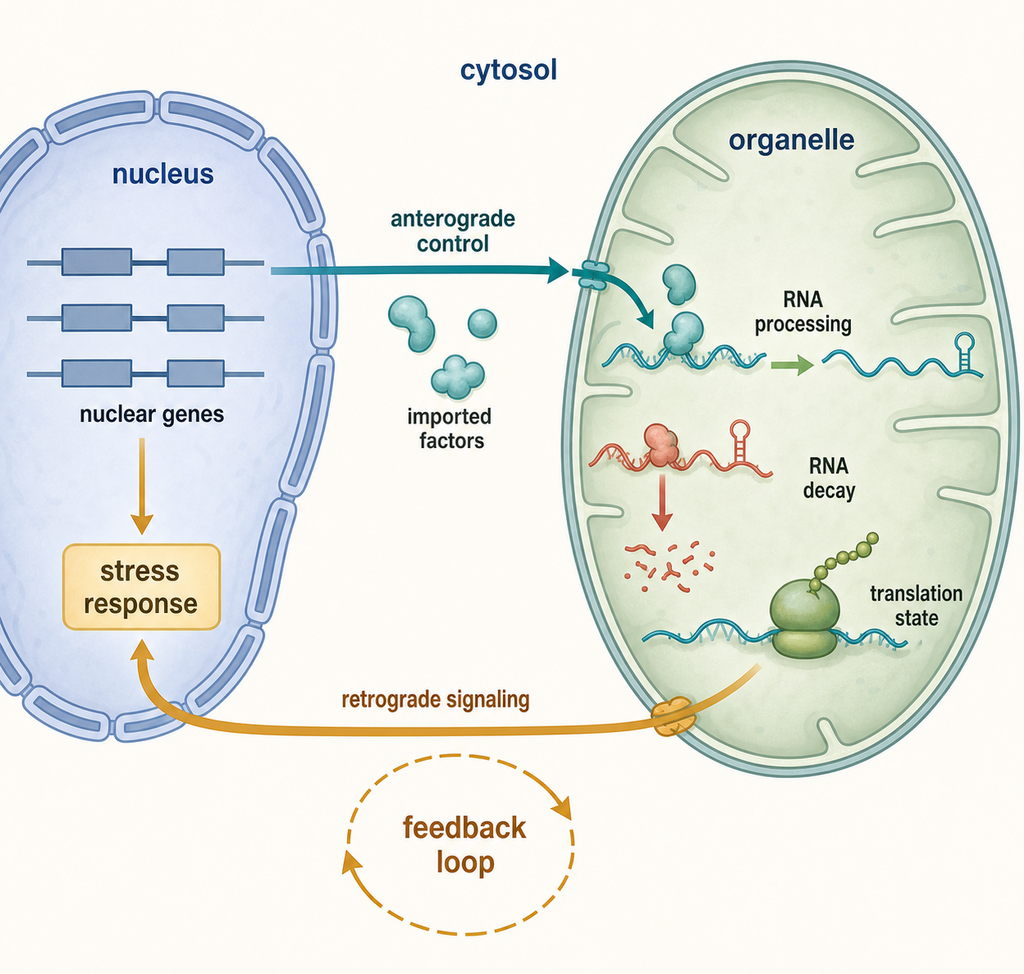
Figure 37.4. Organelle-Nuclear Coordination Loop. Most organellar RNA quality-control factors are nuclear encoded, translated in the cytosol, and imported into mitochondria or chloroplasts, so defects in nuclear genes can manifest directly as organellar RNA processing, stability, or translation phenotypes. Organellar dysfunction feeds back through retrograde signaling to alter nuclear transcriptional programs, creating a bidirectional coordination loop between organellar RNA state and nuclear gene expression. Stress conditions can further modify the activity or condensation state of organellar RNA maturation factors, coupling RNA quality control to environmental and developmental responses.
Anterograde control is nucleus-to-organelle regulation. It includes nuclear transcription of organellar RNA polymerases, ribonucleases, helicases, RNA-binding proteins, tRNA synthetases, editing factors, splicing factors, ribosomal proteins, and assembly factors. If the nucleus reduces production of a mitochondrial RNA helicase, mitochondrial RNA decay can fail. If a plant nuclear mutation disrupts a pentatricopeptide repeat protein, a specific chloroplast or mitochondrial transcript may lose a stabilized end, editing event, or splicing step. Small et al. provide a current synthesis of how plant organellar RNA maturation depends on large families of nuclear-encoded RNA-binding proteins (Small et al. 2023).
Retrograde signaling is organelle-to-nucleus regulation. When mitochondria or chloroplasts experience translation stress, respiratory-chain failure, photosynthetic imbalance, redox stress, or protein import stress, nuclear transcriptional programs change. RNA quality control can be both a cause and a consequence of these signals. For example, loss of a mitochondrial RNA decay factor can impair translation of oxidative phosphorylation subunits, leading to respiratory stress and nuclear stress responses. Conversely, developmental or stress signals from the nucleus can change the abundance of organellar RNA-binding proteins and thereby alter organellar transcript stability.
Coordination also operates at the level of stoichiometry. Many organellar protein complexes contain subunits encoded by both organellar and nuclear genomes. Oxidative phosphorylation complexes in mitochondria and photosynthetic complexes in chloroplasts require balanced production of membrane-embedded organellar subunits and imported nuclear-encoded subunits. RNA surveillance helps prevent translation of defective organellar mRNAs, but excessive decay can also reduce needed subunits. The cell must tune RNA stability, translation, protein assembly, and proteolysis together.
RNA-binding specificity factors are central to this tuning. In plant organelles, pentatricopeptide repeat proteins recognize short RNA sequences and can define transcript ends, recruit or block enzymes, and specify editing sites. Related proteins and maturases support splicing and stabilization. In mammalian mitochondria, RNA granules organize processing, modification, and assembly reactions. The SUV3–PNPase–GRSF1 surveillance axis limits antisense and double-stranded RNA, whereas LRPPRC–SLIRP stabilizes and presents mRNAs to the translation system (Dhir et al. 2018; Santonoceto et al. 2024; Rubalcava-Gracia et al. 2024). These are experimentally separable functions.
The quality-control logic must be distinguished from simple abundance control. A factor that stabilizes an organellar mRNA may protect a productive transcript, but it may also create the mature end that allows translation. A factor that promotes decay may remove defective RNAs, but it may also trim precursors. A defect that lowers one RNA’s abundance can be primary if the factor binds that RNA directly, or secondary if organellar translation collapse destabilizes many transcripts. RNA immunoprecipitation, crosslinking, end mapping, and rescue can help place the factor in the causal pathway.
Organelle-nuclear coordination creates tissue and developmental specificity. A nuclear-encoded mitochondrial RNA factor may be present broadly, yet defects can be most visible in brain, heart, skeletal muscle, liver, or kidney because energy demand and compensatory capacity differ. A chloroplast RNA factor can matter most in photosynthetic tissues or under a particular light or temperature regime. This context dependence is why the molecular RNA phenotype should be measured in the relevant developmental and physiological state, not inferred only from constitutive cultured cells.
37.5. RNautophagy, ribophagy, lysosomal RNA degradation, and bulk organelle recycling
Lysosomal RNA turnover begins with a routing problem. RNA synthesized in the nucleus, cytosol, mitochondrion, or plastid is not automatically accessible to an acidic-compartment nuclease. The RNA must cross a lysosomal membrane directly, enter an autophagic carrier, accompany a ribosome or other RNP, or remain inside an organelle that is itself engulfed. Only then can lysosomal or vacuolar ribonucleases digest it and make nucleosides, bases, phosphate, and sugar products available for salvage or further catabolism. This sequence—selection, delivery, digestion, and metabolic reuse—provides a causal framework and prevents a lysosomal RNA signal from being mistaken for a fully defined pathway.
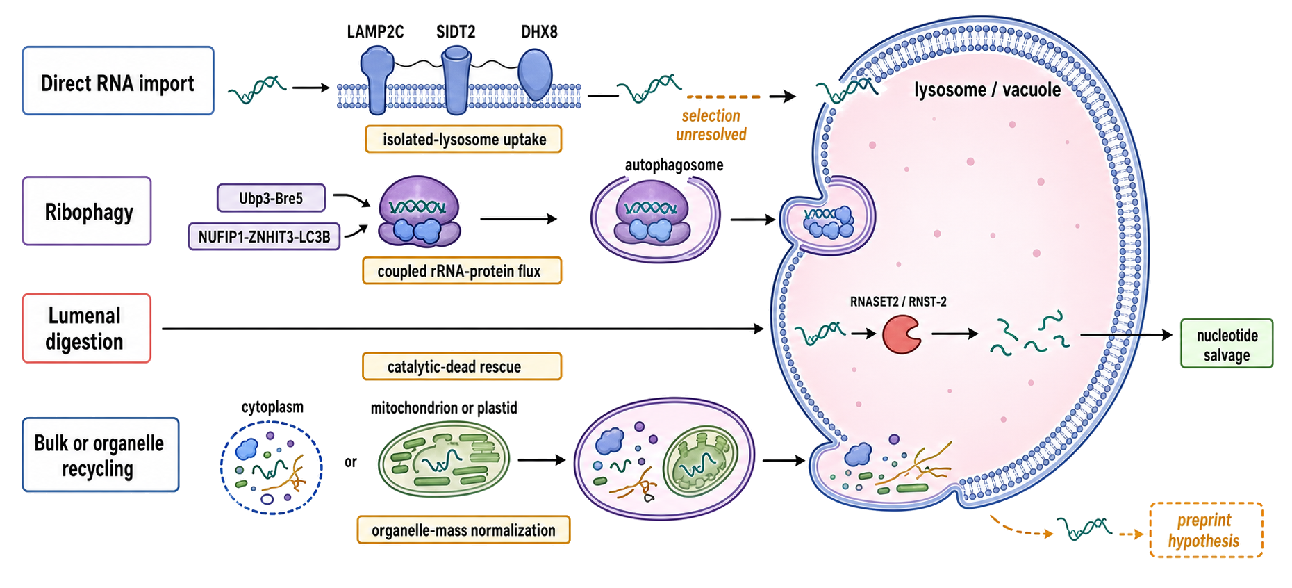
Figure 37.5. Four Routes to Lysosomal RNA Turnover and Their Evidence Gates. Four parallel lanes converge on an acidic lysosomal or vacuolar lumen. Direct-import RNautophagy shows RNA binding at LAMP2C/SIDT2 with DHX8 regulation; ribophagy shows a ribosome linked to yeast Ubp3–Bre5, mammalian NUFIP1–ZNHIT3–LC3B, or the cross-lineage RPL12–ATG8-family receptor layer and enclosed in an autophagosome; lumenal digestion shows RNASET2/RNST-2 cleavage followed by nucleotide salvage; and bulk delivery shows cytoplasm, mitophagy, or chlorophagy carrying RNA as passenger cargo. The RPL12 inset distinguishes experimentally mapped binding from canonical motif prediction and shows yeast Atg1 phosphorylation–Atg11 association without projecting that exact regulatory step onto every lineage. Evidence gates label isolated-lysosome protection, coupled rRNA/protein flux with receptor and core-ATG genetics, nuclease-dead rescue with metabolite tracing, and organelle-mass normalization. Solid arrows indicate demonstrated steps, dashed arrows incomplete substrate selection, and a preprint badge only the 2025 lysosomal RNA-selectivity hypothesis. The design must not imply that SIDT2 is a structurally proven pore or that all routes are universal.
Table 37.5. Pathway-Assignment Matrix for Lysosomal and Autophagic RNA Turnover. Assignment of lysosomal or autophagic RNA turnover requires evidence for substrate selection, delivery, and degradation together with route-specific controls; colocalization or RNA loss alone cannot identify the responsible pathway.
| Candidate route | Selected object | Delivery evidence | Digestion evidence | Decisive controls | Common overclaim |
|---|---|---|---|---|---|
| Direct-import RNautophagy | RNA or RNA feature | Protected uptake into intact isolated lysosomes; LAMP2C/SIDT2 dependence | Acid-dependent loss after entry | Surface stripping; macroautophagy-independent condition; transport- or RNA-binding-defective rescue | RNA near LAMP2 proves direct translocation |
| Ribophagy | Ribosome or subunit | Coupled rRNA and ribosomal-protein flux; receptor and core-ATG dependence | Lysosomal or vacuolar loss of both cargo classes | General flux control; LC3/ATG8-binding mutant; pulse–chase | Any rRNA degradation is ribophagy |
| Lumenal digestion and salvage | RNA already delivered | Not assigned by nuclease loss alone | RNASET2/RNST-2 catalytic dependence; nucleoside production | Localized nuclease-dead rescue; metabolite tracing | RNase loss identifies the import route |
| Bulk cytoplasmic autophagy | Cytoplasmic volume | Nonselective sequestration and core-ATG dependence | General hydrolase dependence | Compare enrichment with bulk cytosolic markers | ATG dependence proves RNA selectivity |
| Mitophagy or chlorophagy | Whole organelle | Organelle-selective delivery | Loss of organelle contents | Normalize RNA to organelle mass, DNA, and membrane proteins | Falling organellar RNA proves RNA-selective autophagy |
Four routes must be kept separate. First, RNautophagy is the name introduced for ATP-dependent direct transport of RNA into mammalian lysosomes, without an autophagosome enclosing the cargo. Second, ribophagy is selective or enriched macroautophagic delivery of ribosomes, including their rRNAs and proteins, to lysosomes or yeast vacuoles. Third, lysosomal RNA degradation is the enzymatic digestion that occurs after delivery and can be blocked even when cargo transport is intact. Fourth, bulk or organelle-selective autophagy carries RNA because it engulfs cytoplasm, mitochondria, plastids, or other RNA-containing material. These routes can operate in the same cell, and an experiment that measures only the endpoint cannot identify which route was responsible.
Direct lysosomal RNA import and the bounded RNautophagy model
The founding RNautophagy experiments incubated purified RNA with isolated mouse lysosomes and measured ATP-dependent uptake and degradation. Fujiwara et al. identified the lysosomal membrane splice isoform LAMP2C as an RNA-binding component and named the process RNautophagy (Fujiwara et al. 2013). Later work implicated SID1 transmembrane family member 2 (SIDT2): SIDT2 perturbation altered RNA uptake, lysosomal targeting motifs were required, and a cytosolic arginine-rich region bound nucleic acid and supported transport in isolated-lysosome and cell assays (Aizawa et al. 2016; Contu et al. 2017; Hase et al. 2020). These studies support a direct-import pathway in the tested mammalian systems. They do not yet establish that all endogenous RNAs cross by the same physical mechanism, that LAMP2C and SIDT2 are obligatory in every tissue, or that “RNautophagy” is a universal category parallel to macroautophagy.
Mechanistic language should match the assay. Uptake of fluorescence- or isotope-labeled RNA into a protease-protected isolated-lysosome fraction supports transmembrane delivery more directly than colocalization in intact cells. ATP dependence and loss after SIDT2 mutation strengthen pathway assignment. Even so, isolated lysosomes may lose cytosolic regulators, and exogenous RNA can differ in structure, modification, protein occupancy, and concentration from an endogenous RNP. Conversely, observing an RNA near LAMP1 or LAMP2 in a cell cannot distinguish binding to the cytosolic lysosome surface, residence in an attached autophagosome, localization inside the lumen, or signal from an incompletely resolved organelle. Protease protection, membrane-impermeant nuclease controls, lumenal quenching, fraction purity, and electron microscopy can narrow those alternatives.
Substrate selectivity remains an active question. LAMP2C and SIDT2 cytosolic regions contain arginine-rich RNA-binding motifs, and early competition assays suggested preferences among substrates rather than uniform uptake (Hase et al. 2015, 2020). Sakai et al. subsequently identified the RNA helicase DHX8 as a guanine-sequence-binding factor that associates with SIDT2 at the cytosolic lysosome surface. DHX8 loss reduced SIDT2-dependent RNA degradation, whereas the study found RNA binding more important than DHX8 ATPase activity for the measured effect (Sakai et al. 2025). This extends the model from a membrane protein alone to a regulated RNA-recognition step, but it does not define a complete endogenous substrate code. Consecutive guanines, RNA structure, RNP occupancy, transcript abundance, and access to lysosomes may all influence what is recovered.
A 2025 study provides a bounded physiological example rather than a general rule. In kidney injury models, a hypoxia-induced tRNA-Asp-GTC 3′-derived RNA formed a G-quadruplex, sequestered PUS7, changed histone-mRNA pseudouridylation, and promoted autophagosome–lysosome turnover of histone mRNAs (Li et al. 2025). The study uses the broader phrase “RNA autophagy” and involves regulatory RNA-triggered delivery through an autophagosome–lysosome route; it should not automatically be recast as direct SIDT2 membrane translocation. The fragment’s biogenesis, structure, and renal biology belong to Chapter 89. Here it illustrates why pathway name, cargo, trigger, and delivery route must be reported separately.
Ribophagy carries an RNP, not naked RNA
Ribosomes are large ribonucleoprotein particles whose rRNA accounts for much of cellular RNA mass. Starvation therefore creates both a proteostasis and a nucleotide-recycling problem. In Saccharomyces cerevisiae, Kraft et al. showed that mature ribosomes are delivered to the vacuole during nutrient starvation and that the selective component of 60S turnover requires the deubiquitylating Ubp3–Bre5 complex (Kraft et al. 2008). General autophagy remained functional in the mutants, while 60S material accumulated relative to controls. This experimental separation was important: starvation can cause nonselective cytoplasmic engulfment, but a cargo-specific genetic requirement supports an enriched ribophagy route. The original results did not prove that every ribosomal subunit follows one receptor pathway or that Ubp3–Bre5 is a mammalian ribophagy module.
In mammalian cells, quantitative lysosome proteomics after mechanistic target of rapamycin complex 1 (mTORC1) inhibition led to a different receptor model. NUFIP1 forms a complex with ZNHIT3, associates with ribosomes under starvation or Torin1 treatment, and binds the autophagosome protein LC3B through a LC3-interacting region. NUFIP1 loss or mutation of its LC3-binding motif reduced starvation-induced lysosomal delivery and degradation of ribosomal components, and reduced survival under prolonged starvation (Wyant et al. 2018). ZNHIT3 accompanies NUFIP1, but NUFIP1 supplies the demonstrated LC3B-interacting motif; calling both proteins equivalent LC3 receptors would overstate the evidence.
More recent work identifies ribosomal protein RPL12 as a receptor layer tested from yeast to mammals. Disrupting the RPL12–ATG8-family interaction caused accumulation of both ribosomal proteins and rRNA, while yeast experiments placed Atg1-dependent RPL12 phosphorylation and enhanced Atg11 association upstream of starvation ribophagy (Chen et al. 2025). RPL12 binding did not rely on the two predicted canonical ATG8-interacting motifs, so a motif prediction alone would have missed the experimentally mapped interaction surface. This cross-lineage model does not erase the Ubp3–Bre5 or NUFIP1–ZNHIT3 results; the factors may act at distinct recognition, signaling, or cargo-delivery steps under different conditions.
Ribophagy evidence should follow both halves of the ribosome. Loss of ribosomal proteins alone could reflect proteasomal turnover or failed assembly, whereas loss of rRNA alone could reflect ribosome-associated quality control or nonlysosomal nucleases. Strong studies measure rRNA and multiple proteins, demonstrate lysosomal or vacuolar delivery, show dependence on core autophagy machinery, and compare a proposed receptor mutant with general flux controls. Pulse–chase labeling can estimate destruction rates; tandem-fluorescent ribosomal reporters can exploit acid-dependent fluorophore quenching; protease-protected lysosome fractions can establish lumenal arrival; and immuno-electron microscopy can visualize sequestration. Each assay has a failure mode. Ribosomal reporters may perturb assembly, lysosome inhibitors can globally alter signaling, and static accumulation can reflect increased delivery or blocked digestion.
Digestion inside the acidic compartment and nucleotide homeostasis
Once RNA reaches the lysosome or vacuole, acid-active ribonucleases must convert the polymer into smaller products. RNASET2 is the sole human member of the conserved T2-family acidic endoribonucleases and has lysosomal localization as well as catalytic activity (Campomenosi et al. 2006). In zebrafish, rnaset2 loss caused rRNA-rich lysosomal storage in neural tissue and modeled the cystic leukoencephalopathy associated with human RNASET2 deficiency (Haud et al. 2011). These findings establish an intralysosomal digestion defect. They do not by themselves specify how the stored rRNA entered lysosomes.
The delivery-versus-digestion distinction is especially clear in C. elegans. The T2-family nuclease RNST-2 is required for lysosomal rRNA degradation during normal development. rnst-2 mutants accumulated both rRNA and ribosomal proteins in enlarged lysosomes, and blocking autophagy suppressed that accumulation, placing autophagic delivery upstream of lumenal digestion. Developmental and lifespan phenotypes were worsened when de novo pyrimidine synthesis was also compromised and were rescued by uridine or cytidine supplementation, linking rRNA destruction to nucleotide homeostasis rather than only waste disposal (Liu et al. 2018). The causal chain is therefore autophagic ribosome delivery, RNST-2-dependent rRNA hydrolysis, and metabolite reuse. It is not direct evidence for LAMP2C/SIDT2-mediated RNautophagy in the nematode.
Nuclease-dead rescue is a decisive control for this layer. If wild-type RNASET2-family enzyme clears lumenal RNA but a properly localized catalytic mutant does not, catalytic digestion rather than a scaffolding effect is implicated. Conversely, a transport mutant can reduce lysosomal RNA without changing lumenal nuclease activity. Measuring nucleosides or isotope-labeled nucleotide return to cellular pools can test salvage directly. Total RNA reduction is a weaker endpoint because RNase loss can activate lysosomal stress and innate sensors, which in turn alter transcription, autophagy, and cell composition. Mouse RNase T2 deficiency, for example, can expose microbial rRNA to TLR13 and reshape macrophage populations (Sato et al. 2025); this is a biological consequence of lysosomal RNA stress, not evidence that immune signaling is the normal delivery mechanism.
Bulk autophagy, mitophagy, and the organelle boundary
Autophagy can remove RNA without recognizing RNA. During nonselective macroautophagy, portions of cytoplasm containing mRNAs, ribosomes, and RNPs are engulfed. During mitophagy, an entire mitochondrion or a mitochondrial fragment is delivered, so its matrix RNAs disappear along with membranes, DNA, and proteins. Chlorophagy or other plastid-quality-control routes can likewise remove chloroplast material. An observed fall in mitochondrial or plastid RNA after activation of organelle-selective autophagy is therefore not evidence for an RNA-selective receptor. The primary selected object may be the damaged organelle.
The boundary also runs in the other direction. Mitochondrial RNA can be degraded inside mitochondria by SUV3–PNPase before any mitophagy occurs. Some mitochondrial RNA can escape or be exposed to cytosolic innate sensors when mitochondrial membrane integrity fails (Dhir et al. 2018). These outcomes—matrix degradation, release, vesicular export, and mitophagic consumption—have different compartments and consequences. Experiments should normalize RNA abundance to organelle mass, membrane proteins, mitochondrial DNA or plastid DNA, and cell number. If RNA falls in exact proportion to organelle mass, whole-organelle clearance is a parsimonious explanation. Preferential loss of one transcript with preserved organelle mass instead motivates tests of RNA-specific processing, export, or decay.
Arabidopsis illustrates the mixture of routes. Sequencing of purified vacuoles recovered mainly short RNA species, including rRNA, tRNA, and chloroplast-derived reads; many chloroplast-derived species were strongly reduced in atg5-1 vacuoles (Hickl et al. 2021). That result supports ATG5-dependent delivery to the vacuole, but a vacuolar endpoint profile samples fragments that survived until isolation. It cannot by itself distinguish chlorophagy, piecemeal chloroplast export, ribosome delivery, or an RNA-selective receptor, and the relatively small compositional effect of RNS2 loss permits redundant lumenal nucleases or compensatory pathways. Photosynthetic tissues add further complications: chloroplast abundance changes with development and stress, and vacuole isolation can carry organellar fragments. Fraction purity, input-normalized enrichment, chlorophyll and organelle markers, RNA-length distributions, and autophagic flux controls are needed before assigning selectivity.
An evidence ladder for pathway assignment
A rigorous experiment first asks where the RNA resides. Fractionation should include cytosolic, lysosomal, mitochondrial, and plastid markers and controls for surface-bound RNA. Imaging should resolve lumen from limiting membrane and apposed autophagosomes. The next question is how cargo arrived. Core ATG dependence and double-membrane sequestration support macroautophagy; direct uptake into isolated lysosomes, persistence when macroautophagy is disabled, and SIDT2/LAMP2C dependence support the direct-import model. A ribosome receptor interaction plus coupled rRNA/ribosomal-protein flux supports ribophagy. Proportional loss of all organelle constituents supports whole-organelle recycling.
The final questions concern digestion and consequence. Lysosomal acidification inhibitors such as bafilomycin A1 or chloroquine can reveal flux, but they also alter mTOR signaling, endosomal traffic, ion homeostasis, and organelle physiology. They should not be the only evidence. Pulse–chase experiments distinguish synthesis from disappearance; wild-type versus catalytic-dead nuclease rescue tests digestion; transport-defective versus RNA-binding-defective SIDT2 or LAMP2C variants test import; and LC3-interaction mutants test receptor logic. Metabolite tracing tests nucleotide salvage. Disease or therapeutic claims require an additional ladder from pathway perturbation to tissue phenotype and independent replication.
A September 2025 bioRxiv preprint used lysosome isolation and RNA profiling to propose that many RNA classes reach lysosomes but that SRP RNAs, Y RNAs, 5′-terminal oligopyrimidine mRNAs, and selected secretory-pathway mRNAs are enriched, with candidate LARP1- and SRP9/14-dependent targeting logic (Ray et al. 2025 preprint). The work is useful as a hypothesis and method map, including candidate roles for RNASET2, PLD3, and RNase A-family enzymes, but it remains a preprint at this chapter’s cutoff. Its selectivity claims should not be treated as consensus until peer review, orthogonal flux measurements, and independent replication resolve isolation bias and the contributions of direct import, macroautophagy, and RNP co-transport.
37.6. Disease links and comparative organismal diversity
Disease links arise when organellar or lysosomal RNA turnover defects impair energy metabolism, nucleotide balance, development, or stress tolerance. Mitochondrial RNA disorders can present as encephalopathy, myopathy, cardiomyopathy, liver disease, growth failure, or multisystem disease. ELAC2 variants provide a direct processing example: affected individuals and mechanistic models connect defective mitochondrial tRNA 3′-end maturation to hypertrophic cardiomyopathy (Haack et al. 2013). SUPV3L1 variants provide a remodeling-and-decay example with abnormal mitochondrial RNA processing in neurodegenerative disease (van Esveld et al. 2022). These cases should not be generalized into one tissue rule; energy demand, developmental timing, genetic background, heteroplasmy, and compensation shape presentation.
Mammalian mitochondrial disease illustrates several mechanisms. A mutation in a mitochondrial tRNA gene can impair tRNA folding, processing, modification, aminoacylation, stability, or decoding. A mutation in a nuclear gene encoding a mitochondrial ribonuclease or helicase can cause accumulation of abnormal RNAs or loss of mature transcripts. A mutation in a transcript-specific stabilizing factor can selectively reduce one respiratory-chain mRNA. In each case, the immediate molecular phenotype is RNA-centered, but the clinical phenotype arises because mitochondrial translation and oxidative phosphorylation fail.
Plant disease and agronomic phenotypes often look different because the relevant outputs include photosynthesis, fertility, stress tolerance, and development. Defects in chloroplast RNA processing can reduce photosynthetic complex accumulation and cause pale, variegated, or growth-impaired plants. Defects in plant mitochondrial RNA processing can affect respiration and are also linked to cytoplasmic male sterility in several systems, where mitochondrial gene-expression changes disrupt pollen development. Small et al. provide a general framework for plant organellar RNA maturation, but final treatment of each phenotype requires crop- and factor-specific references (Small et al. 2023).
Kinetoplastid mitochondria are a comparative warning against universal models. Trypanosomatids contain a kinetoplast, a mitochondrial DNA network with maxicircles and minicircles. Many maxicircle transcripts require guide RNAs, often encoded by minicircles, to direct uridine insertion and deletion. RNA quality control includes guide-RNA integrity, editing complexes, ligation steps, and edited products; an intermediate that would look abnormal in mammalian mitochondria can be normal in a kinetoplastid (Aphasizheva and Aphasizhev 2021). Chapter 51 covers the editing mechanism.
Lysosomal RNA disease illustrates a different compartment. RNASET2 deficiency causes a cystic leukoencephalopathy in humans, and loss of the ortholog in zebrafish causes neuronal lysosomal rRNA storage (Haud et al. 2011). A 2025 report described a single child with biallelic SIDT2 missense variants, cerebellar ataxia, and lysosomal dysfunction; the tested variants reduced SIDT2 interaction with double-stranded RNA, while patient cells and model systems also showed broader autophagy and lysosome abnormalities (Nguyen et al. 2025). The case supports clinical investigation of SIDT2 but is not sufficient to assign the phenotype specifically to failed direct RNA import rather than SIDT2’s wider endolysosomal functions. Therapeutic activation or inhibition of SIDT2, DHX8, NUFIP1, or lysosomal nucleases therefore remains experimental.
Comparative diversity also includes variation in tRNA import, RNA editing, intron content, transcript structures, and nuclease repertoires. Some mitochondria encode many tRNAs; others import most tRNAs. Some plant organelles edit hundreds of cytidines; mammalian mitochondrial RNA editing is limited and different in character. Some chloroplasts retain bacterial-like decay factors; other plastids have reduced or specialized gene-expression systems. These differences matter for annotation. A read ending at a particular site may represent a mature end in one lineage, a decay intermediate in another, or an editing-dependent processing boundary in a third.
Common misconceptions follow naturally from this diversity. Do not assume that organellar RNAs are regulated like nuclear mRNAs. Do not assume that a poly(A) tail means stabilization. Do not assume that a mitochondrial RNA factor has the same substrates in yeast, humans, and plants. Do not assume that all organellar RNA fragments are junk; some are processing intermediates, regulatory RNAs, guide RNAs, or stable protected fragments. Do not infer disease mechanism from RNA abundance alone; connect genotype, RNA processing, translation, organellar function, and tissue phenotype.
Experimental Foundations and Evidence
The strongest organellar RNA turnover studies combine compartment validation with molecular specificity. Compartment validation asks whether the RNA and protein are truly in the organelle rather than cytosolic, nuclear, or bacterial contaminants. This is especially important for mitochondrial RNA fragments because NUMTs, which are nuclear mitochondrial DNA segments, can confuse genomic mapping, and because organelle preparations can contain associated cytosolic RNAs.
RNA-end mapping is essential. Circularized RNA sequencing, rapid amplification of cDNA ends, specialized tail profiling, direct RNA sequencing, and ligation-independent approaches can identify transcript termini and tail composition. However, end-mapping methods are biased by end chemistry. A method requiring 5′ phosphate or 3′ hydroxyl ligation can miss capped, triphosphorylated, phosphorylated, or cyclic phosphate ends. Scacchetti et al. provide a current methodological reminder that blocked 3′ termini can hide tRNA-derived RNAs from standard ligation-dependent assays (Scacchetti et al. 2024).
Genetic perturbation identifies candidate factors. Knockout, knockdown, inducible depletion, temperature-sensitive alleles, organelle-targeting mutants, and catalytic mutants can reveal which proteins affect RNA abundance or processing. The limitation is indirectness. Depleting a mitochondrial helicase may disrupt RNA decay directly, but it may also impair ribosome assembly, translation, organellar membrane potential, or global organelle biogenesis. Rescue with wild-type and catalytic-dead alleles helps separate enzymatic activity from scaffolding or indirect stress.
Biochemistry tests mechanism. Purified enzymes and defined RNA substrates can show nuclease directionality, helicase dependence, tail preference, ligation chemistry, or end-healing requirements. The weakness is substrate realism. A naked RNA oligonucleotide may not represent a structured, edited, modified, protein-bound organellar RNP. In vitro results become stronger when they predict in vivo end changes or genetic phenotypes.
For lysosomal pathways, “inside” must be demonstrated rather than inferred. Isolated-lysosome uptake assays require intactness, latency, and surface-stripping controls. Autophagic flux measurements require comparisons before and after lysosomal blockade, because more RNA or reporter signal in lysosomes can mean faster delivery or slower digestion. Coupled accumulation of rRNA and ribosomal proteins supports ribosome cargo, while nuclease-dead rescue separates lumenal degradation from delivery. Electron microscopy, protease protection, genetics of core ATG factors, receptor-binding mutants, and metabolite tracing form orthogonal evidence layers; no single lysosome image identifies the route.
Systems readouts connect RNA to function. In mitochondria, investigators measure organellar translation, respiratory-chain complex abundance, oxygen consumption, membrane potential, metabolite state, and stress signaling. In chloroplasts, they measure photosynthetic complex accumulation, chlorophyll fluorescence, growth under light regimes, and plastid development. These readouts do not prove an RNA mechanism by themselves, but they show whether the RNA defect matters biologically.
Box 37.2. Minimal Evidence Ladder for an Organellar RNA Surveillance Claim
- Confirm the factor and substrate are localized to the relevant organelle.
- Map the RNA end, tail composition, edit state, or processing defect.
- Show substrate specificity rather than only global organellar stress.
- Perturb the factor and rescue with wild-type and catalytic or binding mutants where possible.
- Measure organellar translation and respiratory or photosynthetic consequences.
- Control for changes in organelle abundance, tissue composition, and contamination.
Biological Contexts Across Organisms
Mammalian mitochondria are compact, gene-dense, and heavily dependent on post-transcriptional processing. Their RNA quality-control problems center on generating correct transcript ends, maintaining transcript-specific stability, removing antisense or double-stranded RNA, and protecting mitochondrial translation. Because mitochondrial gene products are core respiratory-chain subunits, RNA defects can have severe energy consequences.
Plant mitochondria are larger and more complex. They contain many introns, extensive RNA editing, recombination-prone genomes, and numerous nuclear-encoded RNA-binding factors. Their RNA turnover pathways must distinguish slowly maturing transcripts from defective ones. Plant mitochondrial RNA metabolism is also tied to fertility and environmental adaptation.
Chloroplasts and plastids retain bacterial-like features but are integrated into plant development and photosynthesis. Chloroplast transcripts often come from operon-like units, undergo processing into smaller RNAs, and rely on nucleus-encoded specificity factors. Light, developmental stage, plastid type, and stress condition can change the demand for photosynthetic gene expression and RNA quality control.
Kinetoplastid mitochondria are the most dramatic comparative case in this chapter. Their normal mRNA maturation can require extensive editing directed by guide RNAs. Surveillance must operate without destroying normal editing intermediates prematurely. This creates an RNA economy in which small guide RNAs, cleavage-ligation cycles, exonucleases, terminal uridylyltransferases, and editing complexes all shape the mature transcriptome.
Lysosomes and yeast vacuoles add a cross-compartment context. In mammalian cultured cells, direct-import RNautophagy and NUFIP1-dependent ribophagy have been tested most deeply under defined perturbations. Budding yeast established Ubp3–Bre5-dependent starvation ribophagy but lacks a simple one-to-one equivalent of every mammalian receptor. C. elegans shows that developmental rRNA recycling through RNST-2 matters even without starvation. Plants combine cytoplasmic, ribosomal, and organelle-derived RNA delivery to vacuoles. These systems answer different questions and should not be pooled as replicate evidence for a single universal pathway.
Box 37.3. Kinetoplastids as a Boundary Case
- Normal maturation in kinetoplastid mitochondria includes guide RNAs, cleavage, uridine insertion or deletion, and ligation.
- Editing intermediates are not automatically defective RNAs to be removed by surveillance.
- Surveillance must preserve productive intermediates while removing failed or unproductive products.
- See Chapter 51 for the detailed editing mechanism.
Technology, Clinical, and Engineering Links
Clinical genetics increasingly identifies nuclear genes whose products act inside mitochondria. When a variant is found in an RNA helicase, ribonuclease, tRNA-processing enzyme, or RNA-binding protein, the mechanistic question is not simply whether total mitochondrial RNA abundance changes. The better questions are which RNA class changes, which end or processing step changes, whether mitochondrial translation is impaired, which respiratory complexes are affected, and whether patient tissue phenotypes match the biochemical defect.
Plant biotechnology can exploit organellar RNA metabolism because chloroplasts and mitochondria control traits relevant to photosynthesis, stress tolerance, fertility, and transgene expression. Stabilizing a chloroplast transcript may increase protein accumulation, but transcript abundance alone is not enough; translation, complex assembly, and developmental context must also be measured. Conversely, targeting a defective or unwanted organellar RNA for decay could be useful only if specificity is high enough to avoid broad organellar stress.
Sequencing technology is especially important for this chapter. Direct RNA sequencing, long-read cDNA sequencing, tail profiling, and ligation-independent small RNA methods can reveal transcript isoforms and end chemistry missed by conventional short-read RNA-seq. These methods should be paired with careful organellar genome annotation, because organellar genomes include repeats, RNA editing, NUMT-like confounders, and strain-specific structural variation.
Lysosome-centered profiling is promising but unusually sensitive to contamination and endpoint bias. Rapid immunoisolation can limit redistribution, isotope or pulse–chase labeling can measure turnover, and parallel proteomics can test whether RNA travels with an RNP. Claims of therapeutic RNA clearance need endogenous-substrate measurements, dose–response relationships, catalytically or transport-defective controls, and tissue-level safety. Increasing lysosomal RNA digestion could deplete regulatory RNAs or perturb nucleotide and innate-immune homeostasis as well as remove a pathogenic transcript.
Box 37.4. Sequencing Blind Spots from RNA End Chemistry
- Standard adapter ligation requires compatible 5′ and 3′ ends.
- RNAs with 3′ phosphate or 2′,3′ cyclic phosphate ends are not detected by conventional ligation-dependent methods.
- Ligation-independent or end-healing sequencing protocols reveal additional RNA species with blocked termini.
- Absence from a standard small RNA library does not mean absence from the cell.
Box 37.5. Minimum Evidence for a Lysosomal RNA-Selectivity Claim
- Show that RNA is inside the lumen, not on the limiting membrane or in an attached autophagosome.
- Measure time-resolved flux; lysosomal inhibitors have pleiotropic effects and are not sufficient alone.
- Separate direct import, macroautophagic RNP delivery, lumenal digestion, and whole-organelle clearance genetically.
- Use mechanism-defective rescue: transport or RNA-binding mutants, LC3/ATG8-binding mutants, and nuclease-dead mutants.
- Demonstrate enrichment relative to abundance, organelle mass, and bulk cytoplasmic cargo before claiming selectivity.
- Treat purified-vacuole or purified-lysosome RNA profiles as endpoint inventories unless protection and time-resolved delivery controls establish lumenal flux and cargo route.
- Label preprints and single-patient observations as preliminary rather than therapeutic consensus.
Recent Consensus
Current consensus treats organellar RNA turnover as an active part of maturation and gene-expression control. Mammalian mitochondrial RNA degradation prominently involves SUV3–PNPase, plant organellar ends are strongly shaped by nuclear-encoded RNA-binding proteins and ribonucleases, and poly(A)-rich tails can support either degradation or transcript-specific maturation depending on the compartment and RNA (Small et al. 2023; Santonoceto et al. 2024). An abundance phenotype alone is not a decay-rate measurement.
It is also established that lysosomes and vacuoles degrade RNA delivered through more than one route. The evidence supports direct LAMP2C/SIDT2-associated RNA import in defined mammalian systems, selective or enriched ribosome delivery during starvation, and RNASET2-family digestion of lumenal RNA. Autophagic rRNA recycling contributes to nucleotide homeostasis during normal C. elegans development (Liu et al. 2018). These findings do not justify treating RNautophagy, ribophagy, lysosomal nuclease activity, and bulk organelle recycling as synonyms.
The less-settled layer is substrate selection. DHX8 adds a regulator to the SIDT2 pathway, and RPL12 adds a conserved ribophagy-receptor model, but endogenous RNA codes and receptor redundancy remain incomplete (Sakai et al. 2025; Chen et al. 2025). Lysosomal RNA profiling suggests selective cargo classes, yet the broadest dataset is currently a 2025 preprint. Claims about universal physiological roles, human disease mechanisms, or therapeutic manipulation should therefore remain context-qualified.
Open Questions, Controversies, Deprecated Models, and Common Misconceptions
Open questions:
- What determines how organelles distinguish slow maturation from defective maturation? Plant organellar RNAs can require editing, splicing, trimming, and protein binding before becoming functional. A simple timer model would risk destroying legitimate intermediates. The field needs more kinetic measurements that follow individual molecules from transcription through maturation, translation, and decay.
- What determines when failed mitochondrial RNA surveillance progresses from matrix double-stranded RNA accumulation to membrane escape and MDA5–type I interferon signaling? Dhir et al. established this sequence in depletion models and PNPT1-variant fibroblasts, but the determinants of release, tissue specificity, and chronic disease remain unresolved and are treated more fully in chapters on innate sensing and disease.
- What determines how widespread RNA repair is inside organelles? RNA ligation and end-healing chemistry are well established in several RNA biology contexts, and organelles contain many plausible substrates. However, direct organelle-specific repair pathways are not as comprehensively mapped as processing, editing, and decay pathways. Claims should therefore separate established ligase chemistry from organelle-specific inference.
- Which endogenous RNA sequence, structure, modification, or RNP features govern LAMP2C/SIDT2/DHX8-dependent import? Current binding and perturbation data identify candidate determinants but not a universal substrate code.
- How do NUFIP1–ZNHIT3, RPL12, Ubp3–Bre5, bulk autophagy, and ribosome-quality-control pathways divide labor across nutrients, tissues, and organisms? Receptor dependence is context-specific, and multiple pathways may act on different ribosome states.
- How much nucleotide salvage derives from RNA in each tissue? RNST-2 genetics establish developmental importance in C. elegans, but quantitative mammalian flux remains incompletely mapped.
- Which human phenotypes arise specifically from failed lysosomal RNA transport rather than broader lysosomal roles of SIDT2 or LAMP2 isoforms? Current human SIDT2 evidence includes a single biallelic case and should be interpreted cautiously.
Common misconceptions:
- “A poly(A) tail always stabilizes RNA.” In organelles, a poly(A)-rich tail often promotes decay, although mammalian mitochondrial mRNAs show important maturation-linked exceptions.
- “All RNA fragments are degradation waste.” Some organellar fragments are mature small RNAs, guide RNAs, protected processing products, or assay-visible intermediates.
- “Organelles use bacterial RNA decay machinery unchanged.” Organelles retain bacterial ancestry but have evolved extensive nuclear-encoded, lineage-specific RNA-binding and surveillance systems.
- “RNA abundance equals RNA stability.” Organellar RNA abundance changes can reflect transcription, processing, editing, splicing, tailing, degradation, translation, organelle number, or tissue composition.
- “RNautophagy is another name for ribophagy.” RNautophagy denotes direct lysosomal RNA import in a specific literature; ribophagy delivers ribosomal RNPs through autophagic sequestration.
- “RNA detected in a lysosome must have crossed the lysosomal membrane directly.” It may arrive through an autophagosome, with a ribosome, in bulk cytoplasm, or inside an engulfed organelle.
- “More lysosomal RNA proves faster RNA degradation.” Increased signal can instead reflect blocked lumenal nuclease activity, impaired acidification, or stalled flux.
- “Mitophagy selectively recognizes mitochondrial RNA.” Mitophagy selects mitochondrial material; RNA can disappear as a passenger unless transcript-selective evidence is added.