Chapter 70. Regulated and Noncanonical Translation Initiation
Scope Note
This chapter explains how messenger RNA architecture and cell-state signaling regulate ribosome recruitment and start-site choice. It owns upstream open reading frames (uORFs), alternative starts, internal ribosome entry sites (IRESs), ribosome shunting, cap-independent initiation, untranslated-region (UTR) and RNA-binding-protein (RBP) control, the integrated stress response (ISR), mechanistic target of rapamycin (mTOR) signaling, localization- and development-linked initiation, and quantitative measurement of initiation regulation. Chapter 67 owns the conserved initiation apparatus, stepwise canonical mechanism, and domain-level comparison; this chapter introduces only the machinery needed to explain regulated departures from that baseline. Downstream consequences involving elongation, decay, or localization are treated here only when they identify an initiation-control mechanism and are handed to their dedicated chapters for broader pathway coverage.
Executive Summary
Translational regulation is the control of how efficiently, where, when, and under which conditions ribosomes convert an mRNA into protein. For most cellular eukaryotic mRNAs, initiation begins when initiation factors recognize the 5′ cap, recruit the small ribosomal subunit, and allow a preinitiation complex to scan the 5′ untranslated region until an acceptable start codon is selected. That basic pathway is not a passive readout of the coding sequence. The 5′ untranslated region can contain upstream open reading frames, stable RNA structures, protein-binding sites, alternative start codons, and sequence contexts that redirect ribosomes before the main coding sequence is reached. The 3′ untranslated region and poly(A)-associated proteins can alter initiation indirectly by recruiting repressors, activators, localization factors, deadenylases, or microRNA-associated complexes. Coding-region features can also feed back on initiation by changing ribosome traffic, cotranslational quality control, and mRNA stability.
Upstream open reading frames, usually abbreviated uORFs, are short translated regions located upstream of the annotated main coding sequence. A uORF can repress downstream protein production by capturing scanning ribosomes, causing termination before the main start codon, or triggering nonsense-mediated decay when translation termination occurs in a context interpreted as premature. A uORF can also act as a sensor. In some transcripts, amino acid availability, stress-induced phosphorylation of initiation factors, metabolite concentration, or viral infection changes whether ribosomes initiate at the uORF, reinitiate downstream, bypass weak start codons, or stall on a regulatory peptide. Recent high-resolution translation maps and viral studies have reinforced that upstream translation is widespread and context-specific rather than a rare annotation curiosity (Chothani et al. 2022; Lefevre et al. 2024).
Internal ribosome entry sites, or IRESs, and ribosome shunting are alternative routes into translation initiation. An IRES is an RNA element that promotes ribosome recruitment internally, often when cap-dependent initiation is limited. Viral IRESs provide the clearest mechanistic examples, because some viral RNAs fold into structured elements that bind ribosomal subunits and initiation factors directly or with reduced factor requirements. Cellular IRES claims are more difficult to validate because reporter artifacts, cryptic promoters, splicing, RNA cleavage, and altered RNA stability can mimic internal initiation. Ribosome shunting is a related but distinct mechanism in which a scanning ribosome loads near the 5′ end, bypasses a structured segment or upstream region, and resumes scanning or initiation downstream. Both mechanisms show that ribosomes do not always inspect every nucleotide between the cap and a start codon, but both require stringent evidence before being invoked.
Untranslated-region features and RNA-binding proteins form a regulatory grammar. Stable 5′ structures can impede scanning, while moderate structures, protein-remodeled structures, and structured binding platforms can create selective regulatory switches. RNA-binding proteins can repress initiation by blocking ribosome recruitment, scanning, start-codon recognition, or closed-loop communication between the cap and poly(A) tail. They can also activate translation by recruiting initiation factors, remodeling inhibitory structures, protecting RNAs from decay, or coupling localization to local translation. A key principle is that binding is not the same as regulation: a protein-RNA interaction becomes a translational mechanism only when perturbation, rescue, and translation-specific measurements establish a causal effect. The recent literature on 5′ untranslated-region structures and ribosome-associated mRNA fate illustrates why RNA structure, translation, and decay must be interpreted together (Leppek et al. 2018; Hopfler et al. 2023; Kogel et al. 2024).
MicroRNAs and other small RNAs usually regulate translation as part of broader messenger ribonucleoprotein remodeling. Animal microRNAs guide Argonaute-containing complexes to partially complementary sites, often in 3′ untranslated regions. The outcome may include translational repression, deadenylation, decapping, and mRNA decay, with the dominant measurable effect varying by time point, cell type, transcript, and assay. Small interfering RNAs with extensive complementarity often direct slicing rather than primarily translational repression. Bacterial small RNAs use different protein partners and base-pairing rules, but the comparison is useful because bacterial small RNAs can directly expose or occlude ribosome-binding sites. The practical warning is that reduced protein output after small-RNA targeting does not by itself reveal whether initiation, elongation, decay, localization, or protein stability changed.
Stress, nutrient signaling, localization, and development impose higher-order control. In the ISR, HRI, PKR, PERK, or GCN2 phosphorylates eIF2α, converting eIF2 into an inhibitor of its nucleotide-exchange factor eIF2B and lowering ternary-complex availability. ATF4-class leaders translate selectively because their uORFs convert delayed ternary-complex reacquisition into a change in start choice; GADD34-linked dephosphorylation later helps restore bulk initiation. mTORC1 controls another bottleneck by phosphorylating 4E-BPs, releasing eIF4E to assemble with eIF4G and eIF4A in eIF4F. TOP mRNAs connect this cap-factor control to synthesis of ribosomal proteins and translation factors, with LARP1 contributing context-dependent control of translation and stability. These pathways produce selective outputs because individual mRNAs differ in uORFs, cap-proximal sequences, structures, and bound proteins. In embryos, neurons, immune cells, and polarized cells, initiation can remain repressed during transport and activate at a defined place or developmental time. Quantitative interpretation must separate mRNA abundance, initiation frequency, elongation speed, ribosome density, completed protein synthesis, and protein degradation; footprints alone are not direct protein-output measurements.
Concept Inventory
- Cap-dependent initiation: initiation pathway in which the 5′ cap helps recruit initiation factors and the small ribosomal subunit to an mRNA. The scanning preinitiation complex then usually searches the 5′ untranslated region for a start codon.
- Upstream open reading frame (uORF): an open reading frame that begins upstream of the main coding sequence. A uORF may encode a short peptide, function mainly as a translated regulatory element, or represent annotation noise unless translation and regulatory function are shown.
- Upstream translation: ribosome initiation and elongation in a 5′ leader region before the annotated coding sequence. Upstream translation includes uORFs, upstream near-cognate initiation, and alternative N-terminal isoform starts.
- Leaky scanning: bypass of a potential start codon by a scanning preinitiation complex, usually because start-codon context, initiation-factor state, or local structure makes that start codon less favorable.
- Reinitiation: initiation at a downstream start codon after a ribosome has translated and terminated an upstream ORF. Reinitiation depends on uORF length, intercistronic distance, initiation-factor reacquisition, and transcript context.
- Alternative start-site choice: regulated selection among possible AUG or near-cognate initiation sites on one transcript, changing downstream efficiency or the amino terminus of the encoded protein.
- Internal ribosome entry site (IRES): an RNA element that promotes ribosome recruitment internally rather than solely through conventional cap-proximal scanning. Viral IRES elements are better established mechanistically than many proposed cellular IRESs.
- Ribosome shunting: initiation route in which a ribosome loads near the 5′ end, bypasses a segment of the leader, and resumes translation-related scanning or start-site selection downstream.
- Untranslated region (UTR): mRNA segment outside the main coding sequence. The 5′ UTR is especially important for ribosome recruitment and scanning; the 3′ UTR often recruits regulatory proteins and small-RNA complexes.
- RNA-binding protein (RBP): protein that binds RNA with sequence, structure, modification, or context preference. RBPs can regulate translation directly or indirectly through localization, decay, storage, or remodeling.
- MicroRNA (miRNA): small regulatory RNA that guides Argonaute-containing complexes to target RNAs, most often through seed-pairing interactions in animals. Translation repression and mRNA decay are frequently coupled outputs.
- Translation efficiency: an operational measure of protein synthesis per mRNA molecule, often inferred from ribosome profiling normalized to RNA abundance. It is useful but can conflate initiation, elongation, ribosome stalling, and decay.
- Polysome: an mRNA molecule bound by multiple ribosomes. Polysome association usually suggests active translation but does not automatically establish protein production rate or initiation mechanism.
- Stress granule: non-membrane-bound cytoplasmic assembly enriched in nontranslating mRNPs and translation factors during some stress states. Entry into stress granules is influenced by translation status and RNP composition.
- Integrated stress response (ISR): eIF2α-centered signaling program that lowers ternary-complex availability and thereby changes global initiation and uORF-dependent start-site choice.
- mTORC1 initiation control: nutrient- and growth-factor-responsive control of cap-dependent initiation capacity through 4E-binding proteins, S6 kinases, and transcript-selective leader features.
- eIF2B nucleotide exchange: recycling reaction in which eIF2B promotes GDP release from eIF2 so that eIF2–GTP and ternary complex can be regenerated; phospho-eIF2 inhibits this reaction.
- eIF4F: cap-associated complex of eIF4E, eIF4G, and eIF4A that links cap recognition, scaffolding, RNA remodeling, and small-subunit recruitment.
- Terminal oligopyrimidine (TOP) mRNA: mRNA beginning at its cap-proximal end with a cytidine and oligopyrimidine tract, often encoding a ribosomal protein or translation factor and showing mTORC1-sensitive translation.
- LARP1: La-related protein that binds TOP mRNAs and can regulate their cap access, translation, and stability; the balance among these effects is context-dependent.
What to Know Before Reading This Chapter
Readers should know the basic architecture of eukaryotic mRNA: a 5′ cap, a 5′ untranslated region, a coding sequence, a stop codon, a 3′ untranslated region, and usually a poly(A) tail. The 5′ cap is a modified nucleotide structure attached to the 5′ end of many eukaryotic mRNAs. It protects the transcript, helps recruit cap-binding proteins, and contributes to initiation. The poly(A) tail is a stretch of adenosines at the 3′ end that binds poly(A)-binding proteins and influences stability and translation.
The chapter also assumes the core eukaryotic initiation pathway developed in Chapter 67: a small-subunit preinitiation complex is recruited to an mRNA, scans 5′ to 3′, recognizes a start codon, and joins the large subunit. That compact bridge is sufficient here because the present chapter asks how RNA features and signaling alter four variables: recruitment, movement through the leader, start-codon selection, and commitment to productive elongation. Factor composition, assembly order, structural transitions, and comparison with bacterial and archaeal initiation remain primary content of Chapter 67.
Several terms require care. “Translation” is not the same as “ribosome association.” A ribosome footprint can indicate ribosome occupancy, but protein output depends on whether ribosomes initiate, elongate productively, terminate normally, and produce stable protein. “Cap-independent” does not mean factor-independent, structure-independent, or stress-specific. “IRES” should be reserved for an element with evidence for internal initiation, not merely a leader that translates well in a reporter. “Repression” should specify whether the measured output is reduced initiation, reduced elongation, lower mRNA abundance, altered localization, or faster protein degradation.
70.1. uORFs, upstream translation, and regulated start-site choice
An upstream open reading frame (uORF) is a translated region located in the 5′ leader of an mRNA before the main coding sequence. The simplest uORF begins at an AUG, continues through several codons, and terminates before the main start codon. Many upstream translation events are less tidy: they begin at near-cognate codons such as CUG, GUG, or UUG; overlap the main coding sequence in another frame; encode a conserved regulatory peptide; or occur only on transcript isoforms with a particular transcription start site. A sequence that looks like a uORF is therefore not automatically a regulatory element. Functional assignment requires an accurate leader model, evidence for initiation and elongation, and a causal effect on the downstream coding sequence or production of an independent peptide (Chothani et al. 2022; Wethmar 2014).
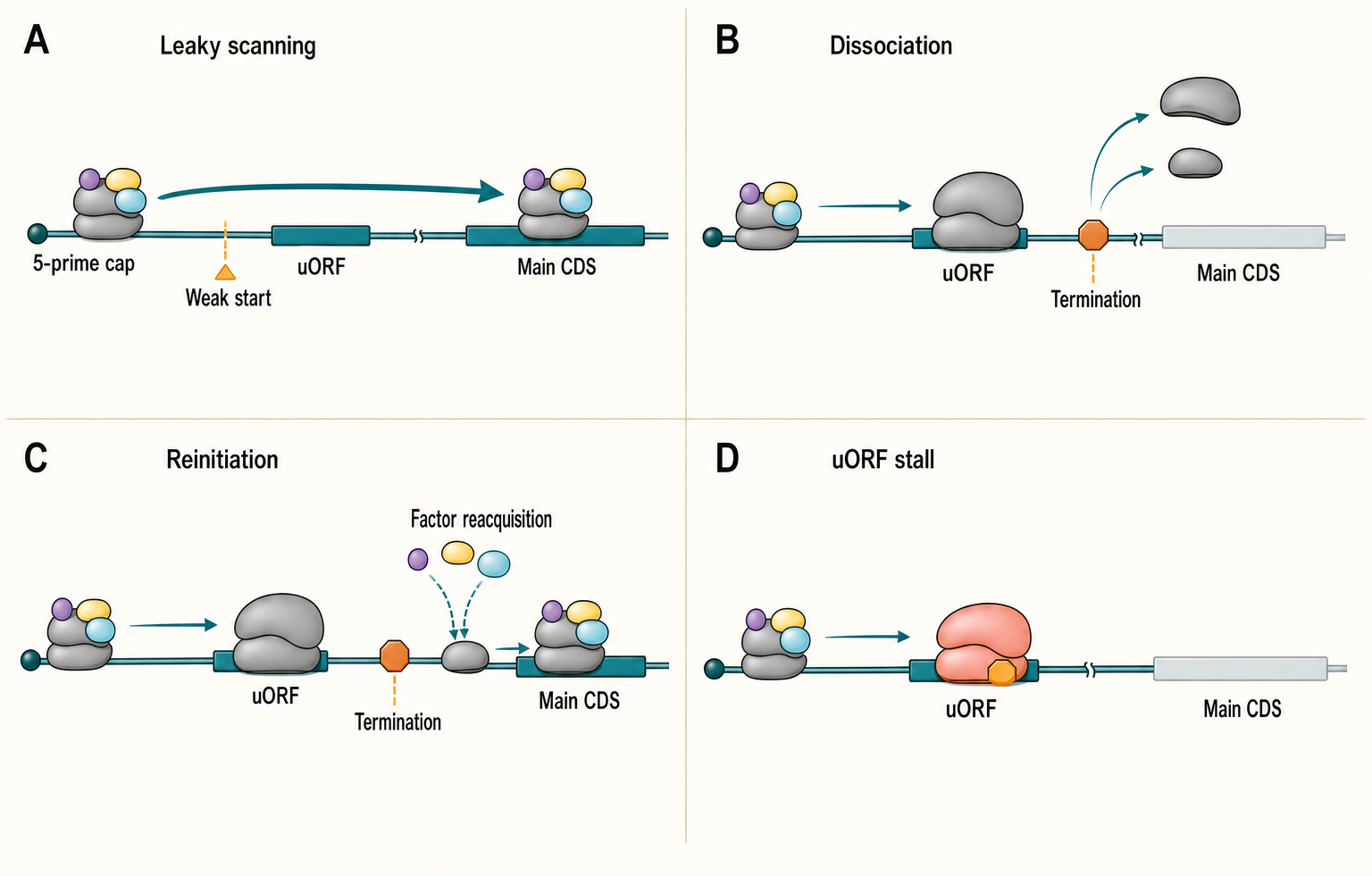
Figure 70.1. uORF Outcomes and Regulated Start-Site Choice. A cap-loaded scanning preinitiation complex can bypass a weak upstream start, initiate and terminate before dissociating, remain associated and reinitiate downstream, stall within a regulatory uORF, or select an alternative start that changes protein amino-terminal identity. Start context, uORF length, termination context, intercistronic distance, RNA structure, ternary-complex availability, and metabolite-sensitive nascent-peptide interactions shift the distribution among these outcomes. A side annotation hands assembly of the canonical scanning complex to Chapter 67 rather than duplicating its machinery diagram.
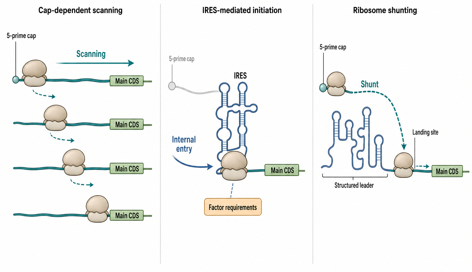
Figure 70.2. Cap-Dependent Scanning, IRES-Mediated Initiation, and Ribosome Shunting. Three mechanistically distinct routes to translation initiation are contrasted side by side. In cap-dependent scanning, the small ribosomal subunit is recruited near the 5′ cap and moves linearly through the leader until a suitable start codon is recognized. The IRES panel branches into a compact dicistrovirus intergenic-region fold with exceptionally low factor requirements, an extended hepatitis C virus-like fold with direct 40S contact and a reduced factor set, and an ITAF-remodeled picornavirus RNP. In ribosome shunting, the complex loads near the 5′ end but bypasses a structured or extended leader segment before resuming start-site selection downstream. Distinguishing these routes requires evidence about where the ribosome is first recruited, how initiation-factor requirements differ, whether cap recognition is necessary, and whether RNA structure remains active only in its protein-bound RNP state.
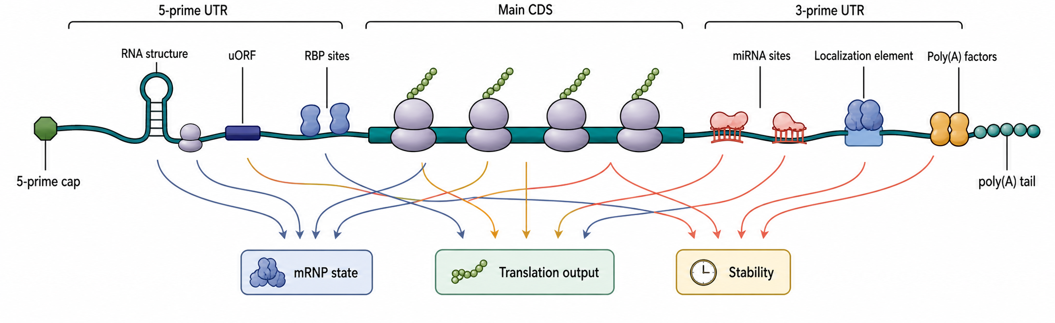
Figure 70.3. UTR Regulatory Grammar. An annotated mRNA map illustrates how multiple regulatory features coexist on a single transcript. The 5′ UTR can carry structures, upstream open reading frames, TOP-like sequence features, alternative starts, and RBP sites that remodel initiation competence. ISR and mTORC1 inputs connect to the leader features they regulate. The 3′ UTR contains microRNA sites, localization elements, and poly(A)-tail-associated factors that affect initiation indirectly. Translation output is determined by the combined messenger ribonucleoprotein and signaling state rather than by one motif in isolation.
Scanning preinitiation complexes are finite molecular machines, not abstract cursors. A complex recruited near the cap moves through the leader while carrying, or becoming competent to carry, the eIF2–GTP–methionyl initiator tRNA ternary complex. Start selection commits the small subunit to initiation, large-subunit joining, and elongation through the selected ORF. After a uORF terminates, both subunits may dissociate; alternatively, the 60S subunit may leave while a 40S-derived complex remains on the mRNA, resumes scanning, reacquires factors and ternary complex, and reinitiates downstream. The probability of that sequence depends on uORF length, the factor state of the post-termination complex, the distance to the next start, and the time required to reacquire ternary complex. These features make reinitiation a kinetic competition rather than a property conferred by the word “uORF” alone.
Three outcomes must be distinguished. In leaky scanning, the scanning complex never initiates at the candidate upstream start, often because the start codon or its surrounding context is weak. In reinitiation, a ribosome translates the uORF, terminates, and a retained small-subunit complex later initiates again. In ribosome stalling, translation of the uORF itself is slowed at elongation or termination, sometimes because a nascent regulatory peptide responds to a metabolite. These mechanisms can yield the same endpoint—less protein from the main coding sequence—but predict different footprints and different responses to start-codon, stop-codon, peptide-sequence, or intercistronic-distance mutations.
A strong uORF AUG close to the cap commonly diverts many scanning complexes. A weak upstream start permits a fraction to scan through, and a very short uORF can favor later reinitiation. Those rules are tendencies, not universal laws. A stable RNA structure can change scanning dwell time; an RNA-binding protein can mask a start or remodel the leader; alternative transcription can add or remove a uORF; and a change in ternary-complex concentration can alter how far a post-termination complex scans before it becomes initiation-competent. The same genomic leader may consequently produce different start-site distributions in different tissues or stress states. Massively parallel leader assays establish broad sequence rules, but endogenous isoform choice and RNP context can shift the quantitative result (Sample et al. 2019; Lewis et al. 2025).
The activating transcription factor 4 (ATF4) leader is the canonical mammalian example of a ternary-complex-sensitive switch. Under basal conditions, ribosomes translate a short permissive uORF1, retain reinitiation competence, quickly reacquire ternary complex, and initiate at inhibitory uORF2, which overlaps the ATF4 coding sequence out of frame. Under stress, phosphorylation of eIF2α lowers the rate at which active ternary complex is regenerated. Many post-uORF1 complexes therefore pass the uORF2 start before becoming competent again and instead initiate at the downstream ATF4 coding sequence (Vattem and Wek 2004; Wek et al. 2023). Stress does not make ribosomes “prefer” ATF4 by recognizing ATF4 protein function; it changes a kinetic interval encoded by the leader.
ATF4 is a model, not a universal template. GCN4 in budding yeast uses four uORFs and related delayed-reinitiation logic, whereas mammalian CHOP/DDIT3, ATF5, GADD34/PPP1R15A, and other stress-responsive transcripts use different combinations of start context, uORF overlap, peptide-dependent effects, bypass, and reinitiation. GADD34 is especially informative because its stress-induced expression helps restore translation: its transcription rises through the ISR, its own leader promotes preferential translation during eIF2α phosphorylation, and the resulting GADD34–protein phosphatase 1 complex dephosphorylates eIF2α (Lee et al. 2009; Young et al. 2015). Thus, uORF logic can control both entry into and recovery from a stress program.
High-throughput data have changed how uORFs are viewed. Ribosome profiling and initiation profiling often reveal ribosome-protected fragments in 5′ leaders, including many translated regions not represented in older coding annotations. A high-resolution human translation map showed widespread translation outside annotated canonical coding sequences, supporting the idea that upstream translation is a common layer of transcript annotation rather than a rare exception (Chothani et al. 2022). Viral RNAs can also encode regulatory upstream ORFs. Zika virus 5′ upstream open reading frames have been reported to affect infection of human brain cells, illustrating that upstream translation can alter viral fitness and host-cell interaction rather than only fine-tune host gene expression (Lefevre et al. 2024).
The evidence basis for a functional uORF should be explicit. Ribosome-protected fragments over a leader support ribosome occupancy, especially when footprints show triplet periodicity, initiation peaks, and coherent start and stop boundaries. Initiation profiling can improve start assignment, but drugs used to arrest initiation can redistribute ribosomes or reveal sites that are rarely used without treatment. Reporter assays in which the uORF start, stop, peptide sequence, or spacing is altered test causality. Endogenous genome editing is stronger than plasmid-only work because it preserves the native transcription start site, cap context, RNP history, and isoform mixture.
No single mutation distinguishes all uORF mechanisms. Removing an AUG abolishes the act of translation but may also alter local RNA structure or an RBP site. Moving the stop codon changes uORF length and reinitiation spacing. Synonymous substitutions can preserve peptide identity while altering elongation kinetics; nonsynonymous substitutions can test peptide identity but also change codon-dependent pausing. A strong design therefore uses multiple orthogonal perturbations and measures leader footprints, main-ORF initiation, mRNA abundance, and nascent protein. Rescue by compensatory structure mutations or by restoring spacing can discriminate RNA, peptide, and scanning effects.
Artifact control is essential. A leader footprint can arise from nonribosomal protection, queued scanning complexes, stalled elongating complexes, an overlapping annotation, or an unrecognized transcript start. A reporter can create a false effect if cloning changes the 5′ end, removes native structure, introduces cryptic transcription or splicing, or changes mRNA abundance. Proteomics may miss a genuine micropeptide because short products are unstable and yield few unique peptides, whereas peptide detection alone does not show cis-regulation. A uORF should therefore be annotated separately as “translated,” “regulatory,” and “peptide-producing” unless evidence supports all three labels.
The boundary between a uORF and an alternative coding sequence can be blurry. Some uORFs encode functional microproteins whose activity is independent of the downstream ORF. Others encode peptides whose function is to regulate the ribosome during translation. Still others may be tolerated translation with no demonstrated function. Conservation of position, start context, peptide sequence, or response supports function, but lack of conservation does not exclude lineage-specific regulation. Conversely, conserved leader sequence can reflect RNA structure rather than peptide function.
Regulated start-site choice extends beyond uORFs. Leaky scanning can expose a downstream in-frame AUG and produce a shorter protein isoform; near-cognate initiation can add an amino-terminal extension; and alternative leaders can change which starts are accessible. Initiation-site profiling, N-terminal proteomics, and targeted editing are complementary: profiling maps candidate starts, proteomics asks whether stable isoforms result, and editing tests whether start context controls phenotype. Chapter 67 owns the assembly and structural mechanics by which initiation factors recognize a start; this section owns how leaders distribute that machinery among competing starts. Chapter 69 begins after initiation and owns programmed frameshifting, stop-codon readthrough, and other recoding events.
70.2. IRESs, ribosome shunting, and cap-independent initiation
Most eukaryotic mRNAs use cap-dependent initiation under favorable growth conditions, but many RNAs can be translated when conventional cap-dependent recruitment is limited or bypassed. Cap-independent initiation is a broad term for initiation that does not rely primarily on standard 5′ cap recognition. It includes internal ribosome entry, factor-reduced viral initiation, initiation promoted by RNA structures or RNA modifications, and other mechanisms. The term should be used carefully because “cap-independent” describes an experimental observation or mechanistic route, not a single molecular pathway.
An internal ribosome entry site is an RNA element that recruits a ribosome internally. The strongest mechanistic examples come from viruses. Some viral IRES elements contain structured RNA domains that position the ribosome at or near the start codon. Some require subsets of canonical initiation factors; others recruit ribosomal subunits with unusually direct RNA-ribosome contacts. Picornavirus, hepatitis C virus-like, and dicistrovirus intergenic-region IRESs illustrate that the label covers several structural and factor-requirement classes rather than one conserved fold (Plank and Kieft 2012). These viral elements show that folded RNA can substitute for parts of the canonical cap-scanning pathway and that viral protein synthesis can persist when host cap-dependent initiation is restricted.
IRES is therefore a functional category, not a universal RNA motif. Dicistrovirus intergenic-region IRESs form compact, pseudoknot-rich folds that can engage ribosomal subunits with exceptionally low initiation-factor requirements. Hepatitis C virus-like IRESs use a more extended domain organization to contact the 40S subunit directly while recruiting a reduced factor set that includes eIF3. Many picornavirus IRESs depend more heavily on initiation factors and on IRES trans-acting factors, or ITAFs, that bind and remodel or stabilize the active RNA conformation. Across these classes, primary sequence, secondary structure, tertiary packing, and protein-dependent RNP remodeling can each determine activity. A computational search for one sequence pattern or one predicted fold therefore cannot identify all IRESs, and structural resemblance alone does not establish internal initiation (Plank and Kieft 2012).
Cellular IRES claims are more controversial. Many cellular mRNAs have been proposed to contain IRES-like elements, especially transcripts translated during stress, apoptosis, mitosis, hypoxia, or differentiation. Some claims are biologically plausible, but the experimental standards are high. A bicistronic reporter can appear to show internal initiation when a cryptic promoter, cryptic splice site, RNA cleavage event, or altered mRNA stability creates a monocistronic message for the downstream cistron. A leader cloned into a reporter may behave differently from the endogenous transcript because RNA structure, RBP occupancy, transcript end formation, and nuclear history differ. A strong cellular IRES claim should therefore combine multiple assays: promoterless controls, splice and RNA integrity checks, endogenous perturbation, structure-function mutation, factor-dependence tests, and translation-specific measurement.
Ribosome shunting differs from internal entry. In shunting, the ribosome generally loads near the 5′ end, scans a short region, bypasses a structured segment or sequence block, and lands downstream. The mechanism can involve leader structures, short upstream ORFs, base pairing, viral or cellular proteins, and spacing constraints. In the cauliflower mosaic virus 35S leader, translation and termination of a cap-proximal short ORF prepare a reinitiation-competent complex to bypass a stable structured region, providing direct mechanistic evidence for a shunt rather than an IRES (Ryabova and Hohn 2000). The important distinction is that shunting retains cap-proximal loading, whereas internal entry does not require ribosome loading at the 5′ end.
Table 70.1. Evidence Standards for Translational-Regulation Claims. Evidence tiers, common artifacts, and preferred controls for major categories of translational-regulation claims discussed in this chapter.
| Claim type | Minimum evidence | Stronger evidence | Common artifacts | Preferred controls |
|---|---|---|---|---|
| uORF represses main ORF translation | Reporter output lower with uORF present; start-codon mutation rescues | Endogenous genome editing increases protein without changing mRNA; ribosome profiling shows uORF footprints and reduced main-ORF initiation | Reporter construction alters 5′ end; start-codon mutation disrupts RNA structure or RBP site | RNA abundance measurement alongside protein; synonymous uORF mutations preserving translation |
| uORF encodes regulatory peptide | Synonymous mutations changing peptide sequence alter regulatory output | Peptide conservation across species; ribosome-tunnel stalling assay; cross-species rescue | Start-codon mutation removes both translation and peptide; conflates act-of-translation with peptide-identity effect | Conservative versus radical amino-acid substitutions; independent peptide detection |
| Cellular leader contains an IRES | Downstream cistron expressed from bicistronic reporter under conditions limiting cap-dependent initiation | Promoterless control passes; RNA integrity, splice, and stability checks pass; endogenous leader perturbation validated | Cryptic promoter, cryptic splice site, or RNA cleavage creates monocistronic downstream message | Promoterless bicistronic construct; Northern blot or long-read RNA check; reverse-orientation leader control |
| Ribosome shunting occurs | Translation requires 5′ region but bypasses internal structured segment; deletion of bypass element reduces downstream expression | Mapped 5′ loading site and downstream landing site; cap-dependence confirmed; factor requirements distinct from internal entry | Leaky scanning, IRES activity, or RNA cleavage misidentified as shunting | Insertional analysis throughout leader; intact 5′ end requirement test; IRES controls |
| UTR structure regulates translation | Predicted or probed stem-loop correlates with reduced reporter translation; disruption mutations partly restore output | Compensatory base-pair rescue restores repression; in-cell chemical probing confirms structure; endogenous protein output measured | Disruption mutations alter RBP sites or sequence context; predicted structure absent in cells | Compensatory rescue mutations; in-cell structure probing; multiple independent disruption sites |
| RBP directly regulates initiation | RBP depletion or overexpression changes translation without equivalent mRNA abundance change; binding site maps to UTR | Binding-site mutation abolishes effect; ribosome profiling with RBP perturbation shows initiation change; binding-dead RBP fails to rescue | RBP affects mRNA stability, nuclear processing, or localization rather than initiation; indirect effects through other targets | Binding-dead RBP rescue; RNA abundance measurement; polysome or ribosome profiling |
| MicroRNA causes translation repression | Seed-matched reporter repressed; seed-match mutation rescues protein output | Early nascent protein decline precedes mRNA decay in time course; endogenous seed-match genome editing recapitulates effect | Indirect network effects through secondary targets; reporter lacks native RBP competition; overexpression forces nonphysiological interactions | Metabolic labeling at early time points; ribosome profiling plus matched RNA-seq; endogenous seed-match perturbation |
| Stress granule localization reflects translation status | mRNA cosegregates with stress granule markers by imaging or fractionation under stress | Translation inhibition or activation shifts granule entry; ribosome profiling shows occupancy change for granule-enriched transcripts | Active and inactive mRNAs can coexist in granules; fixation or fractionation artifacts; granule entry and translation status not identical | Paired polysome or ribosome profiling; drug controls that block or restore translation; granule-marker colocalization with translation reporter |
| ISR or mTORC1 directly regulates initiation of a transcript | Pathway perturbation changes protein synthesis with matched RNA abundance | Endogenous leader-feature mutation removes the selective response; initiation profiling and rescue connect signaling to start choice | Secondary transcription, RNA decay, cell-cycle change, or altered elongation mimics initiation regulation | Time-resolved RNA and nascent-protein measurement; pathway-rescue control; leader-feature perturbation |
Cap-independent initiation and shunting have several biological uses. Viruses use them to compete with host translation, compact regulatory information into structured leaders, and maintain protein synthesis during host shutoff. Cellular mRNAs may use noncanonical initiation during stress states when cap-dependent initiation is inhibited. Some mRNA features can make translation less dependent on cap recognition even without qualifying as a classical IRES. For example, highly structured leaders, modified bases, specialized RBPs, or nearby open reading frames can shift how initiation factors engage an RNA. The chapter boundary with Chapter 69 is important: programmed frameshifting and stop-codon readthrough alter decoding after initiation, whereas IRESs and shunting alter how ribosomes enter the message or select a start region.
The local reference file for this chapter contains recent papers on ribosome-associated mRNA decay, stress granule localization, exosome-ribosome structure, and reading-frame maintenance (Hopfler et al. 2023; Helton et al. 2025; Kogel et al. 2024; Milicevic et al. 2024). These papers do not substitute for canonical IRES and shunting literature, but they support an important modern theme: initiation route, ribosome occupancy, mRNA degradation, and subcellular partitioning are coupled. An mRNA that bypasses one initiation barrier may still be subject to ribosome-associated decay, collision surveillance, or localization control. A translation mechanism should therefore be interpreted in the context of the entire mRNP life cycle.
The main misconception is that poor cap dependence automatically proves internal initiation. A transcript can appear cap-independent because it has a long poly(A) tail, stabilizing RBPs, alternative cap structures, cap-proximal initiation retained under partial inhibition, altered mRNA abundance, or indirect stress effects. Conversely, a genuine IRES may still require initiation factors and may be sensitive to cell state. “IRES” is not a synonym for “translated during stress.” The evidence must show where and how the ribosome is recruited.
Box 70.1. Why Bicistronic Reporters Can Mislead
- A bicistronic reporter contains two cistrons separated by the candidate leader sequence; only genuine internal initiation should produce downstream cistron protein when cap-dependent translation of the upstream cistron is intact.
- Cryptic promoters within the inserted leader can drive transcription of a monocistronic downstream-cistron mRNA, producing protein without any internal ribosome recruitment.
- Cryptic splice sites can generate a shorter mRNA in which the downstream cistron is repositioned near the 5′ cap and translated by normal scanning.
- RNA cleavage events can produce a capped 5′ fragment that contains the downstream cistron, allowing cap-dependent translation to mimic internal initiation.
- Altered RNA stability between bicistronic and monocistronic forms can inflate or deflate apparent IRES activity independently of any initiation mechanism.
- Required controls: promoterless bicistronic construct; Northern blot or long-read RNA sequencing to confirm RNA integrity; reverse-orientation leader as negative control; splice-junction analysis; endogenous transcript validation of the claimed initiation event.
70.3. UTR features, RNA structure, and RBP control of initiation
The untranslated regions of an mRNA are not inert spacers. The 5′ UTR controls how a ribosome is recruited and how easily it reaches a start codon. The 3′ UTR controls translation more indirectly by recruiting proteins, microRNAs, localization factors, decay machinery, and poly(A)-tail regulators. Together, the UTRs define much of the regulatory identity of an mRNA. The same coding sequence can produce different amounts of protein when attached to different leaders or 3′ UTRs, which is why UTR design matters in expression vectors and therapeutic mRNAs.
RNA structure is a central UTR feature. A stable stem-loop near the cap can reduce initiation by preventing cap-binding factors or the preinitiation complex from engaging the mRNA. A stable structure downstream in the 5′ leader can slow scanning, alter start-codon selection, or force ribosomes toward shunting-like routes. However, not every predicted structure blocks translation. Structures fluctuate, proteins remodel them, helicase activity can unwind them, and the position of a structure matters. A weak or transient structure can even support regulation by creating a binding platform for an RBP or by placing start codons in a favorable context. Leppek and colleagues reviewed functional 5′ UTR structures and emphasized that structure discovery must combine prediction, experimental probing, and functional assays (Leppek et al. 2018).
RNA-binding proteins regulate translation through several causal routes. A repressive RBP can bind near the cap and block recruitment, bind within the leader and impede scanning, stabilize an inhibitory structure, recruit deadenylation factors, or promote storage in a nontranslating mRNP state. An activating RBP can recruit initiation factors, bridge the 5′ and 3′ ends, displace a repressor, remodel a structure, or protect the mRNA from decay long enough for translation to occur. Some RBPs are conditional switches: phosphorylation, methylation, ligand binding, phase separation, or localization changes their effect on target mRNAs.
The physical position of the binding site matters. A protein bound directly over a start codon can block start-site recognition. A protein bound upstream can create a roadblock for scanning. A protein bound in the 3′ UTR may affect translation by recruiting a regulatory complex rather than by physically blocking the ribosome. A protein bound in the coding sequence can alter elongation, ribosome collisions, cotranslational targeting, or decay, which may indirectly change apparent translation efficiency. These mechanisms are experimentally separable only when translation measurements are paired with mRNA abundance, ribosome distribution, protein output, and binding-site perturbation.
Specific examples illustrate the coupling between structure, translation, and decay. Tubulin autoregulation involves ribosome-associated recognition of nascent tubulin and mRNA degradation; recent work has clarified mechanisms of ribosome-associated mRNA degradation in this setting (Hopfler et al. 2023). Human exosome-ribosome supercomplex structures show that decay machinery can physically associate with ribosome-linked substrates, reinforcing that translation state and RNA turnover can be part of one regulatory event rather than separate downstream consequences (Kogel et al. 2024). These examples are not simply UTR regulation, but they warn against interpreting reduced protein output as a pure initiation defect when ribosome-associated decay is plausible.
Antisense oligonucleotides and other designed molecules provide useful mechanistic probes. A study of secondary structures that regulate mRNA translation in cardiac hypertrophy used antisense oligonucleotide modulation to connect RNA structure with translational output (Hedaya et al. 2023). Such experiments are powerful because they can perturb structure without changing the whole transcript, but they require controls for RNase H recruitment, steric effects, immune activation, off-target binding, and altered RNA stability. Designed perturbations are strongest when multiple independent oligonucleotides, compensatory mutations, and orthogonal structure assays converge.
A major boundary case is the difference between predicted and observed structure. Computational folding can identify possible stems, but cellular RNA structure depends on temperature, ion conditions, co-transcriptional folding, RNA modifications, helicases, RBPs, ribosome traffic, and compartment. Chemical probing can reveal nucleotide accessibility, but accessibility is influenced by protein binding as well as base pairing. Therefore, a statement such as “this UTR structure represses translation” should specify the evidence: prediction, in vitro probing, in-cell probing, mutational disruption, compensatory rescue, reporter effect, or endogenous protein output.
UTR regulation also intersects with transcript isoforms. Alternative transcription start sites can create shorter or longer 5′ UTRs, adding or removing uORFs and structures. Alternative polyadenylation can shorten or lengthen 3′ UTRs, changing RBP and miRNA site content. A cell-type-specific isoform may therefore translate differently even when the coding sequence is unchanged. This chapter focuses on translation mechanisms, while Chapter 18 covers transcript annotation, Chapter 29 covers alternative polyadenylation, and Chapter 72 integrates full mRNA architecture.
70.4. miRNA and small-RNA effects on translation
MicroRNAs are short regulatory RNAs, usually about 20 to 24 nucleotides long in animals, that guide Argonaute-containing complexes to target RNAs. The most common animal targeting rule uses a seed region near the 5′ end of the microRNA to recognize partially complementary sites, often in the 3′ UTR of an mRNA. Once recruited, the microRNA-induced silencing complex can reduce protein output by repressing initiation, promoting deadenylation, promoting decapping, accelerating mRNA decay, or changing mRNP composition. These outputs are often coupled, so the dominant measurement depends on timing and assay design.
The causal sequence can be described in steps. First, a mature microRNA is loaded into an Argonaute protein. Second, the Argonaute-microRNA complex samples RNAs and stabilizes interactions with sites that match the microRNA seed and have favorable context. Third, the bound complex recruits effector proteins, including factors that interact with poly(A)-tail metabolism and decapping pathways. Fourth, translation may decline before large mRNA abundance changes are detected, or mRNA decay may dominate the steady-state effect. The same target can show an early translation effect and a later RNA abundance effect. A single endpoint measurement cannot resolve that sequence.
MicroRNA regulation is frequently modest at the level of each individual target but broad across a gene network. This makes microRNAs well suited to tuning developmental transitions, cell-state maintenance, immune responses, and stress adaptation. It also creates interpretation challenges. A microRNA overexpression experiment can force nonphysiological interactions. A microRNA knockout can cause indirect effects through transcription, cell-state changes, or feedback loops. Reporter assays with isolated 3′ UTR fragments may exaggerate or miss effects because endogenous transcript abundance, isoform choice, RBP competition, and subcellular localization are missing.
Small interfering RNAs, PIWI-interacting RNAs, bacterial small RNAs, and plant small RNAs follow different rules. Small interfering RNAs with extensive complementarity often direct endonucleolytic cleavage of target RNA, especially in systems with slicing-competent Argonaute proteins. Plant microRNAs frequently have higher complementarity to targets than animal microRNAs and can direct slicing as well as repression. Bacterial small RNAs usually do not use Argonaute; many pair with target mRNAs with help from bacterial RNA chaperones and can directly block or expose the ribosome-binding site. Comparing these systems is useful because it separates the general principle of small-RNA-guided regulation from the specific protein machinery used in each domain or lineage.
Mechanistic work places Argonaute and GW182/TNRC6 at the center of animal microRNA silencing. Argonaute uses the small RNA to recognize the target; GW182/TNRC6 proteins then recruit interactions with poly(A)-binding protein and the PAN2–PAN3 and CCR4–NOT deadenylation machinery. Translation repression and accelerated deadenylation can therefore be coupled outputs of the same target-bound complex rather than mutually exclusive explanations (Chen et al. 2009; Huntzinger et al. 2013). Which output is detected first depends on target, cell type, time resolution, and the method used to measure nascent protein and RNA abundance.
Evidence for small-RNA translational regulation should distinguish target binding from output. CLIP-family data can identify Argonaute binding sites, but binding alone does not prove repression. RNA sequencing can show mRNA abundance changes, but a translation-specific effect requires ribosome profiling, polysome analysis, metabolic labeling, reporter kinetics, or protein measurement. Mutating a seed match in the endogenous 3′ UTR is stronger than deleting a large region because it limits collateral changes. Rescue by restoring the target site or changing the microRNA seed can strengthen causality. Time-course data are especially valuable because translation repression and decay can occur on different time scales.
Several misconceptions recur. First, microRNAs do not always “block ribosomes” physically; many effects are mediated through recruitment of effector complexes that change initiation competence, tail length, or RNA stability. Second, a predicted seed match is not a proven target. Site accessibility, isoform usage, RBP competition, microRNA abundance, and cell type matter. Third, a protein decrease after microRNA expression is not necessarily direct; microRNAs regulate networks, and secondary transcriptional effects are common. Fourth, small-RNA effects on translation are not limited to animals; plants, fungi, protists, bacteria, and viruses contain diverse small-RNA systems, but their mechanisms should not be collapsed into one animal microRNA model.
Box 70.2. Translation Repression Versus mRNA Decay in microRNA Experiments
- microRNAs can reduce protein output through multiple coupled mechanisms: repression of translation initiation, deadenylation, decapping, and mRNA decay, which can occur sequentially on the same target.
- A time-course experiment can distinguish the sequence: early reduction in nascent protein synthesis detectable by metabolic labeling can precede a later decline in mRNA abundance, revealing translation repression as the initial event.
- A steady-state experiment measuring only mRNA abundance cannot determine whether translation repression or RNA decay is primary; both outcomes lower protein levels.
- A single-endpoint protein measurement conflates all mechanisms and provides no information about the order or relative contribution of translation versus decay.
- Endogenous seed-match mutation combined with metabolic labeling and matched RNA-seq across a time course provides the strongest mechanistic distinction.
- The dominant measurable effect varies by microRNA, target transcript, cell type, and time point; no single mechanism applies universally across microRNA-target pairs.
70.5. Integrated stress response, mTOR signaling, localization, and developmental control
Cells rarely regulate translation one transcript at a time in isolation. Stress, nutrients, growth signals, localization, and development change the environment in which all mRNAs compete for ribosomes, initiation factors, RNA-binding proteins, and subcellular space. Regulation is therefore both global and selective. A stress can lower total protein synthesis while increasing translation of a small group of leaders. A developing embryo can store a maternal mRNA and activate it at a defined stage. A neuron can transport an mRNA in a repressed particle and translate it near a stimulated synapse. The global input changes the available machinery; transcript architecture and RNP state determine which mRNAs respond.
The eIF2–eIF2B control point
The integrated stress response (ISR) controls initiation through the eIF2 nucleotide cycle. eIF2 bound to GTP and initiator methionyl-tRNA forms the ternary complex that delivers the initiator tRNA to a preinitiation complex. Following start recognition and GTP hydrolysis, eIF2 leaves in a GDP-bound state. The guanine-nucleotide exchange factor eIF2B releases GDP so that GTP can bind and a new ternary complex can form. eIF2B is therefore not merely another binding partner: it is the dedicated recycling catalyst that determines how rapidly inactive eIF2–GDP returns to the initiation-competent pool (Kenner et al. 2019; Marintchev and Ito 2020).
Four stress-responsive kinases converge on serine 51 of the eIF2α subunit. Phosphorylation changes eIF2 from an eIF2B substrate into a high-affinity inhibitor that occupies a regulatory surface rather than productively undergoing nucleotide exchange. Because eIF2B is less abundant than eIF2, phosphorylation of a fraction of the eIF2 pool can disproportionately depress exchange. The resulting fall in eIF2–GTP and ternary complex reduces bulk initiation and delays reacquisition of ternary complex by scanning post-uORF complexes. Structural and biochemical work has resolved this substrate-to-inhibitor switch and explains why eIF2α phosphorylation can be amplified at the nucleotide-exchange step (Kenner et al. 2019). This chapter owns the regulation and transcript selection; Chapter 67 owns assembly of the ternary complex, preinitiation complex, scanning apparatus, and start-recognition machinery under the canonical baseline.
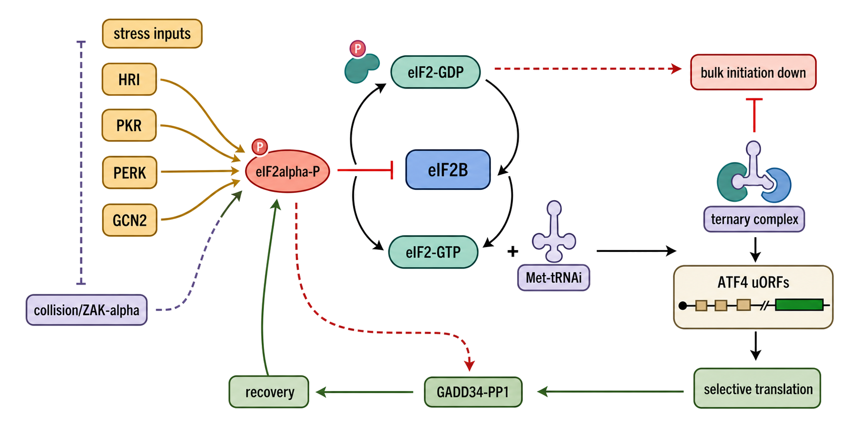
Figure 70.4. The eIF2–eIF2B ISR Cycle from Stress Input to Recovery. A causal cycle begins with eIF2–GDP after start recognition. eIF2B promotes GDP release, GTP binding permits formation of eIF2–GTP–Met-tRNAi ternary complex, and ternary complex supplies the next initiation event. Four color-coded input arms converge on eIF2α serine 51: HRI/EIF2AK1 for heme/iron and selected DELE1-linked mitochondrial stresses; PKR/EIF2AK2 for RNA ligands that productively organize kinase activation; PERK/EIF2AK3 for ER folding stress; and GCN2/EIF2AK4 for amino-acid/tRNA-charging and P-stalk-linked signals. The PKR inset contrasts productive co-occupancy of one RNA with excess-ligand dilution of PKR across separate RNAs. Phospho-eIF2 occupies eIF2B nonproductively, lowers ternary complex, and suppresses bulk initiation. A magnified ATF4 leader shows delayed reinitiation past inhibitory uORF2. ATF4-dependent gene expression and preferential GADD34 translation lead to GADD34–PP1-mediated eIF2α dephosphorylation and recovery. A side boundary points canonical factor assembly to Chapter 67 and distinguishes the catalytic ZAKα–p38/JNK arm from the collision-linked GCN2 intersection owned by Chapter 71.
The ISR is graded and dynamic. “ISR on” and “ISR off” are useful shorthand, but cells experience different amplitudes and durations of kinase activity, phosphatase activity, eIF2B capacity, and ternary-complex depletion. A modest pulse can redistribute initiation without extinguishing protein synthesis. Severe or sustained phosphorylation can prevent synthesis of proteins needed for adaptation, and prolonged ATF4–CHOP output can participate in cell death. The direction of a phenotype therefore depends on stress identity, strength, duration, cell type, and the capacity to recover, not simply on whether phospho-eIF2α is detectable (Wek et al. 2023).
Four upstream kinases, four overlapping input domains
HRI, PKR, PERK, and GCN2 share eIF2α as a substrate but do not constitute four interchangeable labels for generic stress. Their canonical inputs provide a useful map, with important boundary cases.
- HRI/EIF2AK1 was defined as the heme-regulated inhibitor that limits globin synthesis when heme or iron is insufficient in erythroid precursors. This coordination prevents production and aggregation of excess heme-free globin (Han et al. 2001). HRI also relays several mitochondrial perturbations. In the OMA1–DELE1 pathway, mitochondrial stress promotes DELE1 processing and cytosolic signaling to HRI, connecting organelle proteostasis to the ISR (Fessler et al. 2020). HRI should therefore not be described only as an erythroid heme sensor, but neither should every mitochondrial stress response be assigned to HRI without genetic evidence.
- PKR/EIF2AK2 is an interferon-inducible kinase activated by appropriate RNA ligands, classically double-stranded RNA produced during viral infection. RNA binding can organize two or more PKR monomers on one duplex, favoring kinase-domain dimerization, autophosphorylation, and subsequent eIF2α phosphorylation. In a defined in vitro series at 200 mM sodium chloride, 30 base pairs was the shortest duplex that both bound two PKR monomers and activated autophosphorylation; at lower ionic strength, two PKR molecules could occupy a 20-base-pair duplex. Activation was bell-shaped with ligand concentration because excess duplex distributed PKR molecules across separate RNAs rather than concentrating them on one ligand (Lemaire et al. 2008). These values are assay-dependent mechanistic evidence, not a universal cellular length threshold. RNA length, structure, concentration, ionic environment, protein partners, and viral antagonists all influence activation. “Viral RNA” is not synonymous with “PKR activator”: many viral RNAs evade, inhibit, or fail to organize active PKR complexes.
- PERK/EIF2AK3 is an endoplasmic-reticulum transmembrane kinase within the unfolded protein response. Accumulation of folding stress in the ER activates PERK, whose cytosolic kinase domain phosphorylates eIF2α and reduces the influx of newly synthesized client proteins into the secretory pathway (Harding et al. 1999). PERK is one branch of the unfolded protein response; IRE1 and ATF6 have distinct primary outputs. Measuring an ER-stress marker does not establish that a translational effect is PERK-dependent.
- GCN2/EIF2AK4 responds strongly to amino-acid insufficiency and impaired tRNA charging. Uncharged tRNA contributes to activation, and mammalian genetic and biochemical work also implicates the ribosomal P-stalk in coupling an empty decoding demand or stalled translation state to GCN2 (Harding et al. 2019). This dual input logic matters: amino-acid deprivation is more than a bulk metabolite concentration, and GCN2 activation should be tested rather than inferred from starvation treatment alone.
The four input domains overlap. Oxidative stress, mitochondrial dysfunction, infection, proteotoxic stress, and nutrient limitation can activate more than one kinase or change translation through pathways outside eIF2. Kinase knockout or acute depletion, rescue with catalytic mutants, phospho-eIF2α measurement, and matched protein-synthesis assays are needed to assign an input. Inhibitor-only studies are weaker because many stress-pathway inhibitors have dose- and cell-dependent off-target effects.
Selective outputs and programmed recovery
Ternary-complex limitation explains how a general biochemical restriction becomes leader-selective. Most mRNAs lose initiation opportunities. Leaders such as ATF4 instead contain uORF arrangements that translate efficiently only when ternary-complex reacquisition is delayed, as developed in the preceding uORF section. ATF4 protein then drives a transcriptional program involving amino-acid transport and synthesis, redox control, autophagy, feedback regulators, and context-dependent survival or death. Some outputs called “ISR targets” are translated directly through their leaders; others are transcriptional targets of ATF4 or CHOP; still others change secondarily because metabolism, mRNA abundance, or cell state has shifted. These categories should not be merged.
ATF4 is not the only selective translation output. CHOP/DDIT3, ATF5, GADD34/PPP1R15A, and additional messages have stress-responsive leaders, but their mechanisms differ. Some use delayed reinitiation, some use leaky scanning past an inhibitory uORF, and some combine transcriptional induction with preferential translation. A uORF census is not sufficient to predict direction because start context, overlap, spacing, peptide behavior, and transcript isoform all matter. Ribosome profiling during stress has shown pervasive leader translation, but altered leader occupancy can reflect scanning, pausing, or initiation-drug effects unless supported by endogenous perturbation and protein synthesis measurements.
Recovery is part of the mechanism, not an afterthought. ATF4 and related outputs induce GADD34, encoded by PPP1R15A; GADD34 recruits protein phosphatase 1 to promote dephosphorylation of eIF2α. GADD34 expression is itself reinforced by stress-dependent transcription and uORF-regulated translation. As phospho-eIF2α declines, eIF2B-mediated exchange and ternary-complex production recover, allowing broader translation to resume (Novoa et al. 2001; Young et al. 2015). CReP/PPP1R15B provides a more constitutive phosphatase-targeting activity that helps set basal phosphorylation. The ISR is thus a negative-feedback circuit with distinct activation, selective-expression, and recovery phases. An endpoint taken at one hour can describe a different regulatory regime from an endpoint taken after six hours.
mTORC1, eIF4F, and cap-factor competition
Mechanistic target of rapamycin complex 1 (mTORC1) regulates a different limiting interface. mTORC1 integrates amino acids, growth factors, cellular energy, and lysosomal signaling with anabolic growth. One direct translational branch controls eIF4F assembly through eIF4E-binding proteins (4E-BPs). eIF4E binds the 5′ cap; eIF4G is a scaffold that connects cap-bound eIF4E with other factors and the small-subunit recruitment apparatus; and eIF4A is an ATP-dependent DEAD-box RNA helicase that helps the recruitment and scanning machinery negotiate structured leaders. Together these components form the eIF4F complex. Chapter 67 explains how eIF4F participates in canonical recruitment; this section explains how signaling changes access to it.
When mTORC1 activity is low, hypophosphorylated 4E-BPs bind eIF4E and compete with eIF4G, limiting productive eIF4F assembly. Active mTORC1 phosphorylates 4E-BPs through a hierarchical series of sites, weakening their association with eIF4E and expanding cap-dependent initiation capacity. mTORC1 also controls S6 kinases and other translational and metabolic substrates, but phosphorylation of ribosomal protein S6 is not a sufficient proxy for every mTORC1-dependent initiation event. The clearest causal chain is mTORC1 state, 4E-BP phosphorylation and genotype, eIF4E–eIF4G engagement, transcript-specific initiation, and protein output (Thoreen et al. 2012; Liu and Sabatini 2020).
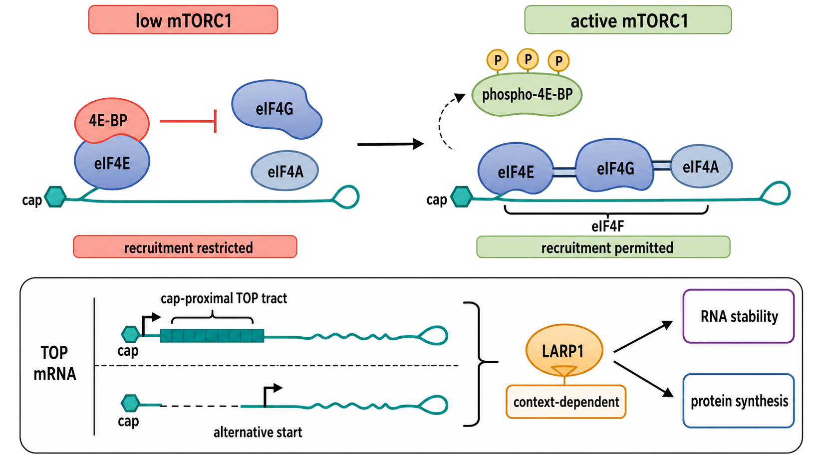
Figure 70.5. mTORC1, 4E-BPs, eIF4F, and TOP-mRNA Fate. Two states are contrasted. With low mTORC1 activity, hypophosphorylated 4E-BP occupies eIF4E and excludes eIF4G; eIF4F assembly and cap-associated recruitment are restricted. With active mTORC1, multisite 4E-BP phosphorylation releases eIF4E, allowing eIF4E, scaffold eIF4G, and helicase eIF4A to assemble as eIF4F. A TOP-mRNA inset places the oligopyrimidine tract at the exact cap-proximal transcription start and shows how alternative starts can create or remove TOP behavior. LARP1 is drawn as a context-dependent regulator of cap access, translation, and stability rather than as the sole pathway switch. Readout icons distinguish phospho-4E-BP, eIF4E–eIF4G association, TOP translation, RNA stability, and phospho-S6.
Sensitivity to cap-factor limitation is not uniform. Leaders with substantial structural or RNP-remodeling requirements may be especially dependent on eIF4A and eIF4F abundance, but leader length or predicted structure alone does not define an mTOR target. Acute, catalytic mTOR inhibition in mammalian cells strongly suppresses a transcript class marked by a 5′-terminal oligopyrimidine (TOP) motif. TOP mRNAs commonly encode ribosomal proteins and translation factors, so their coordinated regulation links nutrient availability to production of the protein-synthesis apparatus itself (Thoreen et al. 2012).
A TOP motif begins at the cap-proximal transcription start and contains a cytidine followed by a pyrimidine-rich tract. This positional definition matters. A pyrimidine-rich sequence buried inside a leader is not automatically a TOP motif, and alternative transcription start sites can create or remove TOP behavior in a tissue-specific manner. TOP regulation is also a continuum rather than an infallible binary classifier; motif composition, exact 5′ end, cap competition, and cellular context tune the response (Philippe et al. 2020).
LARP1 is a major TOP-mRNA regulator but should not be presented as a single unqualified switch. Its C-terminal DM15 region can recognize the cap-proximal TOP sequence and oppose eIF4F assembly, and several biochemical and genetic studies support translational repression when mTORC1 activity is low (Fonseca et al. 2015; Hong et al. 2017). However, LARP1 has also been assigned stabilizing and positive roles, and recent single-molecule and transcriptome analyses found that 4E-BP1/2 dominated acute translational repression while LARP1 preferentially protected TOP mRNAs from degradation in the tested cells (Hochstoeger et al. 2024). The defensible consensus is that 4E-BPs, cap-factor competition, and LARP1 jointly shape TOP-mRNA fate, with the relative contribution of direct translational repression and RNA stabilization depending on context and timescale.
Crosstalk, localization, and development
ISR and mTORC1 are not simple opposites. Amino-acid limitation can activate GCN2 while suppressing mTORC1; ER or viral stress can inhibit initiation without the same nutrient signal; and feedback from ATF4-induced metabolic genes can change amino-acid availability and later mTORC1 activity. Both pathways may suppress bulk initiation, but one limits ternary-complex regeneration and the other restricts cap-factor assembly. Because the molecular bottlenecks differ, an mRNA can be sensitive to one pathway, both, or neither. A factorial experiment that perturbs eIF2 phosphorylation and mTORC1 independently is more informative than treating “translation stress” as one condition.
Stress granules illustrate the connection between initiation and RNP state. These cytoplasmic assemblies are enriched in nontranslating mRNPs and translation-associated proteins under some stresses. An mRNA found in a granule is not necessarily permanently silenced, and absence from a granule does not prove productive translation. Ribosome association can oppose stress-induced granule localization, but imaging, translation reporters, and RNA abundance must be interpreted together (Helton et al. 2025). Granules are a partitioning outcome of a stressed RNP system, not a direct meter for eIF2B or mTORC1 activity.
Localization creates a spatial initiation switch. In polarized cells, neurons, oocytes, embryos, and migrating cells, an mRNA can be packaged with RBPs that recognize localization elements, couple the particle to motors, and keep initiation low during transport. Local signaling can remodel the RNP, phosphorylate a repressor, extend a poly(A) tail, or recruit initiation factors. The resulting protein is synthesized near the structure that needs it, reducing transport distance and allowing stimulus-specific control (Besse and Ephrussi 2008; Fernandopulle et al. 2021). A localized RNA is therefore not merely an RNA observed at a place; a mechanistic localization claim identifies the cis-element, trans-acting RBP or motor, translational state during transit, local activating cue, and local protein output.
Development uses the same logic over longer intervals. Maternal mRNAs can be stored with short poly(A) tails and translational repressors, then activated through cytoplasmic polyadenylation after fertilization. Differentiating cells can change leader isoforms, uORFs, cap-factor availability, mTORC1 state, microRNAs, and localization programs. The composition of a tissue also changes, so bulk translation measurements can confuse cell-type replacement with regulation inside one cell type. Developmental claims are strongest when matched RNA isoforms, ribosome occupancy, nascent protein, poly(A)-tail state, and spatial cell identity are measured.
Evidence, artifacts, disease, and intervention
Mechanistic measurement requires multiple layers. Phospho-specific immunoblotting reports eIF2α phosphorylation but not ternary-complex concentration. ATF4 protein or an ATF4-leader reporter integrates selective initiation but can also reflect transcription, protein turnover, or reporter context. Polysome collapse is consistent with reduced initiation but can also be altered by elongation inhibitors, RNA degradation, or cell lysis conditions. For mTORC1, phospho-S6, phospho-4E-BP, eIF4E–eIF4G association, and translation of TOP reporters measure different nodes. Rapamycin incompletely inhibits some mTORC1 outputs in many settings, whereas ATP-competitive inhibitors have broader and different consequences. Drug identity, dose, exposure time, and genetic controls must be reported.
Ribosome profiling provides transcriptome-wide occupancy but does not by itself measure completed protein. Under acute stress, elongation rates and ribosome run-off change; a transiently occupied leader can appear enriched as coding-region ribosomes depart. Translation-initiation inhibitors can create treatment-specific start peaks, and normalization to an RNA pool whose composition is changing can distort translation-efficiency ratios. Useful orthogonal measures include pulse labeling, quantitative proteomics, polysome fractionation, start-resolved profiling, eIF2B exchange assays, cap-pulldown or proximity assays for eIF4F, and endogenous leader editing. Time resolution is essential because activation and recovery can produce opposite effects at different sampling points.
Disease links follow from the centrality of these control points. Pathogenic variants in eIF2B cause vanishing white matter disease, demonstrating that nucleotide-exchange capacity is physiologically limiting, especially in glial stress responses. Excessive or prolonged ISR signaling is implicated in neurodegeneration, diabetes, cancer, and tissue injury, whereas an insufficient ISR can impair adaptation to infection or proteotoxic and nutrient stress. ISRIB-class compounds stabilize active eIF2B assemblies and can oppose effects of eIF2 phosphorylation in experimental systems, but pathway benefit is context-dependent: releasing the translational brake during unresolved stress can be harmful (Tsai et al. 2018; Marintchev and Ito 2020).
mTOR inhibitors and rapalogs have established clinical uses, yet a drug response cannot be attributed only to translation because mTORC1 also regulates autophagy, metabolism, and organelle programs. Conversely, hyperactive mTOR signaling in cancer, metabolic disease, and genetic syndromes changes many outputs beyond eIF4F. eIF4E–eIF4G and eIF4A are also experimental therapeutic targets, particularly in cancers dependent on structured growth-promoting leaders, but these factors support normal translation and their inhibition can have broad toxicity. Therapeutic interpretation should specify the molecular target, affected transcript class, exposure window, compensatory signaling, and evidence that altered initiation contributes to the phenotype.
Box 70.3. ISR Is Not a Synonym for Ribotoxic Stress
- The integrated stress response is defined here by eIF2α phosphorylation, inhibition of eIF2B nucleotide exchange, ternary-complex limitation, and leader-selective initiation.
- The ribotoxic stress response begins when collided ribosomes activate ZAKα/MAP3K20, which signals prominently through p38 and JNK; collision detection and quality-control mechanisms are owned by Chapter 71.
- GCN2 can respond to ribosome-linked states, and collision stress can produce eIF2α phosphorylation. In the Wu et al. system, ZAKα catalytic activity was required for p38/JNK activation but not for the collision-linked GCN2/eIF2α arm, separating a catalytic ribotoxic-stress output from a noncatalytic pathway intersection.
- Uncharged tRNA and P-stalk-linked inputs can activate GCN2 through routes that need not require ZAKα, so the collision experiment does not make ZAKα a fifth eIF2α kinase or a universal upstream component of GCN2 signaling.
- Discriminating experiment: combine disome/collision profiling, ZAKα and GCN2 genetics, phospho-p38/JNK, phospho-eIF2α, and nascent-protein measurements over a time course.
- Wording rule: report “ZAKα-dependent ribotoxic stress,” “GCN2/eIF2α-dependent ISR,” or both when both are demonstrated; avoid the uninformative umbrella phrase “translation stress pathway.”
Finally, the ISR must be distinguished from the ribotoxic stress response. In mammalian cell lines exposed to elongation inhibitors, intermediate inhibitor doses produced abundant RNase-resistant disomes and maximal p38/JNK and eIF2α phosphorylation, whereas high doses stalled ribosomes more uniformly and reduced the trailing-ribosome collisions needed for these signals. Selective ribosome profiling and fractionation further showed preferential association of ZAKα with colliding disomes. ZAKα kinase activity was required for the p38/JNK arm, but kinase-inactive ZAKα could support the collision-linked GCN2/eIF2α arm in the tested system, consistent with a noncatalytic ribosome-associated role in that branch (Wu et al. 2020).
This evidence establishes pathway intersection without making the pathways interchangeable. GCN2 can also respond to uncharged tRNA and ribosomal P-stalk inputs, so ZAKα is not universally required for every GCN2 response. Conversely, detection of eIF2α phosphorylation during collision stress does not turn ZAKα into a fifth eIF2α kinase. This chapter owns eIF2/eIF2B and mTOR-linked initiation control; Chapter 71 owns ribosome stalling and collision recognition, ZAKα signaling, quality control, and the ribotoxic stress response. The two systems can intersect, but they should not be collapsed into a single generic “translation stress” mechanism.
70.6. Quantitative models and measurement of initiation regulation
Translation is often discussed qualitatively, but experimental interpretation requires quantitative models. The number of protein molecules produced from an mRNA depends on mRNA abundance, initiation frequency, elongation speed, termination efficiency, ribosome recycling, mRNA half-life, protein maturation, and protein degradation. A regulatory feature can change any one of these variables. For example, a 5′ stem-loop may reduce initiation. A coding-region stall may increase ribosome density while reducing completed protein. A microRNA may lower initiation and later accelerate decay. A stabilizing RBP may increase protein output by increasing mRNA lifetime even if translation per unit time is unchanged.
Ribosome profiling is the dominant genome-wide method for mapping ribosome occupancy. In a typical experiment, translating ribosomes protect short mRNA fragments from nuclease digestion; protected fragments are sequenced and mapped to the transcriptome. Triplet periodicity, coding-sequence enrichment, start/stop behavior, and drug-dependent changes help infer translation. Ribosome profiling can reveal uORFs, alternative start sites, pausing, stress-specific programs, and changes in apparent translation efficiency. However, ribosome density is not identical to protein synthesis. Higher density can mean more initiation, slower elongation, ribosome stalling, or defective termination. Lower density can mean reduced initiation, faster elongation, transcript loss, or altered recovery.
Polysome profiling separates mRNAs by the number of ribosomes attached. Heavy polysome association often suggests efficient translation, while monosome or nonpolysome fractions suggest lower translation or storage. The method is valuable for broad shifts but has limited resolution. It cannot easily identify precise start sites, distinguish slow elongation from high initiation, or resolve isoform-specific features without sequencing. Reporter assays offer controlled perturbation of UTRs, uORFs, structures, and RBP sites, but they can be distorted by copy number, promoter context, RNA processing, plasmid backbone, and missing chromatin or localization history. Metabolic labeling and mass spectrometry measure protein synthesis more directly, but sensitivity, protein turnover, and peptide detectability introduce other biases.
Table 70.2. Measurement Methods and Interpretive Limits. Strengths, limitations, and recommended complementary assays for ten methods used to study translational regulation.
| Method | Direct readout | Strengths | Limitations | Best paired assay |
|---|---|---|---|---|
| Ribosome profiling | Ribosome-protected fragment positions and density | Transcriptome-wide; identifies uORFs, stall sites, alternative start sites, and apparent translation efficiency | Footprint density does not equal protein output; elongation and initiation signals are confounded; drug treatment alters ribosome distribution | RNA-seq for translation efficiency ratio; metabolic labeling to confirm protein synthesis |
| Translation initiation profiling | Initiating ribosome positions via drug-based run-off | Identifies start sites transcriptome-wide; separates initiation signal from elongation density | Drug redistributes ribosomes nonuniformly; near-cognate sites may be over- or under-represented depending on drug timing | Standard ribosome profiling; RNA-seq for abundance normalization |
| Polysome profiling | mRNA distribution across ribosome-number fractions | Detects broad shifts in ribosome loading; compatible with sequencing for transcriptome view | Cannot resolve stall positions; high initiation and slow elongation both yield heavy polysome fractions; limited isoform resolution | RNA-seq of gradient fractions; ribosome profiling for nucleotide resolution |
| Reporter assay | Protein or enzymatic output from transfected construct | Controlled UTR and sequence perturbation; suitable for mutagenesis and deletion scanning | Plasmid copy number, promoter context, and missing nuclear history; may not reflect endogenous mRNA behavior | RNA abundance measurement; endogenous genome editing for validation |
| Metabolic protein labeling | Newly synthesized protein over a defined time window | Time-resolved; measures active synthesis rather than steady-state abundance | Labeling period and analogue concentration can perturb translation; protein secretion and maturation affect timing | Ribosome profiling or RNA-seq; protein turnover measurement for steady-state interpretation |
| Mass spectrometry | Protein abundance; synthesis rate with stable isotope labeling | Direct protein measurement; detects post-translational modifications; quantitative with isotope labeling | Peptide detectability biases; protein turnover confounds abundance; low-abundance proteins are often missed | Ribosome profiling or RNA-seq; metabolic labeling for synthesis rate |
| RNA-seq | mRNA abundance across the transcriptome | Transcriptome-wide; detects isoform usage, alternative polyadenylation, and RNA stability changes | Does not measure translation; mRNA abundance and translation efficiency are frequently decoupled | Ribosome profiling for translation efficiency; mass spectrometry for protein-level correlation |
| RNA structure probing | Nucleotide accessibility in vitro or in cells | Maps base-paired and single-stranded regions; can detect protein-protected sites | Accessibility reflects protein binding as well as base pairing; in vitro folding differs from cellular context | Compensatory mutagenesis and rescue; reporter assay to link probed structure to translation output |
| CLIP-family RBP or Argonaute mapping | RNA-protein contact sites at nucleotide resolution | Identifies binding sites transcriptome-wide; maps Argonaute to microRNA target positions | Binding does not imply regulation; crosslinking artifacts; repressive and non-repressive contacts are not distinguished | Ribosome profiling with RBP perturbation; reporter with binding-site mutations |
| Live-cell RNA localization imaging | Subcellular mRNA position and dynamics over time | Single-molecule resolution; time-resolved; visualizes stress granule entry, transport, and local translation | Fluorescent tag may alter RNA behavior; low throughput; colocalization with translation marker required to infer translation | Puromycin-proximity or translation reporter for local synthesis; ribosome profiling for transcriptome-level correlation |
Table 70.3. Four eIF2α Kinases: Canonical Inputs, Boundaries, and Assignment Tests. HRI, PKR, PERK, and GCN2 converge on eIF2α phosphorylation but sense different canonical inputs through different domains; kinase assignment requires perturbation or biochemical evidence beyond a shared integrated-stress-response readout.
| Kinase | Canonical input domain | Mechanistic bridge | Strong assignment evidence | Boundary and common overreach |
|---|---|---|---|---|
| HRI/EIF2AK1 | Heme or iron insufficiency in erythroid precursors; selected mitochondrial stresses | Heme-dependent kinase regulation or OMA1/DELE1-linked signaling to HRI | EIF2AK1 loss and rescue; DELE1/OMA1 epistasis for mitochondrial inputs; phospho-eIF2α and protein-synthesis recovery |
HRI is broader than an erythroid-only kinase, but not every mitochondrial perturbation uses DELE1–HRI |
| PKR/EIF2AK2 | Appropriate double-stranded or structured RNA ligands, often during infection | Productive co-occupancy of one RNA promotes kinase-domain dimerization, autophosphorylation, and eIF2α phosphorylation | EIF2AK2 loss and catalytic rescue; ligand-length and concentration series; direct binding stoichiometry, PKR activation, and phospho-eIF2α |
Viral RNA presence does not prove PKR activation; apparent length thresholds vary with assay conditions, and excess ligand can disperse PKR across separate RNAs |
| PERK/EIF2AK3 | ER protein-folding stress | ER-lumen stress sensing activates cytosolic PERK kinase and reduces new client-protein influx | EIF2AK3 loss and rescue; PERK activation state; phospho-eIF2α; secretory-load and translation assays |
PERK is only one unfolded-protein-response branch; IRE1/ATF6 outputs and other stresses must be separated |
| GCN2/EIF2AK4 | Amino-acid limitation, impaired tRNA charging, and ribosome-linked signals | Uncharged tRNA and ribosomal P-stalk inputs promote kinase activation | EIF2AK4 loss and rescue; tRNA charging measurement; P-stalk perturbation; phospho-eIF2α |
Starvation treatment is not pathway assignment; GCN2 intersection with stalled ribosomes does not make it ZAKα |
Table 70.4. What ISR and mTOR-Pathway Readouts Actually Establish. eIF2α phosphorylation, ATF4 induction, translation changes, and mTOR-pathway readouts support different pathway statements; no single endpoint establishes the initiating stress, kinase identity, or complete translational mechanism.
| Readout | What it supports directly | What it does not establish | Strong orthogonal partner |
|---|---|---|---|
| Phospho-eIF2α immunoblot | Occupancy of the convergent ISR substrate site | Responsible kinase, ternary-complex concentration, or selective translation direction | Kinase genetics plus eIF2B exchange or nascent-protein measurement |
| ATF4 protein or leader reporter | Selective output compatible with ternary-complex-sensitive leader logic | Global ISR amplitude or endogenous ATF4 mechanism if the reporter leader is incomplete | Endogenous leader editing, matched RNA, and start-resolved profiling |
| Polysome collapse | Loss of ribosome loading compatible with reduced initiation | Pure initiation control; elongation, run-off, lysis, and RNA integrity can alter the profile | Pulse labeling and ribosome profiling with time resolution |
| Phospho-4E-BP | Regulation of an mTORC1-linked cap-factor control node | Complete release of eIF4E, eIF4F abundance, or transcript-selective output | eIF4E–eIF4G association and 4E-BP genetic perturbation |
| Phospho-S6 | S6-kinase branch activity | eIF4F assembly or direct initiation regulation of a named mRNA | Phospho-4E-BP, cap pulldown, and nascent-protein measurement |
| eIF4E–eIF4G association | Productive cap-factor assembly state | Translation of every cap-bearing mRNA or TOP specificity | Transcript-resolved profiling and leader perturbation |
| TOP-mRNA ribosome occupancy | Ribosome association of a sequence-defined class | Completed protein output or LARP1-specific causality | Exact 5′-end mapping, 4E-BP/LARP1 genetics, RNA stability, and protein synthesis |
| GADD34 induction and eIF2α dephosphorylation | Engagement of negative feedback and recovery | Resolution of upstream damage or identical recovery across transcripts | Time-resolved ternary-complex proxy, polysomes, and nascent proteomics |
Translation efficiency is commonly calculated as ribosome profiling signal divided by RNA abundance. This ratio is useful as an operational measure, but it should not be treated as a pure initiation rate. If an mRNA has a strong elongation pause, ribosome density may increase even while completed protein decreases. If a transcript is rapidly degraded after translation initiation, steady-state RNA abundance may be low, inflating apparent translation efficiency. If an RBP changes nuclease protection or footprint length, ribosome profiling may change without a true change in translation. Calibration with spike-ins, matched RNA-seq, biological replicates, and orthogonal protein measurements improves interpretation.
Quantitative models can be simple or detailed. A simple model may describe protein synthesis as the product of mRNA abundance and an initiation-rate parameter, with protein degradation as a separate term. A more detailed model includes ribosome traffic along the coding sequence, exclusion between ribosomes, start-codon competition, elongation rates, premature termination, and mRNA decay coupled to ribosome state. For uORFs, a useful model tracks probabilities of cap recruitment, uORF initiation, leaky scanning, uORF termination, reinitiation, and main coding-sequence initiation. For microRNAs, the model may include binding-site occupancy, repression of initiation, deadenylation, decay, and protein turnover. For localization, compartment-specific mRNA abundance and translation rates are needed.
Measurement of RNA quality and abundance remains foundational. RNA integrity affects quantification, and degraded RNA can distort transcript-number estimates (Brisco and Morley 2012). For initiation-focused studies, matched RNA-seq, ribosome profiling, start-resolved profiling, and nascent-protein measurements should be calibrated over the same perturbation interval. Massively parallel leader assays and endogenous translation-initiation profiling can connect sequence features to output, but reporter abundance, transcription-start heterogeneity, and cellular context remain covariates rather than nuisance details (Sample et al. 2019; Lewis et al. 2025).
A rigorous quantitative claim should state the model and the measured variables. “The uORF represses translation” is less informative than “mutation of the uORF start codon increases endogenous protein synthesis without increasing mRNA abundance, and ribosome profiling shows reduced leader footprints with increased main coding-sequence initiation.” “The miRNA represses translation” is less informative than “microRNA induction reduces nascent protein production before detectable mRNA decay, and mutation of the endogenous seed match abolishes the early effect.” The strongest studies combine perturbation, time resolution, endogenous context, ribosome or protein measurement, RNA abundance measurement, and rescue.
Recent Consensus
The current consensus is that mRNA sequence features outside the annotated coding sequence are major determinants of translation. uORFs are widespread and frequently translated, but individual uORFs require validation before being called regulatory or peptide-producing. Ribosome profiling has made upstream translation visible at transcriptome scale, while endogenous editing and protein measurements remain necessary for causal interpretation. 5′ UTR structure is a central regulator of initiation, but structure should be treated as a dynamic cellular property rather than a static computational prediction. RNA-binding proteins and microRNAs act through coupled translation, decay, localization, and storage mechanisms; the clean separation of “translation control” from “mRNA stability control” is often experimentally convenient but biologically incomplete.
There is also broad agreement that viral RNAs provide the clearest examples of robust IRESs and shunting-like strategies, whereas many cellular IRES claims need stringent controls. For the ISR, the established core is eIF2α phosphorylation, inhibition of eIF2B, ternary-complex limitation, leader-selective translation, and phosphatase-linked recovery. For mTORC1, 4E-BP control of eIF4E–eIF4G assembly is a central initiation branch, while TOP mRNAs provide an especially sensitive output. LARP1 is important for TOP-mRNA fate, but its relative roles in translation repression and RNA stabilization vary across systems. Stress and development select among mRNAs by changing initiation-factor availability, RNP composition, localization, and poly(A)-tail state. Quantitative models are moving from single ratios toward integrated descriptions that include initiation, elongation, RNA decay, localization, and protein turnover.
Open Questions, Controversies, Deprecated Models, and Common Misconceptions
Open questions:
- How many translated uORFs encode functional peptides rather than acting only through the act of translation? Some uORF peptides are conserved and mechanistically important, but many upstream translation events may be regulatory without producing a stable peptide, and others may be tolerated noise.
- How prevalent is cellular IRES-mediated initiation? Some cellular examples may be genuine, but historical reporter artifacts mean that each claim needs careful validation.
- What determines how RNA modifications in UTRs quantitatively alter initiation in endogenous transcripts under physiological conditions? Detection maps are improving, but functional causality remains uneven.
- What determines whether ISR activation remains adaptive or becomes maladaptive in a particular cell? Amplitude, duration, feedback recovery, concurrent signaling, and the translated leader repertoire all contribute.
- How do 4E-BPs and LARP1 divide translational and stability control across the complete TOP-mRNA continuum in different tissues and timescales? Recent measurements support both shared and context-dependent functions.
Common misconceptions:
- “Every 5′ leader structure represses translation.” Position, stability, helicase activity, and RBP remodeling matter.
- “Ribosome footprints are the same as protein production.” Footprints report protected RNA fragments, not completed proteins.
- “MicroRNAs only block initiation.” Animal microRNAs frequently couple translational repression with deadenylation and decay.
- “A leader is an IRES because it supports downstream reporter expression.” Cryptic transcription, splicing, RNA cleavage, and stability must be excluded.
- “Stress-granule localization is a direct binary readout of translation state.” Translation and granule partitioning influence each other but are not identical.
- “eIF2α phosphorylation shuts off all translation.” It lowers ternary-complex availability and bulk initiation while permitting selective translation from particular leader architectures.
- “Phospho-S6 proves that mTORC1 changed initiation through eIF4F.” Phospho-S6 reports one pathway branch and must be paired with 4E-BP, eIF4F, and translation measurements.
- “Any ribosome stress is the ISR.” Collision sensing by ZAKα and the ribotoxic stress response is mechanistically distinct, although it can intersect with GCN2 and eIF2α signaling.