Chapter 34. Bacterial and Archaeal RNA Turnover and Small-RNA-Coupled Decay
Scope Note
This chapter explains how bacteria and archaea degrade, process, and surveil RNA molecules after transcription. The emphasis is on messenger RNA decay, degradosome organization, small-RNA-guided target decay, archaeal exosome and non-exosome mechanisms, terminal tailing, and stress-adaptive remodeling of RNA stability. The chapter builds on the enzymology in Chapter 32 and prepares for later chapters on eukaryotic mRNA decay, codon optimality, organellar RNA turnover, bacterial small RNAs, CRISPR systems, phage infection, and RNA therapeutics.
Executive Summary
Bacterial and archaeal RNA turnover is fast, regulated, and mechanistically diverse. In many bacteria, a newly transcribed messenger RNA can be translated while transcription is still proceeding, and its lifetime may be measured in minutes. Decay is therefore not a late disposal step after gene expression has finished. Decay competes with translation initiation, ribosome traffic, RNA folding, transcription termination, small-RNA binding, protein binding, and stress signaling. A bacterial mRNA can be stabilized because ribosomes protect it, destabilized because an endonuclease cuts an exposed single-stranded region, or selectively destroyed because a small RNA base-pairs with it and recruits or exposes decay machinery.
The classical model for bacterial mRNA decay is the Escherichia coli RNase E-centered pathway. RNase E is an endoribonuclease, meaning that it cleaves RNA internally. It prefers accessible, often AU-rich single-stranded regions and is stimulated by some 5′ monophosphorylated RNA ends. RNase E also provides a scaffold for a multiprotein RNA degradosome that includes enzymes and cofactors such as polynucleotide phosphorylase, the RhlB RNA helicase, and enolase in the textbook E. coli complex. This organization lets a cell cut an RNA, remodel structured fragments, and degrade products efficiently. The RNase E-centered degradosome is important but not universal. Many Gram-positive bacteria and other bacterial lineages rely on RNase Y, RNase J, RNase III, PNPase, RNase R, RNase II, and other factors in combinations that differ from the E. coli arrangement. Chapter 32 introduces these enzyme classes; this chapter explains how they operate in bacterial and archaeal physiology.
Small RNAs make bacterial decay sequence-selective. A bacterial small regulatory RNA is usually a short noncoding RNA that controls target RNAs through base pairing, protein sequestration, or both. Hfq, an Sm-like RNA chaperone, stabilizes many small RNAs and accelerates pairing between small RNAs and target mRNAs. ProQ, a FinO-domain RNA-binding protein, supports a partly overlapping and partly distinct set of bacterial RNA interactions. CsrA/RsmA-family proteins regulate translation and stability by binding GGA-rich RNA motifs, while Csr/Rsm small RNAs titrate those proteins away from mRNA targets. Small-RNA-guided decay is therefore not a bacterial equivalent of eukaryotic RNA interference. Bacteria use base pairing and RNA-binding proteins, but the effector logic usually involves translation control, RNase recruitment, mRNA structural remodeling, and target-specific changes in accessibility rather than Argonaute slicing.
Archaeal RNA decay cannot be treated as a simple blend of bacterial and eukaryotic pathways. Many archaea encode RNA exosome-like complexes related to eukaryotic exosomes, but other archaea lack recognizable exosomes and use alternative ribonucleases. Archaeal transcripts can be processed, tailed, stabilized, and degraded in ways that reflect archaeal transcription systems, protein partners, growth temperature, genome organization, and stress conditions. Archaeal small RNAs are diverse, including CRISPR RNAs, C/D box and H/ACA guide RNAs, antisense RNAs, and lineage-specific small RNAs. The supplied chapter bibliography includes a review anchor for archaeal small RNAs, but the current reference set is thin for archaeal exosome enzymology and surveillance mechanisms (Gomes-Filho et al. 2018).
Terminal tailing has different meanings in different organisms. In many eukaryotic mRNAs a long poly(A) tail can stabilize translation-competent mRNA. In bacteria and some organelles, short poly(A), heteropolymeric, or other terminal tails often help decay by creating single-stranded entry regions for 3′ exonucleases. Bacterial poly(A) polymerase I, encoded by pcnB in E. coli, can add tails that promote decay of structured RNA fragments. PNPase can both degrade RNA phosphorolytically and add heteropolymeric tails under some conditions. The correct rule is not “poly(A) stabilizes RNA” or “poly(A) destroys RNA.” Tail length, tail composition, organism, RNA class, associated proteins, and available exonucleases determine the outcome.
Stress, antibiotics, phage, and environmental shifts remodel RNA turnover because cells must rapidly change protein synthesis without waiting for slow dilution of old transcripts. Nutrient limitation, envelope stress, oxidative stress, temperature shifts, iron limitation, biofilm growth, quorum sensing, antibiotic exposure, and phage infection all change bacterial RNA stability programs. Some responses stabilize stress-protective transcripts; others accelerate removal of growth-program transcripts. Small RNAs are prominent in these transitions, and recent pathogen studies illustrate how RNA-RNA interaction mapping and small-RNA perturbation can reveal regulators of infection-relevant phenotypes (Cao et al. 2023; Ruhland et al. 2024). The main evidence caveat is that stress changes transcription, translation, growth rate, and RNA decay at the same time, so abundance changes alone cannot identify decay mechanisms.
Concept Inventory
- RNA turnover: the balance between RNA synthesis and RNA removal. RNA decay is the enzymatic destruction or trimming of RNA. In bacteria and archaea, turnover is often rapid and tightly coupled to translation, transcription termination, operon architecture, RNA folding, and small-RNA regulation. A measured decrease in RNA abundance can reflect lower transcription, faster decay, dilution by growth, altered processing, or a change in cell composition.
- Bacterial degradosome: a physical or functional assembly of RNA decay factors. The best-known example is the E. coli RNase E degradosome, where RNase E acts as both endonuclease and scaffold. Boundary case: not every bacterium has an RNase E-centered degradosome, and not every RNA decay factor physically resides in one stable complex.
- RNase E: a bacterial endoribonuclease central to mRNA decay in many Proteobacteria. RNase E cuts within RNA and can be stimulated by 5′ monophosphorylated ends. RNase Y and RNase J are common comparison points in Gram-positive bacteria. RNase Y is an endonuclease associated with initiation of decay in many Firmicutes, whereas RNase J enzymes can have 5′ to 3′ exonuclease activity and endonuclease activity.
- Polynucleotide phosphorylase: a 3′ to 5′ phosphorolytic enzyme that can degrade RNA and can also add tails under nucleotide and substrate conditions that favor polymerization. RNase II and RNase R are hydrolytic 3′ to 5′ exonucleases; RNase R is especially associated with degradation of structured RNAs and stress-linked substrates in several bacteria. Oligoribonuclease clears short oligoribonucleotides that longer exonucleases leave behind.
- Hfq: a hexameric bacterial RNA chaperone related in fold to Sm and Lsm proteins. Hfq binds many small RNAs and target mRNAs, protects small RNAs from decay, and helps bring complementary RNA segments together. ProQ is a FinO-domain RNA-binding protein that binds structured RNA regions and supports another network of RNA-RNA interactions. CsrA and RsmA are small RNA-binding proteins that regulate translation and stability by binding motifs in target RNAs; Csr/Rsm small RNAs often regulate CsrA/RsmA by sequestration rather than by base-pairing with mRNAs.
- Small-RNA-guided target decay: decay of a target RNA promoted by a regulatory small RNA. In a common Hfq-dependent pathway, the small RNA base-pairs with a target mRNA near the ribosome-binding site or another regulatory region, changes translation initiation or mRNA structure, and promotes RNase E-dependent cleavage or downstream degradation. Boundary case: a small RNA can repress translation without accelerating decay, can activate translation by opening an inhibitory structure, or can be degraded together with its target.
- Archaeal RNA exosome: a multi-subunit 3′ to 5′ RNA-processing and RNA-degradation complex found in many archaeal lineages. It is evolutionarily related to eukaryotic RNA exosome cores, but archaeal compositions and accessory factors differ. Some archaea lack a recognizable exosome and rely on other nucleases, so “archaeal RNA decay” is not synonymous with “archaeal exosome.”
- Polyadenylation-assisted decay: a pathway in which terminal addition of adenosines or mixed nucleotides creates or extends a single-stranded tail that permits exonucleases to engage structured RNA. In bacteria this often promotes decay; in canonical eukaryotic cytoplasmic mRNAs a long poly(A) tail often supports stability and translation. The same chemical word, adenylation, therefore names different biological outcomes.
What to Know Before Reading This Chapter
The first prerequisite is RNA polarity. A transcript has a 5′ end and a 3′ end with distinct chemistry. Bacterial transcripts often begin with 5′ triphosphate ends that can be converted to 5′ monophosphate ends, and that conversion can change susceptibility to decay. Archaeal and bacterial 3′ ends can be trimmed, tailed, or protected by RNA structure and proteins. Chapter 2 covers the chemical notation; Chapter 32 explains exonucleases and endonucleases.
The second prerequisite is bacterial gene organization. Many bacterial mRNAs are polycistronic, meaning that one RNA contains several coding sequences from an operon. Decay can remove a whole operon transcript or can process it into segments with different stabilities. A cleavage in an intercistronic region can make the upstream and downstream coding regions decay at different rates. Chapter 15 introduces operons and transcription units.
The third prerequisite is translation initiation. Bacterial ribosomes typically initiate translation by pairing the 16S rRNA anti-Shine-Dalgarno sequence with a Shine-Dalgarno sequence in the mRNA, although leaderless and noncanonical initiation modes also exist. An mRNA region occupied by ribosomes or initiation factors can be protected from ribonucleases. A small RNA that blocks the ribosome-binding site can therefore repress translation and expose the transcript to decay.
The fourth prerequisite is the difference between direct decay and indirect abundance change. If a small RNA deletion changes mRNA abundance, the mRNA might be a direct base-pairing target, an indirect downstream regulon member, or a transcript whose transcription changes because physiology changed. Strong evidence combines base-pairing prediction, mutational compensation, RNA-RNA interaction mapping, protein dependence, decay-rate measurement, and end mapping. Chapter 5 gives the general evidence ladder.
34.1. Bacterial mRNA decay pathways and degradosome organization
Table 34.1. Major bacterial and archaeal RNA decay factors. Principal enzymes and cofactors involved in bacterial and archaeal RNA decay, with activity, directionality, representative organisms, substrates, and key interpretive caveats.
| Factor | Activity or Role | Directionality | Representative Organisms | Representative Substrates | Key Caveat |
|---|---|---|---|---|---|
| RNase E | Endoribonuclease; degradosome scaffold | Internal cleavage | E. coli, Proteobacteria | mRNAs, sRNA precursors, rRNA precursors | Absent in many Gram-positive bacteria; not a universal bacterial enzyme |
| RNase Y | Endoribonuclease; decay initiator | Internal cleavage | B. subtilis, Firmicutes | mRNAs | Functional analog of RNase E but not a sequence homolog |
| RNase J | Beta-CASP nuclease | 5′-to-3′ exonuclease and/or endonuclease | B. subtilis, some archaea | mRNAs, precursor RNAs | Activity type varies by organism and conditions |
| PNPase | Phosphorolytic exonuclease; reversible polymerase | 3′ to 5′ | E. coli, many bacteria | mRNA fragments, structured RNAs | Can add heteropolymeric tails under some conditions; same enzyme does both |
| RNase II | Hydrolytic exonuclease | 3′ to 5′ | E. coli, bacteria | mRNA single-stranded fragments | Less processive on structured RNA than RNase R |
| RNase R | Hydrolytic exonuclease | 3′ to 5′ | E. coli, many bacteria | Structured RNAs, tmRNA, stress substrates | Associated with stress responses; degrades highly structured RNA that stalls PNPase |
| RNase III | Double-strand endoribonuclease | Internal dsRNA cleavage | E. coli, bacteria, archaea | rRNA precursors, structured mRNA regions | Acts on dsRNA; important for rRNA maturation as well as mRNA decay |
| Oligoribonuclease | Short oligomer exonuclease | 3′ to 5′ | E. coli, bacteria | Di- to pentanucleotides | Clears final short decay products; essential in E. coli |
| Hfq | Sm-like RNA chaperone | N/A (RNA binding) | E. coli, many bacteria | Small RNAs, target mRNAs | Absent or poorly conserved in some bacterial lineages |
| ProQ | FinO-domain RNA-binding protein | N/A (RNA binding) | E. coli, Salmonella, bacteria | Structured RNAs, sRNA–mRNA pairs | Supports a partly distinct network from Hfq; organism distribution varies |
| CsrA/RsmA | Translational repressor; mRNA-binding protein | N/A (RNA binding) | E. coli, P. aeruginosa, many bacteria | GGA-motif mRNA leaders | Controlled by Csr/Rsm sRNAs through protein sequestration, not direct mRNA base-pairing |
| PcnB / poly(A) polymerase I | Non-templated poly(A) tail addition | 3′ addition | E. coli, bacteria | Structured RNA 3′ ends, mRNA decay fragments | Promotes decay; opposite function from eukaryotic cytoplasmic poly(A) polymerase |
| Archaeal exosome | 3′ RNA processing and degradation complex | 3′ to 5′ | Sulfolobus and other archaea | mRNAs, noncoding RNAs, processing intermediates | Absent in some archaeal lineages; distinct from eukaryotic exosome |
| Archaeal beta-CASP / aCPSF1-like | Endonuclease and/or 5′ exonuclease | 5′ to 3′ or internal | Exosome-lacking archaea | mRNAs, noncoding RNAs | Used where exosome is absent; lineage-specific distribution |
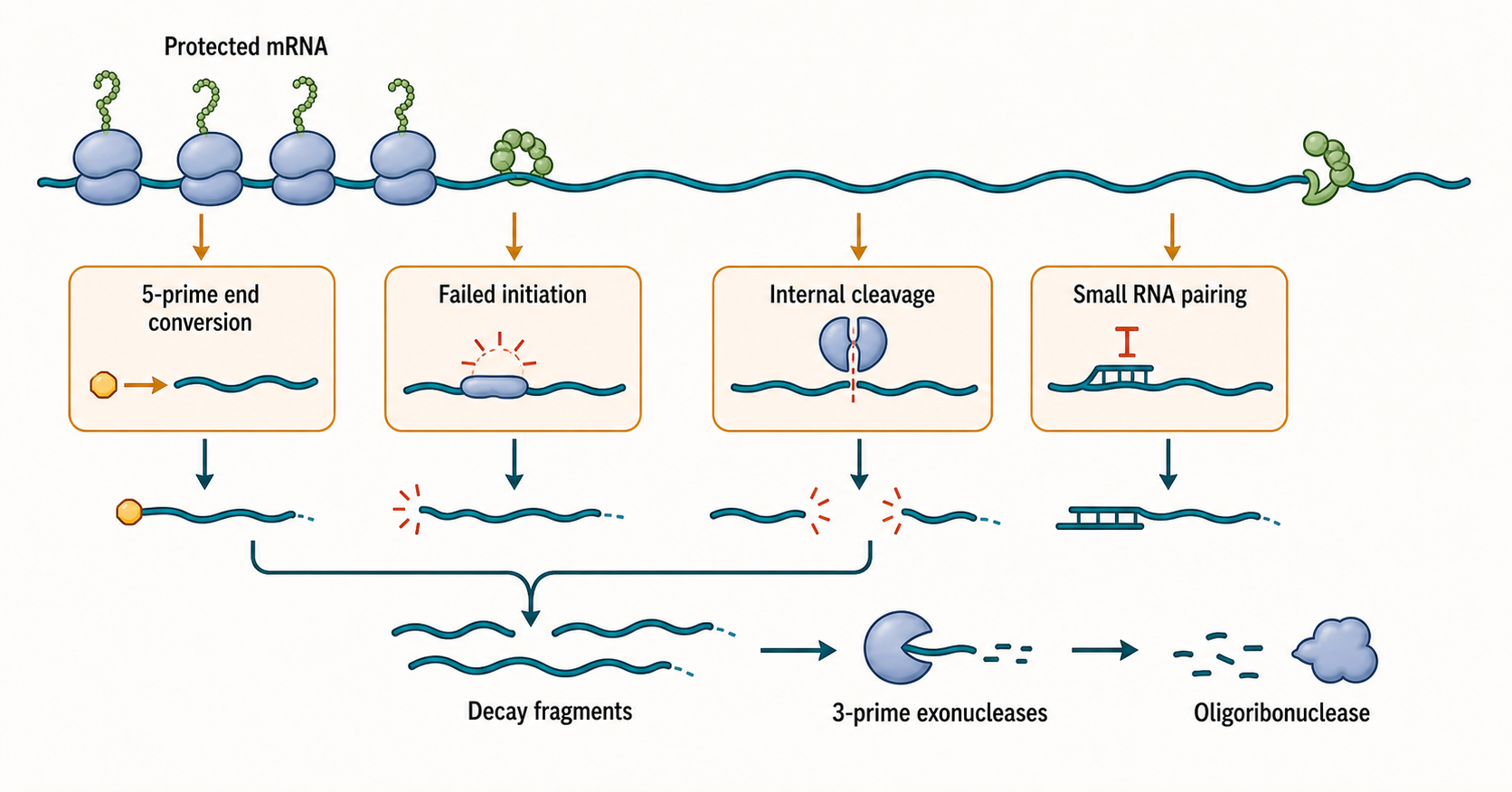
Figure 34.1. Entry points into bacterial mRNA decay. Bacterial mRNA decay can begin when a vulnerable end or internal region becomes accessible. Ribosomes and RNA-binding proteins can protect regions of the transcript, whereas 5′ end conversion from triphosphate to monophosphate, failed translation initiation, internal nuclease cleavage by RNase E or RNase Y, or small-RNA binding can expose the RNA to decay enzymes. Short fragments generated by internal cleavage are finished by 3′ exonucleases such as PNPase, RNase II, and RNase R, and the smallest products are cleared by oligoribonuclease. Decay is therefore a kinetic competition among translation protection, RNA folding, end chemistry, and nuclease access.
Table 34.2. Terminal tailing outcomes across systems. Comparison of tailing enzyme systems, tail types, and biological consequences in bacteria, archaea, eukaryotes, and organelles.
| System | Tail Type | Major Enzymes | Typical Effect | Mechanistic Reason | Exceptions | Related Chapter |
|---|---|---|---|---|---|---|
| Bacterial polyadenylation-assisted decay | Short poly(A) | PcnB (poly(A) polymerase I) | Promotes decay | Short tail creates a 3′ single-stranded entry site for PNPase, RNase II, or RNase R | Highly translated mRNAs may resist tail-promoted decay | Chapter 32 |
| PNPase heteropolymeric tailing | Mixed A/U/C/G | PNPase (reverse reaction) | Promotes decay | Mixed tail provides an exonuclease foothold on structured RNA fragments | PNPase that adds the tail can also degrade the same RNA | Chapter 32 |
| Archaeal exosome-linked tailing | Mixed or short poly(A) | Archaeal exosome (polymerizing mode) | Often promotes 3′ processing and decay | Exosome can switch between polymerizing and degrading modes on the same substrate | Tailing outcomes differ across archaeal lineages | Chapter 29 |
| Eukaryotic cytoplasmic mRNA poly(A) | Long poly(A) | Canonical poly(A) polymerase; PABP-bound | Stabilizes mRNA; supports translation | Poly(A)-binding protein protects tail from deadenylases and promotes ribosome recruitment | Deadenylation by CCR4-NOT or Pan2-Pan3 initiates mRNA decay | Chapter 29 |
| Organellar polyadenylation | Short poly(A) | Organellar poly(A) polymerases | Promotes decay in chloroplasts; variable in mitochondria | Resembles bacterial logic in chloroplasts; outcome depends on organism in mitochondria | Some mitochondrial RNAs require a short poly(A) addition for stability or translation | Chapter 37 |
Bacterial mRNA decay begins with accessibility. A bacterial transcript emerges from RNA polymerase, folds as it is synthesized, and may be engaged by ribosomes before transcription is complete. Regions protected by ribosomes, RNA-binding proteins, or stable secondary structure are less accessible to many nucleases. Regions that are single-stranded, weakly translated, newly cleaved, or poorly protected are more vulnerable. This creates a kinetic competition: translation can protect an mRNA, while failed translation initiation can make the same mRNA an easier decay substrate.
In the E. coli model, RNase E initiates decay of many mRNAs by internal cleavage. RNase E is not a shredder that begins at one end and chews through every transcript. It cuts selected internal sites, often in accessible regions. Once an internal cut occurs, the fragments have new ends. 3′ fragments and structured pieces can be degraded by enzymes such as PNPase, RNase II, and RNase R, while short products are ultimately reduced to mononucleotides by oligoribonuclease and related activities. A 5′ triphosphate end can protect some RNAs from RNase E stimulation, whereas conversion to a 5′ monophosphate can accelerate decay.
The RNA degradosome solves the problem of handoff. In the classical E. coli complex, the C-terminal region of RNase E scaffolds PNPase, the RhlB DEAD-box RNA helicase, enolase, and other partners or regulatory contacts. RNase E cuts; RhlB can help remodel structured RNA using ATP; PNPase degrades fragments phosphorolytically; metabolic proteins may couple RNA decay to cellular state. The complex is also spatially organized, with RNase E membrane association contributing to intracellular localization. The precise functional importance of each partner can differ by substrate and growth condition.
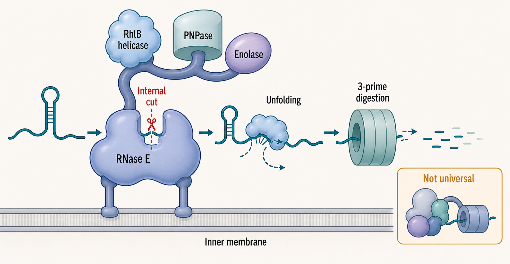
Figure 34.2. The RNase E degradosome as a handoff machine. The classical E. coli degradosome organizes cleavage, remodeling, and exonucleolytic digestion around RNase E, which scaffolds PNPase, the RhlB DEAD-box RNA helicase, enolase, and additional regulatory contacts near the inner membrane. RNase E cuts the RNA internally, RhlB helps unfold structured fragments, and PNPase degrades products phosphorolytically, enabling efficient handoff without releasing substrates into solution. Many bacteria use different nuclease combinations, including RNase Y and RNase J-centered systems in Firmicutes, so degradosome logic should be separated from the specific E. coli parts list.
Not all bacteria run this pathway in the same way. Bacillus subtilis lacks the canonical RNase E-centered degradosome familiar from E. coli. It uses enzymes such as RNase Y, RNase J1/J2, RNase III, PNPase, RNase R, and other ribonucleases. RNase Y can initiate decay by endonucleolytic cleavage, whereas RNase J can degrade from a 5′ end and also cut internally. This difference matters pedagogically because statements such as “bacterial mRNA decay begins with RNase E” are accurate for some model organisms and wrong for others. A reader should ask which bacterial lineage, growth state, and transcript class is being discussed.
Polycistronic transcripts add another layer. An operon mRNA may encode enzymes in a pathway, but each cistron can have a different translation rate, RNA structure, and vulnerability to cleavage. A nuclease cut between coding regions can destabilize one portion while leaving another temporarily intact. Processing can therefore tune stoichiometry within an operon rather than simply remove the entire message. The boundary between processing and decay is not always sharp: a cut that looks degradative for one RNA may generate a functional processed transcript for another.
The evidence for bacterial decay pathways comes from enzyme genetics, catalytic mutants, RNA half-life assays, RNA-end mapping, biochemical reconstitution, and global transcriptomics. Steady-state RNA-seq after RNase depletion identifies candidate substrates but cannot distinguish direct cleavage from changed transcription or stress. RNA-end mapping can identify new cleavage products, but library artifacts and processing intermediates must be controlled. Biochemical assays with purified RNase E, RNase Y, RNase J, PNPase, or degradosome assemblies test direct mechanism but may miss ribosomes, RNA chaperones, membrane localization, and cellular competition.
A common misconception is that bacterial decay is a uniform background process. In reality, decay is transcript-specific and condition-specific. Some mRNAs are destabilized when translation is poor. Some are stabilized by RNA structures, riboswitch ligand binding, or protein occupancy. Some are targeted by small RNAs. Some fragments resist degradation because structured elements or bound proteins block exonucleases. The same cell can therefore contain short-lived regulatory transcripts and long-lived stable RNA precursors at the same time.
34.2. Hfq, ProQ, Csr/Rsm, and small RNA-guided target decay
Bacterial small RNAs are regulatory RNAs that often act after transcription has begun. Many bacterial small RNAs are roughly 50 to 300 nucleotides long, although size boundaries are not strict. Some are encoded between genes and act in trans on separate mRNAs. Others are antisense RNAs encoded opposite their targets. Some regulate by base-pairing with mRNAs, while others bind proteins. The central point for RNA turnover is that a small RNA can change whether a target mRNA is translated, protected, cleaved, or degraded.
Hfq is the best-known bacterial small-RNA chaperone. Hfq forms a ring-shaped hexamer and presents multiple RNA-binding surfaces. It can stabilize small RNAs, bind target mRNAs, and increase the rate at which complementary regions find each other. In a common mechanism, an Hfq-bound small RNA base-pairs near a target mRNA ribosome-binding site. If the pairing blocks translation initiation, ribosomes no longer protect the mRNA leader and coding sequence. RNase E or another nuclease can then cleave the target, and exonucleases finish degradation. In some cases the small RNA is degraded along with the target; in other cases it can act catalytically on multiple targets.
Small RNAs can also activate gene expression. If an mRNA leader folds into a structure that hides the ribosome-binding site, a small RNA can pair with part of the inhibitory structure and open the initiation region. In that case, small-RNA binding can increase translation and indirectly stabilize the mRNA by recruiting ribosomes. This boundary case prevents a common error: bacterial small RNAs are not inherently decay triggers. Their effect depends on where they bind, what structure they alter, and which proteins join the RNP.
Box 34.1. Do not call every small-RNA effect decay
- A small RNA can block translation initiation, causing ribosomes to vacate the mRNA; because ribosome occupancy protects many transcripts from RNase E, the mRNA decays faster as a consequence of lost translation, not from direct nuclease recruitment by the small RNA.
- A small RNA can block translation without causing a detectable change in mRNA half-life, particularly if the transcript retains structure or protein protection that prevents nuclease access.
- A small RNA can open an inhibitory mRNA structure that hides the ribosome-binding site, increasing translation and indirectly stabilizing the transcript by recruiting protective ribosomes.
- A small RNA such as CsrB or RsmZ can titrate a protein regulator such as CsrA/RsmA through multiple decoy binding sites, changing the stability of many target mRNAs indirectly without base-pairing with any of them.
- A small RNA and its target can be co-degraded in a stoichiometric interaction, reducing the pool of active small RNA rather than allowing it to act on multiple targets.
- Small-RNA regulation must be resolved into binding site, translation effect, mRNA half-life change, nuclease dependence, protein cofactor requirement, and in vivo phenotype before labeling any outcome as direct decay.
ProQ expands the small-RNA landscape beyond Hfq. ProQ is a FinO-domain RNA-binding protein that recognizes structured RNA features and supports many RNA-RNA interactions. Some ProQ-associated RNAs overlap with Hfq pathways, but many appear distinct. ProQ-linked regulation reminds the reader that bacterial RNA chaperones are not interchangeable labels. Each protein has its own RNA-binding preferences, organism distribution, and physiological biases.
The Csr/Rsm system uses a different logic. CsrA, called RsmA or related names in many species, is an RNA-binding protein that recognizes short motifs, often including GGA sequences in exposed loops. When CsrA binds near an mRNA translation initiation region, it can repress translation and alter mRNA stability. Csr/Rsm small RNAs contain multiple binding sites for CsrA/RsmA-family proteins. These small RNAs can sequester the protein, reducing its binding to mRNA targets. Thus a Csr/Rsm small RNA often regulates decay indirectly by titrating a protein regulator rather than by base-pairing with each target mRNA.
Pathogenic bacteria show why small-RNA-coupled decay is biologically important. In Pseudomonas aeruginosa, small-RNA regulation can influence chronic and acute infection phenotypes, and recent work identified a small RNA with infection-relevant regulatory effects (Cao et al. 2023). In Klebsiella pneumoniae, global RNA-RNA interactome mapping revealed small-RNA interactions and a regulator linked to cell division (Ruhland et al. 2024). These examples do not mean every interaction causes decay. They show that modern RNA-RNA mapping can discover candidate regulatory pairs that must then be tested for base-pairing dependence, protein cofactors, target stability changes, translation effects, and phenotype.
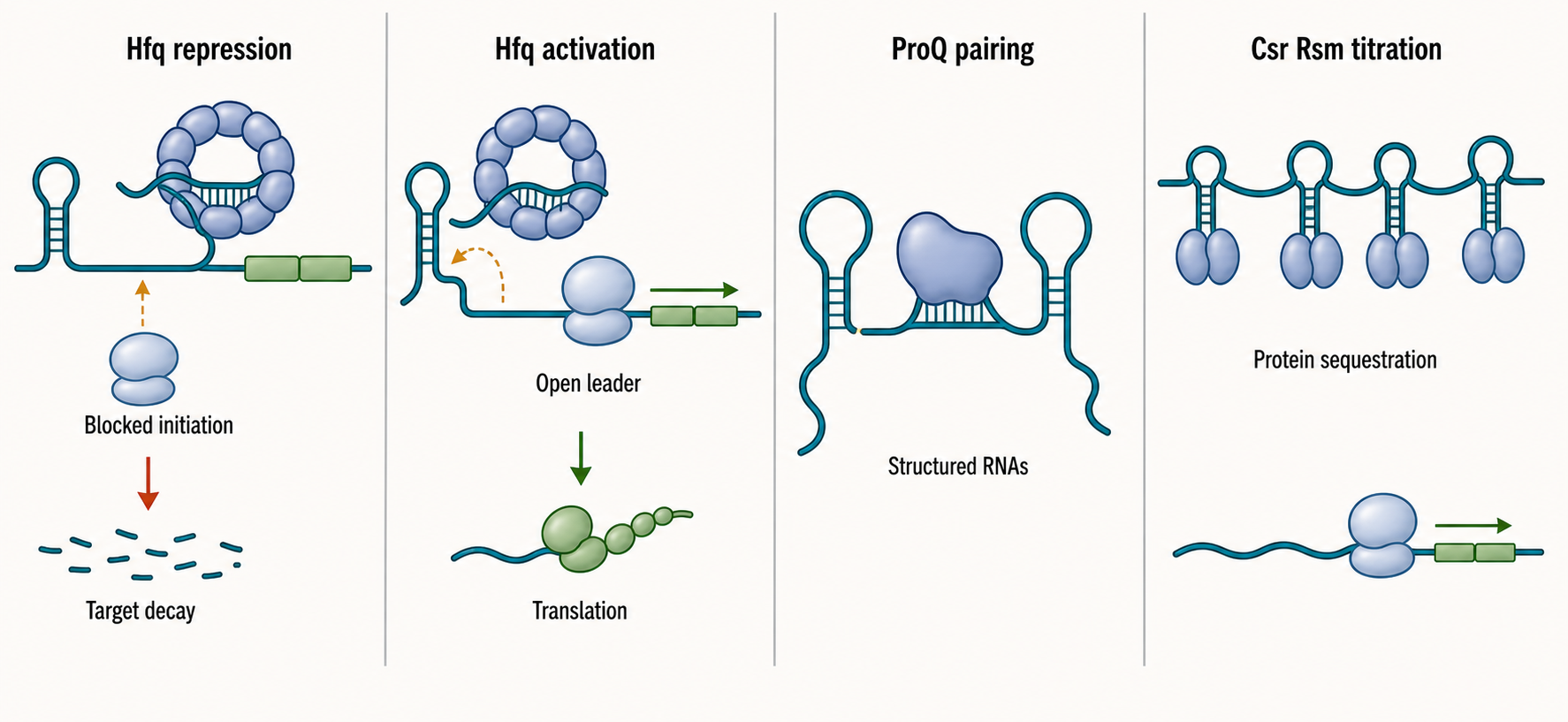
Figure 34.3. Small-RNA-coupled target fates. Bacterial small RNAs regulate target fate through several distinct logics. In Hfq-assisted repression, a small RNA base-pairs near a ribosome-binding site, blocks translation initiation, and exposes the mRNA to RNase E-dependent cleavage and downstream exonuclease degradation. In Hfq-assisted activation, a small RNA opens an inhibitory mRNA structure and recruits ribosomes, increasing both translation output and mRNA stability. ProQ-supported pairing similarly reshapes RNA-RNA interaction networks through structured-RNA binding. Csr/Rsm small RNAs often titrate CsrA/RsmA proteins away from mRNA targets rather than base-pairing with each mRNA directly, so their effect on stability is indirect. Target decay is one possible outcome of small-RNA regulation, not its definition.
Table 34.3. Evidence ladder for small-RNA-guided target decay. Summary of evidence types used to demonstrate that a bacterial small RNA directly guides target mRNA decay, with the limits and required controls for each approach.
| Evidence Type | What It Supports | What It Cannot Prove Alone | Preferred Controls |
|---|---|---|---|
| Small-RNA deletion or overexpression | Change in steady-state target RNA level | Direct base-pairing, decay causality, or regulatory specificity | Include multiple targets; check for indirect regulon members |
| Target abundance change | Altered steady-state level of the candidate target | Whether decay, transcription, or translation is primarily affected | Pair with RNA half-life measurement |
| RNA-RNA interactome hit | Physical proximity or stable pairing of two RNAs in cells | Functional regulatory outcome or direction of causality | Mutational disruption and phenotype rescue |
| Hfq or ProQ dependence | Chaperone-mediated assistance of the RNA-RNA interaction | Which molecular step the chaperone facilitates | Test Hfq/ProQ RNA-binding site mutants |
| Mutational compensation | Direct base-pairing between small RNA and target mRNA | Mechanism of downstream decay or identity of the nuclease | Double-mutant rescue both in vivo and in vitro |
| Half-life measurement | Rate of target RNA decay in vivo | Whether the small RNA directly recruits a nuclease | Compare with and without small RNA; use metabolic labeling or rifampicin with appropriate controls |
| Ribosome profiling | Translation output at the target RNA under regulation | Decay rate or nuclease identity | Pair with mRNA abundance and decay rate data |
| Nuclease end mapping | Cleavage site position and presence of decay intermediates | Which step is rate-limiting or small-RNA-dependent | Compare wild-type and small-RNA or nuclease mutant strains |
| In vitro reconstitution | Sufficiency of listed factors for cleavage or degradation | In vivo relevance under competition from ribosomes and proteins | Include Hfq/ProQ and relevant cofactors; test multiple substrate structures |
| Phenotype rescue | Physiological relevance of the regulatory interaction | Molecular mechanism or identity of primary target | Rescue with both wild-type and base-pairing-defective small RNA |
The experimental standard for small-RNA-guided decay is high because indirect effects are common. Strong evidence includes a predicted or mapped base-pairing region, mutations in the small RNA that disrupt regulation, compensatory target mutations that restore regulation, dependence on Hfq or ProQ when relevant, altered target half-life rather than only altered steady-state abundance, mapped nuclease cleavage or decay intermediates, and physiological rescue. RNA-RNA interactome methods are powerful, but crosslinking or ligation enrichment can capture proximity, abundance, or stable pairing rather than regulatory causality.
Small-RNA-coupled decay should also be distinguished from CRISPR RNA-guided immunity. CRISPR RNAs guide Cas proteins to nucleic acid targets in defense systems; some Cas enzymes target RNA. Hfq- and ProQ-dependent regulatory small RNAs usually control endogenous bacterial gene expression through RNA chaperones and host ribonucleases. The shared word “guide” does not make the mechanisms equivalent. CRISPR systems are treated in later genome-defense chapters; this chapter uses them only as a boundary case for RNA-guided regulation.
34.3. Archaeal exosomes, RNA surveillance, and decay mechanisms
Archaea have bacterial-sized cells and prokaryotic genome organization in many respects, but archaeal information-processing machinery often resembles eukaryotic systems. RNA decay follows this mixed pattern. Some archaeal lineages encode exosome-like complexes related to eukaryotic RNA exosome cores. Other archaeal lineages lack a recognizable exosome and must use alternative nucleases. Archaeal RNA surveillance therefore cannot be inferred by copying either bacterial or eukaryotic diagrams.
The archaeal RNA exosome is a 3′ to 5′ processing and degradation machine. It can bind RNA through a channel-like architecture and degrade or polymerize RNA depending on substrate and nucleotide conditions. In exosome-containing archaea, the complex contributes to RNA processing and decay of mRNAs and noncoding RNAs. In organisms living at high temperature, high salt, or other extreme conditions, RNA structure and protein protection can strongly influence nuclease access.
Exosome-lacking archaea are not decay-deficient; they use different enzymes. Some archaeal species encode beta-CASP family ribonucleases such as aCPSF1-related enzymes, RNase J-like activities, or other nucleases that participate in RNA maturation and decay. The precise enzyme set depends on lineage. This diversity is important because “archaeal” covers methanogens, halophiles, thermoacidophiles, marine Thaumarchaeota, and many other groups. A mechanism established in one archaeon should not be treated as universal without comparative evidence.
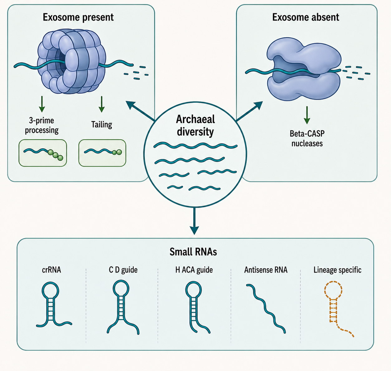
Figure 34.4. Archaeal RNA decay diversity. Archaeal RNA decay combines exosome-like 3′-to-5′ processing in many lineages with alternative nuclease systems in others, and cannot be inferred from either the bacterial or eukaryotic model alone. In exosome-containing archaea, the archaeal exosome degrades mRNAs and noncoding RNAs from the 3′ end and can also polymerize tails that promote further degradation. In exosome-lacking lineages, beta-CASP family nucleases such as aCPSF1-related enzymes take on RNA maturation and decay roles. Archaeal small RNAs include CRISPR RNAs processed from arrays, C/D box and H/ACA guide RNAs for RNA modification, antisense RNAs, and lineage-specific candidates whose connections to mRNA decay remain under investigation.
Archaeal small RNAs are prominent, but their functions are diverse. A review of archaeal small RNAs emphasizes that archaea encode guide RNAs for RNA modification, CRISPR RNAs for immunity, antisense RNAs, and lineage-specific small RNAs with incompletely understood functions (Gomes-Filho et al. 2018). Some archaeal small RNAs likely affect transcript stability or translation, but the current supplied references do not provide enough direct evidence to assign broad decay mechanisms.
RNA surveillance in archaea includes normal maturation and quality control. Pre-rRNAs and pre-tRNAs must be processed; CRISPR arrays must be transcribed and processed into CRISPR RNAs; guide RNAs must assemble with proteins; mRNAs must be cleared when no longer useful. As in bacteria and eukaryotes, a nuclease can be a maturation enzyme for one substrate and a decay enzyme for another. A processing intermediate becomes a quality-control substrate when it fails to acquire the right structure, modification, or protein partner.
Evidence in archaeal RNA decay often comes from a smaller experimental base than in E. coli or yeast. Genetics can be difficult in some archaeal systems, and growth conditions may complicate RNA half-life measurements. Biochemical reconstitution and structural biology have therefore been especially important for archaeal exosome understanding. Transcriptomics can identify RNA abundance changes, but direct decay claims require end mapping, enzyme perturbation, and ideally reconstitution. A cautious chapter should mark archaeal consensus separately from archaeal hypotheses.
One misconception is that the archaeal exosome is simply a primitive eukaryotic exosome. Evolutionary relationship does not mean functional identity. Archaeal complexes can be simpler in subunit composition, different in cofactor use, and embedded in archaeal-specific transcription and translation contexts. Another misconception is that archaea use bacterial RNA decay because they lack nuclei. Archaeal transcription, RNA-binding proteins, RNA modification systems, and exosome-like complexes make that assumption unreliable.
34.4. Polyadenylation-assisted decay and terminal tailing enzymes
Terminal tailing is addition of nucleotides to the 3′ end of an RNA. The added nucleotides can be adenosines, uridines, cytidines, guanosines, or mixed tails, depending on enzyme and organism. Tailing changes the physical substrate available to RNA-binding proteins and exonucleases. In bacterial decay, the most important pedagogical example is polyadenylation-assisted decay: short tails can make structured RNA fragments easier for 3′ exonucleases to engage.
In E. coli, poly(A) polymerase I, encoded by pcnB, can add poly(A) tails to RNA molecules. These tails often promote degradation rather than stability because they provide single-stranded extensions for exonucleases such as PNPase, RNase II, or RNase R. A structured RNA fragment with no accessible 3′ end may resist exonuclease attack. Adding a short tail creates a foothold. Once an exonuclease begins, it may degrade into the structured region, pause, or require helicase assistance.
PNPase is a useful enzyme for understanding why tailing and decay are chemically connected. PNPase degrades RNA phosphorolytically from the 3′ end, but the reaction is reversible in principle. Under some conditions, PNPase can add heteropolymeric tails rather than only remove nucleotides. This duality means that an enzyme assigned to “decay” can also create a decay-promoting tail, and an enzyme assigned to “tailing” can be part of a degradation pathway.
The consequence of a tail depends on the rest of the RNP. A mature eukaryotic cytoplasmic mRNA with a long poly(A) tail and poly(A)-binding proteins is often stabilized and translated efficiently. A bacterial structured RNA fragment with a short unprotected poly(A) tail is often destabilized. An organellar RNA may use polyadenylation as a decay signal in one lineage and as part of maturation in another. Archaeal tailing can be coupled to exosome activity in exosome-containing lineages, but details vary. Chapter 29 treats eukaryotic tailing; Chapter 37 treats organellar cases.
Tails are also evidence. RNA 3′ end sequencing, long-read direct RNA sequencing, and tail-specific methods can reveal tail length and composition. Interpretation requires care. Short tails may be lost during library preparation, added by enzymes after cell lysis, or miscalled because of homopolymer sequencing errors. A tail detected on an RNA does not by itself show whether the tail caused decay, resulted from stalled decay, or marked a processing intermediate. Strong evidence links tailing enzyme perturbation, tail change, RNA half-life change, and downstream nuclease dependence.
Small RNAs can be tailed too. Regulatory small RNAs may be stabilized by Hfq or ProQ and destabilized when unbound. If a small RNA pairs with a target and is co-degraded, its own tailing and decay can affect how many targets it regulates. Csr/Rsm protein-sequestering small RNAs can also be processed or degraded, changing the pool of free CsrA/RsmA protein. Thus terminal tailing contributes not only to cleanup of mRNA fragments but also to the lifetime of regulatory RNAs.
The common misconception is that the phrase “poly(A) tail” has one meaning across biology. It does not. Tail function must be specified by organism, compartment, enzyme, length, nucleotide composition, and protein context. For this chapter, the default bacterial rule is that short unprotected tails often assist decay, but even that rule has exceptions depending on RNA class and physiology.
34.5. Stress, antibiotics, phage, and environmental adaptation
Bacteria and archaea live in changing environments, and RNA turnover is one of the fastest ways to adjust gene expression. If nutrients disappear, a cell benefits from degrading growth-program transcripts and stabilizing stress-response transcripts. If an antibiotic inhibits translation, cell-wall synthesis, DNA replication, or transcription, RNA decay pathways experience altered ribosome protection, stress signaling, and enzyme availability. If phage infects a cell, host and phage RNAs compete for ribosomes and decay machinery. The response is not simply “more decay” or “less decay”; it is transcript-selective remodeling.
Stress can alter decay by changing translation. Translation protects many bacterial mRNAs from nucleases. Antibiotics that stall ribosomes may stabilize some mRNA regions by physically blocking nucleases, while antibiotics or stresses that prevent initiation can expose leaders and coding sequences to decay. Starvation can reduce initiation and change small-RNA expression. Temperature shifts can alter RNA structure and nuclease activity. Oxidative stress can damage RNA or proteins and induce small RNAs that redirect gene expression. Envelope stress and cell-wall stress can induce regulons whose mRNAs have distinct stability programs. Mueller and Levin (2020) provide broad stress context for bacterial cell-wall quality control, but direct RNA decay claims need more specific references.
Small RNAs are major stress-response regulators. Iron limitation induces small RNAs in several bacteria that repress nonessential iron-using proteins, conserving iron for essential processes. Envelope stress, sugar-phosphate stress, quorum sensing, biofilm development, and stationary phase all involve small-RNA networks in model organisms. In pathogens, these networks affect virulence, persistence, and host adaptation. The Pseudomonas aeruginosa infection study and Klebsiella pneumoniae RNA-RNA interactome study are useful modern examples of infection-linked and systems-level small-RNA discovery (Cao et al. 2023; Ruhland et al. 2024).
Antibiotics intersect with RNA turnover in several ways. Some antibiotics directly target the ribosome, changing translation protection and ribosome traffic on mRNAs. Some induce stress responses that alter transcription and small-RNA expression. Some RNA decay enzymes or RNA-binding proteins affect tolerance or persistence by shaping the transcriptome during drug exposure. RNA-targeting antibiotics that bind rRNA are treated in chapters on translation and RNA-targeting drugs; here the relevant point is that antibiotic exposure can make RNA stability measurements difficult because growth rate, transcription, translation, and decay all change together.
Phage infection creates an acute RNA competition. Phages can produce RNAs that overwhelm host gene expression, encode nucleases or host-shutoff factors, change transcription termination, or redirect ribosomes. Host bacteria may use RNases, small RNAs, toxin-antitoxin systems, and CRISPR-associated pathways as part of defense or stress responses. Some phages encode anti-defense factors that alter RNA processing or decay. The chapter should not imply that all phage RNA control is host decay; phage-encoded transcription and translation strategies can dominate.
Environmental adaptation is especially important in archaea. Many archaea live at high temperature, high salt, extreme pH, anaerobic niches, or nutrient-limited marine environments. RNA stability in these contexts depends on RNA structure, protein binding, ion conditions, growth rate, and specialized enzymes. Methanogens and halophiles may use RNA decay differently from thermophiles. The current references include a Methanosarcina mobilome paper that may be relevant to horizontal gene transfer context but does not directly support RNA turnover claims (Saranya et al. 2025). Archaeal stress-linked RNA decay needs a dedicated citation pass.
The strongest way to study stress-linked RNA decay is to measure synthesis and decay separately. A stress RNA-seq experiment at one time point cannot say whether a transcript increased because transcription rose, decay slowed, or a subpopulation expanded. Metabolic labeling, transcriptional pulse-chase methods, RNA-end mapping, ribosome profiling, small-RNA perturbation, and enzyme mutants can separate these possibilities. Growth-rate normalization is essential because slow-growing cells dilute RNAs differently and often remodel global physiology.
Box 34.2. Why stress RNA-seq is not a decay assay
- RNA abundance in a cell is the net result of transcription rate, processing efficiency, translation protection, active enzymatic decay, growth-rate dilution, and cell-population composition; a single steady-state measurement cannot separate these contributions.
- Stress conditions such as nutrient starvation, antibiotic exposure, oxidative stress, temperature shift, or phage infection simultaneously change transcription, translation initiation and elongation, growth rate, RNA folding, nuclease activity, RNA-binding protein levels, and small-RNA expression.
- Growth-rate slowdown alone changes RNA steady-state levels because dilution by cell division is reduced; this can mimic enzymatic stabilization without any change in decay rates.
- Direct evidence for altered decay requires time-resolved measurements such as rifampicin transcription shutoff with appropriate physiological controls, metabolic pulse-chase labeling, RNA 3′-end mapping of decay intermediates, nuclease perturbation, or in vitro reconstitution.
- A stress-induced RNA abundance change is a hypothesis about a possible decay contribution, not itself a decay measurement.
The main biological lesson is that RNA decay is adaptive. Cells use decay to clear obsolete messages, sharpen transitions, remove damaged or untranslated RNA, tune operon outputs, and integrate small-RNA signals with metabolism and stress. The main interpretive lesson is that decay is only one layer of the response. A rigorous analysis must place RNA stability beside transcription, translation, protein stability, cell state, and environmental history.
Experimental Foundations and Evidence
The first foundation is RNA half-life measurement. In bacteria, rifampicin transcription shutoff has often been used to follow RNA disappearance, but rifampicin itself perturbs physiology and can distort decay rates. Metabolic labeling and pulse-chase approaches can be more direct when available, but labeling chemistry, uptake, and growth effects must be controlled. For archaea, labeling methods and genetic tools vary by organism, making cross-species comparisons difficult.
The second foundation is RNA-end mapping. End mapping can reveal RNase E or RNase Y cleavage sites, RNase J 5′ decay intermediates, processed operon segments, small-RNA-induced cuts, and tailing events. A mapped end is not automatically a primary cleavage site. It might be an exonuclease pause, a processed mature end, a library artifact, or a protected fragment. Strong evidence maps both sides of a cleavage event and tests dependence on a candidate nuclease.
The third foundation is small-RNA interaction mapping. Methods that ligate, crosslink, enrich, or sequence RNA-RNA pairs can reveal candidate bacterial small-RNA targets. Ruhland et al. (2024) illustrate this approach in Klebsiella pneumoniae. These methods must be paired with perturbation and mutational tests because abundant RNAs and stable duplexes can be overrepresented, while transient regulatory interactions can be missed.
The fourth foundation is biochemical reconstitution. Purified RNases, RNA chaperones, small RNAs, target RNAs, and degradosome assemblies can test direct mechanism. For example, one can ask whether Hfq accelerates pairing, whether a small RNA changes RNase E cleavage, whether a tail improves PNPase degradation, or whether an archaeal exosome degrades a structured substrate. Reconstitution provides directness, but stripped-down substrates may not represent ribosome-bound, membrane-associated, or stress-state RNPs.
Box 34.3. Poly(A) does not mean one thing
- In many eukaryotic cytoplasmic mRNAs, a long poly(A) tail bound by poly(A)-binding proteins promotes ribosome recruitment and protects the transcript from degradation until the tail is shortened by cytoplasmic deadenylases; tail shortening initiates decay.
- In bacteria such as E. coli, a short poly(A) tail added by PcnB/poly(A) polymerase I is not stably bound by a stabilizing protein; instead it provides a single-stranded 3′ extension that PNPase, RNase II, or RNase R can engage to initiate degradation of otherwise structured RNA fragments.
- PNPase can add heteropolymeric tails containing mixed adenosine, uridine, cytidine, and guanosine residues under conditions where the phosphorolytic reaction runs in reverse; such mixed tails also serve as exonuclease entry sites.
- In organelles such as chloroplasts, short poly(A) addition triggers decay in a logic that resembles the bacterial system; in some mitochondria a short oligo(A) extension is instead required for mRNA maturation or stability.
- In archaeal exosome-containing lineages, tailing by the exosome operating in its polymerizing mode can precede or accompany substrate degradation.
- Always specify organism, cellular compartment, tail length, nucleotide composition, and associated proteins before predicting whether a poly(A) tail stabilizes or destabilizes an RNA.
The fifth foundation is physiology. A decay pathway matters because it changes cell behavior: growth, stress survival, virulence, biofilm formation, phage resistance, antibiotic tolerance, or metabolic adaptation. Physiological claims require separating RNA decay effects from transcriptional regulation and pleiotropic enzyme loss. Rescue experiments and narrow substrate perturbations are especially valuable.
Biological Contexts Across Organisms and RNA Classes
Proteobacteria such as E. coli provide the RNase E-centered reference model. This model is powerful because genetics, biochemistry, and decades of RNA mapping have defined many factors, but it can mislead if treated as universal bacterial biology.
Firmicutes such as Bacillus subtilis show that bacteria can organize mRNA decay around RNase Y, RNase J, and other factors rather than RNase E. This comparison is a useful reminder that enzyme distribution and pathway architecture must be checked before extrapolating.
Pathogenic bacteria use small-RNA and decay systems to adjust virulence, host adaptation, and stress survival. Pseudomonas aeruginosa and Klebsiella pneumoniae examples in the supplied references show that infection-relevant small RNAs and global RNA-RNA interaction maps are active areas of current research (Cao et al. 2023; Ruhland et al. 2024).
Archaea require lineage-specific treatment. Some encode exosome-like machines; some do not. Some have extensive CRISPR RNA systems and small guide RNAs. Some grow under environmental extremes that alter RNA folding and stability. Archaeal RNA decay should therefore be framed as comparative biology rather than as one pathway.
Stable RNAs are included because decay enzymes also process and surveil rRNA, tRNA, CRISPR RNA, and other noncoding RNA precursors. A defective tRNA precursor, an incorrectly processed rRNA fragment, or an unassembled guide RNA can be a decay substrate even though the mature RNA class is normally stable.
Technology, Computational, Clinical, and Engineering Links
RNA decay matters for bacterial biotechnology. Heterologous expression systems, synthetic operons, CRISPR guide expression, RNA sensors, and engineered metabolic pathways can fail because bacterial RNases destroy RNAs or because small-RNA networks create unintended regulation. Stabilizing an engineered RNA may require changing translation initiation, removing RNase sites, altering secondary structure, adding protective protein-binding motifs, or changing host strains.
Antibacterial development can exploit RNA biology, but global inhibition of essential RNases may be toxic or difficult to make selective. A more realistic strategy may target pathogen-specific RNA regulators, virulence-linked small RNAs, RNA chaperone interactions, or stress-dependent vulnerabilities. Such approaches require strong evidence that the target RNA interaction is causal in infection or survival, not merely correlated with a condition.
Computational prediction of bacterial RNA stability must include organism-specific features. Sequence composition, predicted structure, translation initiation strength, codon usage, operon position, small-RNA binding sites, 5′ end chemistry, tailing, and RNA-binding protein motifs may all matter. Models trained on E. coli may not transfer to archaea or Gram-positive bacteria.
RNA-RNA interactome maps are becoming important for bacterial regulatory discovery. They can connect small RNAs to target networks at genome scale, but they require validation and careful normalization. The Klebsiella pneumoniae study in the supplied references provides an example of using global interactome data to uncover a small-RNA regulator of cell division (Ruhland et al. 2024).
Recent Consensus
The current consensus is that bacterial RNA decay is regulated, spatially organized, and tightly coupled to translation and small-RNA control. RNase E-centered degradosomes are central in many Proteobacteria but are not universal. Bacteria use multiple architectures, including RNase Y/RNase J-centered systems in several Gram-positive organisms.
Small RNAs are recognized as major determinants of bacterial RNA fate. Hfq, ProQ, and Csr/Rsm systems represent distinct regulatory logics: RNA chaperone-assisted base pairing, structured-RNA binding networks, and protein sequestration with downstream effects on translation and stability.
Archaeal RNA decay is recognized as mechanistically diverse. Archaeal exosome-like complexes are important in many lineages, but exosome absence in other archaea and the diversity of archaeal small RNAs require lineage-specific evidence.
Terminal tailing is now treated as a context-dependent RNA fate mechanism rather than a universal stabilizing or destabilizing mark. In bacterial decay, short tails often promote degradation of structured RNA fragments, while other systems use tails differently.
Open Questions, Controversies, Deprecated Models, and Common Misconceptions
Open questions:
- What determines how degradosome composition changes across growth conditions, cell locations, and bacterial species? The classical E. coli complex is well known, but transient partners and condition-specific assemblies remain harder to define.
- How many small-RNA interactions primarily regulate translation, how many primarily regulate decay, and how often the two outcomes are inseparable? Because translation protects mRNAs from decay, a small RNA that changes translation can alter stability even without directly recruiting a nuclease.
- What determines how archaeal RNA surveillance is organized in exosome-lacking lineages? Comparative genomics identifies candidate nucleases, but direct substrate maps and biochemical mechanisms remain incomplete for many groups.
Common misconceptions:
- “Bacterial small RNAs are equivalent to eukaryotic miRNAs.” Both can use base pairing to regulate targets, but bacterial small RNAs usually act with Hfq, ProQ, Csr/Rsm proteins, RNases, and translation-initiation control rather than with Argonaute-centered silencing.
- “Polyadenylation always stabilizes RNA.” In many bacterial and organellar contexts, short poly(A) or mixed tails promote decay by giving exonucleases a single-stranded entry site.
Deprecated or weakened claims:
- A deprecated oversimplification is that bacterial RNA decay is merely cleanup. Current evidence supports a more integrated view: RNA decay shapes operon output, stress adaptation, virulence, antibiotic responses, phage competition, and the effective output of transcription.