Chapter 32. RNA Decay Enzymes: Exonucleases, Endonucleases, Exosomes, and Degradosomes
Scope Note
This chapter explains the catalytic machines that cut, trim, and destroy RNA and the multienzyme assemblies that deliver substrates to them. Its primary ownership is ribonuclease mechanism: 5′-to-3′ and 3′-to-5′ exonucleases, endonuclease families, RNA exosomes, degradosomes, catalytic specificity, kinetics, and direct-substrate evidence. It retains pathway-specific treatment of helicase-coupled substrate delivery but hands comparative RNA-motor enzymology to Chapter 55. It likewise distinguishes nuclease-generated ends from the healing and ligation reactions owned by Chapter 33. Chapter 37 owns direct lysosomal RNA import, ribophagy, lysosomal nuclease context, and RNA turnover secondary to organelle-selective or bulk autophagy; those routes should not be inferred from nuclease activity alone. Organism-, compartment-, and pathway-specific chapters explain where these enzyme modules operate.
Executive Summary
RNA decay is not a single reaction. It is a family of chemical and biological processes in which ribonucleases hydrolyze phosphodiester bonds, accessory proteins expose vulnerable RNA regions, and cellular compartments restrict which substrates meet which enzymes. A useful first distinction separates exonucleases from endonucleases. Exonucleases digest RNA from a 5′ or 3′ end, whereas endonucleases cut internally. This distinction is necessary but incomplete because an RNA’s end chemistry, cap status, tail length, structure, protein coating, ribosome occupancy, modification state, and location often determine whether an enzyme can act.
The simplest substrate-recognition rule is that unprotected single-stranded RNA ends are vulnerable. A eukaryotic mRNA with a 5′ cap, a poly(A)-binding protein-coated tail, translating ribosomes, and sequence-specific RNA-binding proteins is protected from many decay enzymes. The same RNA becomes vulnerable when deadenylation shortens the tail, decapping removes the cap, an endonuclease creates an internal entry site, a stalled ribosome recruits quality-control factors, or a surveillance complex exposes the RNA to an exonuclease. Reviews of decapping and exosome biology emphasize that the main specificity problem is not phosphodiester chemistry itself but the regulated presentation of substrates to enzymes (Grudzien-Nogalska and Kiledjian 2017; Weick and Lima 2021).
Decay from the 5′ end usually requires conversion of a protected end into a monophosphorylated end that a 5′ to 3′ exonuclease can engage. In many eukaryotic cytoplasmic mRNAs, deadenylation is followed by decapping, and Xrn1-like exonucleases digest the exposed RNA body. Nuclear Xrn2/Rat1-like enzymes also participate in transcription termination and RNA processing. In bacteria and some archaea, 5′ end status is interpreted differently because many bacterial transcripts begin with triphosphorylated ends and can be converted to monophosphorylated ends before RNase E-dependent decay. RNase J in many bacteria combines endonucleolytic and 5′ exonucleolytic capabilities, illustrating that enzyme names and pathway arrows should not be overgeneralized across organisms (Deikus et al. 2008).
Decay from the 3′ end is dominated in eukaryotes by the RNA exosome, a multi-subunit 3′ to 5′ processing and degradation machine. The exosome can trim normal RNA precursors or degrade defective RNA molecules. Its core scaffold channels RNA to catalytic subunits, but substrate selection depends heavily on cofactors, helicases, adaptor complexes, and RNA-tail features. Recent structural work on a human exosome-ribosome supercomplex shows how ribosome-associated substrates can be positioned for exosome-mediated decay, strengthening the view that decay machines are integrated with translation and surveillance rather than acting only on free RNA (Kogel et al. 2024). Genetic evidence linking EXOSC4 variants to impaired exosome function and neurodevelopmental defects also illustrates why basic ribonuclease machinery has organism-level consequences (Fasken et al. 2024).
Endonucleases create internal cuts that can bypass the need for prior end removal. RNase E in bacteria, RNase Y in many Gram-positive bacteria, RNase L in vertebrate antiviral defense, Integrator cleavage modules in metazoan transcription regulation, MARF1 in selected cytoplasmic decay contexts, and many RNA-processing nucleases all demonstrate the same principle: internal cleavage converts one RNA molecule into fragments with new ends, and those new ends are then finished by exonucleases. Site-specific cleavage can be directed by RNA sequence, structure, guide factors, protein cofactors, modifications, ribosome state, immune signaling, or assembly into larger RNP machines.
Helicase-coupled delivery solves a physical problem without making this a general helicase chapter. Folded or protein-coated RNA often cannot enter a narrow nuclease channel. Mtr4- and Ski2-like motors, adaptors, and degradosome-associated helicases can expose an entry strand or feed RNA toward a catalytic site. Here those motors are discussed only as components of decay assemblies; Chapter 55 compares ATPase cycles, directionality, processivity, family evolution, and non-decay remodeling functions.
Compartmentalization makes decay selective. Nuclear decay machines encounter nascent transcripts, cryptic unstable transcripts, pre-rRNA, pre-tRNA, snRNA precursors, and chromatin-associated RNAs. Cytoplasmic decay machines encounter translating mRNAs, stress-granule-associated RNAs, P-body-associated RNAs, viral RNAs, and quality-control substrates. Bacterial degradosomes cluster enzymes such as RNase E, polynucleotide phosphorylase, enolase, and RNA helicases, often near membranes. Endoplasmic reticulum-associated RNA decay, mitochondrial and chloroplast turnover, and antiviral RNase pathways show that decay machines are positioned where their substrates arise (Ottens et al. 2024; Islam et al. 2021; Watkins and Burke 2024).
The experimental problem is to distinguish direct ribonuclease mechanism from indirect abundance changes. Steady-state RNA-seq after enzyme depletion can reveal candidate substrates but cannot by itself say whether the enzyme cut the RNA directly, altered transcription, changed translation, triggered stress, or destabilized a cofactor. Strong interpretation combines genetics, catalytic mutants, rescue, end mapping, metabolic labeling, in vitro reconstitution, structural biology, and substrate-specific validation. Biochemical assays require equal care: purified enzymes may behave differently from enzyme complexes in cells, and inhibitors or stress treatments may change RNA synthesis and localization as much as decay.
Concept Inventory
- RNA decay: the enzymatic reduction of RNA abundance by phosphodiester cleavage, exonucleolytic digestion, or coupled processing and disposal. RNA turnover is broader because it includes synthesis and decay rates together; a stable steady-state abundance can reflect high synthesis and high decay or low synthesis and low decay.
- Ribonuclease: an enzyme that cleaves RNA. An exonuclease removes nucleotides from an RNA end. A 5′ to 3′ exonuclease begins at a suitable 5′ end and moves toward the 3′ end. A 3′ to 5′ exonuclease begins at a suitable 3′ end and moves toward the 5′ end. An endonuclease cuts internally and thereby creates new RNA ends that can be used by exonucleases.
- Decapping: removal or conversion of a protective 5′ cap so that the 5′ end becomes accessible to decay machinery. In eukaryotic mRNA decay, decapping often creates the entry point for Xrn1-mediated 5′ to 3′ degradation. Chapter 26 treats cap chemistry and cap-binding proteins; this chapter focuses on decapping as an enzymatic entry point into decay.
- RNA exosome: a conserved eukaryotic and archaeal multi-subunit 3′ to 5′ RNA-processing and RNA-degradation complex. In eukaryotes the exosome core is a channel-like scaffold associated with catalytic subunits such as DIS3/RRP44 and RRP6/EXOSC10, with exact composition and localization varying by organism and compartment. The RNA exosome should not be confused with extracellular vesicles named exosomes. Some supplied chapter references concern extracellular vesicle biology (Iqbal et al. 2024; Li et al. 2021) and are terminology cautions rather than direct sources for RNA exosome enzymology.
- Degradosome: a multi-enzyme RNA decay assembly, classically described in bacteria, that brings together an endoribonuclease, exonuclease, helicase, and metabolic or regulatory partners. The best-known example is the Escherichia coli RNA degradosome organized around RNase E. Degradosome architecture varies across bacteria and should not be treated as a single universal complex.
- Helicase-coupled substrate delivery: use of an RNA-dependent ATPase and its adaptors to expose, orient, or feed a structured RNP substrate to a nuclease. The decay-machine context is treated here; the motor-family comparison and mechanochemical terminology are owned by Chapter 55.
- Decay cofactor: a non-catalytic or indirectly catalytic factor that recruits, activates, positions, remodels, tails, or otherwise prepares RNA for a nuclease. Cofactors can dominate substrate specificity even when the nuclease supplies the chemical cleavage activity.
What to Know Before Reading This Chapter
RNA is a directional polymer. The 5′ end and 3′ end are chemically different, and many RNA enzymes are directional. Chapter 2 explains ribose-phosphate chemistry and phosphodiester hydrolysis; here the important point is that ribonucleases must position a scissile phosphate and activate chemistry in an RNA context that is usually folded and protein-bound.
RNA molecules in cells are usually RNPs, not naked polymers. A messenger RNA may carry a cap-binding complex at its 5′ end, poly(A)-binding protein at its tail, ribosomes along its coding sequence, and regulatory proteins on its untranslated regions. A pre-rRNA is embedded in a ribosome-assembly intermediate. A tRNA precursor may be folded, modified, and recognized by processing enzymes. Decay enzymes therefore act on RNP states, not just RNA sequences.
The word “substrate” must be used carefully. A direct substrate is an RNA that an enzyme physically binds and cleaves or digests. An indirect substrate is an RNA whose abundance changes when an enzyme is perturbed but that may not be cut by that enzyme. Many transcriptome-wide studies begin with indirect substrate lists and then narrow the list with end mapping, catalytic mutants, biochemical reconstitution, and rescue.
RNA decay intersects with RNA processing. Trimming a pre-rRNA to a mature end, degrading a cryptic transcript, removing a faulty mRNA, and clearing a viral RNA all use related chemical reactions. The biological outcome depends on how far digestion proceeds, where the RNA is located, whether a mature product remains, and whether the cell treats the RNA as a normal intermediate or a quality-control target.
32.1. RNA-end state, substrate recognition, and catalytic frameworks in RNA degradation
RNA degradation begins with recognition. Recognition does not always mean that a ribonuclease reads a precise sequence motif. More often, recognition is a layered decision in which RNA end chemistry, structure, bound proteins, tail length, translation state, modifications, and subcellular position make an RNA more or less accessible to decay machinery.
Table 32.1. Major ribonuclease classes and recognition logic. Major ribonuclease enzyme classes differ in directionality, entry requirement, representative enzymes, and key caveats relevant to substrate interpretation.
| Enzyme class | Direction or cleavage mode | Entry requirement | Representative enzymes or complexes | Typical substrate contexts | Main caveat |
|---|---|---|---|---|---|
| 5′ to 3′ exonuclease | 5′→3′ exonucleolytic | Accessible 5′ monophosphate end | Xrn1 (cytoplasmic), Xrn2/Rat1 (nuclear), RNase J (bacteria) | Decapped mRNA, transcription termination fragments | Cap blocks entry; requires prior decapping or cleavage |
| Hydrolytic 3′ to 5′ exonuclease | 3′→5′ hydrolysis | Accessible single-stranded 3′ end, often with a 3′ hydroxyl | RNase R, RNase II, DIS3-family enzymes | Tailed transcripts, structured RNAs, exosome substrates | Structural tolerance and processivity differ; RNase R requires a 3′ overhang even when it can traverse downstream structure |
| Phosphorolytic 3′ to 5′ exonuclease | 3′→5′ phosphorolysis, releasing nucleoside diphosphates | Accessible single-stranded 3′ end plus inorganic phosphate | Polynucleotide phosphorylase (PNPase) | Bacterial and organellar RNA decay; degradosome substrates | Duplex structure can stall isolated PNPase; helicase partners and tailing can improve access |
| RNA exosome | 3′→5′ via channel | Single-stranded 3′ region; cofactor and helicase delivery | Exosome core with DIS3/RRP44, RRP6/EXOSC10 | Nuclear surveillance, cytoplasmic mRNA decay, stable RNA maturation | Substrate specificity is determined by cofactors, not intrinsic nuclease selectivity |
| Endonuclease | Internal phosphodiester cleavage | Single-stranded region, structure motif, or guide factor | RNase E (bacteria), RNase Y, RNase L (vertebrate), Integrator, MARF1 | Structured mRNA, antiviral substrates, nascent transcripts | Cleavage site evidence requires both flanking ends and catalytic-mutant confirmation |
| RNA-end phosphatase | Terminal-group hydrolysis rather than chain digestion | RNA bearing a blocked 3′ phosphate | APE1 in defined in vitro assays; pathway-specific end-healing enzymes | Selected cleavage products and damaged RNA ends | Biochemical activity does not establish a general cellular decay route; comparative repair chemistry belongs to Chapter 33 |
| Decapping enzyme | Cap hydrolysis | 5′ cap; often requires prior deadenylation and activators | DCP1-DCP2 complex | Deadenylated mRNAs targeted for 5′ decay | Not a ribonuclease; enables 5′ exonuclease access rather than directly degrading RNA |
| Helicase-coupled decay assembly | ATP-dependent RNP remodeling | NTP; structured or protein-coated substrate | Mtr4 (nuclear), Ski complex (cytoplasmic), TRAMP, NEXT | Structured nuclear RNAs, cryptic transcripts, ribosome-protected substrates | Helicase alone does not cleave RNA; activity depends entirely on nuclease partner |
| Degradosome | Coupled endo- and exonucleolytic | Substrate delivery to scaffold endonuclease | E. coli RNase E + PNPase + RhlB helicase + enolase | Bacterial mRNA and regulatory RNA targets | Architecture varies across bacterial species; not a universal complex |
Table 32.2. Assay evidence ladder for RNA decay mechanism. Each assay provides distinct direct measurements and has characteristic limitations that require follow-up experiments before a direct ribonuclease-substrate conclusion can be drawn.
| Assay | What it measures directly | Strongest supported inference | What it cannot prove alone | Best follow-up |
|---|---|---|---|---|
| Steady-state RNA-seq after nuclease perturbation | Relative RNA abundance change | Candidate substrate or indirect target | Direct cleavage; cannot exclude transcription or localization effects | Metabolic labeling or end mapping |
| Metabolic labeling or time-resolved RNA-seq | RNA synthesis and decay rates separately | Half-life change upon perturbation | Whether the enzyme directly cleaves the RNA | Catalytic-mutant comparison |
| Transcriptional shutoff | RNA abundance decline after transcription block | Apparent half-life estimate | Synthesis-independent rate; method causes cellular stress | Metabolic labeling as less-perturbing alternative |
| 5′ end mapping | Position and chemistry of 5′ RNA ends | Internal cleavage site or exonuclease stall | Whether end arises from endonuclease vs. exonuclease pausing | 3′ end mapping of complementary fragment |
| 3′ end mapping and tail profiling | Position, tail length, and composition at 3′ ends | Trimming extent, tailing events, or cleavage position | Directionality of enzymatic action | Pairing with 5′ end mapping and catalytic mutant |
| Catalytic mutant rescue | Dependence on enzymatic activity versus scaffolding | Separation of nuclease function from structural role | Which RNA molecule is directly cut | In vitro reconstitution with mutant enzyme |
| In vitro reconstitution | Direct cleavage or digestion of defined substrate | Enzyme directly cleaves RNA under defined conditions | In-cell relevance; RNP context may differ substantially | Reconstitution with native cofactors and RNP components |
| Structural biology | Atomic geometry of enzyme-substrate complex | Mechanism of substrate positioning and chemistry | Kinetics, processivity, or in-cell substrate selectivity | Paired kinetic and mutagenesis biochemistry |
| Nascent-transcription and polymerase-occupancy assays | Readthrough transcription and RNA polymerase position downstream of a processing site | A processing or decay factor contributes to transcription termination | Whether the factor acts through RNA digestion, recruitment, or a polymerase-state change | Downstream-fragment mapping plus catalytic-mutant rescue or defined end-chemistry perturbation |
| Imaging of RNP bodies or compartments | Colocalization of RNA or factor with cellular site | Spatial association with a decay compartment | Direct enzymatic activity at that site | Biochemical fractionation and in vitro reconstitution |
Table 32.3. Compartment-specific decay machines. RNA decay machinery is compartment-specific, with each cellular location imposing different substrates, enzymes, recognition logic, and interpretive caveats.
| Compartment or location | Major substrate classes | Representative enzymes or complexes | Recognition logic | Interpretation caveat |
|---|---|---|---|---|
| Nucleus | Nascent transcripts, cryptic unstable transcripts, pre-rRNA, snRNA precursors | Nuclear exosome (DIS3/RRP6), Xrn2/Rat1, Integrator | Chromatin association, RNA tail features, transcription-linked adaptors | Perturbation can alter transcription as well as decay |
| Nucleolus | pre-rRNA, ribosome assembly intermediates | Nuclear exosome, RRP6, TRAMP-tailed substrates | Ribosome assembly progress, tail additions by TRAMP | Many rRNA processing and decay enzymes overlap |
| Cytoplasm | Translating mRNAs, quality-control substrates | Xrn1, cytoplasmic exosome (DIS3), Ski complex | Translation state, tail length, cap status, RBP coating | Abundance reflects translation efficiency as well as decay rate |
| P-body | Repressed or stored mRNAs, MARF1 substrates | DCP1-DCP2, Xrn1, MARF1 | RBP-mediated mRNA routing; condensate composition | Colocalization does not prove active decay versus storage |
| Endoplasmic reticulum | ER-targeted mRNAs, secretory pathway substrates | ER-associated decay factors, stress-responsive cleavage factors | ER stress, ribosome stalling on ER membrane | Not a simple extension of bulk cytoplasmic decay |
| Bacterial membrane-associated degradosome | Bacterial mRNA, regulatory RNA targets | RNase E + PNPase + helicase (E. coli) | Degradosome scaffolding; regulatory RNA base-pairing | Degradosome composition varies across bacterial species |
| Mitochondrion or chloroplast | Organellar mRNA, rRNA, tRNA precursors | OEX1 (plant), PNPase, organellar RNases | Organelle-specific processing signals; R-loop context | Organellar pathways differ substantially from nuclear or cytoplasmic systems |
| Antiviral response compartments | Viral RNA, cellular RNA | RNase L, RNase L-induced bodies | 2′,5′-oligoadenylate activation during innate immune response | RNase L is stress-induced; not a housekeeping decay enzyme |
Table 32.4. Processing versus degradation outcomes. The same RNA classes and enzymes can produce either a useful mature product or complete degradation depending on substrate context, and distinguishing these outcomes requires specific experimental evidence.
| RNA class | Processing or decay enzyme | Productive outcome | Degradation outcome | Evidence needed to distinguish |
|---|---|---|---|---|
| pre-rRNA | Nuclear exosome, RNase MRP, endonucleases | Mature 18S, 5.8S, and 28S rRNA | Degradation of aberrant or surplus precursors | Pulse-chase labeling, end mapping, perturbation of assembly factors |
| pre-tRNA | RNase P, RNase Z, CCA-adding enzyme | Mature tRNA with correct ends and modifications | Degradation of misprocessed or hypomodified tRNA | End mapping, modification-state analysis, tailing assays |
| snRNA precursor | Integrator cleavage, exosome | 3′-processed mature snRNA | Truncated or defective precursor degradation | Integrator catalytic-mutant comparison, end mapping |
| mRNA | Xrn1, exosome, endonucleases | Complete translation and normal programmed turnover | Accelerated decay via surveillance or regulatory signals | Half-life assay, catalytic-mutant rescue, ribosome profiling |
| Cryptic nuclear transcript | TRAMP, nuclear exosome | None (this substrate class is non-productive) | Complete degradation by nuclear exosome | RNA-seq after TRAMP or exosome depletion, end mapping |
| Viral RNA | RNase L, Xrn1, host decay factors | None (from host defense perspective) | Fragmentation and clearance during antiviral response | Infection time-course, RNase L activation assay, fragment end mapping |
| Organellar transcript | Organellar RNases, PNPase, OEX1 | Mature organellar mRNA or rRNA | Degradation of aberrant or surplus transcript | Organellar RNA fractionation, end mapping, mutant complementation |
The most general recognition signal is exposure of a compatible RNA end or single-stranded region. A 5′ to 3′ exonuclease cannot efficiently begin on a capped mRNA unless a decapping enzyme first removes the cap or creates an equivalent accessible end. A 3′ to 5′ exonuclease cannot digest through a strongly protected 3′ end unless tail shortening, RNP remodeling, or endonucleolytic cleavage produces an entry point. An endonuclease may require a single-stranded loop, a stem-loop shape, a bound guide factor, a stalled ribosome, or a protein cofactor that positions the active site.
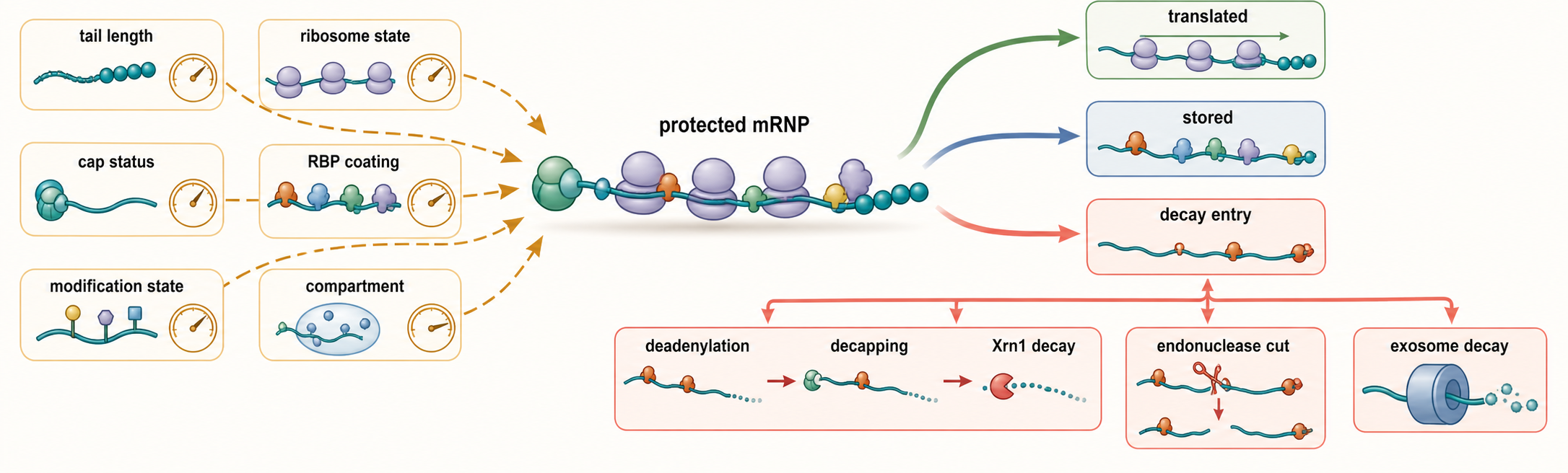
Figure 32.1. Decision points in RNA decay substrate recognition. A protected mRNP can remain translated, be stored, or enter decay depending on tail length, cap status, ribosome state, RNA-binding proteins, modification state, and compartment. Deadenylation can promote decapping and Xrn1-mediated 5′ to 3′ decay; endonucleolytic cleavage can create new exonuclease entry sites; exposed 3′ ends can be delivered to the RNA exosome for 3′ to 5′ degradation.
A concrete example is a cytoplasmic eukaryotic mRNA undergoing general deadenylation-dependent decay. First, regulatory proteins and translation state influence recruitment of deadenylase complexes that shorten the poly(A) tail. Second, loss of poly(A)-binding protein reduces circularized or protected mRNP architecture. Third, decapping activators help the DCP1-DCP2 decapping enzyme remove the protective cap. Fourth, Xrn1 engages the exposed 5′ monophosphate and digests the mRNA body. The pathway is often drawn as a linear arrow, but in cells it is a kinetic competition among translation, storage, deadenylation, decapping, endonucleolytic cleavage, and 3′ decay (Grudzien-Nogalska and Kiledjian 2017).
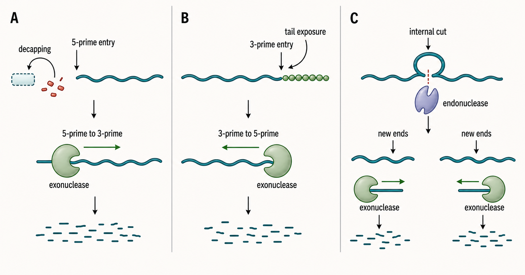
Figure 32.2. Exonuclease polarity and entry requirements. A 5′ to 3′ exonuclease requires a compatible 5′ end, often created by decapping or internal cleavage; a 3′ to 5′ exonuclease requires an accessible 3′ end, often created by tail shortening, trimming, or endonucleolytic cleavage. An inset should compare a 5′-phosphorylated downstream transcript that can support XRN2/Rat1-coupled termination with a 5′-hydroxyl product that is a poor XRN substrate, and compare a 3′ hydroxyl with a 3′ phosphate that may require end healing. The visual should distinguish end position from end chemistry and show that endonuclease fragments are not automatically ready for exonuclease cleanup.
Substrate recognition can also be negative recognition: normal RNAs are protected because they have the right RNP architecture. A mature tRNA is compactly folded, heavily modified, and aminoacylated or bound by tRNA-binding proteins. A pre-rRNA in a productive assembly intermediate is protected by ribosome biogenesis factors. A translating mRNA is partly shielded by ribosomes. When these protective features fail, the same RNA class can become a decay substrate. Fluoropyrimidine-triggered decay of hypomodified tRNA in yeast illustrates how modification state can affect RNA stability, although the supplied reference supports a specific stress and organism context rather than a universal tRNA rule (Gorlitz et al. 2024).
RNA modifications and structure complicate recognition. A modification such as pseudouridine can change RNA folding, protein binding, or nuclease susceptibility. In bacterial RNase E assays, substrate fold, pseudouridylation, and degradosome organization influence endonuclease activity, showing that the physical state of the RNA and the architecture of the decay machine can change cleavage outcomes (Islam et al. 2021). This is a useful caution for readers: a ribonuclease consensus site inferred from short naked substrates may not predict cleavage in a folded RNP.
Recognition is often compartmental. Nuclear exosome cofactors encounter nascent and chromatin-associated RNAs. Cytoplasmic decapping factors encounter mRNPs that have exited translation or surveillance pathways. Endoplasmic reticulum-associated decay factors encounter membrane-bound translation and secretory-pathway stress substrates (Ottens et al. 2024). Bacterial degradosomes may be organized near membranes, coupling RNA decay to local translation, metabolism, or RNA processing. A substrate that is biochemically cleavable in vitro may never meet the enzyme in vivo.
Do not overgeneralize the phrase “RNA decay signal.” A short poly(A) tail, a 5′ monophosphate, an AU-rich element, a stalled ribosome, a double-stranded viral RNA feature, or an exposed 3′ tail can all promote decay in some contexts. None is a universal destruction tag. The correct question is which enzyme or cofactor recognizes the feature in which organism, compartment, and RNP state.
32.2. 5-prime-to-3-prime exonucleases and decapping-coupled substrate access
A 5′ to 3′ exonuclease digests RNA starting at a compatible 5′ end. For many substrates the decisive step is not the exonuclease chemistry but making the 5′ end compatible. In eukaryotic cytoplasmic mRNAs, the 7-methylguanosine cap blocks ordinary 5′ exonucleolytic entry. Decapping removes that protection and creates an RNA end that Xrn1-like enzymes can degrade.
The causal sequence in decapping-coupled decay is conceptually straightforward. A mature mRNA first loses stabilizing features, often through deadenylation and reduced translation. Decapping activators and repressors then alter access of the decapping enzyme to the cap. DCP2 carries catalytic decapping activity in the core eukaryotic decapping complex, with DCP1 and additional factors regulating activity and substrate selection. Once the cap is removed, Xrn1 digests the RNA from 5′ to 3′. Reviews of selective mRNA decay emphasize that decapping is regulated, transcript-selective, and coupled to RNA-binding proteins rather than a passive terminal step (Grudzien-Nogalska and Kiledjian 2017).
Xrn enzymes are processive because they remain associated with a substrate through many nucleotide-removal cycles. Processivity matters biologically: once a decay intermediate is committed to Xrn1, the RNA body can be rapidly cleared, reducing accumulation of partially degraded fragments. In the nucleus, Xrn2/Rat1-like enzymes also act after cleavage events associated with transcription termination and processing. Cleavage downstream of a polyadenylation signal can leave an uncapped RNA attached to elongating RNA polymerase II; digestion of that downstream RNA gives the exonuclease a route toward the polymerase, the core of a torpedo model of termination.
The paired 2004 yeast and human studies support this causal sequence with complementary evidence rather than with transcript abundance alone. In budding yeast, Rat1 and its cofactor Rai1 localized near gene 3′ ends, loss of their function stabilized RNA downstream of poly(A) sites without detectably blocking cleavage or polyadenylation in the tested assay, and a catalytically inactive Rat1 D235A mutant failed to restore termination. In a transfected human β-globin reporter, XRN2 depletion increased polymerase readthrough and stabilized a downstream product made by a co-transcriptional cleavage element. A hammerhead ribozyme that left a 5′ hydroxyl did not substitute for the element that generated a 5′ phosphate, consistent with the end-chemistry requirement of XRN2 (Kim et al. 2004; West et al. 2004). These experiments establish cleavage-coupled exonuclease action in the tested systems; they do not imply that every RNA polymerase II terminator uses an identical cleavage element, recruitment route, or balance between exonuclease-driven and polymerase-intrinsic changes.
Bacteria illustrate a different 5′ logic. Many bacterial transcripts begin with 5′ triphosphates, and conversion to a 5′ monophosphate can promote RNase E-dependent decay. RNase E is primarily an endoribonuclease, but its preference for monophosphorylated substrates connects 5′ end state to internal cleavage. In Bacillus subtilis and many other bacteria that lack the same RNase E-centered architecture, RNase J enzymes can combine endonucleolytic and 5′ exonucleolytic activity. Work on Bacillus subtilis RNase J1 and trp leader RNA turnover demonstrated that RNase J1 can participate in both internal cleavage and 5′ exonucleolytic degradation in a real bacterial RNA substrate context (Deikus et al. 2008).
Boundary cases are important. Decapping does not always mean immediate complete destruction, because decapped RNAs can sometimes be stored, recapped in specialized contexts, or cleaved by additional pathways. Conversely, endonucleolytic cleavage can create a new 5′ end that bypasses canonical decapping. Some viral and immune pathways generate RNA fragments whose ends recruit different nucleases from those used in ordinary mRNA turnover. The pathway diagram “deadenylation, decapping, Xrn1” is therefore a core model, not a universal law.
Assays for 5′ decay must measure end state, not only abundance. A decrease in mRNA abundance after adding a decay factor may reflect reduced transcription, deadenylation, decapping, endonucleolytic cleavage, or exonuclease digestion. Cap analysis, 5′ end sequencing, decapping-enzyme mutants, Xrn-sensitive fragment accumulation, and metabolic labeling can distinguish these steps. Chapter 26 covers cap analysis in more detail.
32.3. 3-prime-to-5-prime exonucleases, exosome cores, and catalytic cofactors
A 3′ to 5′ exonuclease digests RNA from a compatible 3′ end. The eukaryotic RNA exosome is the central example, but the exosome is not simply a free nuclease. It is a multi-subunit machine whose channel, catalytic subunits, cofactors, and compartment-specific adaptors together determine substrate fate.
The exosome core resembles a ring or barrel through which single-stranded RNA can pass. In eukaryotes, catalytic activity is associated mainly with DIS3/RRP44-family and RRP6/EXOSC10-family nucleases, with differences between nuclear, cytoplasmic, and organism-specific complexes. The core can support both processing and degradation. Processing means controlled trimming to make a mature RNA end, as in some stable RNA maturation events. Degradation means extended digestion until the RNA is eliminated. The same chemical polarity can therefore produce opposite biological outcomes: maturation of a useful RNA or destruction of an unwanted RNA.
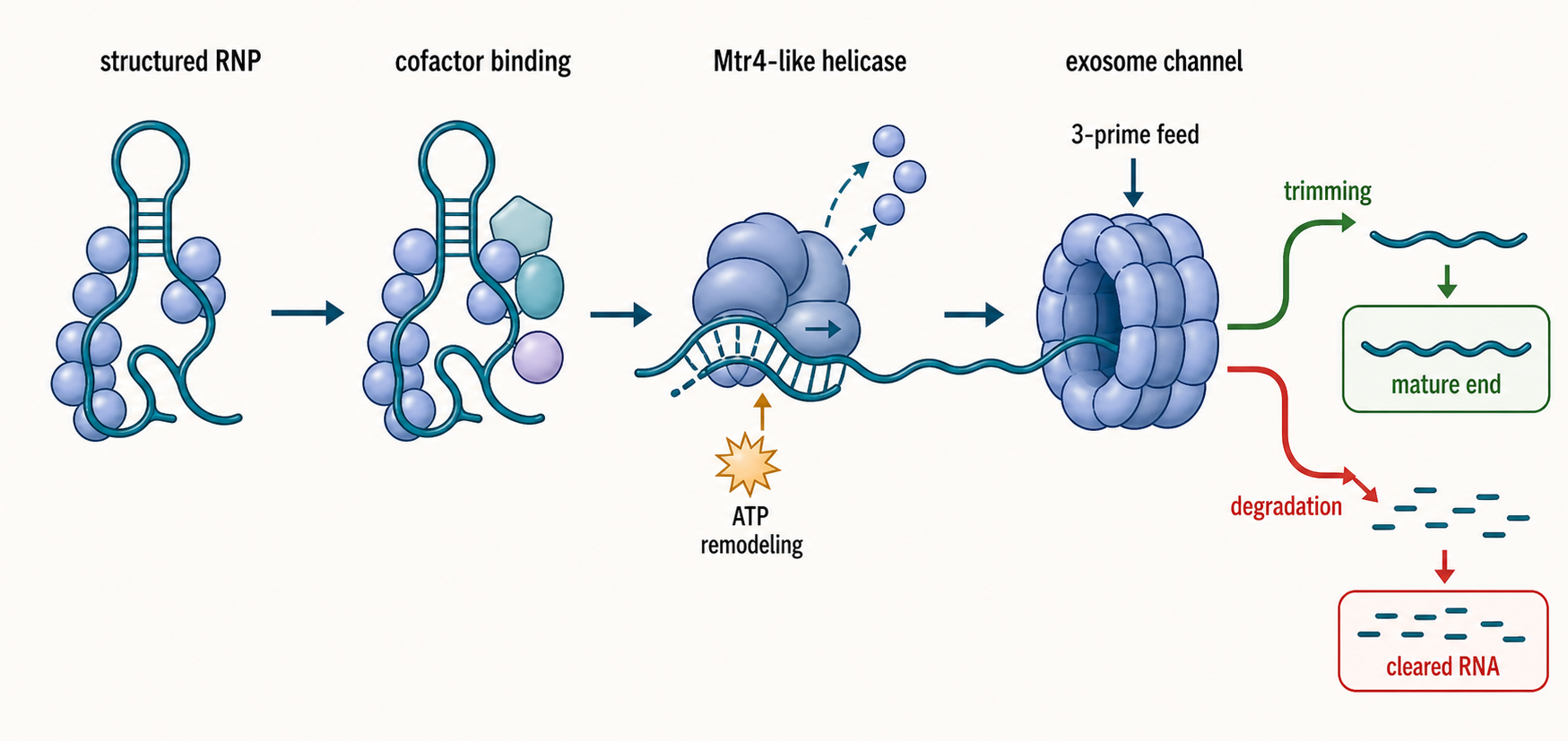
Figure 32.3. RNA exosome and helicase handoff. Exosome cofactors bind a structured or protein-coated RNA, an Mtr4-like helicase remodels the RNP, and the RNA is fed into the exosome channel for 3′ to 5′ trimming or degradation. The visual owns the nuclease-directed handoff and RNA path, not the comparative ATPase cycle or motor-family classification developed in Chapter 55. The same machinery can produce mature RNA ends or complete degradation depending on substrate identity, cofactor context, and compartment.
Substrate delivery is the key specificity step. The exosome needs RNA with a suitable single-stranded region and must often receive that substrate from a helicase-containing cofactor. Mtr4-family helicases in nuclear pathways, Ski-associated factors in cytoplasmic pathways, and adaptor complexes such as TRAMP, NEXT, or related assemblies help capture RNA, remodel RNPs, and feed RNA toward the exosome. Weick and Lima describe RNA helicases as hubs that orchestrate exosome-dependent 3′ to 5′ decay, a phrasing that captures the modern view: exosome function is an organized handoff system rather than isolated exonuclease activity (Weick and Lima 2021).
A concrete eukaryotic example is exosome activity on ribosome-associated mRNA decay substrates. Structural work on a human exosome-ribosome supercomplex showed how a decay substrate can be coupled to the ribosome and exosome machinery, supporting a model in which ribosome state, RNA path, and exosome engagement are physically coordinated (Kogel et al. 2024). The result is not merely that “the exosome degrades mRNA”; it is that exosome access can be integrated with translation-state surveillance and RNP architecture.
The nuclear exosome also prevents accumulation of nascent or aberrant RNAs, as described in Chapter 31. MYCN-linked studies provide a specific human example in which an oncogenic factor can connect the nuclear exosome to RNA polymerase II-associated RNA control. MYCN recruits nuclear exosome machinery to RNA polymerase II and acts as an RNA-binding accessory factor of the nuclear exosome targeting complex in tested systems (Papadopoulos et al. 2022; Papadopoulos et al. 2024). These findings should be interpreted in their cancer-relevant contexts, not generalized to all exosome recruitment.
Archaeal exosomes are related but not identical to eukaryotic exosomes. Bacterial 3′ decay often uses different enzymes, and even enzymes with the same polarity need not perform the same chemistry. Polynucleotide phosphorylase (PNPase) processively removes RNA in the 3′ to 5′ direction by phosphorolysis, releasing nucleoside diphosphates; an accessible single-stranded 3′ segment promotes entry, whereas duplex structure can stall the isolated enzyme. Association with an RNA helicase in a degradosome can improve access to structured substrates. RNase R, by contrast, is a hydrolytic RNase II-family exonuclease that can traverse substantial RNA structure when the substrate presents a suitable 3′ overhang. Thus, “3′ to 5′ exonuclease” specifies polarity but not reaction chemistry, product identity, structural tolerance, or assembly dependence (Arraiano et al. 2010). Chapter 34 treats bacterial and archaeal diversity more fully.
Human disease emphasizes the importance of exosome integrity. A biallelic variant in EXOSC4 that impairs RNA exosome function and translation has been associated with neurodevelopmental defects (Fasken et al. 2024). Such disease links are biologically plausible because exosome activity affects many RNA classes, but they also create interpretation challenges. A phenotype from an exosome subunit variant may reflect altered mRNA decay, stable RNA maturation, translation, stress responses, or developmental cell-state changes. Strong disease mechanism requires connecting the variant to specific molecular defects and rescue.
Box 32.1. Scientific Caution: RNA Exosome Is Not Extracellular Vesicle Exosome
- The RNA exosome is a multi-subunit ribonuclease complex that processes and degrades RNA in the nucleus and cytoplasm.
- Extracellular vesicle exosomes are membrane-bound vesicles secreted by cells that can carry RNA cargo.
- References about exosome-derived miRNAs, vesicle delivery, or cell-to-cell RNA transfer concern extracellular vesicles and should not be cited as evidence for RNA exosome catalysis or substrate selection.
- When reading the literature, check whether “exosome” refers to the ribonuclease complex or the secreted vesicle; context and citation genealogy usually clarify this, but the terminology collision is common.
32.4. Endonuclease families, site-specific cleavage, and product-end chemistry
An endonuclease cuts within an RNA molecule rather than chewing from an end. Endonucleolytic cleavage is powerful because it creates new RNA ends. A single internal cut can convert a protected full-length RNA into two fragments: one fragment may expose a 3′ end for exosome-like decay, and the other may expose a 5′ end for Xrn-like decay. Endonucleases therefore often initiate decay even when exonucleases finish it.
Bacterial RNase E is a central model. RNase E can cleave internal sites in bacterial RNAs and can organize a degradosome with accessory enzymes. Its activity depends on RNA sequence, structure, 5′ end state, and complex organization. Experiments on RNase E showed that pseudouridylation, substrate fold, and degradosome organization can alter endonuclease activity (Islam et al. 2021). This result makes a general teaching point: endonuclease specificity is not determined by a short linear consensus alone.
Metazoan endonucleases provide different examples. RNase L is activated in antiviral innate immunity by 2′,5′ oligoadenylates produced downstream of interferon-induced OAS enzymes. Once activated, RNase L cleaves cellular and viral RNAs, producing fragments that can affect RNA abundance and immune signaling. Work on RNase L-induced bodies and flavivirus subgenomic RNAs illustrates how endonuclease activation can reorganize RNA fate in infection-linked contexts (Watkins and Burke 2024). RNase L should not be treated as a housekeeping mRNA-decay enzyme; it is a regulated immune nuclease with strong stress and antiviral context.
Box 32.2. Mechanism Box: How an Endonuclease Commits an RNA to Exonuclease Cleanup
- A cofactor, RNA structure, immune signal, ribosome stall, or sequence feature exposes an internal cleavage site.
- The endonuclease cuts the RNA at that site.
- The upstream fragment gains a new 3′ end; the downstream fragment gains a new 5′ end. The illustration should label whether those termini are hydroxylated, phosphorylated, or cyclic-phosphorylated.
- If an end is chemically incompatible with the finishing enzyme, a phosphatase, kinase, or cyclic-phosphate-opening activity first heals the end.
- 3′ to 5′ exonucleases (such as the RNA exosome) can then clear the upstream fragment, while 5′ to 3′ exonucleases (such as Xrn1) can clear a downstream fragment with a compatible 5′ end.
- Accumulation of one fragment in an exonuclease mutant, loss of the cleavage product in an endonuclease mutant, and sensitivity to end-healing perturbation together confirm the sequence of events.
The Integrator complex is another metazoan example, best known for roles in cleavage of nascent transcripts such as small nuclear RNA precursors and promoter-proximal RNAs. Recent work on the assembly mechanism of Integrator’s RNA cleavage module provides structural and mechanistic support for the idea that endonucleolytic cleavage can be embedded in transcription-associated machines (Sabath et al. 2024). Integrator cleavage is not simply “RNA destruction”; depending on substrate and context, cleavage can promote maturation, termination, or decay.
MARF1-mediated mRNA decay illustrates how endonucleolytic decay can intersect with cytoplasmic RNP organization. P-bodies directly regulate MARF1-mediated mRNA decay in human cells, showing that decay outcomes can depend on cytoplasmic condensate-like compartments or granules rather than only on enzyme presence (Brothers et al. 2022). This example also warns against interpreting P-bodies as passive trash bins. P-bodies can be sites of storage, repression, remodeling, or decay depending on substrate and factor composition.
Some proteins cross the usual boundary between cleavage, end processing, and digestion. Human apurinic/apyrimidinic endonuclease 1 (APE1) is best established as a DNA-repair enzyme, but purified APE1 also removed 3′ phosphate groups from three defined RNA substrates and removed a terminal ribonucleotide from an RNA overhang. Active-site substitutions and naturally occurring APE1 variants reduced the RNA phosphatase signal, supporting assignment to APE1 rather than a contaminating nuclease. The same study found only weak 3′ to 5′ exoribonuclease activity on unstructured poly(A), poly(C), and poly(U) substrates and required micromolar-range enzyme concentrations (Chohan et al. 2015). APE1 therefore demonstrates that RNA product-end chemistry can be enzymatically remodeled and that one protein can combine several nuclease-related activities; the experiments do not establish APE1 as a general processive cellular 3′ exonuclease. Product-end healing and ligation are developed comparatively in Chapter 33.
Endonuclease interpretation is especially vulnerable to artifact. A sequencing peak at a cleavage site may arise from genuine enzymatic cleavage, reverse-transcription stops, RNA structure, fragmentation during library preparation, or selective stabilization of a fragment. Strong site-specific cleavage evidence combines mapping of both new ends, nuclease perturbation, catalytic mutants, in vitro cleavage, rescue, and kinetic timing. Chapter 5 gives the general evidence logic; this chapter applies it to ribonuclease mechanism.
32.5. Degradosomes, exosome-associated assemblies, and helicase-coupled substrate delivery
A nuclease rarely encounters a naked, pre-positioned RNA in a cell. Structured RNA, ribosomes, RNA-binding proteins, and stable RNP architecture can obstruct an active site or the narrow channel of a processive nuclease. Decay assemblies solve this access problem by combining recognition, remodeling, cleavage, and fragment transfer. The mechanistic question for this section is not whether an associated protein is generically called a helicase, but how the assembly converts an inaccessible substrate into a geometry that the catalytic subunit can use.
The nuclear RNA exosome illustrates sequential delivery. An adaptor first recognizes an RNA class or an aberrant RNP state. A cofactor complex binds the adaptor and RNA. An Mtr4-family motor captures a single-stranded region and can remodel local structure or protein occupancy. The RNA is then directed through or around the exosome core toward DIS3/RRP44 or RRP6/EXOSC10, depending on substrate and system. ATP hydrolysis may accelerate remodeling and directional feeding, but adaptor binding can also determine which substrate is admitted. Thus, a catalytic-subunit mutation, an ATPase mutation, and an adaptor-interface mutation can produce different substrate spectra even when all reduce decay.
Cytoplasmic exosome pathways use analogous but nonidentical logic. Ski2-like motors and the Ski complex couple translating or translation-associated substrates to the exosome. Ribosome state, a protruding RNA end, Ski-complex engagement, and exosome access form a physical handoff chain. Structural snapshots can show alignment of these components, but processive decay must be established by kinetic or product-distribution evidence. Chapter 55 explains how Ski2-like motors differ mechanochemically from local DEAD-box remodelers and directional DEAH/RHA enzymes.
Bacterial degradosomes combine activities on a scaffold. In the classical Escherichia coli assembly, RNase E supplies endonucleolytic activity and a long scaffold that recruits polynucleotide phosphorylase, the RhlB RNA helicase, enolase, and context-dependent partners. Internal cleavage generates new ends; remodeling exposes structured fragments; exonucleolytic activity removes products. This organization can increase local effective concentration and substrate transfer, but it does not guarantee channeling of every intermediate. Demonstrating channeling requires kinetics or intermediate capture that distinguishes direct transfer from diffusion and rebinding.
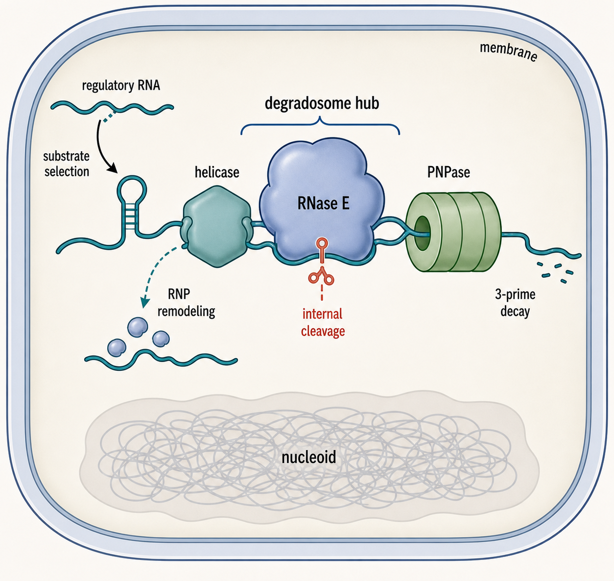
Figure 32.4. Bacterial degradosome architecture as a reaction hub. A bacterial degradosome combines RNase E-mediated internal cleavage, helicase-mediated substrate presentation, 3′ exonuclease activity from polynucleotide phosphorylase, and regulatory RNA-guided substrate selection in a single physical assembly. The teaching focus is how assembly changes nuclease access and product transfer; helicase mechanochemistry is handed to Chapter 55. Physical organization near the membrane or nucleoid affects substrate access and decay efficiency.
Degradosome is a family-level architectural term rather than one invariant composition. Many bacteria lack the canonical E. coli combination or build assemblies around RNase Y, RNase J, or other scaffolds. Membrane association can change encounter rates and separate decay from the nucleoid, while stress and regulatory RNAs can recruit or exclude particular substrates. Chapter 34 owns the lineage-specific pathways; this chapter uses them to teach how multienzyme organization changes a ribonuclease reaction network.
Guide and adaptor RNAs can alter delivery without becoming catalytic components. A bacterial small RNA can base-pair with a target, block translation, expose an RNase site, or recruit an RNA chaperone that contacts a decay factor. The causal chain must distinguish base pairing, RNP assembly, cleavage, and exonuclease cleanup. Bandyra et al. (2024) provide a recent example of regulatory-RNA cooperation with a degradosome, but no single bacterial small-RNA mechanism should be generalized across all Hfq-, ProQ-, or Csr/Rsm-associated systems.
The ownership boundary is deliberate. This section explains Mtr4, Ski, and RhlB only where they deliver RNA to decay enzymes. ATPase-cycle comparison, directionality, processivity, conserved motifs, single-molecule motor measurements, helicase inhibitors, disease variants, R-loop remodeling, and non-decay RNP machines belong to Chapter 55. RNA fragments that require phosphorylation, cyclic-phosphate opening, or ligation after cleavage pass to Chapter 33.
32.6. Specificity, kinetics, structures, regulation, inhibitors, and pathway handoffs
Ribonuclease specificity has at least four layers. Intrinsic specificity arises from active-site geometry, electrostatics, end-state requirements, and contacts with RNA sequence or structure. Assembly specificity arises from adaptors, scaffolds, partner enzymes, and substrate-delivery motors. Spatial specificity arises because enzyme and RNA occupy the same compartment or local translation environment. Kinetic specificity arises because a weakly preferred substrate can nevertheless dominate when it is abundant, long-lived, or repeatedly presented. A cleavage motif by itself therefore rarely predicts an in-cell substrate.
Exonuclease kinetics must separate binding, initiation, individual catalytic steps, pausing, dissociation, and rebinding. Processivity is the number or distance of catalytic events completed per productive binding event; it is not synonymous with a high bulk rate. A distributive enzyme can appear fast at high enzyme concentration because fragments rebind rapidly, while a processive enzyme can appear slow if initiation is rate limiting. Time courses with resolved product lengths, enzyme titrations, pulse-chase designs, and single-molecule trajectories help distinguish these models.
Endonuclease assays require equal care. A single-turnover experiment with enzyme in excess measures chemistry and conformational steps after productive complex formation more directly than a multiple-turnover assay dominated by substrate binding or product release. Steady-state parameters such as apparent kcat and Km are model-dependent summaries, not automatically microscopic rate constants or binding affinities. For an RNP enzyme with obligatory cofactors, the active concentration of assembled complex—not total protein added—is the relevant denominator.
Structures define possible reaction geometries. X-ray crystallography and cryogenic electron microscopy can reveal catalytic metals, bound phosphate mimics, RNA-channel dimensions, cap or end recognition, and adaptor interfaces. A structure assigned as substrate, intermediate, or product needs chemical justification: cleavage-resistant analogs, catalytic mutations, metal substitutions, and sample preparation can trap off-pathway states. Linking structures to stopped-flow, quench-flow, isotope, product-distribution, or single-molecule measurements converts a static pose into a mechanistic cycle.
Regulation can change abundance, localization, assembly, post-translational state, or substrate presentation. Decapping activators regulate access to DCP2; exosome adaptors redirect the same catalytic core to different RNA classes; innate immune signaling activates RNase L; degradosome assembly and localization alter bacterial substrate encounters. Chronic depletion often triggers transcriptional and stress compensation, so acute degrons, catalytic rescue, and matched expression controls are preferable when the claim concerns direct regulation.
Inhibitors can test mechanism or expose therapeutic dependencies, but pleiotropy is a central risk. A compound that stabilizes an RNA may inhibit a nuclease, disrupt an adaptor, deplete ATP, block translation, change transcription, or cause stress. Strong target validation combines biochemical inhibition against purified or reconstituted enzyme, cellular target engagement, resistant alleles or rescue, acute kinetics, and substrate-end readouts. Broad inhibition of a housekeeping nuclease is likely toxic; context-selective cofactors or pathogen-divergent enzymes may offer greater selectivity.
Pathway ownership follows biological context. Nuclear surveillance is developed in Chapter 31, bacterial and archaeal turnover in Chapter 34, eukaryotic mRNA decay in Chapter 35, organellar and lysosomal/autophagic RNA turnover in Chapter 37, stress and disease regulation in Chapter 38, helicase enzymology in Chapter 55, end healing and repair in Chapter 33, and quantitative kinetic design in Chapter 124. The lysosomal handoff is mechanistic as well as organizational: detecting an RNA-degrading enzyme inside a lysosome establishes a candidate terminal chemistry, not how the RNA or ribosome reached the compartment, whether delivery was selective, or whether a whole organelle was recycled. These handoffs prevent one general enzyme chapter from becoming a catalog of every place where RNA is degraded.
32.7. Biochemical assays, kinetic models, perturbations, and interpretation limits
RNA decay assays ask at least four different questions: how much RNA is present, how fast RNA is made, how fast RNA is destroyed, and where cleavage or digestion occurs. No single assay answers all four.
Steady-state RNA-seq measures abundance after synthesis, processing, localization, and decay have already acted. If an RNA accumulates after a ribonuclease is depleted, the RNA is a candidate substrate or indirect target. The experiment does not prove direct cleavage. Metabolic labeling and transcriptional shutoff can estimate half-life, but transcriptional shutoff can cause stress and alter decay pathways. Time-resolved RNA-seq can separate synthesis and degradation more effectively, but model assumptions and labeling efficiency matter. Plant RNA-seq guidance and broader biochemical-computational reviews are useful reminders that library design, normalization, replicate structure, and statistical modeling shape biological interpretation (Upton et al. 2023; Du et al. 2024).
Box 32.3. Scientific Caution: Abundance Change Is Not Direct Cleavage
- RNA accumulation after nuclease depletion may reflect direct loss of cleavage, altered transcription, altered processing, altered localization, stress responses, compensation by other factors, or changes in cell composition.
- Direct-substrate claims require end mapping at the specific cleavage site, dependence on catalytic activity (not just protein presence), rate measurements, biochemical reconstitution with purified components, and rescue or orthogonal perturbation.
- A transcriptome-wide accumulation list is a hypothesis; each candidate on that list requires further mechanistic evidence before it can be called a direct substrate.
RNA end mapping is central for ribonuclease mechanism. A 5′ monophosphate peak at a reproducible position can suggest endonucleolytic cleavage or exonuclease stalling. A 3′ end peak can suggest cleavage, trimming, or tailing. Paired evidence from both sides of a cut is stronger than one end alone. Cap-sensitive methods distinguish capped, decapped, triphosphorylated, and monophosphorylated ends. Long-read direct RNA sequencing can preserve isoform and tail context, but error profiles and modification effects complicate precise end calling.
Biochemical reconstitution tests direct mechanism. A purified ribonuclease, defined RNA substrate, cofactors, ATP, metal ions, and controlled reaction times can reveal cleavage requirements, directionality, processivity, and inhibitor sensitivity. The limitation is that a naked RNA in a tube may not mimic a cellular RNP. The strongest experiments reconstitute increasing layers of native context, such as helicase plus exosome, degradosome assembly, ribosome-bound substrate, or RNA-binding adaptor.
Structural biology explains geometry. Cryo-electron microscopy, X-ray crystallography, crosslinking, and biochemical footprinting can show how an RNA enters an enzyme, how cofactors position a substrate, or how a ribosome-associated RNA is delivered to a nuclease. Structural work on the human exosome-ribosome supercomplex is a current example (Kogel et al. 2024). Structural snapshots should be connected to kinetics because a captured state may be a substrate, intermediate, product, stalled complex, or off-pathway assembly.
Inhibitors are useful but can be blunt. A small molecule or stress treatment that reduces RNA decay can also alter transcription, translation, localization, protein stability, or cell viability. Viral polymerase assay development and RNA-targeted drug-discovery reviews are adjacent reminders that biochemical screening requires careful counterscreens and target validation, even when the immediate subject is not a cellular ribonuclease inhibitor (Chen et al. 2025; Xiao et al. 2023). For decay enzymes, a convincing inhibitor study should show target engagement, catalytic effect, dose response, rescue or orthogonal perturbation, and substrate-specific readouts.
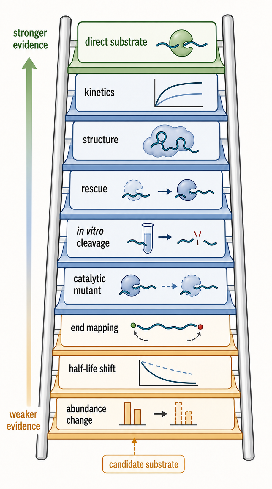
Figure 32.5. Evidence ladder for direct ribonuclease substrates. Evidence for a direct ribonuclease substrate progresses from RNA abundance change after perturbation, to metabolic-labeling half-life measurement, to 5′ and 3′ end mapping, to catalytic-mutant dependence, to in vitro biochemical reconstitution, to rescue and structural or kinetic mechanism, with each step providing stronger support for direct enzymatic action.
The most reliable interpretation combines levels: genetics identifies candidate factors; catalytic mutants separate scaffolding from nuclease activity; end mapping locates cleavage; metabolic labeling estimates rates; biochemical reconstitution tests direct action; structural biology explains geometry; and rescue or orthogonal perturbation tests causality. Chapter 5 supplies the general evidence ladder; RNA decay is one of the fields where that ladder is most necessary.
Experimental Foundations and Evidence
The first foundation is enzyme perturbation. Knockout, knockdown, degron-mediated depletion, catalytic mutation, dominant-negative expression, and acute inhibition can reveal RNAs controlled by a decay factor. Acute methods are usually easier to interpret than chronic loss because chronic loss can trigger compensation and stress. Catalytic mutants are especially informative when a factor has both enzymatic and scaffolding functions.
The second foundation is RNA-end and fragment analysis. Ribonucleases leave molecular traces: new 5′ ends, new 3′ ends, shortened tails, terminal additions, cleavage fragments, or protected decay intermediates. Mapping these features can distinguish initiation from completion. For example, an endonuclease mutant may eliminate a specific internal cleavage product, whereas an exonuclease mutant may cause fragments to accumulate because finishing decay is blocked.
The third foundation is reconstitution. Purified RNase J1 acting on trp leader RNA, RNase E activity in different degradosome contexts, APE1 tested against defined RNA-end chemistries, exosome cofactors with defined substrates, and ribosome-exosome structural assemblies all demonstrate different levels of reconstitution (Deikus et al. 2008; Chohan et al. 2015; Islam et al. 2021; Weick and Lima 2021; Kogel et al. 2024). Reconstitution is powerful because it tests direct mechanism, but the substrate and enzyme concentration must be chosen carefully. A detectable activity on a short naked oligonucleotide at high enzyme concentration establishes biochemical capability, not its rate, substrate share, or physiological importance in an RNP-rich cell.
The fourth foundation is systems measurement. Transcriptome-wide half-life measurements, RNA-seq after perturbation, ribosome profiling, CLIP-like protein-RNA interaction maps, and single-cell measurements can reveal pathway scope. These methods are hypothesis generators unless paired with mechanism. RNA abundance changes can reflect altered synthesis, altered localization, altered cell composition, or altered processing rather than direct RNA decay.
Biological Contexts Across Organisms and RNA Classes
Bacteria use RNA decay to adjust rapidly to nutrient shifts, stress, phage infection, and regulatory RNA programs. RNase E-centered degradosomes dominate the textbook model for many Proteobacteria, but Gram-positive bacteria and other lineages use different nuclease combinations such as RNase J and RNase Y. Chapter 34 expands this diversity.
Archaea possess exosome-like 3′ processing machines in many lineages and also have archaeal-specific RNA processing and decay logic. Archaeal systems are important because they clarify evolutionary relationships among RNA-processing complexes, but they should not be forced into either bacterial or eukaryotic categories.
Eukaryotic nuclei use the exosome, Xrn2/Rat1-like factors, Integrator, transcription-coupled adaptors, tailing enzymes, and RNA helicases to distinguish maturation from disposal. Eukaryotic cytoplasm uses deadenylation, decapping, Xrn1, cytoplasmic exosome cofactors, endonucleases, P-body factors, ribosome-associated quality-control factors, and stress-regulated pathways. The same mRNA can pass through multiple possible decay routes depending on translation state, codon optimality, RNA-binding proteins, and stress.
Stable noncoding RNAs require decay enzymes too. Pre-rRNA processing, pre-tRNA quality control, snRNA and snoRNA maturation, and degradation of defective stable RNP precursors all use nucleases. A common misconception is that decay enzymes only remove failed mRNAs. In reality, many essential stable RNPs are made by controlled cutting and trimming, and failed processing intermediates must be eliminated.
Technology, Computational, Clinical, and Engineering Links
RNA therapeutics and synthetic RNA design must account for decay enzymes. A therapeutic mRNA needs cap chemistry, untranslated-region design, coding-sequence choices, tail properties, and modification patterns that support the desired balance of translation, stability, and immune avoidance. An siRNA or antisense oligonucleotide intentionally recruits nuclease or RNA-silencing pathways. A synthetic regulatory RNA may fail because cellular ribonucleases remove it before it acts. Chapters 26, 29, 36, 62, and 116 treat therapeutic design details.
Computational models of RNA decay should separate features that affect synthesis from features that affect destruction. Codon optimality, RNA structure, modification state, RBP binding, tail length, subcellular localization, and translation state can all correlate with half-life. Causality requires perturbation and rate measurements, not only a predictive model. Chapter 36 covers codon optimality-mediated decay in detail.
Clinically, mutations in exosome subunits, helicases, and RNA-processing factors can cause developmental, neurological, immune, or cancer phenotypes. EXOSC4 variant work provides one direct example for exosome dysfunction (Fasken et al. 2024). Helicase reviews document broader disease links (Bohnsack et al. 2023). However, disease association does not identify the causal RNA substrate. Mechanistic clinical interpretation must connect variant, molecular activity, substrate class, cell type, and phenotype.
Drug discovery against RNA decay factors is possible but challenging. Shared enzymes often process many RNA classes, so inhibition can be toxic or pleiotropic. Selective targeting may require context-specific cofactors, disease-specific dependencies, or substrate-specific recruitment rather than global nuclease blockade. RNA-targeted small-molecule discovery provides useful conceptual parallels because target validation and off-target RNA effects are central concerns (Xiao et al. 2023).
Box 32.4. Handoff Box: What a Decay Chapter Must Say About a Helicase
- RNA helicases in decay pathways can act by locally remodeling secondary structure, displacing RNA-binding proteins, clamping RNA in a single-stranded state, recruiting adaptor proteins, or feeding RNA into a nuclease channel.
- Many decay-relevant helicases do not processively unwind long double-stranded RNA in the way a DNA replication helicase unwinds duplex DNA.
- Mechanistic descriptions should specify the actual helicase action observed (local remodeling, protein displacement, substrate handoff) rather than defaulting to the generic term “unwinding.”
- This chapter owns the consequence for nuclease access and RNA routing; Chapter 55 owns comparative ATPase cycles, translocation, processivity, family classification, and motor assays.
Box 32.5. Scientific Caution: Tailing Can Stabilize or Destabilize
- A long poly(A) tail on a mature eukaryotic mRNA promotes PABP binding, translation, and protection from decay.
- Short oligo(A) additions, mixed tails, or poly(U) additions can create decay handles that recruit the nuclear exosome or bacterial and organellar decay machines.
- The consequence of a tail depends on the tail composition, length, proteins that bind it, and the cellular compartment in which the RNA resides.
- Do not generalize from cytoplasmic mRNA poly(A) biology to nuclear, bacterial, or organellar tailing without checking context.
Recent Consensus
The current consensus is that RNA decay enzymes are regulated molecular machines, not generic RNA-destroying background activities. Substrate selection depends on RNA end state, RNP architecture, compartment, cofactors, and competing maturation or translation pathways. Exonucleases and endonucleases cooperate: internal cuts create new ends, decapping enables 5′ exonucleases, tailing or trimming can expose 3′ ends, and helicases feed structured substrates into nuclease channels.
The RNA exosome is now understood as a cofactor-directed processing and decay platform whose functions span nuclear surveillance, cytoplasmic decay, and stable RNA maturation. Helicases are central organizers of exosome-dependent decay. Bacterial degradosomes are spatially and compositionally organized assemblies whose architecture affects substrate recognition and cleavage. Endonucleases such as RNase E, RNase L, Integrator-associated nucleases, and MARF1-related pathways show that internal cleavage is often the initiating event that commits RNA fragments to downstream exonucleases.
Consensus is weaker on how to assign every transcriptome change to a direct nuclease substrate. The field increasingly treats RNA abundance data as a starting point that must be refined by end mapping, acute perturbation, catalytic separation, reconstitution, and structural or kinetic validation.
Open Questions, Controversies, Deprecated Models, and Common Misconceptions
Open questions:
- What determines how decay machines choose between trimming and complete degradation? The same exosome-associated activity can mature an RNA precursor or destroy an RNA, and the switch likely depends on RNA length, structure, adaptor identity, modification state, and kinetic competition with RNP assembly.
- How do decay compartments such as P-bodies, stress granules, ER-associated sites, bacterial membrane-associated degradosomes, and viral replication compartments alter reaction rates? Microscopy can show colocalization, but colocalization does not prove cleavage. Biochemistry can show cleavage, but purified reactions may miss compartment effects.
Common misconceptions:
- “A nuclease-depletion RNA-seq experiment identifies direct substrates.” It identifies RNAs whose abundance depends on the perturbation. Direct substrate status requires additional evidence.
- “Exosome always means extracellular vesicle.” In RNA enzymology, the RNA exosome is a ribonuclease complex. Extracellular vesicle exosomes can carry RNAs and influence disease or therapy, but that is a different subject and should not be used as evidence for RNA exosome catalysis.
- “Endonucleases and exonucleases define separate pathways.” In many real pathways, an endonuclease creates ends, exonucleases finish digestion, helicases remodel the substrate, and cofactors determine where the reaction happens.
- “Any newly exposed RNA end is immediately usable by the exonuclease of matching polarity.” End identity includes chemical state as well as position: XRN-family enzymes favor a 5′ monophosphate rather than a 5′ hydroxyl, and a 3′ phosphate can block enzymes that require a 3′ hydroxyl until an end-healing activity acts.
Deprecated or weakened claims:
- A deprecated oversimplification is the idea that RNA decay is only a cleanup pathway after gene expression has finished. Current evidence supports a more integrated view in which decay enzymes shape transcription termination, translation, immune signaling, stable RNP biogenesis, stress responses, and genome stability.