Chapter 103. Neuronal and Glial RNA Integration in Plasticity, Neurodevelopment, and Disease
Scope Note
This chapter integrates RNA regulation across neurons and glia, linking localization and local translation to splicing, editing, granules, electrophysiology, plasticity, neurodevelopment, and disease. It owns neural and glial system-level interpretation: how several RNA layers combine within a cell type, compartment, circuit state, developmental window, or pathology. Chapter 74 owns generic transport machinery and localization grammar, Chapter 75 owns membrane and organelle targeting, and Chapter 76 owns comparative developmental or cell-type local-translation programs. This chapter summarizes those prerequisites only when needed to connect an RNA event to neural physiology or disease.
Executive Summary
Neurons and glia use RNA regulation to solve problems created by nervous-system geometry, developmental timing, and activity-dependent adaptation. A neuron can place synapses hundreds or thousands of micrometers from the soma, so relying only on protein synthesis in the cell body would delay responses and make individual synapse regulation imprecise. Neurons therefore transport selected mRNAs as ribonucleoprotein particles, keep many of them translationally repressed during travel, and activate translation locally in dendrites, axons, growth cones, and synaptic compartments when cues such as synaptic activity, neurotrophins, injury, or guidance signals alter local signaling. Local translation is not a niche exception to central protein synthesis. It is a recurring mechanism for synaptic plasticity, axon growth, presynaptic function, injury responses, and circuit remodeling, although the identity of locally translated mRNAs varies by cell type, developmental stage, compartment, and assay.
Glia also have spatial RNA biology. Astrocytes extend fine processes around synapses and blood vessels; oligodendrocytes build myelin at distant membrane domains; microglia alter RNA programs during surveillance, synaptic pruning, inflammation, and neurodegeneration. Glial mRNAs can localize to peripheral processes, and local translation can support rapid remodeling of synaptic contacts, myelin maintenance, metabolic support, and immune-like responses. Neuron-glia RNA regulation includes signaling from neuronal activity to glial transcription and translation, glial modulation of neuronal RNA programs, extracellular vesicle and Arc-like RNA transfer mechanisms, and disease-associated feedback between stressed neurons and reactive glia.
Neurodevelopment depends on RNA processing as much as RNA abundance. Alternative splicing changes protein domains, untranslated regions, localization signals, and regulatory elements during neuronal maturation. Neural microexons are very short alternative exons, often three to twenty-seven nucleotides long, that can remodel protein interaction surfaces without disrupting reading frames. A-to-I RNA editing by ADAR enzymes changes selected adenosines so they are read as guanosines by sequencing and translation machinery, with notable effects on receptors, ion channels, and immune recognition of endogenous double-stranded RNA. These processes are especially important in the nervous system because neuronal identity, synapse formation, excitability, and circuit refinement require finely tuned protein isoforms and RNA regulatory landscapes.
Many neurological diseases expose the fragility of RNA regulation. RNA-binding proteins such as TDP-43, FUS, hnRNP proteins, DDX3X, and others can mislocalize, aggregate, or lose nuclear function, producing altered splicing, transport, translation, and RNA surveillance. Repeat expansions can generate toxic RNAs, sequester RNA-binding proteins, alter splicing, trigger repeat-associated non-AUG translation, and promote somatic expansion in vulnerable neurons. RNA granules normally support dynamic storage and translation control, but persistent or compositionally abnormal granules can intersect with neurodegenerative pathology. These mechanisms should not be collapsed into a single granule disease model: gain of toxic RNA function, protein aggregation, loss of RBP function, DNA repair-driven repeat instability, nucleocytoplasmic transport defects, and glial stress responses can contribute in different proportions across diseases.
Modern methods make neuronal and glial RNA biology unusually rich but also artifact-prone. Single-cell and single-nucleus RNA-seq classify neural cell states, but dissociation and nuclear sampling alter what can be measured. Spatial transcriptomics and multiplexed RNA imaging preserve tissue geography but differ in sensitivity, target breadth, and resolution. Live imaging can follow RNA movement and translation reporters, but tags and overexpression can perturb localization. Patch-seq links electrophysiology, morphology, and transcriptomes in the same cell, but low RNA input and sampling bias require cautious interpretation. The best evidence combines spatial localization, biochemical association, activity or perturbation, local protein synthesis measurement, and functional readout.
Concept Inventory
- Local translation: protein synthesis from an mRNA near a subcellular site of function rather than only near the nucleus or soma. In neurons, local translation often refers to protein synthesis in dendrites, dendritic spines, axons, growth cones, presynaptic boutons, or injury sites. In glia, it can refer to translation in astrocytic peripheral processes, oligodendrocyte processes and myelin-associated domains, or microglial processes. The term does not prove that every ribosome, factor, or amino acid source is locally restricted; it means the mRNA template is translated in a compartment where the protein can act quickly or selectively.
- Neural spatial RNA program: the combined distribution, translation state, processing history, and functional output of RNAs in neuronal or glial compartments. Generic motors, adaptors, cis elements, anchoring, and release are defined in Chapter 74.
- Synaptic plasticity: an activity-dependent change in synaptic strength, structure, or molecular composition. Long-term potentiation and long-term depression are major experimental models, but plasticity also includes homeostatic scaling, presynaptic adaptation, spine remodeling, inhibitory synapse regulation, and developmental refinement.
- Neural microexons: very short alternatively spliced exons enriched in neuronal transcripts. Because many microexons have lengths divisible by three, they can insert a few amino acids into protein interaction regions without frameshifts. Their inclusion is often regulated by neural splicing factors and can be altered in neurodevelopmental disorders.
- A-to-I RNA editing: conversion of adenosine to inosine in RNA by adenosine deaminases acting on RNA. Inosine pairs like guanosine during sequencing and translation, so editing can recode codons, alter splice sites, reshape RNA structures, or prevent innate immune sensors from misclassifying endogenous double-stranded RNA as viral-like RNA.
- RNA granules: non-membrane RNP assemblies that concentrate RNAs and proteins. Neuronal transport granules, stress granules, processing bodies, and disease-associated inclusions overlap in components but are not identical entities. Granule membership does not automatically imply functional condensation, irreversible aggregation, or pathology.
- Repeat expansion RNA: RNA transcribed from genomic loci with expanded short tandem repeats, such as CAG, CUG, GGGGCC, or CGG repeats. Repeat-containing RNAs can form structures, bind RBPs, accumulate as nuclear foci, be translated by canonical or repeat-associated non-AUG mechanisms, or contribute indirectly through effects on chromatin and DNA repair.
What to Know Before Reading This Chapter
The chapter assumes familiarity with mRNA processing, RNA-binding proteins, translation initiation, untranslated regions, alternative splicing, and basic nervous-system cell types. A neuron has a cell body, dendrites that receive many inputs, an axon that sends output, and synapses that convert electrical and chemical signals into changes in molecular state. Glia include astrocytes, oligodendrocytes, microglia, ependymal cells, and lineage states such as oligodendrocyte precursor cells. Glia are not passive support cells: they regulate synapse formation, myelination, metabolism, immune responses, extracellular ions and neurotransmitters, vascular coupling, and circuit stability.
Several interpretive cautions are important. First, transcript abundance in a compartment is not the same as local translation. A localized mRNA may be stored, transported, degraded, or translated. Second, a transcript detected in bulk neuropil or synaptosome preparations may derive from neuronal processes, glial processes, blood vessels, mitochondria, or contamination unless the assay is carefully controlled. Third, activity-dependent RNA changes can reflect transcription, splicing, transport, translation, stabilization, decay, or cell-state composition. Fourth, disease-associated RNA granules and inclusions are not automatically causal; they can be causes, modifiers, protective storage states, or downstream markers depending on the system.
Running examples in this chapter include beta-actin mRNA localization in growth cones and dendrites, Arc mRNA and protein in activity-dependent plasticity and intercellular RNA transfer, CPEB-family regulation of local translation, neural microexon inclusion during neuronal maturation, ADAR editing of neuronal transcripts, TDP-43 and FUS pathology in amyotrophic lateral sclerosis and frontotemporal dementia, CUG and GGGGCC repeat expansion mechanisms, and spatial transcriptomic maps of neuronal and glial states in tissue.
103.1. Integration of RNA localization and local translation in synaptic plasticity
Neuronal RNA localization begins with a physical problem. A mature neuron can have a soma in one region, dendritic branches receiving thousands of synaptic inputs, and an axon projecting across long distances. If all proteins were synthesized near the soma and then shipped outward, the neuron would lose temporal precision and synapse specificity. Local translation gives a neuron a way to convert local signals into local protein production. A stimulated dendritic spine can alter translation of nearby mRNAs, an injured axon can synthesize proteins needed for retrograde signaling and repair, and a growth cone can translate guidance-response proteins at one side of the cone to bias movement.
Transport provides the spatial prerequisite, but the neural question is how localization becomes physiology. A transcript must arrive or be retained in a neuronal compartment, remain competent for regulated translation, respond to activity or injury, and produce a protein whose local action changes synaptic strength, morphology, organelle state, or signaling. The cis elements, RBPs, motors, anchoring, and release steps are developed in Chapter 74, while endosome- and mitochondria-coupled targeting examples are developed in Chapter 75.
Beta-actin mRNA is a classic example because its localization can support cytoskeletal remodeling in growth cones and dendritic compartments. The point is not that beta-actin explains all local translation, but that it shows the causal chain that must be established: an mRNA contains localization information, an RBP-dependent transport mechanism moves or anchors the transcript, a signal triggers local translation, the protein product acts near its site of synthesis, and perturbing the localization or translation event changes cellular behavior. Similar logic applies to mRNAs encoding receptors, scaffolding proteins, kinases, phosphatases, cytoskeletal regulators, mitochondrial proteins, and presynaptic components.
Synaptic plasticity provides a functional context for local translation. Long-term potentiation and long-term depression can require new protein synthesis, and experiments using local translation inhibitors, compartmentalized cultures, ribosome profiling, reporter imaging, and genetic perturbations have shown that dendritic translation contributes to durable changes in synaptic strength. Arc is a central activity-regulated example. Arc mRNA is induced by neuronal activity, transported into dendrites, and locally translated in ways connected to synapse remodeling. Arc protein also has unusual virus-like ancestry and can assemble capsid-like particles that mediate intercellular RNA transfer in experimental systems, a finding that links synaptic plasticity to broader questions about RNA movement between cells.
Presynaptic translation was once more controversial because axons were often treated as protein-receiving cables, but axonal and presynaptic mRNAs are now well supported in many systems. Axonal translation can regulate guidance, branching, mitochondrial maintenance, injury responses, and presynaptic transmission. For example, CPEB-family proteins can activate axonal translation of mRNAs relevant to glutamatergic transmission, and local control can alter presynaptic plasticity. The evidence is strongest when local mRNA, local ribosomes, nascent peptide labeling, perturbation of a specific transcript or RBP, and electrophysiological or morphological consequences are linked in the same model.
RNA surveillance and decay also operate locally. Nonsense-mediated decay factor UPF1 can associate with STAU2-containing neuronal RNA granules and influence synaptic plasticity. This example cautions against treating transport granules as simple storage containers. A granule can carry translational repressors, activators, decay factors, helicases, and scaffold proteins. The state of an mRNP particle can shift as it moves from soma to dendrite, as it pauses near a synapse, or as it responds to activity. Local RNA regulation is therefore a network of transport, anchoring, translation, decay, and remodeling rather than a one-directional delivery route.
Evidence for local translation has several recurrent limitations. Synaptosome and neuropil fractions can be contaminated by glial fragments or somatic material. Reporter constructs can overrepresent localization motifs or saturate RBPs. Translation inhibitors can affect cellular metabolism beyond local protein synthesis. Ribosome profiling can be difficult in small compartments. Imaging of nascent peptides requires careful controls for diffusion of completed proteins. The strongest conclusions combine orthogonal evidence and include rescue experiments in which restoring local translation of a specific mRNA restores the phenotype.
103.2. Glial RNA programs and neuron-glia regulatory integration
Glial RNA biology is not simply neuronal RNA biology in another cell type. Astrocytes, oligodendrocytes, and microglia have different morphologies, developmental origins, physiological functions, and disease responses. Astrocytes extend fine processes that ensheath synapses, regulate neurotransmitter uptake, buffer ions, release metabolic substrates, and coordinate vascular responses. Oligodendrocytes produce myelin sheaths and maintain long membrane extensions that wrap axons. Microglia survey tissue, prune synapses during development, respond to damage, and enter disease-associated states. Each geometry creates a rationale for spatial RNA control.
Astrocytic local translation is particularly relevant at perisynaptic processes. These fine processes can be far from the astrocyte soma and can remodel rapidly in response to neuronal activity. Localized mRNAs and translational machinery in astrocyte processes could support local production of cytoskeletal regulators, transporters, signaling proteins, and metabolic enzymes near active synapses. The field has evidence from mRNA localization studies, translating-ribosome approaches, and imaging, but the interpretation is often more difficult than in large neuronal dendrites because astrocyte processes are thin, highly branched, and interwoven with neuronal structures. A transcript detected near a synapse may be neuronal, astrocytic, oligodendrocytic, microglial, vascular, or extracellular unless cell-type assignment is explicit.
Oligodendrocyte RNA localization supports myelin biology. Myelin is a massive specialized membrane system, and oligodendrocyte processes must deliver proteins and lipids to sheaths associated with particular axons. Local translation of myelin-related mRNAs can support sheath formation, maintenance, and adaptation. This mechanism allows local axon-glia signals to shape individual internodes rather than forcing the oligodendrocyte to regulate all sheaths uniformly. Boundary cases matter: compact myelin has low cytoplasmic volume, but oligodendrocytes retain cytoplasmic channels, paranodal regions, and process domains where RNA and translation can operate.
Microglial RNA regulation is often studied through cell-state transcriptomics rather than subcellular translation, but spatial RNA control may matter for process extension, phagocytosis, synaptic contact, and inflammatory signaling. Microglial activation states can change mRNA abundance, splicing, RNA stability, translation, and secreted signals that feed back onto neurons and astrocytes. Single-cell and spatial studies in Parkinson disease, neuropathic pain, and other contexts show that glial states are not uniform across tissue. However, a disease-associated transcriptomic state is not itself a mechanism; it becomes mechanistic evidence only when linked to perturbation, cellular function, and disease-relevant outcome.
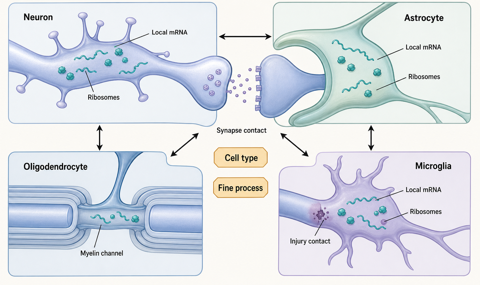
Figure 103.2. Spatial RNA regulation across neurons, astrocytes, oligodendrocytes, and microglia. Glial local translation is plausible and increasingly supported, but cell-type assignment and fine-process resolution are essential.
Neuron-glia RNA regulation includes signaling in both directions. Neuronal activity can alter astrocyte and oligodendrocyte RNA programs through neurotransmitters, ions, growth factors, extracellular matrix signals, and metabolic cues. Glia can influence neuronal RNA programs by controlling extracellular glutamate, potassium, cytokines, cholesterol, lactate, myelination, synapse elimination, and inflammatory tone. Extracellular vesicles and RNA-containing particles provide another possible layer, although claims about functional RNA transfer require especially careful controls. Arc-derived capsid-like particles show that a neuronal protein can package RNA and mediate transfer in experimental systems, but this finding should not be generalized to all extracellular RNA without evidence for packaging specificity, delivery, stability, and recipient-cell function.
Glial local translation also intersects with disease. Reactive astrocytes and disease-associated microglia can express RNA programs that modify neuronal survival, synapse stability, and inflammatory signaling. Oligodendrocyte stress and myelin dysfunction can alter axonal metabolism and neuronal vulnerability. In neurodegeneration, glial transcriptomes may be protective in one phase and damaging in another. For example, glial lipid handling, oxidative stress responses, and tau-linked interactions can modify neuronal pathology. The lesson for RNA biology is that cell-type-resolved and spatially resolved data are necessary but not sufficient: the causal question is which RNA regulatory event changes which glial function and how that function affects neurons.
Table 103.1. Glial RNA regulation by cell type and compartment. Astrocytes, oligodendrocytes, microglia, and other glial cells regulate RNA in distinct nuclear, somatic, and process-localized compartments; cell-type identity and compartment-specific evidence are required before assigning function.
| Glial cell type | Relevant compartment | RNA regulatory process | Example biological function | Main evidence types | Major interpretive caveat |
|---|---|---|---|---|---|
| Astrocyte | Peripheral or perisynaptic fine process | Localized mRNAs with ribosome-associated or cue-responsive local translation | Synapse modulation, transporter remodeling, cytoskeletal remodeling, metabolic support | smFISH, proximity-specific ribosome labeling, translating-ribosome assays, imaging | Fine processes interweave with neuronal and vascular structures; cell-type assignment needs explicit markers |
| Oligodendrocyte | Process, myelin-associated cytoplasmic channel, paranodal domain | Localized mRNAs and regulated translation near selected sheaths | Sheath formation, myelin maintenance, axon-specific adaptation | mRNA and protein imaging, compartmental assays, developmental perturbation | Compact myelin has little cytoplasm; process or channel RNA must be separated from bulk sheath signal |
| Microglia | Surveilling process, phagocytic cup, synapse or lesion contact | State-dependent RNA abundance, splicing, translation, and possible process-local regulation | Synaptic pruning, phagocytosis, inflammatory signaling, debris response | Single-cell or single-nucleus RNA-seq, spatial transcriptomics, perturbation, imaging | A transcriptomic activation state is not a mechanism without linked perturbation and function |
| Oligodendrocyte precursor cell | Axon-contacting process and differentiating lineage compartment | Translation and splicing shifts during maturation and response to axon cues | Lineage progression, process extension, initiation of myelination | Single-cell developmental trajectories, spatial assays, genetic perturbation | Maturation state can mimic regulation; lineage staging and cell-cycle controls are needed |
103.3. Alternative splicing, microexons, RNA editing, and neurodevelopment
Neurodevelopment requires cells to generate many molecular identities from a shared genome. Transcription determines which genes are expressed, but post-transcriptional processing determines which isoforms, regulatory elements, and protein domains are produced. Alternative splicing is therefore central to nervous-system development. It can include or skip cassette exons, choose alternative splice sites, retain introns, select alternative first or last exons, or alter untranslated regions. These decisions can change protein localization, interaction partners, channel properties, receptor signaling, cytoskeletal regulation, or RNA localization.
Neuronal splicing programs are controlled by RNA-binding proteins that recognize short sequence motifs and act from position-dependent binding sites. RBFOX, NOVA, PTBP, MBNL, CELF, QKI, SR proteins, hnRNP proteins, and other factors contribute to cell-type-specific and developmental splicing programs. The same factor can activate or repress exon inclusion depending on whether it binds upstream, within, or downstream of an exon. Developmental transitions often involve coordinated changes in multiple splicing regulators, so an isoform switch should not be attributed to a single factor unless the evidence shows direct binding, perturbation response, and rescue or mechanistic specificity.
Neural microexons are a striking specialization. A microexon may add only one to nine amino acids, but those amino acids can alter protein interaction surfaces in synaptic proteins, cytoskeletal regulators, trafficking factors, and signaling molecules. Because many neural microexons preserve the reading frame, they can fine-tune protein networks rather than create entirely new proteins. Microexon inclusion is often high in neurons and increases during maturation. Disrupted microexon regulation has been associated with autism spectrum disorder and other neurodevelopmental contexts, although association does not prove that every altered microexon is causal. Mechanistic priority should go to microexons with defined protein-domain effects, developmental timing, cell-type specificity, and functional perturbation data.
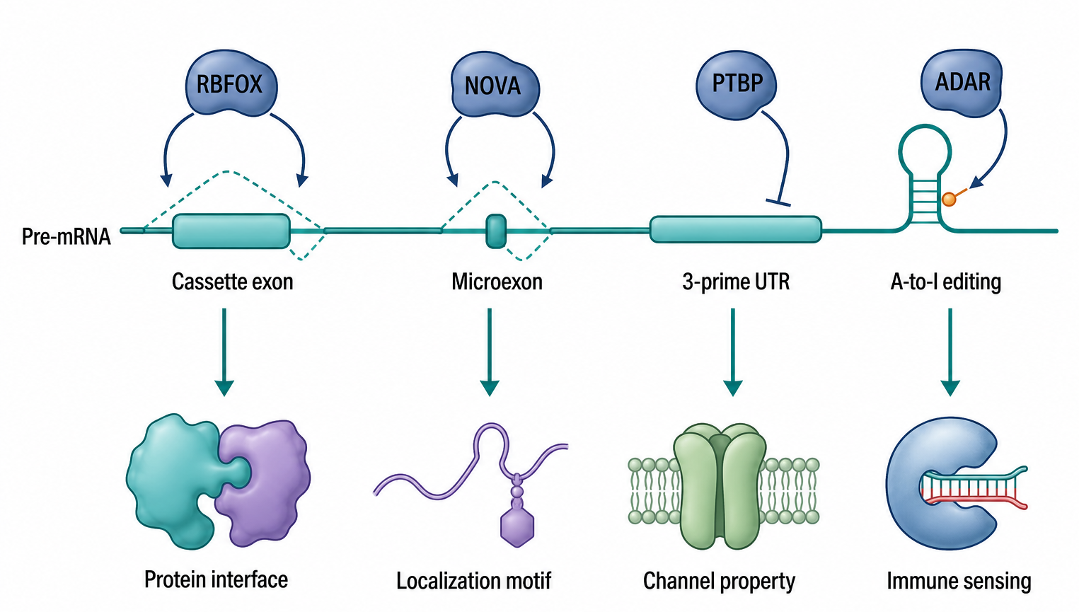
Figure 103.3. Neurodevelopmental isoform control by splicing, microexons, UTR choice, and RNA editing. Isoform-level mechanisms should be linked to target identity, developmental timing, and functional assay.
Alternative splicing also modifies untranslated regions and thereby links isoform choice to RNA localization and translation. A coding exon change may be the most visible output, but an alternative 3′ UTR can add or remove localization elements, microRNA sites, RBP-binding motifs, and stability elements. In neurons, long 3′ UTR isoforms are common and can support compartment-specific RNA regulation. Thus a neurodevelopmental splicing program can alter both protein structure and the RNA’s future life as a transported, stored, translated, or degraded molecule.
A-to-I RNA editing adds another layer. ADAR enzymes bind double-stranded RNA structures and deaminate adenosine to inosine. In coding regions, this can recode proteins. In untranslated regions and introns, editing can alter RNA structure, splicing, nuclear retention, stability, or immune recognition. The nervous system contains several well-known recoding events in transcripts related to excitability and synaptic function, and editing levels can change across development. Editing of endogenous double-stranded RNA also prevents inappropriate activation of innate immune sensors. This immune-discrimination role is important in neurons and glia because chronic innate immune activation can affect development, synapse function, and degeneration.
The evidence base for splicing and editing is method-dependent. Short-read RNA-seq can quantify annotated splice junctions but may miss full isoform structure. Long-read sequencing can connect distant exons and UTRs but has different error profiles and throughput constraints. CLIP methods can map RBP binding but do not by themselves prove regulation. Editing calls require distinction from genomic variants, mapping artifacts, paralogous sequences, and sequencing errors. Developmental studies must also separate true maturation programs from changing cell-type composition. The best studies combine cell-type-resolved transcriptomics, perturbation of splicing or editing factors, isoform-aware validation, and phenotypic assays such as synapse formation, excitability, migration, or behavior.
Box 103.2. From Neural Isoform Change to Developmental Mechanism
Use four questions to evaluate a proposed neurodevelopmental isoform mechanism. What molecule changed: a coding exon, a microexon, an untranslated-region isoform, an A-to-I editing site, or a mixture of transcript models? Where and when did the change occur: progenitor, migrating neuron, maturing excitatory neuron, interneuron, astrocyte lineage cell, organoid, or postmortem tissue? What physical output follows from the change: altered protein interaction surface, receptor conductance, RNA localization element, miRNA site, splice-site choice, or innate immune sensing? Finally, what causal test links the isoform to development: endogenous perturbation, rescue with the correct isoform, cell-type-specific manipulation, or a phenotype such as neurite extension, synapse formation, excitability, migration, or behavior? Association with autism, epilepsy, or intellectual disability is a starting point. Mechanism requires matching the RNA event to a testable molecular and cellular consequence.
Neurodevelopmental disease links should be stated with calibrated confidence. Mutations in splicing regulators, helicases, chromatin factors that indirectly alter RNA processing, and RNA-binding proteins can produce intellectual disability, epilepsy, autism-related phenotypes, or structural brain defects. DDX3X syndrome illustrates how a broadly acting RNA helicase can produce nervous-system phenotypes through effects on translation, RNA metabolism, stress responses, and development. However, broad RNA regulators have many targets. A disease mechanism should not be reduced to one transcript unless that target has strong genetic, biochemical, and rescue evidence.
103.4. RNA granules, RBP pathology, repeat expansions, and neurodegeneration
RNA granules are essential to neuronal RNA logistics. Transport granules move transcripts through dendrites and axons. Stress granules assemble when translation initiation is inhibited and untranslated mRNPs accumulate. Processing bodies are enriched for decay and repression factors. Germ granules and other specialized RNP bodies appear in distinct biological contexts. These assemblies share components, but they differ in composition, triggers, dynamics, and function. A rigorous discussion should specify which granule is being measured, how it was detected, whether it is liquid-like, gel-like, solid-like, or simply clustered by microscopy, and whether the granule state changes RNA fate.
Neurons may be unusually vulnerable to RNP disturbance because they are long-lived, polarized, and dependent on local RNA control. RNA-binding proteins with low-complexity domains can support reversible assembly but can also form persistent aggregates under stress or mutation. TDP-43 and FUS are major examples in amyotrophic lateral sclerosis and frontotemporal dementia. These proteins normally function in nuclear RNA processing, splicing, transport, stability, and other RNP processes. Disease can involve loss of nuclear function, gain of cytoplasmic toxicity, altered stress-granule dynamics, impaired nucleocytoplasmic transport, and secondary effects in glia. The same inclusion seen by pathology can reflect several molecular failures at once.
hnRNP proteins, DDX-family helicases, and other RBPs broaden this principle. HNRNPF/H proteins contribute to splicing and RNA regulation and are linked to disease contexts. DDX3X mutations cause a neurodevelopmental syndrome with complex cellular consequences. The mechanistic lesson is that RBPs are not generic RNA glue. Each RBP has binding preferences, domain architecture, subcellular distribution, regulated interactions, and target classes. A mutation can alter phase behavior, binding specificity, enzymatic activity, localization, or protein-protein interaction. Disease interpretation requires matching the molecular defect to RNA-processing outputs and cellular phenotypes.
Repeat expansion disorders provide a second major route from RNA biology to neurological disease. Expanded repeats can act at the DNA, RNA, and protein levels. At the RNA level, repeats can form structures that bind and sequester RBPs, producing splicing defects and other RNA-processing changes. CUG repeat RNA in myotonic dystrophy is a paradigmatic toxic RNA because it can sequester MBNL proteins and alter splicing programs. GGGGCC repeat expansions in C9orf72-linked amyotrophic lateral sclerosis and frontotemporal dementia can generate sense and antisense repeat RNAs, RNA foci, dipeptide repeat proteins through repeat-associated non-AUG translation, and changes in nucleocytoplasmic transport and stress pathways. CGG repeats in fragile X-associated disorders and CAG repeats in polyglutamine diseases add further variation.
Box 103.3. Do Not Collapse Repeat and RBP Disease Mechanisms
Several RNA-linked disease mechanisms can appear in the same patient tissue or model system. RNA foci support a toxic RNA or RBP-sequestration hypothesis only when the bound proteins, affected RNA-processing outputs, and rescue logic are defined. Cytoplasmic TDP-43 or FUS inclusions support RBP mislocalization, but the key downstream question may be loss of nuclear splicing regulation, gain of cytoplasmic toxicity, altered transport, or stress-granule persistence. Repeat-associated non-AUG translation supports production of repetitive peptides, but peptide toxicity must be separated from the repeat RNA that encoded them. Somatic repeat expansion points to DNA repair and repeat instability, even though the consequences may include toxic transcripts and proteins. Glial activation can amplify or buffer each route. The most useful disease model names the mechanism being tested and states which alternative routes remain possible.
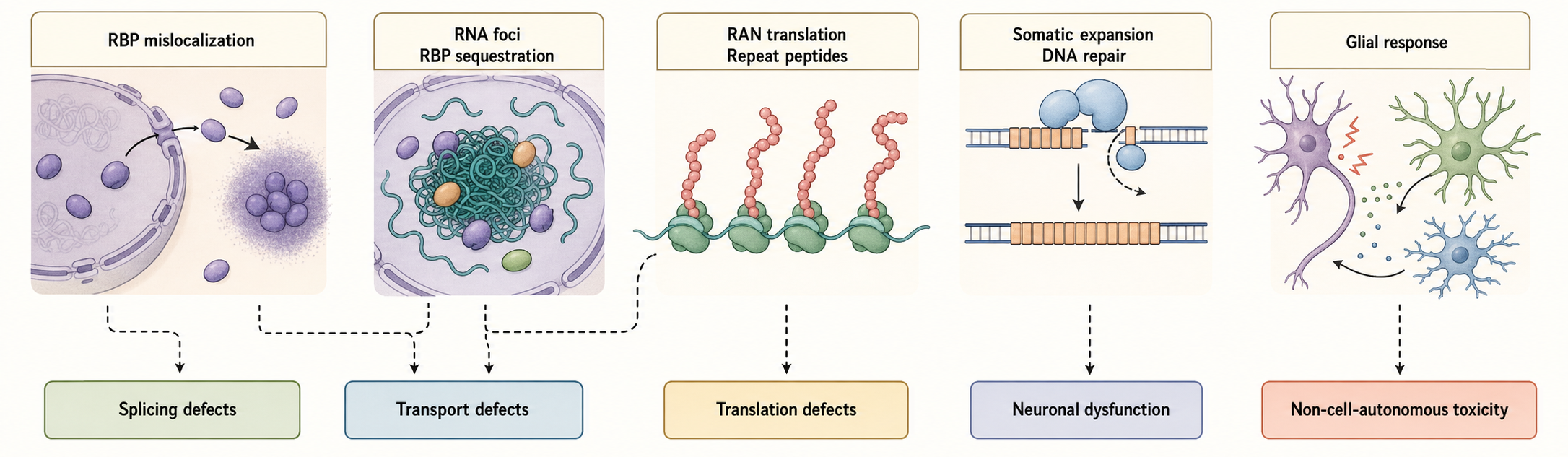
Figure 103.4. RNA-centered mechanisms in neurodegenerative repeat and RBP disease. Granule pathology, toxic RNA, toxic protein, and DNA repeat instability can coexist but are separable mechanisms.
Somatic repeat expansion is increasingly important for neurodegeneration. In Huntington disease, CAG repeat length in vulnerable neurons can continue to expand during life, and long somatic expansions can drive disease progression. This is not only an RNA problem because DNA repair and repeat instability are central, but RNA biology enters through transcript toxicity, protein production, antisense transcription, and therapeutic strategies such as antisense oligonucleotides. Suppressing modifiers such as MSH3 can reduce somatic repeat expansion in model systems, illustrating how nucleic-acid metabolism can be targeted upstream of downstream RNA and protein toxicity.
RBP pathology and repeat expansion mechanisms converge on granules but should not be forced into a single framework. Stress granules can recruit TDP-43, FUS, hnRNPs, translation factors, and repeat RNAs; repeat-associated dipeptide proteins can alter granule dynamics; and chronic stress can make normally reversible assemblies more persistent. Yet granule formation can also be protective by storing untranslated mRNAs during stress. The central causal questions are whether a disease-linked granule changes target RNA splicing, localization, translation, or decay; whether preventing abnormal granule persistence improves cell function; and whether the intervention preserves normal RNP biology.
Glia modify neurodegenerative RNA phenotypes. Astrocytes and microglia respond to neuronal stress, clear debris, regulate inflammation, and can amplify or buffer toxicity. Oligodendrocytes and myelin affect axonal energy balance. Single-cell sequencing of human disease tissue can reveal disease-associated neuronal states and glial activation states, but postmortem data are late-stage snapshots. A complete disease model should integrate cell-autonomous neuronal RNA defects with non-cell-autonomous glial responses and with tissue-level timing.
103.5. Multimodal evidence integration across imaging, single-cell, spatial, and electrophysiology
Neural RNA claims are strongest when several measurement axes intersect. Cell or nucleus sequencing identifies a molecular state; imaging or spatial assays place that state in tissue or a cellular process; isoform-aware sequencing resolves splicing and editing; ribosome or nascent-chain assays test translation; electrophysiology and morphology describe neural function; and perturbation establishes causal direction. None of these axes substitutes for the others. A disease-associated cluster without location or perturbation is a state description, while a local RNA spot without synthesis evidence is a localization observation.
The integration problem begins with mismatched sampling units. Single-nucleus RNA-seq is practical for frozen human brain but underrepresents cytoplasmic and localized RNA. Dissociated single-cell data preserve more cytoplasm but lose anatomy and can induce stress. Spatial sequencing preserves neighborhoods but may combine several cells or fail to resolve dendrites, synapses, and glial fine processes. Multiplexed imaging offers higher spatial precision for selected targets but narrower transcript coverage. A defensible analysis states which RNA compartment each modality measures and does not treat nuclear, somatic, process-local, and whole-cell abundance as interchangeable.
Functional modalities add a second alignment problem. Patch-seq links electrophysiology, morphology, and RNA from the same recorded neuron, but recording stress, low RNA recovery, and sampling bias can distort the transcriptome. Ribosome profiling, translating-ribosome purification, or proximity-specific ribosome labeling test translation at population, cell-type, or compartment scales, yet ribosome association does not guarantee completed protein. Live RNA or nascent-chain imaging adds temporal and subcellular information but can perturb RNA behavior through tags or overexpression. The useful question is which missing link a modality supplies, not which method is generally best.
Table 103.2. Multimodal evidence integration for neural RNA mechanisms. Sequencing, spatial, translational, physiological, and perturbational modalities contribute different links between RNA identity, neural cell type, location, state, and function; no single modality completes the causal chain.
| Method class | Primary RNA question | Spatial or functional link | Strength | Common limitation | Best paired validation |
|---|---|---|---|---|---|
| Single-cell RNA-seq | Cell-state transcriptome in dissociated neural cells | Cell identity, developmental trajectory, perturbation or disease state | Broad discovery of neuronal and glial populations and rare states | Dissociation stress, loss of tissue anatomy, cell-composition confounding | Spatial validation, marker imaging, perturbation or rescue |
| Single-nucleus RNA-seq | Nuclear transcriptome from frozen or postmortem tissue | Human disease tissue access and archived sample mapping | Practical for fragile cells and clinical or archival specimens | Underrepresents cytoplasmic, localized, and actively translated RNA | Cytoplasmic RNA assay, spatial assay, protein or ribosome readout |
| MERFISH, seqFISH, or smFISH | Position of selected RNAs in cells or tissue | Cell, process, neighborhood, and subcellular transcript location | High spatial precision with molecule-level or targeted multiplex readout | Limited target set, optical crowding, segmentation of fine processes | Orthogonal probe set, perturbation, protein or nascent-translation readout |
| Spatial sequencing platforms | Transcriptome linked to tissue coordinates | Anatomical layers, lesion neighborhoods, region-specific glial states | Broad tissue maps with preserved topology | Spot or capture resolution may be too coarse for synapses and glial fine processes | Multiplexed RNA imaging, histology, cell-type deconvolution |
| Live RNA imaging | RNA transport, pausing, anchoring, or reporter translation over time | Dynamic movement in dendrites, axons, growth cones, or glial processes | Direct temporal evidence for transport and cue response | Tags, overexpression, or reporters can perturb localization, decay, or translation | Endogenous tagging, rescue, fixed smFISH, nascent-peptide imaging |
| Ribosome profiling or ribosome tagging | Ribosome-associated or translated RNA pool | Cell-type or compartment translation depending on enrichment design | Links RNA abundance to translation and can reveal uORFs or codon occupancy | Contamination, low input, and ribosome occupancy without protein-output proof | Local nascent-protein imaging, transcript-specific perturbation, tagging controls |
| Patch-seq | RNA state after electrophysiology from the same cell | Transcriptomic identity linked to firing properties and morphology | Multimodal single-cell classification and ion-channel transcript links | Low RNA recovery, recording-induced stress, sampling bias | Morphology reconstruction, targeted in situ validation, replicate recordings |
Perturbation is what converts a multimodal association into a mechanism. A candidate synaptic RNA program should be perturbed in the relevant cell type and developmental or activity window, followed by measurements of RNA location or processing, local protein output, electrophysiology or morphology, and rescue. A candidate glial mechanism should connect a spatial or single-cell state to glial function and then to a neuronal or circuit consequence. A microexon or editing mechanism should connect isoform identity to protein or RNA function, excitability or development, and phenotype. Perturb-seq can reveal convergent RNA states, but convergence becomes causal only when direct targets, timing, and physiological outcomes are separated from secondary stress or composition changes.
The chapter therefore uses evidence matrices rather than method tutorials. For synaptic plasticity, the minimum linked variables are transcript identity, compartment, activity state, translation, and synaptic function. For glial regulation, they are glial cell type, process or neighborhood, RNA program, glial function, and neuronal consequence. For neurodevelopmental isoforms, they are event identity, lineage and time, molecular output, excitability or morphology, and rescue. For disease granules or repeats, they are molecular species, cell type, pathology stage, RNA-processing consequence, functional phenotype, and competing mechanism. Detailed imaging, transport, single-cell, and spatial protocols remain with their dedicated method and process chapters.
Biological Contexts Across Development, Circuits, and Disease
Neural RNA programs change across time. During early neurodevelopment, RNA regulation helps progenitors exit the cell cycle, migrate, extend neurites, choose synaptic partners, and mature electrophysiologically. Splicing regulators switch isoform programs as neurons differentiate. Local translation in growth cones helps axons respond to guidance cues. Astrocytes and oligodendrocyte lineage cells mature with region-specific RNA programs. Microglia shift from developmental synapse remodeling to adult surveillance and disease-associated responses.
In the adult nervous system, RNA regulation supports plasticity and maintenance rather than only development. Activity-regulated transcription produces immediate-early gene RNAs, but long-lasting circuit changes require post-transcriptional sorting, translation, and decay. Sleep, learning, stress, injury, inflammation, and aging can all alter RNA programs. The same RNA regulator can have different consequences in excitatory neurons, inhibitory interneurons, astrocytes, oligodendrocytes, or microglia because the target transcriptome and cellular physiology differ.
Disease contexts should be interpreted through timing and cell type. A neurodevelopmental disorder may result from altered RNA processing during a narrow developmental window, even if the gene remains expressed later. A neurodegenerative disorder may begin with subtle RNA mislocalization or splicing defects long before cell death. Glial activation may be adaptive early and harmful later. Therapeutic RNA interventions, including antisense oligonucleotides and RNA editing approaches, must therefore consider the relevant cell type, delivery route, developmental timing, target engagement, and off-target RNA networks.
Technology, Computational, Clinical, and Engineering Links
Neuronal and glial RNA biology has direct links to therapeutics and technology. Antisense oligonucleotides can alter splicing, promote RNase H-mediated degradation, block toxic RNA interactions, or reduce production of harmful proteins. Splicing-modifying therapies for neurological disease illustrate the clinical relevance of isoform control, while repeat-expansion disorders motivate strategies that target repeat RNAs, somatic instability modifiers, or disease proteins. RNA editing tools based on ADAR recruitment or CRISPR-dCas13 systems are being developed to manipulate editing and splicing, but delivery, specificity, immune activation, reversibility, and cell-type targeting remain major barriers.
Computational analysis is essential because neural RNA data are high-dimensional and spatial. Isoform quantification, microexon detection, RNA editing calling, RBP motif analysis, trajectory inference, spatial neighborhood modeling, and multimodal integration all require assumptions. In neural tissue, the most common computational errors include confusing cell-type proportion with within-cell regulation, treating nuclear RNA as equivalent to whole-cell RNA, overinterpreting inferred trajectories as lineage, and calling editing or splicing changes without adequate mapping controls. Computational models are useful when they are tied to validation and when uncertainty is carried forward into biological interpretation.
Recent Consensus
The current consensus is that local translation is a major, regulated feature of neuronal biology rather than an experimental artifact, but the exact local translatome is context-specific. Dendritic, axonal, and presynaptic translation contribute to plasticity, growth, maintenance, and injury responses. Glial RNA localization and local translation are real and biologically plausible, especially in astrocytic and oligodendrocyte processes, but glial subcellular RNA biology is less mature and requires careful cell-type and compartment assignment.
There is also broad consensus that alternative splicing, microexon regulation, and RNA editing are central to nervous-system development and function. These processes tune protein interaction networks, excitability, immune discrimination, and RNA localization. Neurodevelopmental disorders can arise from altered RNA regulators, but broad regulators demand target-specific caution. In neurodegeneration, RBP dysfunction, RNA granule dysregulation, repeat expansion RNA toxicity, RAN translation, and glial responses are established mechanisms, although their relative importance differs by disease and stage.
Open Questions, Controversies, Deprecated Models, and Common Misconceptions
Open questions:
- How many synapse-specific plasticity events require translation of a defined local mRNA rather than broader dendritic protein synthesis?
- Which glial mRNAs are translated locally in vivo, and which glial functions depend on this local synthesis?
- How often does extracellular RNA transfer between neural cells produce physiologically meaningful regulation rather than clearance or bystander uptake?
- Which microexon changes in neurodevelopmental disorders are causal, compensatory, or secondary?
- Which disease granules should be dissolved therapeutically, and which are protective stress responses?
Common misconceptions:
- “A localized neural transcript is necessarily translated where it is detected.” Localization can represent transport, anchoring, storage, decay, or translational readiness; local synthesis requires direct evidence.
- “A disease-associated single-cell cluster is a mechanism.” A cluster is a state description until spatial context, temporal order, perturbation, and functional consequences establish causality.
- “A neural microexon associated with autism is necessarily causal.” Event-specific molecular output, developmental timing, endogenous perturbation, and rescue are required.
- “A-to-I editing matters only when it changes a codon.” Editing can also alter RNA structure, processing, stability, localization, and immune discrimination.
- “Glia are background contamination in neuronal RNA studies.” Glia are active RNA-regulated participants in synapse function, development, circuit homeostasis, and degeneration.