Chapter 45. Y RNAs and Vault RNAs: RNP Assembly, Regulatory Fragments, Function, and Evidence
Scope Note
This chapter treats Y RNAs and vault RNAs as two families of small RNA polymerase III transcripts whose biological identities cannot be reduced to either “stable RNP component” or “small-RNA precursor.” It explains their full-length RNPs, processed fragments, functions, and evidence problems. Broad stable-RNA fragments belong to Chapter 89; spliceosomal snRNAs to Chapter 27; the specialized U7 snRNP and replication-dependent histone mRNA 3′-end pathway to Chapter 29; sno/scaRNAs to Chapter 42 and Chapter 49; 7SK to Chapter 24; SRP RNA to Chapter 75; pathway-specific RNase P/MRP roles to Chapter 39 and Chapter 42; and comparative RNase P/MRP catalytic-RNA mechanism to Chapter 9. U7 is an Sm-like RNP, but that compositional resemblance does not make it part of Y- or vault-RNA biology. These boundaries keep the analysis centered on Y and vault RNAs.
Executive Summary
Y RNAs and vault RNAs are short, structured, abundant noncoding RNAs transcribed by RNA polymerase III in many eukaryotes. Both families form multiple molecular populations. A full-length Y RNA can bind Ro60 through a conserved lower stem, bind La through its newly synthesized oligo(U) 3-prime end, exchange those proteins during maturation, and recruit additional proteins through a more variable loop. A full-length vault RNA can associate with the large major-vault-protein particle, but much of the cellular RNA can remain outside that particle and bind other proteins. Each family can also yield shorter fragments. The correct unit of interpretation is therefore a specified RNA form in a specified complex, compartment, organism, and condition.
Human Y RNAs are commonly represented by Y1, Y3, Y4, and Y5, encoded by RNY1, RNY3, RNY4, and RNY5. They share a stem formed by pairing their 5-prime and 3-prime regions, an internal bulge recognized by Ro60, an upper stem, and a divergent loop. La binds the 3-prime oligo(U) tract of nascent transcripts. Ro60 is a toroidal RNA-binding protein, encoded by TROVE2, whose outer surface binds the Y-RNA stem while its central cavity can engage single-stranded ends of misfolded RNAs. The surfaces are not functionally independent: a misfolded substrate can contact the outer ring as its single-stranded end enters the cavity, so a bound Y RNA can overlap and sterically regulate the substrate route. This geometry supports separable but coupled ideas: Y RNA stabilizes and localizes Ro60-containing particles, whereas Ro60 can participate in recognition or routing of defective RNAs. The bacterial Ro60/Y-RNA literature provides especially direct examples in which a Y RNA gates the Ro60 cavity or tethers Ro60 to an exoribonuclease. Vertebrate RNA-surveillance models are strongly supported structurally and biochemically but remain less completely connected to substrate turnover in living cells.
Y RNAs have also been implicated in chromosomal DNA replication. Depletion and add-back experiments established a Y-RNA requirement for efficient establishment or maintenance of active forks in defined vertebrate cell-free systems. The founding reconstitution study did not distinguish an initiation step from an early elongation requirement, although later literature often describes the activity as replication initiation. Yet Ro60-, La-, or nucleolin-containing Y RNPs were dispensable in the same class of assay, showing that the replication-active form is not simply the canonical Ro RNP. The most defensible statement is that Y RNAs or related stem-bulge RNAs support replication in specific vertebrate and nematode systems; the exact step, direct molecular target, and universality of this requirement remain unresolved.
Ro60 and La are major autoantigens. Ro60-containing immune complexes can deliver self RNAs to endosomal sensors, and Ro60 also controls endogenous retroelement RNAs and inflammatory gene expression. These results connect RNA metabolism to innate and adaptive autoimmunity, but they do not mean that every anti-Ro antibody response is driven by a Y RNA or that Y RNA abundance alone causes disease. A causal model must distinguish the antigenic protein, associated RNA species, cell-death or release pathway, receptor, responding cell, and inflammatory outcome.
Vault RNAs are named for their association with vault particles, giant cytoplasmic RNP assemblies whose shell is built from repeated major vault protein (MVP) subunits and that can contain telomerase-associated protein 1 (TEP1), PARP4, and vault RNA. RNA-location studies place vault RNA near the particle caps, but vault RNA is neither the main structural material of the shell nor obligatorily particle-bound. The human VTRNA1 paralogs have distinct expression and protein-binding behaviors. The frequently discussed VTRNA2-1/nc886 locus is a boundary case: historical annotation places it in the extended vault-RNA family, but its association with canonical vault particles and its functional identity differ from those of VTRNA1 RNAs. Reports should state the exact locus and should not use “vault RNA” as if all paralogs were interchangeable.
Some full-length vault-RNA mechanisms are unusually concrete. VTRNA1-1 binds the autophagy receptor SQSTM1/p62 and restrains p62 oligomerization, thereby modulating selective autophagy. Structure–function work identified the flexible RNA region and p62 residues required for this interaction. Other studies link VTRNA1-1 to apoptosis resistance, lysosome biology, and protein redistribution during viral infection. These are molecule- and context-specific mechanisms, not evidence that the entire vault particle performs the same function.
Processed fragments require a separate evidence ladder. Distinct Y-RNA fragments occur in cells, extracellular particles, and biofluids; extracellular RNase 1 can produce characteristic Y-RNA fragments after release. Vault RNAs can be processed into small RNAs, and NSUN2-dependent 5-methylcytosine alters VTRNA1-1 processing and partner choice. Some products associate with Argonaute or influence differentiation. Nevertheless, a short read mapping to a Y RNA or vault RNA does not by itself establish regulated processing, microRNA identity, or function. Size-resolved validation, native-end evidence, biogenesis perturbation, stoichiometry, protein association, and sequence-specific rescue are required.
The central evidence rule of this chapter is form-specific causality. A claim about a gene locus, full-length RNA, RNP, processed fragment, extracellular species, autoantibody, or vault particle must be tested at the level it names. Conflating those levels is the major source of overstatement in this field.
Concept Inventory
A Y RNA is a structured small noncoding RNA family member defined by conserved architecture and, in canonical systems, the ability to bind Ro60. A Ro RNP is a molecular complex containing Ro60 and an associated RNA, commonly a Y RNA but sometimes another RNA substrate; Ro RNP and Y RNA are therefore not synonyms. A vault RNA is a Pol III transcript related to RNAs first found in vault particles. A vault particle is the much larger MVP-based RNP assembly; the particle and the RNA are distinct experimental objects.
The chapter uses full-length RNA for a mature molecule that retains the defining terminal stem and major domains of its family. A processed fragment is a shorter product whose sequence derives from that RNA. “Derived from” is a statement of sequence origin, not of pathway or function. Particle occupancy means the fraction of a named RNA population physically associated with a named particle under specified conditions. Riboregulation means direct regulation of protein behavior by RNA binding, exemplified by VTRNA1-1 control of p62 oligomerization.
What to Know Before Reading This Chapter
RNA polymerase III commonly terminates at a short run of thymidines in DNA, producing an RNA with a short 3-prime oligo(U) tract. La recognizes such nascent ends and can protect Pol III transcripts during early maturation. The exact mature end, protein occupancy, and intracellular location can change after transcription. Consequently, detecting a genomic locus, primary transcript, mature RNA, and protein-bound RNA requires different assays.
RNA structure is not decoration. Pairing between the 5-prime and 3-prime ends of a Y RNA creates the lower stem recognized by Ro60, while a flexible loop can recruit other proteins or become accessible to cleavage. Likewise, a vault RNA’s stem-loop architecture creates protein-binding and processing surfaces. A mutation that preserves abundance but disrupts a structural surface can be more informative than a knockdown that reduces every molecular form.
Three running examples organize the chapter. The first is human Y3 RNA in a Ro60-containing particle. The second is human Y1 or another Y RNA in a cell-free DNA-replication assay, where the active RNA population can be separated from canonical Ro RNPs. The third is human VTRNA1-1, which can reside in a vault-associated pool, bind p62 as a full-length riboregulator, or be processed into smaller products. Each example asks the same question: which molecular form was actually perturbed and measured?
45.1. Y RNA families, structures, Ro60 and La binding, and Y RNP assembly
Y RNAs were discovered through their association with the Ro60 and La autoantigens. In humans, four canonical genes produce Y1, Y3, Y4, and Y5 RNAs, generally about 80-110 nucleotides long. Other vertebrates have different complements: mice retain fewer canonical family members than humans, and amphibian systems contain related variants. Bacterial Y RNAs are more diverse and can contain extensions or tRNA-like domains. The family is therefore recognized by conserved structural relationships and Ro60 binding rather than by a universal sequence or a fixed gene count.
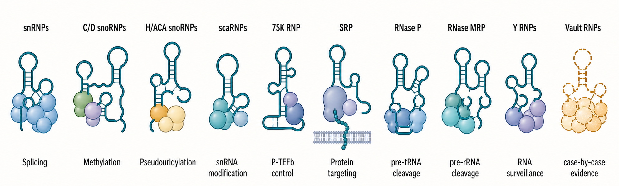
Figure 45.1. Modular architecture and assembly states of a canonical vertebrate Y RNA. “Y-RNA assembly is modular and dynamic. La recognizes the nascent oligo(U) end, Ro60 recognizes a conserved bulged stem, and other proteins engage variable regions; these interactions define an ensemble rather than one immutable particle.”
The canonical vertebrate fold has four functional regions. The lower stem pairs the 5-prime and 3-prime portions of the RNA and contains conserved bulges. An upper stem extends from that core. A loop, whose sequence and length differ substantially among Y RNAs, projects from the stem. Finally, the 3-prime end commonly carries a short oligo(U) tract left by Pol III termination. This organization allows one molecule to present several non-equivalent surfaces: Ro60 recognizes the lower-stem architecture, La recognizes the oligo(U) end, and other RNA-binding proteins can recognize the loop or upper stem.
Ro60 is a ring-shaped protein built from HEAT-like repeats in a TROVE domain plus a von Willebrand factor A-like domain. Mutational and structural experiments show that the Y-RNA lower stem binds on the outside of the ring. A 39-nucleotide minimal Y RNA retained high-affinity binding, and mutational plus chemical-probing experiments showed that specific base pairs, a singly bulged nucleotide, and an opposing three-nucleotide bulge widen a major groove recognized by Ro60. A later crystal complex contained a Y-RNA fragment on the outer ring and a separate single-stranded RNA in the central cavity; mutating Ro60 residues then showed that a modeled misfolded pre-5S substrate uses the cavity and part of the outer Y-RNA-binding surface. The binding routes are therefore topologically different but overlapping, not two independent sites. A bound Y RNA can anchor Ro60 while sterically limiting access of another RNA, whereas Y-RNA dissociation can expose the broader substrate path. The structure supplies a gating model rather than direct evidence for turnover of every physiological substrate.
La recognizes a different stage and feature. As Pol III finishes transcription, La binds the 3-prime oligo(U) end of the nascent Y RNA. This interaction protects the end against exonucleolytic attack and can assist early folding and maturation. Ro60 and La can coexist in some preparations, but their occupancy is not fixed for the lifetime of every molecule. End trimming, structural maturation, nuclear export, stress, and competition with other proteins can redistribute Y RNAs among RNP states. “The Y RNP” is therefore shorthand for an ensemble, not one invariant particle.
Assembly can be described causally. First, Pol III transcribes the Y-RNA gene and terminates to produce a uridine-rich 3-prime end. Second, La binds the nascent end and protects it. Third, folding brings the 5-prime and 3-prime regions together to create the Ro60-binding stem. Fourth, Ro60 loads onto that stem and can change RNA stability or localization. Fifth, Y-RNA-specific and condition-specific partners bind the variable loop or other exposed elements. Finally, exportins, retention factors, and stress-dependent remodeling determine whether an RNP is predominantly nuclear, cytoplasmic, extracellular, or associated with another organelle or complex.
Y-RNA binding is itself a localization switch for Ro60. A Y-RNA-binding-defective Ro60 mutant accumulated in mammalian nuclei, and depletion of mouse Y3 or both major mouse Y RNAs caused nuclear enrichment of Ro60; the Y1-only depletion was inconclusive because Ro60-bound Y1 fragments remained. Mouse–bacterial Ro60 chimeras mapped stress-responsive nuclear accumulation to HEAT-repeat sequences overlapping the Y-RNA-binding surface, and ultraviolet or oxidative stress used similar determinants. These results support masking of nuclear-accumulation information by bound Y RNA, but they do not exclude an additional effect on export, nor did the mapped sequence behave as a simple autonomous classical nuclear-localization signal. This result is stronger than simple colocalization because it connects a defined binding interaction to cellular redistribution. It also warns against interpreting total-cell Ro60 abundance as a direct measure of nuclear RNA-surveillance activity.
Not every Y-RNA-like locus produces a canonical RNP. Vertebrate genomes contain many Y-RNA-derived pseudogenes, often created by retrotransposition. Short reads can map ambiguously among canonical genes, pseudogenes, and processed fragments. Conversely, highly divergent bacterial Y RNAs may be missed by sequence-only searches even when structural and protein-association evidence is persuasive. Comparative annotation should combine conserved secondary structure, promoter and terminator features, synteny when informative, Ro60 association, mature-size validation, and phylogenetic distribution.
The strongest evidence for assembly uses multiple assays. Northern blotting establishes mature size. End mapping establishes the actual termini. RNA immunoprecipitation or ultraviolet-crosslinking assays establish protein association, but immunoprecipitation alone cannot distinguish direct binding from co-membership in a larger complex. Mutational binding assays locate RNA elements. Structural studies establish contact geometry. Cell imaging or fractionation establishes localization only when contamination and fixation artifacts are controlled. A complete assembly model connects these outputs rather than asking one assay to support all of them.
The conceptual boundary to other stable RNPs is important. Sm and Lsm assembly on spliceosomal snRNAs is covered in Chapter 27, guide-RNP assembly for snoRNAs and scaRNAs in Chapter 42 and Chapter 49, and 7SK assembly around P-TEFb regulation in Chapter 24. Y RNPs share the general principle of an RNA scaffold recruiting a protein core, but Ro60/La recognition, Y-RNA structural variation, and multi-state assembly are their own mechanistic system.
45.2. Y RNP functions in RNA surveillance, DNA replication, innate sensing, and autoimmunity
The phrase “Y-RNA function” includes at least four mechanistically distinct claims: regulation of Ro60-based RNA surveillance, support of chromosomal DNA replication, modulation of stress or innate sensing, and participation in autoimmune antigenic complexes. These functions can use different RNA regions and different RNP populations. Their evidence bases should be kept separate.
Ro60 recognizes misfolded or aberrant RNAs through structural features rather than through a single nucleotide sequence. Biochemical work showed binding to variant 5S ribosomal RNA precursors, and structural work visualized single-stranded RNA entering the Ro60 cavity. The simplest surveillance model has three steps: an aberrant RNA presents an accessible single-stranded end; Ro60 binds or channels that end; and another nuclease or degradation machinery processes the substrate. Y RNA can regulate access to the cavity, stabilize Ro60, localize it, or connect it to another protein.
The bacterial literature makes the partner step concrete, but it also separates two mechanisms that are easy to merge. In D. radiodurans, about 40% of 23S rRNA retained terminal extensions at 30 degrees Celsius in the experiments reviewed here. Shifting cells to 37 degrees Celsius made maturation efficient through Y-RNA-free Rsr together with RNase II and RNase PH; deleting the Y RNA accelerated Rsr-dependent maturation, showing that Yrn1 inhibits rather than tethers the nuclease in this pathway. In a separate degradation state, Yrn1 binds Rsr through its conserved stem and polynucleotide phosphorylase (PNPase) through its other module, producing the double-ringed RYPER complex that degrades structured RNAs more efficiently. Other bacterial Y RNAs can gate substrate entry or carry tRNA-mimic domains. “Y RNA” therefore describes a modular family whose second domain can inhibit access, recruit a nuclease, or mimic tRNA, not one invariant vertebrate-like mechanism.
One should not transfer every bacterial mechanism unchanged to mammals. Mammalian Ro60 binds misfolded RNAs and affects RNA populations, but direct substrate-to-product chains in intact cells are less completely resolved. The conserved inference is architectural: Y RNA controls access, localization, or partner recruitment around a toroidal RNA-binding protein. The organism-specific questions are which substrates are encountered, which nuclease completes degradation, and under which stresses the pathway becomes rate limiting.
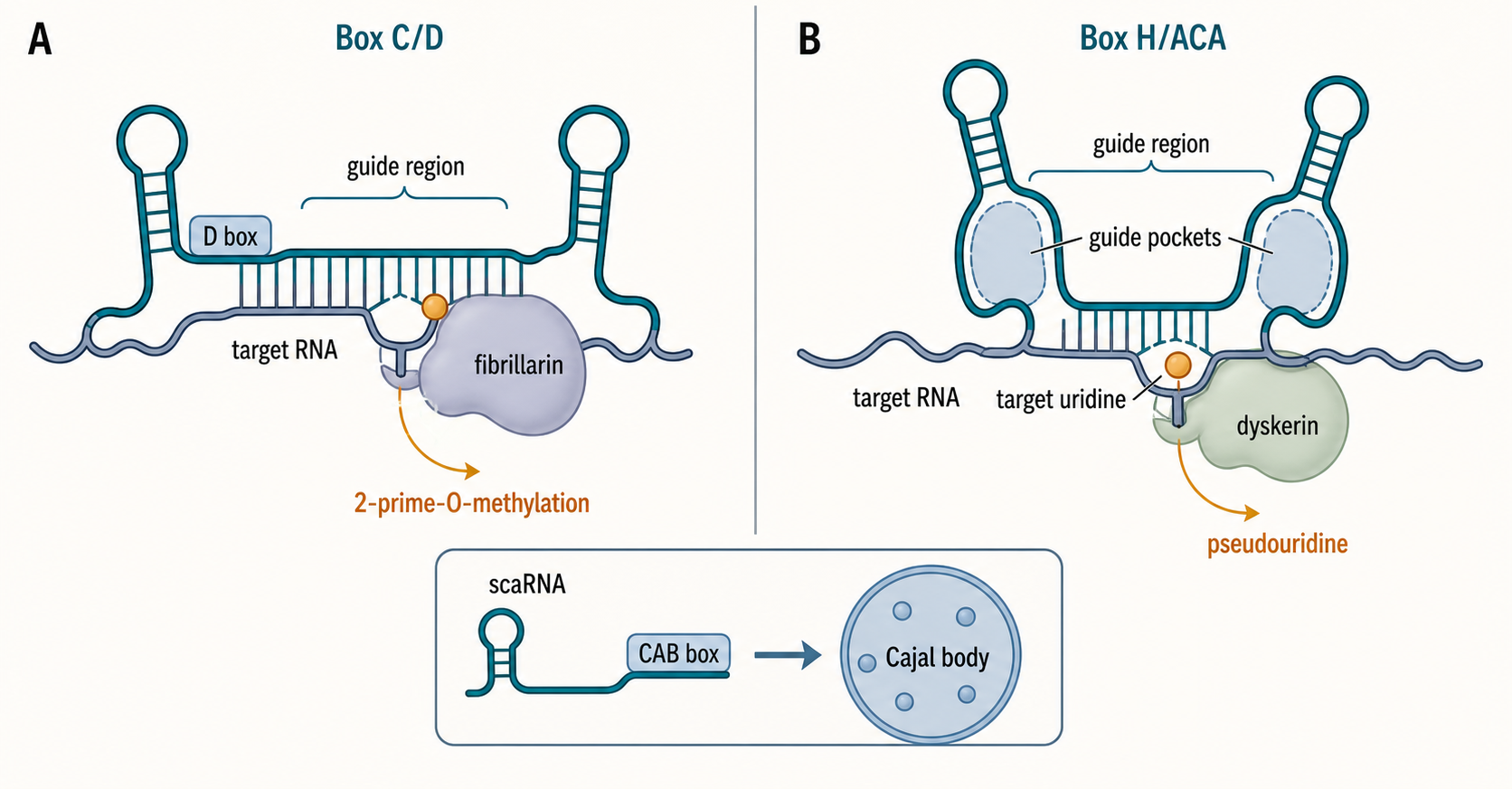
Figure 45.2. Functional branching of Y RNA across surveillance, replication, and immune presentation. “The same RNA family can occupy distinct molecular states. Surveillance, replication, and immune presentation have different partners, compartments, and evidence limits.”
DNA replication involves a different Y-RNA state. In a human cell-free system, antisense-directed degradation of Y1, Y3, or Y4 reduced semiconservative replication in late-G1 template nuclei, and purified recombinant Y RNA restored activity. Each of the four human Y RNAs could substitute in the reconstitution assay. Mutations that abolished Ro60 binding retained activity, a 3-prime-blocked Y1 RNA remained active and therefore did not act as a DNA primer, and a sequence-scrambled Y1 designed to preserve the predicted overall fold was inactive. These experiments establish sequence-dependent Y-RNA activity during the establishment or maintenance of active forks, but the original assay could not resolve whether the RNA acts at initiation, early elongation, or both. Related stem-bulge RNAs in nematodes can support analogous assay outputs, suggesting that the relevant property may be conserved architecture plus sequence information rather than vertebrate Ro60 binding alone.
The replication evidence is strongest for necessity and add-back sufficiency within defined extract systems. It is weaker for a complete molecular mechanism. Immunodepletion of Ro60-, La-, or nucleolin-containing Y RNPs did not remove replication activity, and excess Ro60 or La did not prevent Y-RNA-dependent replication. Thus, canonical Ro RNP formation is not required for the assayed replication function. This negative result separates two often-conflated models: the same RNA family can support RNA surveillance when bound to Ro60 and replication in a different complex.
Box 45.1. A negative result that separates two Y-RNA mechanisms
- Required elements: claim before test; immunodepleted complexes; retained replication activity; revised model with a distinct replication-active Y-RNA population; unresolved partner question.
Claims that Y RNAs are universally essential replication factors should remain qualified. Extract depletion can remove associated molecules; antisense reagents can trigger degradation or steric effects; and add-back concentrations may exceed physiological free-RNA concentrations. The founding cellular experiment achieved a five- to tenfold Y1 reduction and observed about a threefold decrease in bromodeoxyuridine-positive cells, but bromodeoxyuridine incorporation reports DNA synthesis and cell-cycle occupancy, not origin firing specifically. Reduced proliferation can also arise from stress or innate sensing rather than a direct replication block. A rigorous in vivo test would combine acute, isoform-aware depletion, cell-cycle-resolved phenotyping, direct measurements of origin firing and fork progression, rescue with wild-type and structure-disrupting mutants, and identification of the replication-proximal protein partner.
Innate sensing introduces another layer. Endosomal Toll-like receptor 7 can detect single-stranded RNA delivered in immune complexes, and cytosolic sensors can respond to RNA structure or end chemistry. Ro60-containing particles released during cell injury or apoptosis can be captured by antibodies or B-cell receptors, internalized, and delivered to endosomes. The RNA cargo can then provide a receptor ligand while the protein provides antigenic specificity. This two-component logic helps explain how an RNA-binding autoantigen connects adaptive recognition to innate cytokine production.
Ro60 also binds endogenous Alu retroelement RNAs. Ro60 loss increased Alu RNAs and interferon-regulated expression, while anti-Ro60-positive lupus immune complexes contained Alu RNA. In a transfected healthy-donor peripheral-blood-mononuclear-cell assay, an Alu-derived Ro60-binding motif induced cytokines, pharmacologic inhibition implicated endosomal Toll-like-receptor signaling, and the tested Y RNAs did not induce cytokines under the same conditions. That negative result is system-specific, but it reinforces why this study cannot be cited as direct evidence that a canonical Y RNA is the inflammatory ligand. Ro60 biology is broader than Y-RNA biology, and an immunoprecipitated Ro60 complex can contain more than one RNA class.
Autoantibodies against Ro60 and La are clinical biomarkers in systemic lupus erythematosus, Sjögren disease, and related settings; maternal antibodies are associated with neonatal lupus manifestations. Seropositivity shows that the immune system recognizes the protein complex. It does not demonstrate that the Y RNA caused tolerance breakdown, nor that anti-Ro antibodies inhibit a specific Y-RNA function. Mechanistic claims require evidence about the released RNP form, RNA cargo, receptor, responding immune-cell type, and downstream signaling.
Y RNAs and Y-RNA fragments are often detected extracellularly. Their abundance can reflect protection by proteins or vesicles, selective release, extracellular cleavage, or high intracellular abundance. Extracellular RNase 1 can cleave released full-length Y RNAs into characteristic fragments, showing that at least some biofluid products arise after secretion rather than through an intracellular dedicated biogenesis pathway. This finding changes biomarker interpretation: a reproducible fragment can report extracellular nuclease environment as much as the state of the source cell.
A useful evidence ladder separates five levels. Level one is co-occurrence: Y RNA, Ro60, an immune phenotype, or DNA synthesis changes in the same condition. Level two is physical association or spatial proximity. Level three is perturbation of a nominated RNA form with a matched biochemical output. Level four is rescue by a sequence or structure that restores the specific interaction. Level five is reconstruction of the causal chain, including partner, substrate, product, and compartment. RNA-surveillance and replication claims occupy different positions on this ladder, and broad disease associations should not be promoted to level five without additional work.
45.3. Vault RNA gene families, structures, vault-particle association, and protein partners
Vault RNAs are small structured Pol III transcripts, commonly about 80-140 nucleotides, found in diverse but not all eukaryotic lineages. Mammals encode several paralogs. Human nomenclature distinguishes VTRNA1-1, VTRNA1-2, and VTRNA1-3 and often includes the more divergent VTRNA2-1/nc886 locus in historical or extended classifications. Because paralogs differ in expression, processing, partner binding, and particle association, “vtRNA” without a locus name is insufficient for a mechanistic claim.
Vault RNAs were named after their recovery with vault particles. A vault particle is a very large, barrel-shaped cytoplasmic assembly built primarily from many copies of MVP. TEP1 and PARP4 can associate with the shell, and a smaller amount of vault RNA localizes near the cap regions. In the landmark localization experiment, combined RNase A and RNase T1 treatment reduced particle RNA below detection and a 22-angstrom cryogenic-electron-microscopy difference reconstruction placed the lost density at the cap ends; the same study modeled a nearby 16-fold density ring as the TEP1 WD40 region. The resolution and subtraction strategy establish regional localization, not nucleotide-level contacts or fixed RNA stoichiometry. Later 3.5-angstrom crystallography assigned 78 MVP chains to the rat-liver shell, revising an earlier 96-copy cryo-EM model and illustrating that particle stoichiometry is method- and model-dependent. Vault RNA is therefore a genuine particle component, but the MVP shell can assemble without the RNA, and the RNA is not a continuous scaffold equivalent to ribosomal RNA in the ribosome.
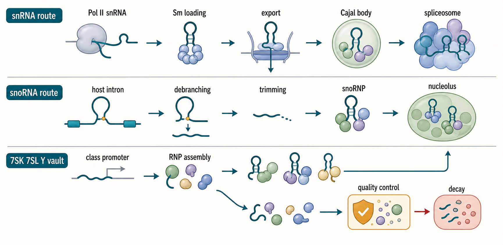
Figure 45.3. Vault particle, particle-bound RNA, and particle-independent vault-RNA complexes. “Vault RNA is a genuine vault-particle component, but vault-RNA biology is not restricted to the particle. Particle occupancy is paralog-, cell-, and condition-dependent.”
Particle association is fractional. Only part of a cellular vault-RNA pool may co-sediment or co-purify with vaults, and the fraction can depend on paralog, cell type, extraction conditions, and stress. Conversely, MVP abundance is not a proxy for VTRNA1-1 abundance or function. The field’s earliest name encourages a false equivalence: “vault RNA function” is not automatically “vault-particle function.” Every study should measure both RNA and particle components if it proposes a particle-dependent mechanism.
Vault-RNA genes also have distinctive transcriptional logic. The rat vault-RNA promoter combines internal Pol III elements with upstream elements that act synergistically, demonstrating that these genes cannot always be treated as generic tRNA-like templates. Human paralogs may differ in chromatin state and transcription-factor input. Pol III termination yields a structured RNA with a uridine-rich 3-prime end, after which folding, protein binding, modification, and processing determine the mature populations.
The common secondary-structure model contains paired terminal regions and a central domain with stem-loops or flexible segments. The terminal stem supports overall folding, whereas variable central regions contribute paralog-specific partner recognition. For VTRNA1-1, chemical probing and binding experiments identified a flexible central loop important for direct interaction with SQSTM1/p62. The experimental lesson is general: computational folding alone does not establish the conformation present in a protein-bound cellular RNP. Mutational rescue should preserve global stability while testing the nominated local surface.
TEP1 provides one particle-associated protein connection. Structural localization and biochemical work place TEP1 close to vault RNA at the cap. Yet TEP1 also participates in telomerase-related contexts, and the presence of TEP1 in a pulldown does not prove that a detected RNA was inside an intact vault. MVP, TEP1, and PARP4 should be assayed together with size, sedimentation, or imaging evidence when intact-particle membership is the claim.
VTRNA1-1 binding to p62 is a different type of complex. p62 is an autophagy receptor whose oligomerization helps organize cargo for selective autophagy. Ultraviolet crosslinking, immunoprecipitation, and binding of an in-vitro-transcribed parent-length RNA to purified p62 establish a direct VTRNA1-1–p62 interaction. Antisense depletion, overexpression, locus deletion, p62 epistasis, and autophagic-flux measurements then showed that VTRNA1-1 restrains p62 oligomerization and downstream interactions with LC3B and GABARAP. Starvation lowers both steady-state VTRNA1-1 and the p62-bound RNA pool, releasing this restraint. Follow-up structure–function experiments identified p62 residues Lys7 and Arg21 in the PB1 region and an RNA flexible loop as important for specific binding and riboregulation. This chain—direct binding, mapped surfaces, perturbation, and functional consequence—is a mature mechanistic example. Nevertheless, the 2019 cellular perturbations targeted the locus or a shared RNA sequence and therefore could also change VTRNA1-1-derived products. Failure to detect MVP in the p62 preparation supports a particle-independent model only indirectly; the particle state of the active cellular RNA was not formally resolved.
VTRNA1-1 has also been linked to apoptosis resistance. In Epstein–Barr-virus-associated B-cell systems and additional cell lines, increased VTRNA1-1 reduced apoptotic responses, and MVP depletion did not reproduce the phenotype, arguing for an RNA function outside the intact vault particle. This evidence is stronger than an expression correlation but does not imply that all VTRNA paralogs protect all cell types. Dose, cell state, viral proteins, and the perturbation route all affect interpretation.
A 2025 primary study established a more specific infection-associated pathway. Selected picornaviruses, alphaviruses, and betacoronaviruses induced VTRNA1-family expression, whereas several other tested viruses did not; ultraviolet inactivation and replication inhibition showed specifically that encephalomyocarditis-virus and Sindbis-virus induction required productive replication. Combined deletion of VTRNA1-1, VTRNA1-2, and VTRNA1-3 reduced Sindbis-virus replication in HEK293 cells and encephalomyocarditis-virus and Mengovirus replication in A549 cells; VTRNA1 re-expression rescued the phenotype, and antisense depletion independently reduced encephalomyocarditis-virus replication. In the same systems, endogenous-RNA affinity purification and protein-centered immunoprecipitation identified hnRNP C and ELAVL1 as VTRNA1 partners. Loss of the three VTRNA1 loci selectively impaired infection-associated cytoplasmic redistribution of both proteins during Sindbis-virus and encephalomyocarditis-virus infection and reduced their association with Sindbis viral RNA. This causal chain is stronger than expression correlation, and blocking residual type I interferon signaling did not restore Sindbis-virus replication in the knockout cells. Its boundaries are equally important: the perturbations did not assign the phenotype to one VTRNA1 paralog, did not distinguish full-length parents from derived products, did not establish a direct RNA-export-adaptor mechanism, and did not formally resolve whether the active RNA pool was outside intact vault particles.
VTRNA2-1/nc886 is a nomenclature boundary case with practical consequences. Some databases and papers call it a vault RNA; others emphasize that its sequence, particle association, epigenetic regulation, and PKR-related functions justify treating nc886 as a distinct Pol III RNA. The safe convention in this chapter is to name it VTRNA2-1/nc886, state the database or annotation version, and avoid using results from this locus as generic evidence for VTRNA1 paralogs or vault particles.
Species comparisons also require care. Vault particles are patchily distributed among eukaryotes, paralog counts vary, and homologous-looking RNAs can acquire lineage-specific partners. The conserved principle is that structured Pol III RNAs can occupy both particle-bound and particle-independent pools. The detailed partner network must be demonstrated in each system. Comparative RNA-family annotation is developed in Chapter 12, while broader Pol III transcription belongs to Chapter 21.
45.4. Full-length Y and vault RNAs versus processed fragments and small-RNA effectors
Short reads from Y-RNA and vault-RNA loci are abundant in many small-RNA data sets. They may represent regulated cleavage products, turnover intermediates, extracellular nuclease products, library-selection artifacts, or degradation during handling. A fragment’s sequence origin is often clear, but its biogenesis and function are not. The broad biology of stable-RNA-derived fragments belongs to Chapter 89; this section focuses on the parent–product distinction needed to interpret Y and vault RNA experiments.
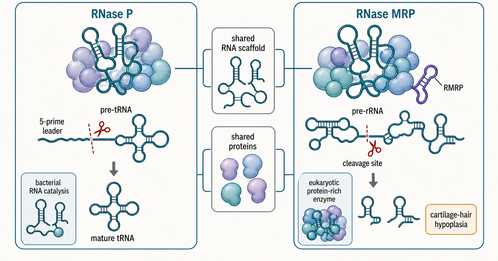
Figure 45.4. Parent–product logic for Y RNAs and vault RNAs. “A mapped fragment reports sequence origin. Regulated biogenesis, microRNA-like activity, extracellular processing, and turnover are alternative models that require different tests.”
Full-length and fragment assays have different eligibility filters. A Northern blot with a probe in the terminal stem can show both the intact molecule and selected fragments if the gel resolves them. Standard reverse-transcription PCR may amplify a short internal region without distinguishing an 100-nucleotide parent from a 25-nucleotide product. Small-RNA sequencing selects fragments by size and end chemistry and can exclude the parent entirely. Long-RNA sequencing can do the reverse. A report should therefore avoid saying “Y RNA increased” when the measured analyte was one 5-prime fragment.
Y-RNA fragments often derive from reproducible loop-proximal cleavage regions and can become enriched extracellularly. This reproducibility is evidence against completely random degradation, but not proof of a dedicated intracellular biogenesis enzyme. In endothelial-cell extracellular-vesicle preparations, small-RNA profiling found enriched Y-RNA fragments alongside other RNA products. In a separate system, full-length Y RNAs released into the non-vesicular extracellular space were cleaved by RNase 1, and RNase 1 loss reduced characteristic products. Together these studies show that release, carrier association, and extracellular processing are separable variables.
Calling a Y-derived fragment a microRNA requires more than length. Canonical microRNAs have defined Drosha- and Dicer-related precursor logic, Argonaute loading, and target repression rules. Some Y fragments associate with Argonaute or have miRNA-like sizes, while other analyses find that major Y-derived populations do not depend on canonical miRNA machinery. A functional claim should test the relevant processing enzyme, Argonaute occupancy, seed-dependent target repression at endogenous concentrations, and rescue. Historical labels such as “Y-RNA-derived miRNA” should be read as hypotheses unless those criteria are met.
Vault RNAs also yield shorter products. In the 2009 VTRNA1-1 study, the historical “vRNA1” transcript produced approximately 23-nucleotide products called svRNAa and svRNAb. Drosha depletion did not reduce them, partial Dicer depletion did, and recombinant Dicer cut the parent into some but not all observed products. svRNAb was enriched with Argonaute 2 and 3, repressed a complementary reporter, and was linked by antisense inhibition plus reporter-site tests to regulation of CYP3A4 in MCF7 cells. This is product-specific evidence for a noncanonical, Drosha-independent and Dicer-sensitive small-RNA route, not proof that every vault fragment is a microRNA. The products were much less abundant than miR-16 in that data set, Dicer knockdown was incomplete and pleiotropic, and the named assays did not provide a general endogenous stoichiometry standard. Later work showed that NSUN2 deposits 5-methylcytosine on vault RNA and that loss of NSUN2 changes processing into Argonaute-associated products. Together these studies connect precursor processing, effector association, and covalent modification while showing why pathway perturbations must be interpreted at the exact product level.
VTRNA1-1 provides a sharper modification mechanism. NSUN2-dependent methylation at cytosine 69 changes the balance of processed products. SRSF2 preferentially binds the unmethylated RNA and counteracts processing, and perturbations of the pathway influence epidermal differentiation. The causal model has explicit steps: NSUN2 changes the chemical state; methylation changes protein affinity; partner occupancy changes cleavage or protection; fragment abundance changes; and the altered RNA population contributes to a differentiation phenotype. Rescue with methylation-competent and binding-site mutants is more informative than simply correlating NSUN2 with fragment counts.
Full-length VTRNA1-1 and its fragments can have different functions simultaneously. The parent can bind p62 through a flexible region and regulate oligomerization, whereas cleavage through or near that region can destroy the p62-binding surface and create a different small RNA. A perturbation targeting the common sequence may remove both forms. Similarly, a Y-RNA antisense oligonucleotide can reduce the parent, alter Ro60 localization, and change fragment production at once. Form-specific reagents and size-resolved readouts are therefore essential.
End chemistry can reveal biogenesis. A cleavage product with a 5-prime monophosphate and 3-prime hydroxyl is compatible with some small-RNA ligation protocols; a cyclic phosphate or 5-prime hydroxyl may be invisible until repaired. Parallel libraries with selective end treatment can distinguish populations, but treatment erases native chemistry after conversion. Dedicated end, tail, and cleavage profiling is covered in Chapter 127, and method-specific recovery of structured small RNAs in Chapter 126.
Stoichiometry also matters. A highly abundant parent can generate a reproducible fragment that represents less than one percent of molecules. Overexpressing a fragment mimic to micromolar levels can create target repression that endogenous production never achieves. The relevant quantities are parent copies, fragment copies, fraction bound to the nominated protein, and concentration in the compartment where the target resides. Unique molecular identifiers correct some amplification duplication but do not correct extraction, ligation, reverse-transcription, or mapping biases.
An evidence ladder for a functional fragment has six rungs. First, a size-resolved assay confirms a discrete molecule. Second, end mapping shows reproducible termini and native chemistry. Third, handling controls exclude ex vivo cleavage. Fourth, a nuclease, modification, or protective protein perturbation changes formation without simply destroying the parent transcript. Fifth, a physical interaction or molecular target is demonstrated at endogenous abundance. Sixth, a sequence- or structure-specific rescue restores the phenotype. Many biomarker reports reach the first two rungs; comparatively few establish the complete causal chain.
Box 45.2. Six-rung evidence ladder for a functional Y- or vault-RNA fragment
- Required elements: discrete size; precise native ends; handling controls; nuclease or modification dependence; endogenous partner/target engagement; sequence- or structure-specific rescue; side label indicating where biomarker validity can be established without effector function.
Fragments can still be valuable biomarkers without being effectors. A Y-RNA fragment in plasma may integrate tissue release, cell death, carrier binding, and extracellular RNase activity. A vault-RNA fragment may report NSUN2 activity or differentiation state. Diagnostic utility requires analytical reproducibility and clinical validation, whereas mechanistic function requires molecular causality. These are legitimate but different scientific goals.
45.5. Transcription, end processing, modification, localization, turnover, and stress responses
The life cycle of a Y RNA or vault RNA begins at a Pol III transcription unit but branches rapidly. Promoter occupancy sets primary output; termination creates a uridine-rich 3-prime end; La or other factors protect the nascent RNA; folding creates protein-binding surfaces; modification changes structure or partner affinity; trafficking distributes the RNP; and nucleases generate turnover products or regulated fragments. Measuring only steady-state abundance collapses all of these rates into one number.
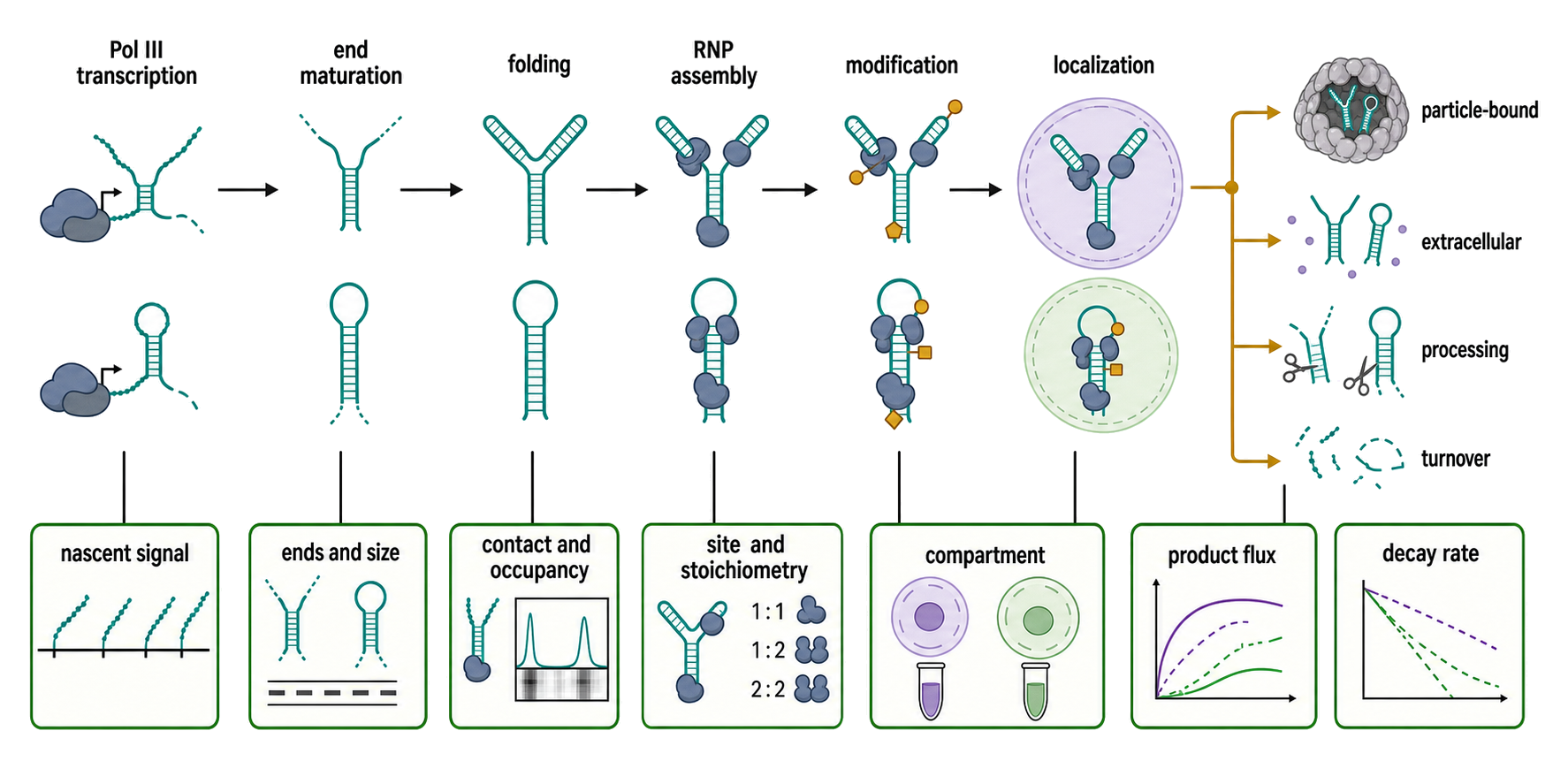
Figure 45.5. Lifecycle-resolved measurement of Y and vault RNAs. “Steady-state abundance collapses several kinetic processes. Stage-matched measurements are required to identify whether transcription, maturation, occupancy, trafficking, processing, or turnover changed.”
For Y RNAs, early La binding couples 3-prime-end recognition to stabilization. Ro60 binding to the mature terminal stem further stabilizes many Y-RNA populations and regulates Ro60 localization. The variable loop remains available for partner exchange and cleavage. A change in total Y RNA after Ro60 depletion could reflect loss of stability rather than reduced transcription. Conversely, increased nascent Pol III signal without increased mature RNA can indicate processing or turnover limitation.
Y-RNA localization is dynamic. Canonical Ro RNPs occur in cytoplasmic and nuclear pools, and stress can promote Ro60 relocalization. Y-RNA binding affects the accessibility of Ro60 localization information. Extracellular Y RNA may be associated with protein complexes, vesicles, lipoprotein-like particles, or non-vesicular carriers. Fractionation claims need density gradients, protease and nuclease protection tests, detergent controls, particle markers, and recovery controls; a high-speed pellet is not synonymous with an extracellular vesicle.
For vault RNAs, transcription varies among paralogs and cell states. Promoter architecture can combine internal and external elements. After transcription, the RNA partitions among vault particles, free or smaller protein complexes, and processed populations. The same locus can therefore respond to a signaling pathway through transcription while its functional effect is mediated by altered protein binding or cleavage.
5-Methylcytosine illustrates how modification changes fate rather than merely marking an RNA. NSUN2 modifies selected cytosines in vault RNAs. Loss of modification can shift processing and Argonaute association, while VTRNA1-1 C69 methylation changes SRSF2 binding and small-RNA production. Bisulfite conversion, crosslinking-based methyltransferase mapping, and direct mass-spectrometric confirmation have different error profiles. Structure can protect cytosines from conversion, and a methyltransferase-trapping method can capture binding without quantifying final stoichiometry.
Stress responses are family- and form-specific. In D. radiodurans, shifting from 30 to 37 degrees Celsius exposes efficient 23S rRNA maturation by Y-RNA-free Rsr, RNase II, and RNase PH; Yrn1 inhibits that maturation state even though the same RNA positively tethers Rsr to PNPase in a distinct degradation complex. Starvation reduces parent-length and p62-bound VTRNA1-1 and permits greater p62 oligomerization and autophagic activity, although the responsible transcriptional or post-transcriptional step remains unresolved. Productive encephalomyocarditis-virus and Sindbis-virus infection induces VTRNA1 transcription, and VTRNA1-family loss impairs infection-associated cytoplasmic redistribution of hnRNP C and ELAVL1 in those tested systems. These examples should not be compressed into a generic statement that stress “activates Y/vault RNA.” The direction and consequence depend on the RNA, partner, and stress, and induction of an RNA by one virus does not establish that the same RNA is required by every virus.
Turnover can be protective, constitutive, or signaling-related. Protein binding shields terminal stems and ends from nucleases. Endonucleolytic cleavage can remove a protein-binding domain and commit the RNA to degradation, or it can generate a stable product protected by a new RNP. Extracellular cleavage adds another compartment. Pulse–chase labeling, metabolic labeling with Pol III-aware controls, transcriptional shutoff, and acute degron-based partner depletion can separate synthesis from stability more effectively than a single steady-state comparison.
Table 45.1. Lifecycle step, direct observable, and common overinterpretation. Match each causal step to a direct measurement and state what the measurement does not show.
| Lifecycle step | Direct observable | Useful method | Common overinterpretation |
|---|---|---|---|
| Pol III transcription | Nascent signal at named locus | Nascent RNA and Pol III occupancy | Mature RNA increased |
| 3-prime-end maturation | Terminal position and chemistry | Direct end mapping, Northern sizing | One internal amplicon is full length |
| RNP assembly | Direct contact and occupancy | Calibrated crosslinking, binding mutants | Co-IP proves direct binding |
| Modification | Site and stoichiometry | Orthogonal chemical or MS evidence | Writer binding proves modification |
| Localization | Compartment-resolved molecules | Imaging plus controlled fractionation | Pellet equals vesicle or vault particle |
| Processing | Parent-to-product flux | Time course, nuclease genetics, end mapping | Fragment abundance proves regulated biogenesis |
| Turnover | Decay rate of a defined form | Pulse–chase or acute shutoff | Steady-state level equals synthesis rate |
A quantitative life-cycle experiment uses stage-matched controls. Nascent transcription is measured at the locus. Full-length RNA is measured by a size-resolved assay. Ends are mapped directly. Protein occupancy is measured by calibrated crosslinking or immunoprecipitation. Localization is measured by imaging or carefully controlled fractionation. Fragments are measured with end-aware libraries. RNA half-life is estimated after a perturbation whose own stress effects are characterized. No single readout substitutes for the complete set.
The main boundary case is that Pol III transcription itself responds to growth, stress, and transformation. A cancer-associated increase in a Y RNA or vault RNA can be part of broad Pol III activation rather than selective regulation of one locus. Comparing the target with several Pol III transcripts, measuring promoter occupancy, and testing paralog-specific regulation can distinguish those models. General Pol III mechanisms are treated in Chapter 21.
45.6. Functional evidence, disease causality, annotation, profiling, and orthogonal validation
The field combines unusually strong structural and biochemical observations with a large association literature. A useful evaluation begins by naming the causal object: gene, primary transcript, full-length mature RNA, specified RNP, processed fragment, vault particle, extracellular carrier, autoantibody, or downstream protein. Each object requires a different perturbation and readout.
For full-length RNA function, the best perturbation preserves neighboring genes and distinguishes paralogs. CRISPR deletion of a compact Pol III locus can disrupt local chromatin or nearby regulatory elements. Antisense oligonucleotides may block protein binding as well as promote degradation. RNA interference reagents are poorly matched to some nuclear or highly structured small RNAs and can introduce small-RNA-like off-targets. A rescue transcript must reproduce native ends, modification, abundance, folding, and localization. Expression from a generic plasmid can fail all five conditions.
For RNP function, partner depletion alone is insufficient when the protein has other RNA clients. Ro60 binds Y RNAs, misfolded RNAs, and retroelement transcripts. MVP forms the vault shell but does not account for every VTRNA1-1 function. p62 regulates autophagy through many protein interactions. A mechanistic experiment should test whether an RNA-binding-defective protein mutant separates the nominated RNP function from the protein’s other roles.
For fragments, the assay must distinguish parent and product. Short-read counts should be paired with Northern blotting or another size-resolved method. End chemistry and handling controls should be reported. Canonical microRNA claims require pathway genetics and Argonaute-dependent target evidence. General fragment biology and nomenclature belong to Chapter 89, but Y/vault studies should explicitly report which evidence rung they reach.
Annotation is a major source of error. Y-RNA pseudogenes create multi-mapping, and reads can be too short to distinguish Y-family members. Vault-RNA paralogs share sequence, while historical aliases such as vtRNA2-1, nc886, and pre-miR-886 can cause databases to assign the same reads to different feature classes. Reference builds can omit alternative loci or use different mature boundaries. Reports should provide the genome build, annotation release, feature sequences, allowed mismatches, multi-mapping policy, and count unit.
Structured and modified RNAs are also method-biased. Stable terminal stems can inhibit adapter ligation. Protein-bound ends may be inaccessible. Modifications can stop reverse transcription. A standard poly(A)-selected transcriptome can miss the RNA entirely, while a 15-35-nucleotide small-RNA library can measure only fragments. Specialized recovery and mapping strategies are treated in Chapter 126. A negative sequencing result should be stated as “not recovered under this protocol,” unless an orthogonal assay establishes absence.
Table 45.2. Assays for full-length RNA, RNP, fragment, and extracellular species. Compare what common assays can and cannot resolve.
| Assay | Strongest observable | Dominant limitation | Orthogonal partner |
|---|---|---|---|
| Northern blot | Size-resolved parent and selected fragments | Sensitivity and paralog cross-hybridization | End-aware sequencing or targeted RNase protection |
| RT-qPCR | Sensitive internal-sequence abundance | Loses molecule length and can share RT bias | Northern blot or direct hybridization |
| Small-RNA sequencing | Eligible short products and ends | Excludes parent; ligation and mapping bias | Full-length Northern and treatment contrasts |
| Crosslinking/IP | Protein-proximal RNA region | Antibody, crosslink chemistry, indirect membership | Purified binding and mutant rescue |
| smFISH | Subcellular location of probe-accessible RNA | Short target and structure limit probe design | Biochemical fractionation with recovery controls |
| Particle purification | Co-fractionation with vault or carrier | Contamination and dissociation/reassociation | Density, nuclease protection, and imaging |
| Autoimmune serology | Antibody specificity and titer | Does not identify RNA cargo or receptor signaling | Immune-complex RNA profiling and sensor assay |
Orthogonal validation is strongest when its dominant artifact differs from that of discovery. Northern blotting provides size but can cross-hybridize among paralogs. Reverse-transcription PCR is sensitive but often loses size information. Single-molecule fluorescence in situ hybridization provides localization but can struggle with very short targets and structure. Crosslinking maps protein contact but depends on nucleotide photochemistry and antibody quality. Mass spectrometry can validate modified nucleosides but usually loses sequence context unless combined with targeted digestion. Combining two assays that share the same reverse-transcription step is less orthogonal than it appears.
Disease causality requires a matched ladder. Expression association identifies a candidate. Temporal association asks whether the RNA changes before the phenotype. Perturbation tests necessity or sufficiency in a model. Rescue tests specificity. Direct binding and structure–function analysis identify mechanism. Animal or clinical genetics test organismal relevance. Prospective cohorts test biomarker utility. Cancer survival correlations, autoimmune serology, and extracellular fragment classifiers are valuable at their appropriate rung but should not be described as proof of a cellular mechanism.
Ro60 autoimmunity illustrates the need for component resolution. Anti-Ro60 antibodies recognize protein epitopes; associated RNAs can stimulate innate receptors; Ro60 loss can change endogenous retroelement RNA and interferon pathways. These facts support a coupled model, but the dominant RNA ligand may differ across patients and immune complexes. Clinical anti-Ro positivity does not measure Y-RNA abundance, and a Y-RNA fragment classifier does not measure autoantibody function.
Vault-RNA cancer studies face similar confounding. Pol III activity, cell-cycle composition, apoptosis resistance, autophagy, viral status, and tumor purity can all change vtRNA measurements. VTRNA1-1 perturbation has direct evidence in p62 binding and apoptosis contexts, but a tumor-cohort association for VTRNA1-2 cannot inherit that mechanism. Paralog identity and molecular form must remain explicit.
The best reporting unit is a claim matrix. Rows specify RNA form and locus. Columns specify abundance, size, termini, modification, protein occupancy, compartment, perturbation, rescue, molecular output, cellular phenotype, organism, and evidence limitations. Missing cells remain visible rather than being smoothed into a single narrative. This structure supports both human reading and claim-level computational reuse.
Table 45.3. Form-specific claim matrix for functional and disease evidence. Make missing causal links visible and prevent evidence from one molecular level from being assigned to another.
| Named causal object | Required identity evidence | Required perturbation | Matched outcome | Typical confounder |
|---|---|---|---|---|
| Y-RNA gene | Locus and promoter | Locus-specific editing | Nascent transcription | Nearby chromatin or pseudogene reads |
| Full-length Y RNA | Mature size and ends | Form-preserving depletion/rescue | Partner or replication output | Fragments removed simultaneously |
| Ro RNP | Direct Ro60 contact and composition | RNA-binding-defective separation | Surveillance substrate/product | Other Ro60 RNA clients |
| Vault particle | MVP shell plus TEP1/PARP4/RNA evidence | Particle-specific perturbation | Particle-proximal function | Free vault RNA and MVP-independent effects |
| Full-length VTRNA1-1 RNP | Full length plus named partner | Binding-surface mutant | p62 oligomerization or matched pathway | Paralog and fragment effects |
| Infection-associated VTRNA1 RBP complex | Named paralogs, RNA size, direct contact, and compartment | Paralog- and form-specific depletion/rescue | hnRNP C/ELAVL1 redistribution, viral-RNA association, and virus titer | Triple-locus perturbation conflates paralogs and products |
| Processed fragment | Discrete size, ends, and formation route | Biogenesis-specific perturbation | Endogenous target engagement | Degradation and mimic overexpression |
| Extracellular species | Carrier fraction and protection | Release or nuclease perturbation | Recipient-cell or biomarker output | Processing after collection |
| Autoimmune complex | Antigen, RNA cargo, and receptor | Component-resolved blockade | Cell-type-specific cytokine response | Heterogeneous immune-complex cargo |
Experimental Foundations and Evidence
The foundational evidence for Y RNP assembly combines mutational binding analysis and structural biology. Ro60 recognizes a conserved, bulged Y-RNA helix, and structural work separated outer-surface Y-RNA binding from central-cavity engagement of aberrant RNA. Localization experiments then connected Y-RNA binding to Ro60 distribution. These three evidence classes answer different questions: contact specificity, physical architecture, and cellular consequence.
Functional evidence for RNA surveillance is strongest in bacterial systems where genetic interactions, defined nucleases, precursor accumulation, and stress dependence can be connected. Vertebrate surveillance is supported by substrate binding and structural compatibility, but a full catalogue of physiological substrates and executing nucleases remains incomplete.
Replication evidence is based on depletion and add-back in cell-free systems. The founding study also excluded direct DNA priming and Ro60 binding, demonstrated interchangeability of the four human Y RNAs in reconstitution, and showed that predicted fold alone was insufficient when Y1 sequence was scrambled. Its key negative follow-up showed that canonical Ro60-, La-, and nucleolin-containing RNPs were dispensable. Together they argue for a replication-active Y-RNA population distinct from the surveillance RNP, while leaving the direct partner and exact initiation-versus-elongation step unresolved.
Vault-particle evidence combines biochemical purification and particle reconstruction. RNase-sensitive density near the caps establishes regional physical association but not nucleotide-level contact or invariant stoichiometry. Later 3.5-angstrom structural synthesis resolved the 78-chain MVP shell and contextualized earlier, lower-resolution component assignments. VTRNA1-1–p62 work goes beyond association by combining cellular crosslinking and immunoprecipitation, purified-protein binding to parent-length RNA, reciprocal RNA gain and loss, locus deletion, p62 epistasis, and mapped RNA and protein determinants with oligomerization and autophagy outputs. Those experiments strongly support parent-RNA riboregulation but do not directly quantify the particle occupancy of the active cellular pool.
The infection study adds a different causal chain. Triple VTRNA1-locus deletion, re-expression rescue, and independent antisense depletion connect VTRNA1-family expression to replication of named alpha- and picornaviruses. RNA antisense purification–mass spectrometry and protein-centered immunoprecipitation identify hnRNP C and ELAVL1 as partners; controlled fractionation and single-cell imaging place the relevant change at infection-associated cytoplasmic redistribution; and reduced viral-RNA association plus genetic interaction with RBP depletion connects localization to the proviral output. However, reverse-transcription readouts and family-wide perturbations do not resolve the active RNA’s length, ends, individual paralog, or vault-particle state, and the negative type I interferon tests apply only to the named cellular infection systems.
Fragment evidence spans several levels. Sequencing establishes recurrent products, but RNase 1 genetics and extracellular cleavage experiments identify one formation route for Y fragments. NSUN2, SRSF2, methylation, and differentiation experiments identify a regulated vault-RNA processing route. Neither mechanism should be generalized to every fragment from the respective family.
Biological Contexts Across Organisms, Cell Types, and Perturbations
Vertebrate Y RNAs emphasize Ro60/La assembly, autoimmunity, and replication assays. Bacterial Y RNAs emphasize stress-responsive RNA maturation and nuclease tethering. These are homologous systems with different experimental strengths. Conservation supports shared architectural principles, while divergence warns against assuming a single substrate or partner set.
Vault RNAs are eukaryotic and vary by lineage and paralog. Human VTRNA1-1 is the best-developed mechanistic example for p62, autophagy, apoptosis, and modification-dependent processing. The 2025 RNA-virus study instead perturbed VTRNA1-1, VTRNA1-2, and VTRNA1-3 collectively, so its hnRNP C/ELAVL1 localization mechanism cannot be assigned to VTRNA1-1 alone. Evidence for other paralogs is more uneven. Cell type matters because Pol III transcription, protein partners, stress programs, and extracellular release differ among epithelial cells, immune cells, neurons, and tumors.
Perturbation context can reverse interpretation. Starvation changes the VTRNA1-1–p62 axis; heat stress exposes the bacterial Ro60 pathway; apoptosis and extracellular nucleases reshape Y-RNA products; infection changes vault-RNA expression and protein localization. The relevant comparison is not merely treated versus untreated, but the molecular form and pathway state before and after perturbation.
Technology, Computational, Clinical, and Engineering Links
Y RNAs and vault RNAs are useful stress tests for RNA measurement technologies. Their compact folds, Pol III ends, modifications, paralogs, pseudogenes, RNP protection, and processed products expose weaknesses in extraction, adapter ligation, reverse transcription, annotation, and normalization. A robust platform should recover defined full-length and fragment standards, preserve end information, and report family-level counts when locus-level identification is impossible.
Clinically, anti-Ro serology is established, whereas Y- and vault-RNA biomarkers remain under active validation. Analytical performance, preanalytical handling, carrier fraction, renal or hepatic clearance, inflammatory state, and treatment must be separated from disease specificity. A fragment can be a good classifier without being a causal effector.
Vault particles have also inspired delivery nanotechnology because their MVP shells can be reconstituted and loaded. That engineering topic should not be used as retrospective proof of native vault function. Native particle biology and engineered cargo delivery answer different questions, even when they use the same shell.
Recent Consensus
Y RNAs are structured Pol III transcripts that form multiple RNP states. Ro60 binds a conserved bulged lower stem, La binds the uridine-rich nascent 3-prime end, and other proteins can recognize variable regions. Ro60 is an RNA-quality-control protein with additional RNA clients, so Y-RNA and Ro60 functions overlap but are not identical. Bacterial Y RNAs provide established gating and nuclease-tethering mechanisms; the complete vertebrate surveillance pathway is less resolved.
Y RNAs support replication in defined vertebrate cell-free systems, and related stem-bulge RNAs support replication and development in nematodes. In the founding human reconstitution study, the requirement lay in establishment or maintenance of active forks, but initiation could not be separated from early elongation. The replication-active RNA need not be in a canonical Ro60/La RNP. The exact replication step, direct molecular target, and universality of the pathway remain open.
Vault RNAs are genuine vault-particle components, but most functional claims concern specific paralogs in particle-independent complexes. VTRNA1-1–p62 riboregulation is supported by direct binding, structural determinants, perturbation, and functional readouts. NSUN2-dependent modification can alter vault-RNA processing and partner selection. These mechanisms should not be assigned automatically to all vault RNAs or to the intact vault particle.
During Sindbis-virus and encephalomyocarditis-virus infection in defined human cell lines, the VTRNA1 family supports cytoplasmic redistribution of hnRNP C and ELAVL1 and viral replication independently of a detectable requirement for type I interferon signaling. This is a collective VTRNA1-locus result, not proof that one paralog, one RNA molecular form, or the intact vault particle controls RBP trafficking in every viral infection.
Y-RNA and vault-RNA fragments are real molecular populations, yet fragment function must be demonstrated separately from parent function. Extracellular cleavage, handling, end chemistry, and library bias can shape observed products. Disease and biomarker studies require form-specific measurement and orthogonal validation.
Open Questions, Controversies, Deprecated Models, and Common Misconceptions
Open questions:
- What protein or replication intermediate directly recognizes the conserved replication-active Y-RNA structure in vertebrate cells?
- Which mammalian RNAs are physiological Ro60-surveillance substrates, and which nucleases execute their degradation in each compartment?
- How are Y RNAs exchanged among La, Ro60, replication-associated proteins, extracellular carriers, and other partners?
- What fraction of each vault-RNA paralog occupies intact vault particles in defined cell types and stresses?
- Does a particular full-length VTRNA1 paralog directly promote hnRNP C or ELAVL1 export during infection, or does the phenotype arise through a derived RNA, another RNP component, or an upstream trafficking pathway?
- Which vault-RNA modifications are stoichiometric and causal, and how do they change folding, partner affinity, and cleavage?
- Which Y- and vault-RNA fragments act at endogenous abundance, and which are turnover or extracellular-processing products?
- How should
VTRNA2-1/nc886be classified across databases while preserving both historical continuity and mechanistic distinction? - Which disease-associated RNA changes are causes, compensatory responses, cell-composition effects, or consequences of altered Pol III output?
Controversies:
- The importance of Y RNAs in DNA replication is supported in cell-free and selected cellular systems, but whether the RNA acts directly at origin firing, early fork progression, or both—and how broadly the requirement applies in vivo—remains disputed.
- Some Y- and vault-derived small RNAs have Argonaute association or target effects, whereas other abundant fragments are independent of canonical microRNA biogenesis. “MicroRNA-like” is therefore product-specific, not a family-wide classification.
- The historical inclusion of nc886 in the vault-RNA family conflicts with evidence that its particle association and regulatory behavior differ from canonical VTRNA1 paralogs.
- Vault particles correlate with drug resistance in several systems, but MVP, full-length vault RNA, processed fragments, and cell-state covariates can contribute independently.
Deprecated or weakened claims:
- A single immutable “Ro RNP” performs all Y-RNA functions. Current evidence supports multiple Y-RNA RNP states with distinct partners and activities.
- Every RNA called a vault RNA functions within the giant vault particle. Particle association is partial, paralog-dependent, and unnecessary for several demonstrated RNA mechanisms.
- A 20-30-nucleotide read from a structured RNA is automatically a microRNA. Size and Argonaute association alone do not establish canonical biogenesis or endogenous target regulation.
Common misconceptions:
- “Y RNA and Ro60 are the same functional unit.” Y RNA can act in Ro60-containing surveillance RNPs and in Ro60-independent replication-associated populations; Ro60 also binds non-Y RNAs.
- “Anti-Ro60 antibodies prove that Y RNA initiates autoimmunity.” The antibodies recognize protein, and associated RNAs can contribute innate stimulation, but the initiating ligand and causal sequence vary by context.
- “Vault RNA abundance measures vault-particle abundance.” Much vault RNA is outside the particle, and MVP shells can assemble without RNA.
- “All human vault-RNA paralogs are interchangeable.” VTRNA1 paralogs and VTRNA2-1/nc886 differ in sequence, regulation, partners, processing, and particle association.
- “Any virus that induces vault RNA depends on the same vault-RNA mechanism.” Viral induction is selective, and causal VTRNA1 requirements, partner trafficking, innate-sensing effects, and even the direction of the phenotype differ among tested virus–cell systems.
- “A reproducible extracellular Y-RNA fragment must be produced inside the source cell.” RNase 1 can generate characteristic fragments after extracellular release.
- “An RNA-seq count reports total Y RNA or vault RNA.” Library size selection, end chemistry, structure, modification, and multi-mapping determine which molecular form becomes countable.
- “Removing a parent RNA proves the fragment caused the phenotype.” Parent depletion commonly changes full-length RNP functions, fragment production, partner localization, and innate sensing simultaneously.