Chapter 152. miRNA Therapeutics: Mimics, Anti-miRs, Target Protectors, Delivery, and Clinical Pharmacology
Scope Note
This chapter is the book’s primary owner for microRNA-directed therapeutic chemistry, delivery, pharmacology, biomarkers, and clinical evidence. It treats miRNA drugs as a therapeutic class distinct from single-target antisense oligonucleotides and siRNAs; explains how replacement, inhibition, target-site blocking, and decoy strategies alter gene-regulatory networks; and uses clinical successes, setbacks, and discontinued programs to shape next-generation design. The chapter-local bibliography contains verified coverage-repair anchors migrated from the retired duplicate chapter, while precise claim-level support for several named clinical programs remains an explicit curation gap.
Executive Summary
MicroRNA therapeutics intervene in a regulatory layer where one short RNA can tune many transcripts and where each transcript can integrate signals from many RNA-binding factors. A miRNA replacement therapy supplies a synthetic or expressed miRNA-like guide when an endogenous tumor-suppressive or homeostatic miRNA is lost. A miRNA inhibitor does the opposite: it binds an endogenous mature miRNA and prevents that miRNA from repressing its targets. Sponges, tough decoys, and related expressed inhibitors use multiple binding sites or structured decoy motifs to sequester miRNA-loaded Argonaute complexes, usually for local or experimental settings rather than conventional systemic dosing. Target-site blockers occupy selected miRNA recognition elements on a chosen mRNA, preserving the rest of the miRNA network.
The therapeutic appeal is network-level: miRNA drugs may correct a pathological program rather than one isolated gene product. The therapeutic risk is also network-level: on-target activity can spread across dozens or hundreds of transcripts, with context-dependent effects that are difficult to predict from seed matches alone. Successful design therefore requires more than choosing a disease-associated miRNA. It requires understanding the miRNA’s endogenous abundance, Argonaute loading, target-site accessibility, cell-type distribution, disease-state directionality, dose-response behavior, and the resilience of the affected pathway.
Clinical history has made the field more sober. Liver-enriched miR-122 inhibition demonstrated that a miRNA can be drugged in humans and that viral and metabolic phenotypes can be modulated by anti-miRs, but safety and program-specific liabilities have limited translation. miR-34a replacement embodied the promise of restoring a broad tumor-suppressive program, but severe immune-related toxicities halted development. Other programs in fibrosis, cancer, dermatology, and viral disease have supplied useful lessons about delivery route, immune activation, patient stratification, pharmacodynamic biomarkers, and the need to distinguish target engagement from clinical benefit. The current consensus is not that miRNA therapeutics failed as a class, but that they require unusually disciplined pharmacology: narrow disease hypotheses, tissue-aware delivery, chemical designs that manage innate immune sensing and off-target hybridization, and biomarkers that report network response rather than only oligonucleotide exposure.
Concept Inventory
- MicroRNA: A mature miRNA is a short endogenous regulatory RNA, typically about 22 nucleotides long, that guides Argonaute-containing RNA-induced silencing complexes to partially complementary target sites, most often in 3′ untranslated regions of mRNAs. The seed region, usually nucleotides 2 to 8 of the miRNA, is a major determinant of target recognition, but pairing outside the seed, RNA structure, site number, site context, Argonaute availability, and competing RNA-binding proteins can modify repression.
- miRNA mimic: A synthetic duplex designed so that one strand resembles an endogenous mature miRNA and is loaded into Argonaute as the guide strand. The passenger strand is designed to be removed, degraded, or poorly loaded. A mimic is used for replacement or gain-of-function therapy.
- miRNA replacement: A therapeutic strategy that restores a miRNA whose reduced expression contributes to disease, for example a tumor-suppressive miRNA that has been deleted, epigenetically silenced, or diluted during oncogenic reprogramming.
- Anti-miR: A single-stranded oligonucleotide that binds an endogenous miRNA through complementarity and prevents productive target recognition. Anti-miRs may be called antagomirs, antimiRs, miRNA inhibitors, or LNA anti-miRs depending on chemistry and historical context.
- Antagomir: Historically, a cholesterol-conjugated, chemically modified antisense oligonucleotide designed to inhibit a mature miRNA in vivo. In current usage, the term is sometimes used loosely for anti-miR oligonucleotides, but chemistry-specific naming is preferable.
- Locked nucleic acid: A ribose-constrained nucleotide analog that increases duplex thermal stability and nuclease resistance. LNA-containing anti-miRs can bind short miRNAs with high affinity but require careful design to avoid excessive tissue accumulation or unintended hybridization.
- Sponge or decoy: An RNA containing repeated miRNA-binding sites that competes with natural targets for miRNA-loaded Argonaute complexes. Expressed sponges and tough decoys are widely used as research tools and may be relevant to gene-therapy-style interventions, but systemic drug development is more challenging.
- Target-site blocker: A steric-blocking oligonucleotide that binds a specific miRNA recognition element on a specific transcript. It protects one target mRNA from one miRNA without globally inhibiting the miRNA.
- Multi-target pharmacology: The intended therapeutic logic in which a miRNA-directed agent changes a coordinated set of transcripts or pathways. In miRNA therapeutics, multi-target action can be beneficial when it matches disease biology and harmful when it crosses tissue, immune, metabolic, or developmental boundaries.
What to Know Before Reading This Chapter
This chapter assumes that the reader understands basic RNA polarity, base pairing, oligonucleotide chemical modification, and the miRNA pathway described in Chapter 84 and Chapter 85. The key bridge concept is that a miRNA drug acts through endogenous small-RNA machinery rather than through a conventional receptor or enzyme active site. A mimic must be recognized by cellular loading machinery, retained in Argonaute, and guide repression of target mRNAs. An inhibitor must bind a mature miRNA with enough affinity, duration, and tissue exposure to stop that miRNA from acting. A decoy must compete with natural targets. A target-site blocker must occupy one cis-regulatory element on one transcript.
For therapeutic reasoning, the reader should separate three questions. First, is the disease mechanism actually sensitive to the miRNA in the relevant human cells? Second, can a drug reach those cells in a form that engages the miRNA pathway without unacceptable toxicity? Third, does the resulting network response improve patient outcomes rather than merely changing molecular biomarkers? Many disappointing miRNA programs have looked plausible under the first question but failed under the second or third.
152.1. miRNA Replacement and Mimic Design
MicroRNA replacement therapy starts from a simple therapeutic idea: if loss of a miRNA helps create a disease state, supplying a functional substitute might restore repression of a pathological gene program. The clearest conceptual examples come from cancer biology, where some miRNAs behave as tumor suppressors in particular contexts. A tumor-suppressive miRNA can be lost through chromosomal deletion, promoter methylation, altered transcription-factor activity, defective processing, or selection against a regulatory state that restrains proliferation. Replacement does not mean that the disease has no other driver. It means that the missing miRNA contributes enough to the pathological network that restoring miRNA activity could shift the cell state in a useful direction.
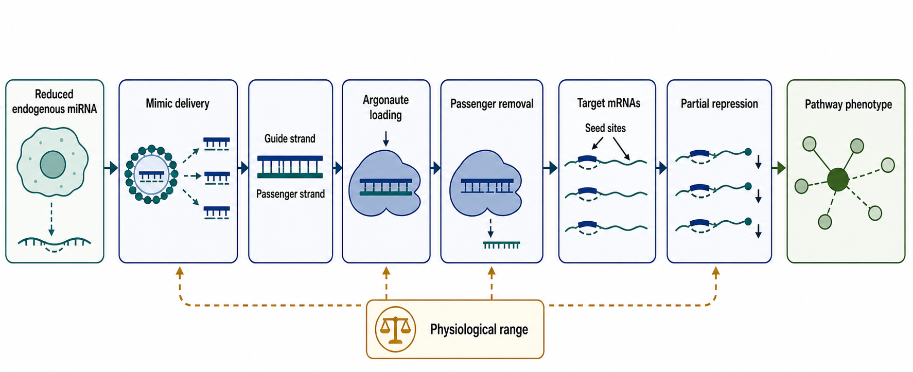
Figure 152.1. miRNA Replacement Workflow from Lost Endogenous miRNA to Mimic-Loaded Argonaute. “miRNA replacement supplies a guide strand intended to restore a missing regulatory program. The therapeutic mechanism depends on delivery, correct Argonaute loading, physiological dose range, and repression of a disease-relevant target set.”
A miRNA mimic is usually a short RNA duplex. One strand is intended to become the guide strand loaded into Argonaute; the other is a passenger strand that supports duplex recognition during loading but should not accumulate as an active guide. The design problem is therefore not merely sequence identity to an endogenous mature miRNA. The mimic must have the right duplex end asymmetry, strand modifications, thermodynamic profile, purity, length distribution, and formulation behavior. If the wrong strand loads into Argonaute, the drug can create an unintended miRNA-like activity. If the intended guide is overrepresented relative to endogenous miRNA abundance, repression can extend beyond physiological targets, including weak seed matches that would not be substantially regulated at endogenous concentrations.
The seed region gives replacement therapy much of its power and much of its uncertainty. A seed-matched family, such as a let-7 family member, can regulate many transcripts that share short recognition motifs. In a favorable case, this distributes therapeutic pressure across a pathway: oncogenes, cell-cycle regulators, survival factors, or fibrotic mediators may be partially reduced together. In an unfavorable case, the same seed logic represses transcripts that maintain viability in normal tissue or immune cells. The pharmacological unit is not one target. The pharmacological unit is a guide strand, an Argonaute-loaded complex, and the transcriptome context encountered by that complex.
Table 152.1. Design Variables for miRNA Mimics. Give readers a structured way to evaluate mimic design.
| Design variable | Mechanistic purpose | Failure mode | Evidence or assay |
|---|---|---|---|
| Guide sequence identity | Match the intended mature miRNA guide, especially seed and disease-relevant pairing logic. | Wrong isomiR or engineered guide changes the target set and weakens replacement claims. | Mature-guide sequence check, isomiR profiling, seed-dependent reporter or endogenous target response. |
| Passenger-strand suppression | Prevent the support strand from becoming an unintended active guide. | Passenger loading creates a second miRNA-like activity with unrelated targets. | Argonaute small-RNA sequencing, guide/passenger strand ratios, passenger-target reporter assay. |
| Duplex asymmetry | Bias Argonaute loading toward the therapeutic guide strand. | Symmetric or misbiased duplex favors wrong-strand loading or weak loading. | Terminal stability analysis, strand-specific Argonaute loading, guide reporter potency. |
| Chemical modification | Improve stability and immune silence while preserving RISC-compatible guide chemistry. | Excess modification impairs loading, alters targeting, or causes chemistry-linked toxicity. | Serum stability, cytokine panels, Argonaute loading, target-repression reporter assays. |
| Dose range | Restore regulatory tone without expanding into supraphysiological seed repression. | RISC saturation, endogenous miRNA competition, or broad weak-site repression. | Dose-response transcriptomics, proteomics, Argonaute occupancy, tolerability markers. |
| Argonaute loading | Place the guide in functional RNA-induced silencing complex rather than only inside cells. | Uptake without cytosolic release or loading gives exposure without mechanism. | AGO immunoprecipitation followed by guide qPCR or sequencing, target-site reporter response. |
| Target-set validation | Confirm direct disease-relevant targets and downstream pathway correction. | Predicted seed targets or transfection artifacts are mistaken for therapeutic mechanism. | Argonaute CLIP, early time-course expression, target-site mutagenesis, rescue experiments. |
| Formulation compatibility | Deliver an intact duplex to the right cells while preserving strand balance. | Endosomal trapping, strand loss, particle toxicity, or exposure in wrong tissue. | Particle QC, duplex integrity, biodistribution, endosomal escape, AGO loading in target tissue. |
The first design step is selecting a disease hypothesis. A replacement candidate should have convergent evidence that the miRNA is lower in the disease-relevant cell population, that experimentally restoring the miRNA shifts disease phenotypes, and that important targets respond in the expected direction. Bulk tissue differential expression is not sufficient. A tumor sample with low miR-34a, for example, may contain changing proportions of tumor cells, stromal cells, immune infiltrates, and necrotic tissue. A fibrosis sample with altered miRNA abundance may reflect both injury response and cell composition. Replacement design should therefore use cell-type-resolved data, perturbation experiments, rescue logic, and pathway readouts rather than relying only on association.
The second design step is choosing whether to match an endogenous miRNA exactly or to engineer a related guide. Exact replacement preserves known biology but inherits all natural targets, including potentially undesirable ones. Engineered guides can reduce passenger loading, improve potency, change chemical stability, or avoid selected seed-mediated liabilities, but they may no longer be replacement therapies in the strict biological sense. They become designed small-RNA drugs that borrow miRNA machinery. The distinction matters because mechanistic claims about restoring a lost endogenous program are strongest when the guide sequence, abundance range, and target engagement resemble the endogenous miRNA in the relevant tissue.
Chemical modification is constrained by Argonaute loading and target recognition. In siRNA therapeutics, extensive 2′ sugar modifications, phosphorothioate linkages, and conjugation patterns can be tuned for stability and delivery. A miRNA mimic has less freedom at positions that participate in guide loading and seed pairing. Modifications may reduce nuclease degradation and innate immune activation, but excessive modification can impair RISC loading or alter target selection. The passenger strand can often carry modifications that bias strand selection or prevent passenger-mediated repression. The guide strand must preserve a conformation and chemistry that Argonaute can use.
Dose is an unusually important part of mimic design. Endogenous miRNAs usually repress individual targets modestly, often by changing mRNA stability and translation through multiple weak interactions. A mimic delivered at very high levels can saturate Argonaute, compete with endogenous miRNAs, and drive supraphysiological repression of transcripts with weak seed matches. This is not simply “more efficacy.” It is a change in mechanism. At low to moderate exposures, a mimic may restore a missing regulatory tone. At high exposures, it may become a broad seed-directed knockdown reagent with toxicity that is difficult to assign to one target.
Box 152.1. When Is a Mimic Really Replacement?
A therapeutic mimic is strongest as replacement when five conditions hold. The mature guide sequence should match the endogenous miRNA species that is lost in the disease-relevant cell type. The delivered dose should approximate a range that restores regulatory tone rather than overwhelming Argonaute. The guide strand, not the passenger strand, should be the main Argonaute-loaded species. Early target responses should include validated endogenous targets in the expected direction. The downstream phenotype should be rescued or phenocopied by manipulating those targets or target sites.
If these conditions are not met, the molecule may still be useful, but the mechanism should be described more cautiously as engineered miRNA-like regulation. That distinction matters for safety: supraphysiological guide abundance can repress weak seed sites, compete with endogenous miRNAs, and create network effects that natural expression of the miRNA would not produce.
Evidence for mimic activity should include both proximal and distal readouts. Proximal readouts show that the guide strand enters cells, is loaded into Argonaute, and represses reporter or endogenous transcripts containing relevant sites. Distal readouts show that cell-state phenotypes change: reduced proliferation, altered apoptosis, restored differentiation, decreased fibrotic activation, or changed viral replication. Strong evidence links these levels by showing seed-dependent effects, rescue through target-site mutation or target re-expression, and concordant transcriptomic signatures. Weak evidence shows only that a mimic changes many mRNAs in transfected cells, because transfection itself, immune stimulation, and nonphysiological dose can produce broad expression changes.
The main boundary case is a disease-associated miRNA that is reduced but not causally limiting. Many miRNAs change during disease because cells change state. A miRNA may be a marker of differentiation rather than a driver. Replacing such a miRNA can still produce phenotypes in experimental systems, especially at high dose, but that does not prove a clinically useful therapeutic window. Another boundary case is a miRNA that acts beneficially in one cell type and harmfully in another. A tumor-suppressive miRNA in malignant epithelial cells might also repress immune activation or tissue repair genes in surrounding cells. Replacement therapy is therefore inseparable from delivery specificity.
This section connects to Chapter 84 for the normal miRNA pathway, Chapter 85 for miRNA turnover and Argonaute dynamics, Chapter 149 for modified oligonucleotide chemistry, and Chapter 156 for formulation constraints. The most important lesson is that replacement is a pathway-restoration strategy only when the guide, dose, cell type, and target network are all aligned.
152.2. miRNA Inhibition by Antagomirs, LNA Inhibitors, Tough Decoys, and Sponges
MiRNA inhibition uses the opposite logic from replacement. Instead of supplying a missing guide, the therapeutic agent reduces the activity of an endogenous miRNA that contributes to disease. The most direct inhibitor is an anti-miR oligonucleotide: a single-stranded sequence complementary to the mature miRNA. When the anti-miR binds the miRNA, the miRNA cannot productively pair with target mRNAs. Depending on chemistry and biology, the bound miRNA may be sequestered, degraded, tailed and trimmed, or otherwise removed from active Argonaute pools. The practical goal is reduced repression of the miRNA’s natural targets in the tissue where the miRNA is pathogenic.
Antagomirs were among the first in vivo demonstrations that a mature miRNA could be inhibited pharmacologically. Classic antagomir designs used cholesterol conjugation and chemical stabilization to improve tissue uptake and nuclease resistance. The term remains historically important, but modern anti-miR development is better described by chemistry and format: LNA-containing inhibitors, 2′ modified oligonucleotides, phosphorothioate backbones, short high-affinity anti-miRs, conjugated anti-miRs, or formulated anti-miRs. This precision avoids the misconception that all miRNA inhibitors share the same distribution, potency, or toxicity.
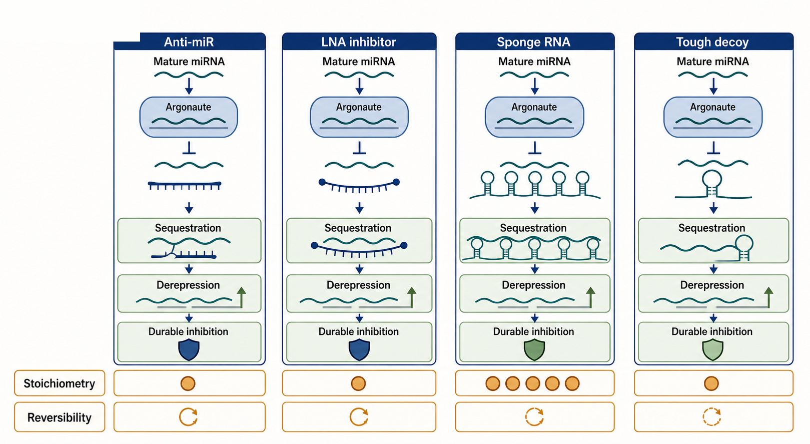
Figure 152.2. Four Modes of miRNA Inhibition. “miRNA inhibitors differ in chemistry, stoichiometry, duration, and reversibility. All reduce productive miRNA-target engagement, but they do not share identical pharmacology.”
Locked nucleic acid anti-miRs exploit the short length of mature miRNAs. Because an anti-miR target is only about 22 nucleotides long, high affinity is useful. LNA nucleotides constrain the sugar and increase duplex stability, allowing short inhibitors to bind strongly. High affinity can improve potency and duration, but it also raises design hazards. A very stable oligonucleotide can bind partially complementary RNAs, accumulate in tissues, interact with proteins through phosphorothioate chemistry, or persist long after a transient pharmacodynamic effect would be preferable. The benefit is strong target engagement; the boundary is that high-affinity chemistry does not make sequence specificity automatic.
The liver-enriched miRNA miR-122 is a central example because it links normal physiology and viral infection. miR-122 is abundant in hepatocytes, contributes to liver gene-expression identity, and is used by hepatitis C virus RNA in a way that supports viral RNA stability and replication. Inhibiting miR-122 can therefore reduce viral replication while also perturbing host hepatic regulation. This example teaches why miRNA inhibition can be clinically plausible: the target is abundant, tissue-enriched, mechanistically connected to disease, and measurable through viral and host biomarkers. It also teaches why chronic inhibition must be cautious: the same miRNA has normal functions, and long-term network effects may not be fully predicted by short-term viral response.
Table 152.2. miRNA Inhibitor Formats and Pharmacological Tradeoffs. Compare antagomirs, LNA anti-miRs, other modified anti-miRs, sponges, and tough decoys.
| Format | Molecular form | Main mechanism | Strengths | Main liabilities | Best-fit use case |
|---|---|---|---|---|---|
| Antagomir | Cholesterol-conjugated, chemically stabilized single-stranded anti-miR. | Binds mature miRNA and prevents productive target engagement. | Historical in vivo proof that mature miRNAs can be inhibited. | Broad distribution, chemistry-linked effects, and loose terminology across designs. | Tissue-accessible inhibition with a measurable pharmacodynamic marker. |
| LNA anti-miR | Short locked-nucleic-acid-containing antisense oligonucleotide. | High-affinity binding sequesters or disables the mature miRNA. | Potent binding to short miRNAs and potentially durable target engagement. | Partial-complement binding, tissue accumulation, and long duration may limit reversibility. | Abundant pathogenic miRNA where sustained inhibition is acceptable. |
| Other modified anti-miR | 2′-modified, phosphorothioate, conjugated, or formulated single strand. | Chemistry-dependent sequestration, derepression, or miRNA turnover. | Chemistry, route, and duration can be tuned for the tissue hypothesis. | Platform-specific immune sensing, protein binding, delivery, and metabolism risks. | Programs needing adjustable exposure rather than maximal affinity alone. |
| Sponge RNA | Expressed RNA with repeated miRNA-binding sites. | Competes with natural targets for miRNA-loaded Argonaute complexes. | Can inhibit seed families and reveal long-term loss-of-miRNA phenotypes. | Requires high expression, correct localization, favorable stoichiometry, and reversibility control. | Experimental or local vector settings where cell-selective expression can be verified. |
| Tough decoy | Vector-expressed structured RNA with accessible miRNA-binding sites. | Durable sequestration or disabling of selected miRNA-loaded complexes. | Strong, long-lived inhibition can be restricted by promoter or vector tropism. | Vector immune response, dose control, tropism, and difficult reversal. | Gene-therapy-like local inhibition after a strong safety rationale. |
| TDMD-inducing target mimic | Highly complementary target-like RNA or oligonucleotide design. | Triggers tailing, trimming, and degradation of susceptible miRNAs. | Can actively reduce miRNA abundance rather than only compete for binding. | Context-dependent biology; not all miRNAs or sites induce degradation. | Specialized programs after cell-specific target-directed decay validation. |
Sponges and decoys inhibit miRNAs by presenting competing binding sites. A sponge RNA contains multiple miRNA recognition elements, often imperfectly paired so that it binds miRNA-loaded Argonaute without being immediately cleaved like a perfectly complementary target. A tough decoy, or TuD, uses a structured RNA architecture with accessible miRNA-binding sites designed for potent inhibition after expression from a vector. These tools are especially powerful in experimental biology because they can inhibit miRNA families, be expressed in selected cells, and reveal phenotypes of long-term miRNA reduction. Therapeutic use is more complicated because vector delivery, expression control, insertional risks, immune response, and reversibility become part of the drug profile.
The mechanism of a sponge differs from that of a stoichiometric anti-miR. A chemically synthesized anti-miR is dosed as a molecule with defined pharmacokinetics. An expressed sponge is produced inside cells, and its inhibitory capacity depends on promoter strength, transcript stability, subcellular localization, number and affinity of binding sites, Argonaute occupancy, and degradation of the decoy transcript. If a sponge is too weak, natural targets outcompete it. If a sponge is too strong or broadly expressed, it can deplete miRNA activity outside the intended tissue. Because miRNA families share seed sequences, a sponge designed against a seed family may inhibit several related miRNAs. That can be a feature or a liability.
Target-directed miRNA degradation provides another conceptual route to inhibition. Natural target RNAs with extensive complementarity to the miRNA 3′ region can trigger miRNA tailing, trimming, and decay in some contexts. Artificial targets can be designed to exploit this pathway, but the biology is context-dependent. Not every binding site induces degradation, not every miRNA is equally susceptible, and the proteins controlling tailing and trimming vary by cell type. This area is important because it shifts the design question from passive sequestration to active remodeling of miRNA abundance.
Inhibitor evidence should be interpreted carefully. A rise in predicted target mRNA abundance is supportive but not definitive, because stress responses, transfection artifacts, and secondary transcriptional effects can also increase mRNAs. Stronger evidence includes physical engagement of the mature miRNA, loss of Argonaute association with target sites, derepression of reporters with intact but not mutated sites, dose-dependent target derepression, and phenotypic rescue by reintroducing a resistant miRNA or altering key target sites. For clinical candidates, pharmacodynamic biomarkers should include direct miRNA engagement or target derepression in accessible tissue, not only plasma exposure.
The main misconception is that inhibiting a miRNA automatically increases all of its predicted targets. Prediction algorithms often begin with seed matches, conservation, and site context, but endogenous repression depends on transcript abundance, site accessibility, 3′ UTR isoforms, competing RNA-binding proteins, and cell state. A target with a perfect-looking seed site can be insensitive in one cell type and responsive in another. Conversely, modest derepression across many real targets may be biologically important even when no single transcript changes dramatically.
This section links to Chapter 85 for miRNA stability and target-directed miRNA degradation, Chapter 150 for antisense chemistry, Chapter 151 for comparison with siRNA knockdown, and Chapter 158 for pharmacodynamic assessment. The design principle is that a miRNA inhibitor is not only an antisense molecule. It is an intervention in an endogenous regulatory circuit whose normal activity may be beneficial in some tissues and harmful in others.
152.3. Target-Site Blockers and Multi-Target Pharmacology
Target-site blockers occupy a middle position between global miRNA inhibition and conventional antisense targeting. A target-site blocker is an oligonucleotide designed to bind the mRNA sequence that contains a particular miRNA recognition element. Because the blocker occupies the cis-regulatory site, the miRNA cannot bind that site, but the miRNA remains free to regulate other targets. This approach is sometimes called target protection. It is conceptually attractive when the disease phenotype depends on miRNA repression of one especially important transcript, while global inhibition of the miRNA would be too broad.
The simplest example is a transcript whose expression is pathologically low because a specific miRNA site is overactive or newly created. A sequence variant in a 3′ untranslated region can create or strengthen a miRNA site, producing allele-specific disease risk. In that setting, a target-site blocker can be designed to cover the pathogenic site. Unlike an RNase H gapmer, the blocker is not meant to degrade the mRNA. Unlike an siRNA, it is not meant to guide cleavage. Its mechanism is steric: hybridize to the mRNA region and prevent a regulatory RNP from occupying the site. Because the mRNA remains intact, target-site blockers belong mechanistically with steric-blocking oligonucleotides discussed in Chapter 150.
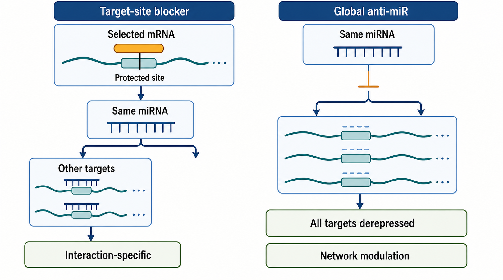
Figure 152.3. Target-Site Blocking Protects One Interaction Rather Than the Whole miRNA Network. “Target-site blockers protect a selected transcript from a selected miRNA interaction. This increases specificity at the interaction level but gives up broad network modulation.”
Target protection offers specificity at a different level from anti-miR specificity. A global anti-miR asks: which miRNA should be inhibited? A target-site blocker asks: which miRNA-target interaction should be interrupted? This distinction matters because the same miRNA can repress transcripts with opposing biological consequences. For example, a miRNA may restrain a harmful inflammatory mediator in one cell type while also repressing a protective epithelial gene in another. Global inhibition would remove both effects. A target-site blocker can, in principle, protect the beneficial transcript without releasing the entire target network.
The tradeoff is that target-site blockers lose part of the multi-target advantage that motivates many miRNA therapeutics. If disease biology truly depends on coordinated modest regulation of many transcripts, protecting one site may be insufficient. The correct design depends on the causal architecture. A monogenic or site-variant mechanism favors target protection. A pathway-state mechanism, such as oncogenic dedifferentiation or fibrotic activation, may favor mimic or anti-miR strategies that influence multiple nodes. The field should therefore avoid treating “multi-target” as automatically superior. Multi-target pharmacology is useful only when the target set maps onto a disease-controlling network and leaves enough therapeutic window.
Table 152.3. Choosing Between Global miRNA Modulation and Target-Site Protection. Help readers reason from disease architecture to therapeutic format.
| Disease architecture | Preferred format | Rationale | Biomarker priority | Main risk |
|---|---|---|---|---|
| Single pathogenic target-site variant | Target-site blocker. | One harmful cis-regulatory interaction can be protected without inhibiting the whole miRNA. | Allele or transcript-specific mRNA/protein restoration and site occupancy. | Sequence variation, isoform choice, or incomplete delivery to the mRNA compartment. |
| Disease-cell pathway state | Mimic or anti-miR, depending on the causal direction of miRNA change. | Coordinated modest regulation of several nodes may match the disease program. | Cell-type-specific miRNA state, direct targets, pathway signature, and functional response. | On-target network toxicity or treating a compensatory signature as causal. |
| Host-factor viral dependence | Anti-miR. | Inhibiting a host miRNA can reduce viral RNA support while avoiding viral escape at one enzyme. | Viral load, host target derepression, liver function, and duration of miRNA inhibition. | Normal host-miRNA functions and changing therapeutic alternatives. |
| One protective transcript repressed by miRNA | Target-site blocker. | Preserves a beneficial transcript while leaving other miRNA targets regulated. | Protected transcript/protein without broad derepression of unrelated targets. | The protected interaction may be insufficient to shift disease phenotype. |
| Broad inflammatory or oncogenic signature | Cautious network modulation with mimic or anti-miR only after bounded validation. | Multi-node effects may be useful if direct targets map to the disease-driving circuit. | Early direct targets, pathway output, immune or cell-state readouts, and safety markers. | Secondary apoptosis, immune activation, or cell-composition shifts can mimic mechanism. |
Multi-target pharmacology in miRNA therapy has three layers. The first layer is direct target engagement: a miRNA guide or inhibited miRNA changes transcripts with accessible sites. The second layer is pathway propagation: direct changes alter transcription factors, signaling proteins, metabolic enzymes, or RNA-binding proteins that then change additional genes. The third layer is tissue-system response: immune cells, stromal cells, vasculature, liver, kidney, and disease tissue respond to the changed pathway and to the drug material itself. Clinical benefit or toxicity emerges from all three layers, not from the direct seed-target layer alone.
Network-level interpretation requires both computational and experimental discipline. Seed enrichment among downregulated transcripts after mimic treatment can support on-target activity, but it cannot by itself prove therapeutic mechanism. A transcriptome signature may be dominated by secondary apoptosis, interferon activation, cell-cycle arrest, or cell-type shifts. Time-course experiments help separate early direct effects from later secondary effects. Argonaute crosslinking, reporter assays, target-site mutagenesis, and rescue experiments help identify direct targets. Single-cell or spatial assays can show which cells respond. Proteomics can reveal effects that mRNA measurements miss. The strongest network models integrate several evidence types rather than treating target prediction as a substitute for biology.
Box 152.2. Evidence Ladder for a Direct miRNA Target
Use an evidence ladder before treating a transcript as a direct therapeutic target. At the lowest tier, the transcript has a computationally predicted seed site or appears in a disease-associated pathway map. A stronger tier shows that the transcript changes shortly after mimic or anti-miR dosing, before broad secondary responses dominate. Stronger still is physical evidence, such as Argonaute binding near the relevant site in the right cell type. Sequence-specific evidence comes from reporter assays, target-site mutation, or a target-site blocker that selectively protects the interaction. The highest tier links the target to phenotype: restoring or removing the target changes the disease-relevant response.
No single rung is decisive in all contexts. Transcript abundance, untranslated-region isoforms, protein turnover, RNA-binding proteins, and cell state can make a real site weak or a predicted site irrelevant.
Dose-response modeling is also different for multi-target miRNA drugs. A single-target enzyme inhibitor may have a relatively interpretable relationship between occupancy, pathway inhibition, and efficacy. A miRNA mimic can repress some targets at low dose, additional weaker targets at higher dose, and endogenous miRNA networks indirectly if Argonaute is saturated. A miRNA inhibitor can derepress high-sensitivity targets first and low-sensitivity targets later or at higher exposure. The therapeutic window may therefore be defined by which parts of the target set respond at a given dose. More complete miRNA modulation is not necessarily better.
Patient genetics and transcript isoforms can alter target-site pharmacology. Alternative polyadenylation can shorten 3′ untranslated regions and remove miRNA sites in proliferating cells or cancers. RNA editing, somatic mutation, germline variants, and RNA-binding protein occupancy can change site accessibility. A target that is miRNA-regulated in a reference cell line may not carry the same 3′ UTR or regulatory context in patient tissue. For target-site blockers, sequence variation at the binding site can directly affect drug binding. For mimics and inhibitors, variation across the target network can change both efficacy and toxicity.
The most important caveat is that network diagrams can make weak evidence look mechanistically complete. A pathway map assembled from predicted targets, differential expression, and disease associations is a hypothesis generator. It is not proof that a miRNA drug will benefit patients. A clinically useful model should name the disease cell type, the direction of miRNA change, the direct targets most likely to mediate benefit, the biomarkers that should move, the tissues where harm is expected, and the evidence that changing the network improves function in vivo.
This section hands off to Chapter 142 for target prediction and quantitative regulatory models, Chapter 106 for single-cell and spatial response profiling, Chapter 136 for perturbational screening, and Chapter 158 for pharmacodynamic modeling. The central lesson is to treat multi-target pharmacology as a design hypothesis that must be bounded, measured, and tested, not as a slogan.
152.4. Delivery, Tissue Targeting, Innate Immune Effects, and Toxicity
Delivery is often the difference between a plausible miRNA mechanism and a viable drug. Naked RNA duplexes and single-stranded oligonucleotides are vulnerable to nuclease degradation, renal clearance, poor membrane permeability, and uptake into compartments that do not communicate efficiently with the cytosol. A mimic must reach the cytosol and load into Argonaute. An anti-miR must reach the compartments where mature miRNA-loaded Argonaute can be bound or where miRNA turnover can be altered. A target-site blocker must reach the mRNA compartment at sufficient concentration. Delivery is therefore not an afterthought added to a sequence. Delivery shapes mechanism.
Systemic delivery has historically favored the liver because hepatocytes and liver nonparenchymal cells encounter blood-borne oligonucleotides and nanoparticles efficiently. This makes liver-enriched miRNAs and liver diseases attractive early indications, but it also means that liver toxicity and liver pharmacology can dominate even when the intended disease tissue is elsewhere. Local delivery, such as intratumoral, intradermal, ocular, inhaled, or surgically accessible administration, can reduce systemic exposure but narrows the indications. Viral-vector expression can provide durable decoy or replacement activity in selected tissues but introduces questions of immune response, dose control, reversibility, and vector tropism.
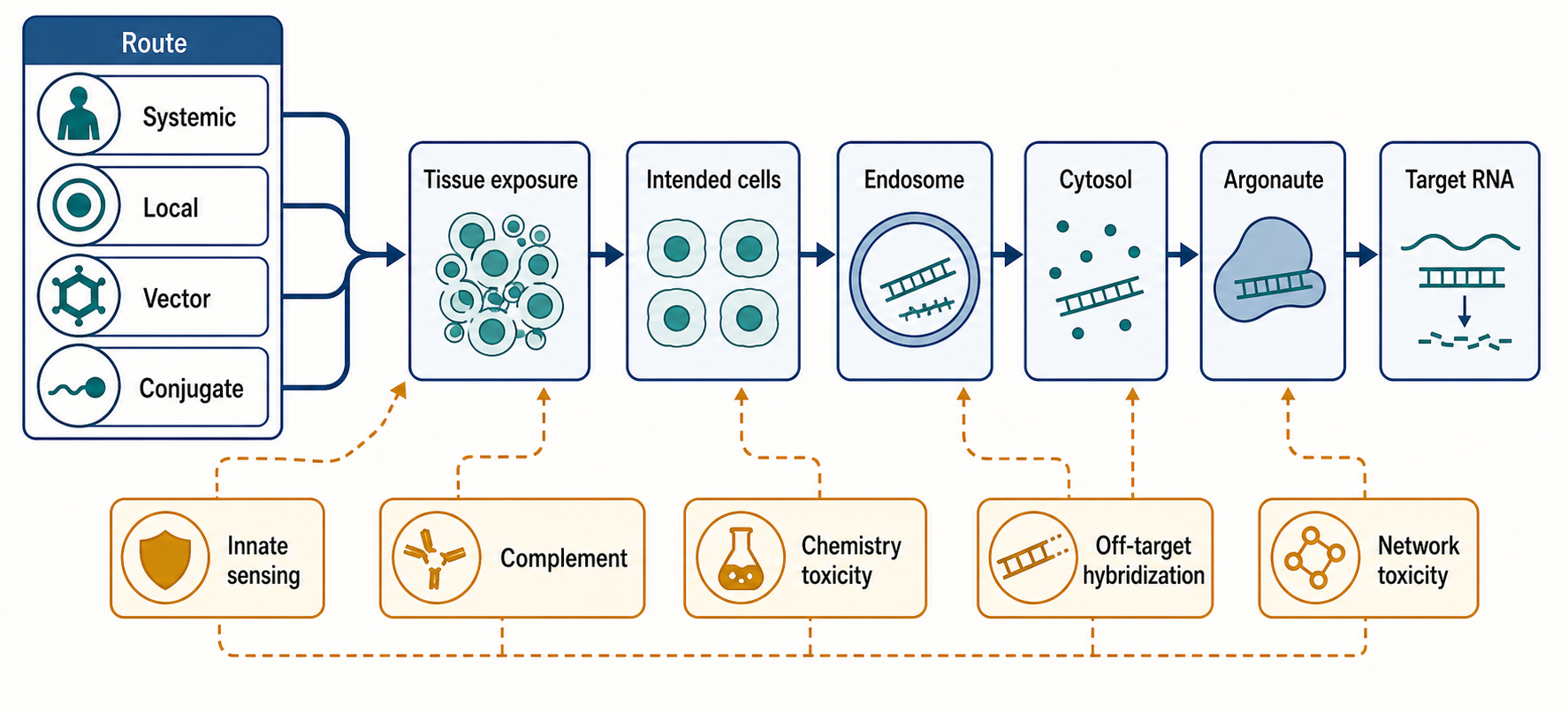
Figure 152.4. Delivery and Toxicity Decision Tree for miRNA Therapeutics. “Functional delivery requires the drug to reach the right cell and the right intracellular compartment. Toxicity can originate from sequence, chemistry, formulation, route, or intended network activity.”
Lipid nanoparticles and related formulations can encapsulate miRNA mimics or inhibitors, protect them in circulation, promote cellular uptake, and support endosomal escape. Their pharmacology is determined by ionizable lipid chemistry, helper lipids, cholesterol, polyethylene glycol-lipid behavior, particle size, surface charge, protein corona, route of administration, and tissue vascularization. For a miRNA mimic, formulation must preserve duplex integrity and avoid preferential loss of one strand. For an inhibitor, formulation must balance uptake with release. For both, only a small fraction of internalized material may escape endosomes, so apparent cellular uptake can overestimate functional delivery.
Box 152.3. Uptake Is Not Functional Delivery
For miRNA therapeutics, delivery should be parsed into checkpoints. Exposure means the formulation or oligonucleotide reaches a tissue. Uptake means cells internalize the material, often into endosomes. Escape means enough material reaches a compartment that communicates with Argonaute or target mRNAs. Pathway engagement means the guide loads into Argonaute, the anti-miR binds the mature miRNA, or the blocker occupies the intended mRNA site. Functional response means direct targets and disease-relevant pathways move in the expected direction.
Assays should match the checkpoint. Fluorescence microscopy, tissue qPCR, and biodistribution can show exposure or uptake, but they do not prove endosomal escape or Argonaute loading. AGO immunoprecipitation, target derepression, seed-dependent repression, site protection, and early time-course pharmacodynamics are closer to mechanism.
Table 152.4. Toxicity Sources in miRNA Therapeutics. Separate toxicity sources that are often conflated.
| Toxicity source | Mechanistic origin | Example concern | Monitoring strategy | Mitigation strategy |
|---|---|---|---|---|
| On-target network toxicity | Intended miRNA modulation affects normal tissues or homeostatic pathways. | Liver metabolism, immune tolerance, cardiac function, or tissue repair shifts. | Normal-tissue pharmacodynamics, organ-function tests, transcriptomics, proteomics. | Tissue targeting, lower exposure, local route, or narrower patient selection. |
| Hybridization-dependent off-target toxicity | Guide, passenger, or anti-miR binds unintended RNAs. | Unplanned repression or derepression unrelated to the disease hypothesis. | In silico complementarity screen, sequence controls, transcriptomics, proteomics. | Redesign seed or antisense sequence, suppress passenger strand, tune modifications. |
| Chemistry-dependent toxicity | Backbone or sugar chemistry alters protein binding, tissue retention, or complement. | Phosphorothioate protein binding, renal or hepatic accumulation, complement signal. | Chemistry-class toxicology, plasma protein binding, complement and histopathology panels. | Optimize modification pattern, reduce total oligo burden, or switch chemistry class. |
| Formulation-dependent toxicity | Lipid, polymer, nanoparticle, or conjugate properties drive exposure and inflammation. | Infusion reaction, liver or spleen exposure, endosomal stress, particle instability. | Particle QC, biodistribution, cytokines, complement, histopathology. | Tune lipid mix, size, PEG behavior, route, dose schedule, or use conjugate/local delivery. |
| Innate immune activation | RNA motifs, duplex features, impurities, formulation injury, or cell stress activate sensors. | TLR7/8, RIG-I-like receptor, PKR, OAS/RNase L, cytokine, or interferon response. | Cytokine panels, interferon-stimulated genes, immune-cell assays, timing versus dose. | Remove impurities, use tolerated modifications, adjust formulation, slow or local dosing. |
| Procedure or route toxicity | Administration route, injection volume, schedule, or tissue access causes injury. | Local inflammation, ocular or intrathecal adverse effects, inhaled-route irritation. | Site exams, imaging when appropriate, route-specific labs, adverse-event timing. | Choose less injurious route, limit volume, space doses, and standardize procedure. |
Conjugates can improve tissue targeting for some oligonucleotide formats. GalNAc conjugation targets the asialoglycoprotein receptor on hepatocytes and has transformed siRNA and antisense delivery to liver. Whether a specific miRNA inhibitor or mimic can use the same principle depends on format, chemistry, and required intracellular compartment. Cholesterol and other hydrophobic conjugates can alter distribution and protein binding. Peptide, antibody, aptamer, polymer, and extracellular-vesicle approaches remain active areas of exploration, but each must be judged by functional delivery, not by uptake alone.
Innate immune sensing is a central safety issue for miRNA therapeutics. Cells have receptors and enzymes that detect RNA features associated with infection or damage, including endosomal Toll-like receptors that recognize single-stranded RNA, cytosolic RIG-I-like receptors that detect certain duplex or triphosphorylated RNAs, protein kinase R responses to double-stranded RNA, and OAS/RNase L pathways. Synthetic oligonucleotides and formulations can activate complement, cytokines, inflammasome-related pathways, or infusion reactions. Chemical modification can reduce sensing, but it may also change potency, distribution, protein binding, and metabolism. Immune stimulation can be especially hard to interpret in oncology because some immune activation may look beneficial while also causing systemic toxicity.
The clinical experience of miR-34a replacement is an important cautionary example. A miR-34a mimic was designed to restore a tumor-suppressive program and was formulated for systemic delivery. Development was halted after serious immune-related adverse events. Without overinterpreting from a thin local reference file, the lesson for this chapter is mechanistic rather than program-specific: a mimic that is biologically plausible can fail because delivery material, RNA chemistry, innate immune activation, patient condition, disease burden, or network-wide target effects narrow the therapeutic window.
Toxicity can arise from several separable sources. Hybridization-dependent on-target toxicity occurs when the intended miRNA modulation affects normal tissues. Hybridization-dependent off-target toxicity occurs when the oligonucleotide binds unintended RNAs. Chemistry-dependent toxicity can arise from phosphorothioate protein binding, tissue accumulation, complement activation, or class-specific effects of modified nucleotides. Formulation-dependent toxicity can arise from lipid, polymer, or nanoparticle components. Procedure-dependent toxicity can arise from route and schedule. Network-dependent toxicity occurs when modest regulation of many transcripts destabilizes a homeostatic process, such as liver metabolism, immune tolerance, cardiac function, or tissue repair.
Preclinical toxicology must therefore be designed around mechanism. Rodent studies are useful but may not capture primate-specific immune sensing, human miRNA target-site conservation, or human disease-state vulnerability. Nonhuman primates can improve distribution and immune assessment for some platforms but still do not reproduce all human target networks. In vitro cytokine assays can detect some immune liabilities but may miss tissue-level interactions. Transcriptomics can detect pathway perturbation but can also overcall adaptive responses without functional consequence. Good toxicology programs combine sequence-level risk assessment, chemistry class knowledge, formulation controls, immune assays, biodistribution, histopathology, and pharmacodynamic target engagement.
The delivery boundary for miRNA therapy is sharper than for many single-target drugs because a small amount of drug in the wrong cells may create a broad regulatory change. A liver-targeted anti-miR might be acceptable if liver is the intended tissue. The same exposure might be a liability if the therapeutic goal is a tumor outside the liver and hepatic miRNA inhibition changes lipid metabolism or stress responses. A mimic intended for tumor cells might affect immune cells, endothelial cells, or hepatocytes if the delivery vehicle distributes broadly. Tissue targeting is therefore not only a way to increase efficacy; it is a way to preserve the biological meaning of the therapeutic hypothesis.
This section connects to Chapter 108 for innate immune RNA sensing, Chapter 156 for lipid nanoparticles and endosomal escape, Chapter 157 for conjugates and tissue targeting, and Chapter 158 for toxicology and regulatory science. The core design rule is that miRNA therapeutic development should begin with a delivery-aware disease hypothesis rather than adding delivery after a miRNA has been selected from expression data.
152.5. Patient Selection, Biomarkers, Pathway Response, and Network-Level Interpretation
Patient selection is necessary because miRNA therapeutics are conditional interventions. A patient is most likely to benefit when the disease tissue depends on the miRNA-regulated network that the drug changes, when the relevant cells are reachable, and when the patient’s baseline state leaves room for therapeutic correction. A tumor with low expression of a tumor-suppressive miRNA may be a candidate for replacement only if the low expression occurs in malignant cells and if restoring that miRNA affects vulnerabilities that the tumor still retains. A viral infection that depends on a host miRNA may be a candidate for inhibition only if the virus remains sensitive and the host tissue can tolerate reduced miRNA activity.
Baseline miRNA abundance is an obvious biomarker, but it is not enough. miRNAs measured in plasma may come from blood cells, damaged tissue, extracellular vesicles, lipoproteins, or technical contamination. Tissue miRNA measurements may reflect cell composition. Small RNA sequencing can be biased by adapter ligation, RNA modifications, and isomiR heterogeneity. Quantitative PCR assays depend on normalization choices. In situ hybridization provides spatial context but may be less quantitative. A robust patient-selection biomarker should identify the disease-relevant cell type, the mature miRNA species, and the direction of causal dependence.
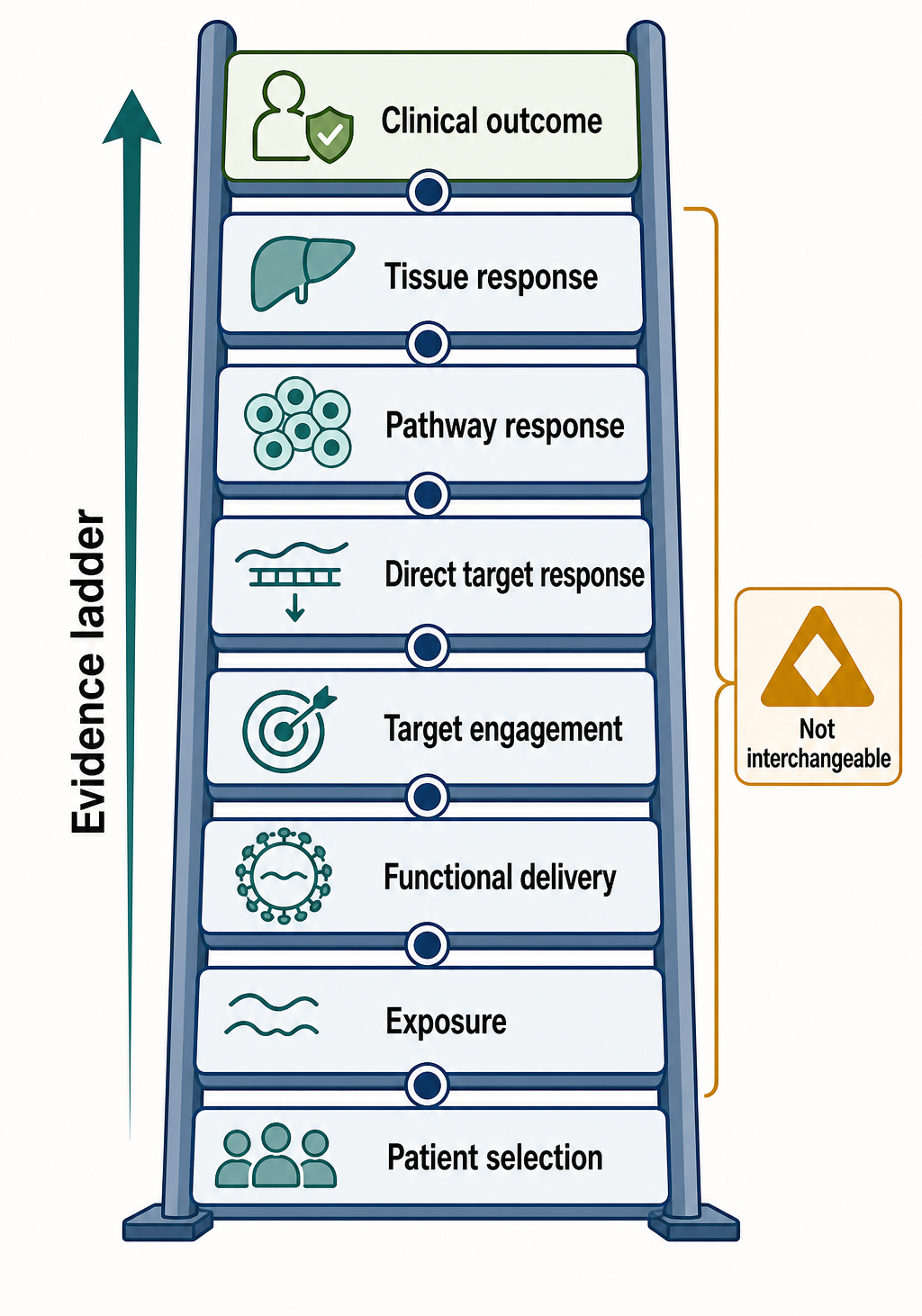
Figure 152.5. Biomarker Ladder from Exposure to Clinical Benefit. “A miRNA therapeutic trial should not treat exposure, target engagement, pathway response, and clinical benefit as interchangeable endpoints.”
Pharmacodynamic biomarkers report whether the drug changes the intended biology. For an anti-miR, biomarkers may include increased levels of validated target mRNAs or proteins, decreased availability of the mature miRNA, altered Argonaute occupancy, or pathway outputs downstream of derepressed targets. For a mimic, biomarkers may include seed-enriched downregulation of targets, repression of specific disease mediators, or restoration of a differentiation or stress-response signature. For a target-site blocker, the best proximal marker is protection of the specific transcript or protein without broad derepression of other targets. For a sponge or decoy, biomarkers must show durable inhibition in the cells expressing the decoy.
The difference between target engagement and pathway response is critical. A drug can bind the miRNA but fail to alter disease because the target network is buffered, redundant, or irrelevant in that patient. A mimic can repress predicted targets but fail to change tumor growth because the tumor has bypassed those targets. An anti-miR can derepress mRNAs but not proteins if translation, protein turnover, or feedback loops compensate. A pathway biomarker should therefore be closer to the disease mechanism than a single predicted target. In fibrosis, that might mean extracellular matrix production, myofibroblast activation, or tissue stiffness. In oncology, it might mean cell-cycle arrest, apoptosis, immune infiltration, or tumor burden, depending on mechanism.
Network-level interpretation should include directionality. If a miRNA is high in disease tissue, it may be pathogenic, compensatory, or merely a marker of a cell type that expands during disease. Inhibiting a compensatory miRNA can worsen disease even if the miRNA is elevated. If a miRNA is low in disease tissue, replacing it may help, but low expression can also be a consequence rather than a driver. Perturbation experiments are needed to distinguish these possibilities. Ideally, patient selection should use biomarkers that show both baseline dependence and expected response after dosing.
Responder analysis must also account for delivery heterogeneity. Nonresponse can mean the disease mechanism was wrong, the patient lacked the relevant miRNA state, the drug did not reach the tissue, endosomal escape was inefficient, the dose was limited by toxicity, or the biomarker sampled the wrong compartment. Clinical trials should avoid collapsing all nonresponse into one interpretation. Paired biopsies, circulating biomarkers, imaging, and tissue-specific pharmacokinetic measurements can help, but each adds feasibility and ethical constraints.
Multi-target drugs also require careful safety biomarkers. A narrow single-target drug can often be monitored through target pathway markers and organ function tests. A miRNA drug may need broader transcriptomic, proteomic, immune, liver, renal, coagulation, and cytokine monitoring, especially early in development. However, broad monitoring can generate ambiguous signals. A transient interferon-stimulated gene signature, for example, may reflect formulation immune sensing, tissue injury, or desired antitumor immunity. Interpretation should be anchored to timing, dose, comparator controls, and clinical signs.
Biomarker-guided trial design should be conservative in early programs. Enrich for patients whose disease tissue expresses the relevant miRNA state. Include a pharmacodynamic sampling plan that can fail the mechanism early. Use dose escalation that considers network expansion at higher exposure, not just conventional maximum tolerated dose. Prespecify which target or pathway changes count as evidence of mechanism. Avoid claiming that broad transcriptomic movement is beneficial unless it aligns with functional outcomes. When possible, include washout or reversibility information, because sustained miRNA network modulation can have delayed effects.
The most common misconception is that a miRNA signature is equivalent to a therapeutic target. A signature may be diagnostically useful while being therapeutically irrelevant. Conversely, a therapeutically important miRNA may not be the most differentially expressed miRNA in a disease dataset. Therapeutic prioritization depends on causality, druggability, delivery, safety, and measurable response. Differential expression is a starting point, not a decision rule.
This section connects to Chapter 125 and Chapter 130 for transcriptomic measurement, Chapter 142 for small RNA regulatory models, Chapter 106 for single-cell and spatial profiling, and Chapter 158 for clinical pharmacology. The practical lesson is that patient selection and pharmacodynamic biomarkers should be designed as tests of the disease mechanism, delivery hypothesis, and network-response model together.
152.6. Clinical History, Failures, Lessons Learned, and Next-Generation Design
The clinical history of miRNA therapeutics is best read as a series of mechanism tests rather than as a simple success-or-failure ledger. The field has shown that mature miRNAs can be inhibited in humans, that miRNA-like mimics can be delivered in clinical settings, and that pharmacodynamic effects can be measured. It has also shown that plausible disease associations do not guarantee clinical benefit, that immune and delivery liabilities can dominate, and that broad regulatory mechanisms require unusually careful dose and patient selection.
Miravirsen and other miR-122 inhibitor programs provided early proof that anti-miR pharmacology could be clinically meaningful. The disease logic was unusually strong: hepatitis C virus uses liver miR-122 to stabilize and replicate viral RNA, and hepatocytes are reachable by systemic oligonucleotide exposure. Viral load is a direct pharmacodynamic and clinical biomarker. This made the program a clear test of whether an endogenous miRNA could be inhibited for therapeutic effect. Later hepatitis C therapy moved rapidly because direct-acting antivirals achieved high cure rates, changing the clinical need. The lesson is not only scientific but strategic: a miRNA therapy can be overtaken if the indication becomes well served by safer, simpler, or more potent drugs.
RG-101 and related miR-122 inhibitor experience reinforced both promise and caution. Potent viral reductions supported the biological target, but safety concerns and evolving standards affected development. Host-factor targeting can offer a high barrier to viral resistance, but host-factor inhibition also carries host biology risk. A host miRNA is not a viral enzyme. The therapeutic window must account for the normal role of the miRNA, the duration of inhibition, the reversibility of the chemistry, and the availability of alternative treatments.
MRX34, a liposomal miR-34a mimic, became the cautionary example for replacement therapy. miR-34a connects to p53-regulated tumor-suppressive programs in many experimental systems, and restoring miR-34a was a compelling idea for cancer. The clinical program was halted after severe immune-related adverse events. Several lessons follow. First, a tumor-suppressive network in cell culture does not ensure systemic tolerability. Second, a mimic and its delivery system can activate immune pathways that are not captured by target prediction. Third, oncology patients may have disease burden, prior therapies, and immune states that magnify toxicity. Fourth, broad pathway restoration must be dosed and monitored as a multi-system intervention, not as a clean replacement of a missing molecule.
Other miRNA programs have explored fibrosis, dermatology, oncology, and inflammatory disease. For example, replacement of miR-29 family activity has been investigated because miR-29 represses multiple extracellular matrix genes and is often discussed in fibrosis biology. Local or tissue-targeted approaches may be better suited to some fibrotic indications than broad systemic exposure. Anti-miR strategies against oncogenic or inflammation-linked miRNAs have also been explored. Some candidates have advanced and then been discontinued, often for combinations of efficacy, safety, delivery, commercial, and strategic reasons. A rigorous chapter should not infer molecular failure from discontinuation alone unless the underlying data show lack of target engagement or harmful biology.
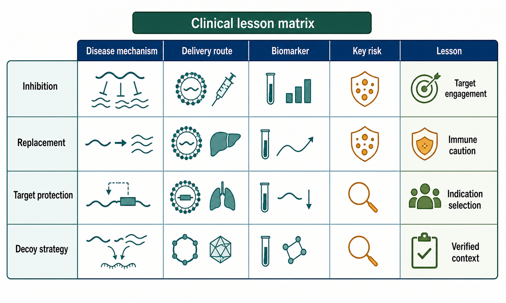
Figure 152.6. Clinical Lesson Matrix for miRNA Therapeutics. “Clinical history should be interpreted as mechanism evidence. Program discontinuation can reflect efficacy, safety, delivery, competition, or strategy, so each case requires verified context.”
Clinical failures have supplied recurring design lessons. The first is to start with a disease mechanism that is specific enough to be falsified. “This miRNA is dysregulated in cancer” is too broad. A stronger hypothesis states that a defined cancer subtype has reduced miR-X in tumor cells, that restoring miR-X represses a named vulnerability network, that delivery reaches those cells at a tolerable dose, and that a biomarker will show pathway correction. The second lesson is to build delivery and toxicology into the therapeutic hypothesis at the beginning. If the intended cells cannot be reached selectively, the miRNA may be the wrong modality for that indication.
The third lesson is to define the desirable target set. For a mimic, which targets are expected to drive benefit, which targets could drive toxicity, and which tissues express them? For an anti-miR, which derepressed targets are beneficial, and which could cause harm? For a target-site blocker, why is one interaction sufficient? For a decoy, why is durable inhibition acceptable? A target list should be more than predicted seed matches. It should include evidence tiers: direct physical binding, early expression response, protein response, phenotypic rescue, in vivo relevance, and human disease correlation.
The fourth lesson is to respect reversibility and duration. Some oligonucleotide chemistries produce long tissue half-lives. That can be useful for infrequent dosing but risky if an immune or metabolic phenotype emerges late. Viral-vector decoys may be even harder to reverse. Replacement therapies may need transient pulses or local administration rather than continuous systemic exposure. Next-generation programs should match duration to disease biology: acute antiviral intervention, chronic metabolic modulation, episodic fibrosis flare control, local tumor injection, or durable genetic-like modulation each implies a different risk profile.
The fifth lesson is that clinical endpoints must match mechanism. A miRNA therapy that changes a biomarker but not a patient-relevant endpoint may still be biologically active but clinically inadequate. Conversely, a modest clinical signal without mechanism biomarkers is hard to optimize. Early trials should therefore be designed to learn: confirm exposure, confirm target engagement, confirm pathway response, identify dose-limiting biology, and decide whether the indication remains worth pursuing.
Next-generation design is likely to be more selective and more integrated. Better small RNA sequencing, single-cell profiling, spatial transcriptomics, perturbational screens, and proteomics can identify patient subsets and disease-cell dependencies. Improved chemistry and formulation can reduce immune stimulation and increase tissue exposure. Conjugates, local delivery, biodegradable nanoparticles, and ligand-targeted systems may narrow distribution. Machine learning can help rank targets and predict off-target risks, but computational models must be trained and validated against experimental perturbations. The field should prefer smaller, stronger mechanism programs over broad claims that one miRNA will treat an entire disease category.
The open question is whether miRNA therapeutics will become a large general drug class or a specialized set of high-value interventions. The specialized route is currently more defensible: indications where a miRNA has strong causal evidence, the relevant tissue is reachable, the target network is measurable, and existing therapies leave room for improvement. Examples may include local tissue remodeling, liver host-factor biology, selected cancers with delivery solutions, and genetically or transcriptomically defined target-site mechanisms. Broader systemic replacement remains possible, but it must overcome the same immune, delivery, and network-toxicity barriers that shaped earlier programs.
This section connects to Chapter 153 and Chapter 154 for adjacent RNA therapeutic modalities and to Chapter 158 for regulatory interpretation. The clinical lesson is not that miRNAs are undruggable. It is that a miRNA drug must be developed as a network pharmacology product whose sequence, chemistry, delivery, indication, biomarkers, and safety plan are inseparable.
Recent Consensus
Recent Consensus, Open Questions, and Common Misconceptions
Current consensus can be summarized cautiously. First, mature miRNAs are druggable in principle. Anti-miRs can inhibit endogenous miRNAs, and mimics can create miRNA-like repression when delivered to cells. Second, target prediction alone is not sufficient for therapeutic design. Direct target evidence, disease-cell context, dose response, and in vivo pharmacology are required. Third, delivery and immune activation are central determinants of clinical viability. Fourth, multi-target regulation is both the rationale and the liability of miRNA therapeutics. Fifth, local, tissue-targeted, or biomarker-enriched indications are more plausible near-term paths than broad systemic correction of complex diseases.
Open Questions, Controversies, Deprecated Models, and Common Misconceptions
Open questions:
- How much miRNA activity must be changed to alter disease without destabilizing normal tissue?
- Which target changes mediate benefit, and which are passengers?
- How can programs measure Argonaute-loaded drug rather than total tissue oligonucleotide?
- Which chemical modifications best balance stability, potency, immune silence, and correct RISC behavior for mimics?
- When does target-directed miRNA degradation become a reliable therapeutic mechanism?
- How should regulators evaluate drugs whose intended mechanism is distributed across many transcripts?
Common misconceptions:
- “MiRNA differential expression is the same as therapeutic causality.” Differential expression is an association; therapeutic causality requires target engagement, disease mechanism, and benefit evidence.
- “All predicted seed targets are real targets in a patient tissue.” Target prediction must be filtered by expression, site accessibility, Argonaute loading, tissue context, and validation.
- “A mimic is safe because it matches an endogenous miRNA.” Concentration, compartment, strand selection, and delivery material can make the exposure nonphysiological.
- “An anti-miR is specific because it binds a short sequence.” Chemistry, partial complementarity, tissue accumulation, and normal miRNA functions all matter.
- “Multi-target pharmacology is inherently better than single-target pharmacology.” It is better only when the target set is the disease mechanism.
Deprecated or weakened claims:
- The idea that a miRNA sponge can be interpreted simply by counting binding sites without considering expression, accessibility, Argonaute occupancy, and degradation. Another weakened model is the use of unfiltered ceRNA logic to infer that any RNA with shared miRNA sites will meaningfully compete in vivo. Competition requires sufficient abundance, localization, binding affinity, and stoichiometric relationship to the miRNA pool. Therapeutic sponges should be evaluated with this quantitative caution.
- The most constructive framing is that miRNA therapeutics sit between oligonucleotide drugs and systems pharmacology. They use nucleic acid chemistry, but their intended effect is not just hybridization. They use endogenous regulatory networks, but their dose, distribution, and safety are drug-engineering problems. A mature program must be fluent in both languages.