Chapter 41. tRNA Modifications, Stress Responses, tRNA Fragments, and Disease Links
Scope Note
Transfer RNAs are not simple adapters made only of four canonical ribonucleotides. Mature tRNAs are compact, heavily modified RNA molecules whose chemical marks help establish folding, decoding accuracy, aminoacylation, stress responsiveness, and resistance or sensitivity to cleavage. This chapter explains how tRNA modifications are installed, why anticodon-loop and body modifications have different biological consequences, how stress can remodel the tRNA modification landscape, why tRNA fragments require cautious interpretation, and how modification defects connect to human disease without turning every altered mark into a proven causal biomarker.
Executive Summary
Mature tRNAs contain dozens of chemically distinct modifications. Some modifications are nearly universal, such as pseudouridine and ribose methylations at conserved structural positions; others are lineage-specific, organelle-specific, anticodon-specific, or condition-dependent. Modification enzymes, often called writers, include methyltransferases, pseudouridine synthases, deaminases, transglycosylases, sulfur-transfer systems, radical S-adenosylmethionine enzymes, and multicomponent mitochondrial enzymes. These enzymes do not decorate tRNAs randomly. They recognize sequence, local structure, precursor or mature processing state, subcellular compartment, and sometimes previous modifications. Because tRNA folding and decoding depend on the combined chemical state of a molecule, a single missing modification can have effects that range from nearly silent in one condition to lethal or disease-associated in another.
Anticodon-loop modifications have especially direct effects on translation. The anticodon loop is the region of tRNA that reads messenger RNA codons in the ribosomal A site, and modifications near positions 34 and 37 alter codon recognition, reading-frame maintenance, and decoding speed. Wobble-position 34 modifications can expand, restrict, or tune base-pairing with synonymous codons, whereas position 37 modifications stabilize the anticodon loop and help prevent frameshifting. The strongest mechanistic evidence comes from modification-enzyme genetics, ribosome profiling, reporter assays, biochemical decoding experiments, and mitochondrial disease genetics. Even so, the phrase “changes translation” is too broad: a mark may affect a subset of codons, a subset of tRNA isoacceptors, a subset of tissues, or a stress condition rather than the whole proteome uniformly.
Body modifications are marks outside the anticodon loop, including modifications in the D loop, variable loop, T loop, acceptor stem, and other structural regions of tRNA. These marks often shape tRNA folding, thermal stability, aminoacylation efficiency, processing, subcellular trafficking, and vulnerability to quality-control decay or endonucleolytic cleavage. In many systems, body hypomodification exposes tRNAs to surveillance pathways or makes tRNAs more easily fragmented under stress. Because body marks can affect global tRNA abundance and folding, their phenotypes can be indirect: altered protein synthesis may reflect loss of a stable tRNA pool rather than an anticodon-specific decoding defect.
Stress responses can reprogram tRNA modification in at least four separable ways. First, cells can change expression, localization, or activity of modification enzymes. Second, metabolic cofactor availability can change because many modification reactions depend on S-adenosylmethionine, folates, iron-sulfur clusters, sulfur donors, flavins, or mitochondrial metabolites. Third, stress can select for translation of codon-biased transcripts whose decoding depends on particular modified tRNAs. Fourth, stress can trigger tRNA cleavage, producing tRNA-derived fragments that may inhibit translation, interact with RNA-binding proteins, or behave as small regulatory RNAs in some contexts. These mechanisms are not equivalent and should not be merged into a single epitranscriptomic stress story.
tRNA-derived fragments are real and abundant in many datasets, but signaling claims vary in evidential strength. A tRNA fragment may be a regulated product of a specific nuclease, a decay intermediate, a stable protected fragment bound to a protein, a sequencing artifact enriched by modified bases, or a contaminant from abundant mature tRNAs. Strong functional claims require precise mapping of fragment ends, demonstration that the fragment is not merely a byproduct, evidence for a dose-relevant effector mechanism, and rescue or perturbation experiments that distinguish the fragment from its parent tRNA and from the modifying enzyme that may control both.
Disease links are strongest where genetics, biochemistry, and tissue physiology converge. Mitochondrial tRNA modification defects can impair mitochondrial translation and cause tissue-selective disease. Mutations or dysregulation of tRNA-modifying enzymes are associated with neurological disorders, developmental phenotypes, cancer, immune phenotypes, and pathogen fitness. Cancer studies frequently report altered expression of writers and altered tRNA fragment profiles, but expression correlation alone is not proof that a writer drives cancer through a specific tRNA modification. Detection limits are central: modifications can block reverse transcriptase, bias ligation, change fragmentation efficiency, confound small-RNA mapping, or escape antibody-based detection. Orthogonal validation by mass spectrometry, calibrated sequencing, genetics, and biochemical mechanism is the standard for high-confidence disease interpretation.
Concept Inventory
- Transfer RNA modification: a covalent chemical change to a tRNA nucleotide after transcription. Modification can alter base-pairing, stacking, hydration, local charge, metal binding, enzyme recognition, or nuclease sensitivity.
- Writer: an enzyme or enzyme complex that installs a modification. The term is convenient but should not imply a reversible regulatory circuit unless an eraser, reader, and dynamic stoichiometry have been shown.
- Anticodon-loop modification: a mark in or near the anticodon loop, especially at wobble position 34 or position 37, that commonly affects decoding.
- Body modification: a mark outside the anticodon loop that commonly affects tRNA folding, maturation, stability, localization, or quality control.
- Hypomodification: reduced or missing modification at one or more sites. Hypomodification can be caused by mutation, stress, cofactor limitation, altered tRNA processing, organelle dysfunction, or technical under-detection.
- tRNA fragment: a small RNA derived from tRNA. Major classes include tRNA halves produced by anticodon-loop cleavage, 5′ and 3′ tRNA-derived fragments, internal tRNA fragments, and fragments from precursor tRNA leader or trailer regions.
- Signaling claim: a claim that a tRNA fragment or modification actively changes a cellular pathway, rather than merely correlating with stress, disease, or tRNA turnover.
- Detection limit: a technical or interpretive constraint that prevents direct measurement of a modification, fragment, stoichiometry, or causal mechanism.
What to Know Before Reading This Chapter
tRNAs are transcribed as precursor molecules and then processed into mature adapters that carry amino acids to the ribosome. A mature cytosolic tRNA is typically about 70-90 nucleotides long, folds into a cloverleaf secondary structure, and packs into an L-shaped tertiary structure. The acceptor stem at one end is aminoacylated by an aminoacyl-tRNA synthetase. The anticodon loop at the other end pairs with an mRNA codon during translation. Chapter 39 covers transcription, processing, aminoacylation, and quality control; this chapter focuses on chemical maturation and its consequences.
Numbering matters because tRNA biologists use conserved positional notation. Position 34 is the wobble nucleotide, the first anticodon nucleotide in the tRNA but the nucleotide that pairs with the third base of the mRNA codon. Position 37 is immediately 3′ of the anticodon and is not itself paired with the codon, but it stacks near the anticodon and helps preserve the reading frame. Positions in the tRNA body are interpreted relative to the canonical tRNA cloverleaf, although mitochondrial and unusual tRNAs may deviate from the canonical fold.
The reader should also separate four levels of explanation. Chemistry describes what bond or group is added. Enzymology describes which enzyme installs the mark and how substrate specificity is achieved. Molecular function describes how the modified tRNA behaves in folding, decoding, aminoacylation, or cleavage. Cellular phenotype describes how altered tRNA chemistry changes growth, stress survival, development, or disease. Many weak claims skip from enzyme expression to disease phenotype without measuring the modified tRNA or the direct molecular consequence.
41.1. tRNA modification chemistry and writers
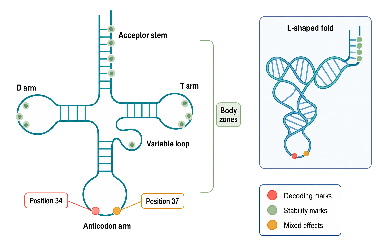
Figure 41.1. Chemical and Functional Map of a Mature tRNA. A canonical tRNA cloverleaf showing the acceptor stem, D arm, anticodon arm, variable loop, and T arm, with an inset of the L-shaped tertiary structure. Position 34 (the wobble nucleotide) and position 37 (the anticodon-adjacent stabilizing position) are highlighted in the anticodon loop, while body modification zones in the D loop, T loop, variable loop, and acceptor stem are marked separately. Color coding distinguishes modifications associated primarily with decoding, those that primarily support structural stability, and marks with mixed or condition-dependent effects. The acceptor stem terminates in separate 5′ and 3′ ends rather than a hairpin-like closure, with the unpaired 3′ acceptor end extending slightly beyond the 5′ end in both the cloverleaf and tertiary-fold inset.
tRNA modification chemistry spans simple methylation, base isomerization, base exchange, deamination, thiolation, reduction, complex side-chain construction, and incorporation of metabolite-derived groups. The modified nucleotide is part of the RNA chain; the modification usually changes a base or ribose after transcription rather than replacing the phosphodiester backbone. Common examples include pseudouridine, in which uridine is isomerized so that the base attaches to ribose through a carbon-carbon bond; 2′-O-methylation, in which the ribose hydroxyl is methylated; N1-methyladenosine and N7-methylguanosine, in which methyl groups change base charge or hydrogen-bonding behavior; inosine, produced by adenosine deamination; thiolated uridines, in which sulfur substitutes for oxygen; and hypermodified nucleosides such as queuosine and wybutosine that are assembled through multistep pathways.
Table 41.1. Representative tRNA Modification Chemistries, Writers, and Evidence Caveats. Selected modifications illustrating the chemical diversity of tRNA maturation, the writer systems responsible, and the interpretive cautions relevant to each class.
| Modification | Common position or region | Writer class | Main molecular effect | Evidence caveat |
|---|---|---|---|---|
| Pseudouridine (Ψ) | Multiple body positions; D loop, T loop | Pseudouridine synthase | Structural stabilization; improved stacking and hydration of tRNA fold | Sequencing detection requires CMC chemical treatment or calibrated nanopore models |
| 2′-O-methylation | Variable body and structural positions | 2′-O-methyltransferase | Ribose protection; reduced nuclease sensitivity; altered backbone flexibility | Causes reverse-transcription pausing; position requires modification-aware sequencing |
| Inosine (I34) | Wobble position 34 | Adenosine deaminase acting on tRNA (ADAT) | Expanded decoding of synonymous codons ending in U, C, or A | ADAT edits multiple isoacceptors; decoding effect is codon-pair and tRNA species specific |
| Thiolated uridine (s²U or ms²U34) | Wobble position 34 | Sulfur-relay system (URM1 pathway in eukaryotes; TusA–TusE in bacteria) | Wobble pairing restriction; metabolic coupling to sulfur and oxidative stress | Stoichiometry varies with nutrient and stress state; LC-MS/MS required for reliable quantification |
| Queuosine (Q34) | Wobble position 34 in Tyr, His, Asp, Asn tRNAs | Queuine transglycosylase (TGT) | Decoding modulation; possible influence on frameshifting and translational accuracy | Q is salvaged from diet or microbiota; tissue and organism levels vary and are not always experimentally controlled |
| Wybutosine (yW) or related mark | Position 37, adjacent to anticodon 3′ side | Multi-enzyme pathway (TYW1–TYW5 in eukaryotes) | Anticodon loop stabilization; frameshift suppression in phenylalanine tRNA | Complex multistep biosynthesis; stoichiometry and pathway completeness differ by species |
| N1-methyladenosine (m1A58) | Position 58 in T loop | TRMT6–TRMT61A methyltransferase complex | tRNA structural stabilization; influences tRNA fragment modification state | m1A at some positions blocks reverse transcription; fragment studies require end-resolved modification validation |
| Mitochondrial taurine-modified uridine (τm5U or τm5s²U) | Wobble position 34 of mitochondrial tRNAs | MTO1–GTPBP3–MTU1 enzyme system | Decoding of mitochondrial codons; structural support in noncanonical mt-tRNA folds | Defects linked to mitochondrial disease; quantifying stoichiometry in isolated mitochondria is technically demanding |
Table 41.2. tRNA Fragment Classes and Interpretation Standards. Major classes of tRNA-derived fragments, their sequence features, probable origins, common detection methods, and the minimum evidence expected before ascribing a regulatory function.
| Fragment class | Defining sequence feature | Likely biogenesis route | Common assay | Minimum evidence for function | Common artifact |
|---|---|---|---|---|---|
| 5′ tRNA half | Starts at mature 5′ end; 3′ end falls in or near anticodon loop | Stress-activated endonuclease (e.g., angiogenin in mammals) | Small-RNA sequencing; northern blot with size marker | Precisely mapped 3′ end; stress-correlated abundance; nuclease identified; effector binding shown | Abundant class; easily confused with shorter 5′-tRFs or degradation intermediates |
| 3′ tRNA half | 5′ end in anticodon loop; terminates at mature 3′ CCA | Stress-activated endonuclease paired with 5′-half production | Small-RNA sequencing; northern blot | Precisely mapped 5′ cleavage site; physiological concentration; effector and rescue evidence | CCA tail ambiguity; 3′ ends subject to exonucleolytic trimming |
| 5′ tRF | Short fragment from 5′ mature end; 3′ end outside anticodon loop | Regulated or opportunistic nuclease; processing | Small-RNA sequencing with end mapping | 3′ end distinct from tRNA-half cleavage; unique tRNA gene assignment; defined effector | Short length increases multi-mapping across near-identical tRNA gene families |
| 3′ CCA tRF | 3′ fragment retaining CCA end, derived from mature tRNA body | Exonucleolytic or endonucleolytic processing of mature tRNA | Small-RNA sequencing with CCA-aware alignment | CCA terminus confirmed; parent tRNA identified; not attributable to aminoacyl-tRNA decay | Terminal CCA contamination from tRNA turnover; often conflated with 3′ trailer fragments |
| 3′ trailer tRF | Sequence 3′ of CCA in precursor tRNA; no mature end | Pre-tRNA processing byproduct | Small-RNA sequencing with precursor-aware reference | Confirmed precursor origin; 5′ end mapped past CCA; distinct from mature fragments | Frequently mismapped as mature-tRNA-derived if reference excludes precursor sequences |
| Internal tRF | Internal tRNA segment lacking either mature 5′ or 3′ end | Endonucleolytic cleavage or degradation | Small-RNA sequencing; end-specific validation | Both ends precisely mapped; not attributable to multi-mapping, adapter dimers, or ligation artifacts | Rarest class; most vulnerable to misassignment; requires orthogonal validation |
Writers recognize tRNAs by combining sequence and structure. A modification enzyme may read a local base, a stem-loop shape, a distance from the anticodon, the folded L shape, or the presence of a precursor feature such as an intron or a 3′ trailer. Some enzymes act on many tRNAs at a conserved position. Others are restricted to a tRNA family, an anticodon, an organelle, or a lineage. The term writer is therefore a shorthand for a large biochemical category, not a claim that all modifications are regulated in the same manner. A methyltransferase that uses S-adenosylmethionine, a pseudouridine synthase that flips a uridine into an active site, and a sulfur-relay system that requires protein persulfides or iron-sulfur chemistry are different mechanistic systems even if all install tRNA marks.
Many marks are installed hierarchically. A first modification can stabilize the local fold needed for a second enzyme. Conversely, a processing defect can prevent modification because the writer recognizes a mature end, a correctly spliced anticodon loop, or a folded tertiary surface. This dependency explains why mutation of a processing factor can mimic a modification defect and why loss of a writer can generate a mixed population of mature, unstable, misfolded, and fragmented tRNAs. When a sequencing dataset reports that a modification is lower in a stress condition, the immediate possibilities include lower writer abundance, lower writer activity, loss of a cofactor, altered tRNA processing, altered tRNA stability, changed tRNA isoacceptor expression, or altered detectability. A strong interpretation must distinguish these alternatives.
Mitochondrial tRNA writers illustrate why compartment matters. Human mitochondrial tRNAs are encoded by the mitochondrial genome, have unusual structures compared with many cytosolic tRNAs, and support translation of a small but essential set of oxidative phosphorylation proteins. Their modifications include taurine-containing uridine derivatives and methylations that are important for decoding and structural stability. Because mitochondrial translation defects can reduce respiratory-chain function, mitochondrial tRNA modification defects can have organism-level consequences, especially in high-energy tissues. However, mitochondrial phenotypes often reflect combined effects of tRNA mutation, RNA processing, aminoacylation, ribosome interaction, and organelle stress rather than a single isolated chemical mark.
41.2. Anticodon-loop modifications and decoding
The anticodon loop is the most direct interface between tRNA chemistry and the genetic code. During elongation, an aminoacyl-tRNA enters the ribosomal A site as part of a ternary complex with elongation factor and GTP. The anticodon must pair with the mRNA codon quickly and accurately enough to support translation, yet flexibly enough to allow synonymous codons to be read by a limited tRNA set. Modifications around the anticodon tune this balance.
The wobble base at position 34 is central because it pairs with the third codon base. Inosine at position 34 can pair with more than one codon-ending base, expanding decoding capacity. Modified uridines at position 34 can restrict or bias wobble pairing, improve reading of A- or G-ending codons, or prevent inappropriate pairing. Thiolation and side-chain modifications at wobble uridines are common examples in bacteria and eukaryotes. The exact effect depends on the tRNA species and codon box, so one should not say simply that wobble modification “increases translation.” It may increase accuracy for one codon set, reduce misreading of another, alter ribosome dwell time, or change translation of a codon-biased transcript subset.
Position 37, adjacent to the anticodon on the 3′ side, often stabilizes the anticodon loop and helps maintain reading frame. Bulky or charged modifications at position 37 can improve stacking and suppress frameshifting, especially at codon contexts that are prone to slippage. Wybutosine derivatives in eukaryotic phenylalanine tRNA and threonylcarbamoyladenosine-related modifications are well-known examples of position-37 chemistry that influences decoding fidelity. Loss of a position-37 mark can manifest as codon-specific translation defects, frameshift susceptibility, proteostasis stress, or growth defects, depending on the system and assay.
Recent primary studies show how anticodon-loop marks can link codon bias to cell-state programs. For example, m3C32 modification has been reported to affect serine codon-biased mRNA translation, cell-cycle behavior, and DNA-damage response in a mammalian context. Such studies are valuable because they move beyond measuring a mark and test translation output. They also require careful interpretation. A codon-biased translation effect can arise from modified-tRNA decoding, but it can also be influenced by tRNA abundance, aminoacylation, ribosome pausing, mRNA stability, or stress pathways that change translation initiation. The strongest causal chain measures the modification, perturbs the writer, rescues the enzymatic activity or tRNA substrate when possible, measures codon-specific translation, and controls for changes in tRNA abundance.
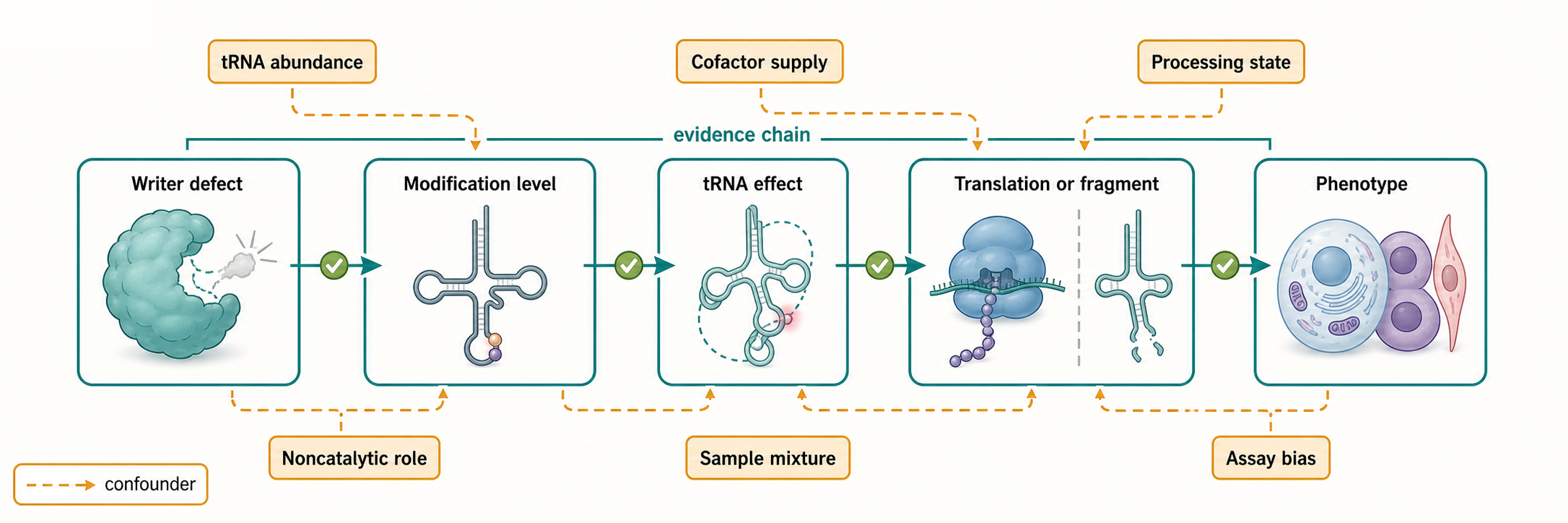
Figure 41.2. From Writer Defect to Phenotype: The Causal Chain. A five-step flow diagram tracing the causal chain from a writer enzyme defect to an observable phenotype, with steps covering writer activity change, modification stoichiometry, tRNA molecular effect, translation or fragment profile change, and cellular or disease phenotype. Side arrows mark common confounders—changes in tRNA abundance, metabolic cofactor availability, tRNA processing, noncatalytic writer functions, sample composition, and assay bias—that must be distinguished before assigning a phenotype to a specific chemical modification.
Mitochondrial anticodon modifications are clinically important because mitochondrial decoding uses a compact tRNA set and a genetic code that differs from the standard nuclear code. Taurine-containing wobble modifications in mitochondrial tRNAs help decode codons correctly in the organelle. Defective installation or recognition of these marks can impair mitochondrial protein synthesis and contribute to disease phenotypes. The lesson is not that all anticodon modification defects cause disease, but that the ribosome, codon table, tRNA structure, and tissue energy demand determine whether a chemical defect becomes a clinical phenotype.
41.3. Body modifications and tRNA stability
Body modifications are marks outside the anticodon loop. They occur in the D loop, D stem, variable loop, T loop, acceptor stem, and other structural regions. These marks usually do not contact an mRNA codon directly. Instead, they help the tRNA fold into a stable tertiary structure, preserve local geometry for aminoacylation or ribosome binding, prevent inappropriate base-pairing, or influence recognition by processing and quality-control enzymes.
The distinction between anticodon-loop and body modifications is useful but not absolute. A body mark can alter decoding indirectly by changing the abundance of a tRNA available to the ribosome. A body mark can also affect fragmentation, and a fragment derived from a body-hypomodified tRNA can influence stress phenotypes independently of translation. Conversely, an anticodon mark can affect tRNA stability if the anticodon loop becomes more nuclease-sensitive. The categories identify the usual first-order mechanism, not a rigid functional boundary.
Body hypomodification can expose tRNAs to decay. In eukaryotes, unstable hypomodified tRNAs may be degraded by rapid tRNA decay or related surveillance pathways, especially when high temperature, oxidative stress, or processing defects destabilize folding. In bacteria, modification defects can influence tRNA quality control, translation stress, and pathogen adaptation. A missing methyl group or pseudouridine does not have to change a codon interaction to matter; by shifting a tRNA folding equilibrium, it can reduce the mature tRNA pool. The phenotype then resembles a shortage of a specific charged tRNA rather than a decoding-error phenotype.
The body-stability principle also explains why tRNA disease genetics can be difficult. A variant in a tRNA gene can alter a base that is normally modified, a base that a writer uses for recognition, or a stem pair that establishes the modification substrate. If the mature tRNA is low, the causal defect may be reduced aminoacylation, reduced modification, misfolding, increased decay, or a combination. Separating these mechanisms requires measuring precursor processing, mature tRNA abundance, aminoacylation state, modification stoichiometry, folding or thermal stability, and translation output. A single northern blot or small-RNA sequencing profile rarely resolves the mechanism alone.
Body modifications are also linked to tRNA fragment production. Some modifications protect tRNAs from cleavage; others influence which fragments accumulate after cleavage. Methylation can block or slow some nucleases, and structural stabilization can make an anticodon loop less accessible. In stress conditions, cleavage enzymes such as angiogenin in mammalian systems can produce tRNA halves, but the observed fragment profile depends on parent tRNA abundance, modification state, nuclease activity, RNA-binding proteins, and library preparation. Therefore, a change in fragment abundance may report stress-dependent cleavage, altered parent tRNA abundance, altered modification, or altered sequencing recovery.
41.4. Stress responses and reprogramming
Stress reprogramming means that the tRNA modification landscape, tRNA abundance, tRNA charging, or tRNA fragmentation pattern changes in response to environmental or cellular stress and contributes to altered gene expression or survival. Stresses include oxidative stress, nutrient limitation, heat, infection, antibiotics, unfolded protein stress, DNA damage, inflammatory signaling, and drug exposure. A tRNA modification response can be adaptive, maladaptive, or simply a consequence of damaged metabolism.
One mechanism is regulated writer activity. A cell can increase or decrease the abundance, localization, post-translational modification, or substrate access of a tRNA-modifying enzyme. In bacteria, tRNA modifications have been linked to virulence, antibiotic response, and adaptation to host-associated stresses. In parasites, tRNA modification reprogramming has been linked to drug resistance; a reported example is tRNA modification remodeling in artemisinin-resistant Plasmodium falciparum. These examples show that tRNA chemistry can sit inside stress-response networks rather than only serving as housekeeping maturation.
A second mechanism is metabolic coupling. Many tRNA modification reactions depend on metabolites that change during stress. S-adenosylmethionine supplies methyl groups; folate-linked one-carbon metabolism supports some methylation chemistry; sulfur-relay systems depend on sulfur trafficking; iron-sulfur enzymes are sensitive to oxidative and metabolic state; mitochondrial marks depend on organelle metabolism and import of nuclear-encoded enzymes. Thus, stress can reduce modification without directly regulating a writer gene. In this case, the tRNA modification profile is both a molecular output of metabolism and a potential cause of translation changes.
A third mechanism is selective translation. If a stress-response transcript set is enriched for codons that depend on a specific modified tRNA, changing that modification can favor or disfavor translation of those transcripts. This idea is attractive because it provides a bridge from chemistry to gene-expression programs. It also creates a common overinterpretation: codon enrichment plus writer perturbation is not enough to prove selective translation. A causal claim should show that codon-biased mRNAs change translation efficiency, that the relevant tRNA modification changes, and that changes are not explained only by altered mRNA abundance, translation initiation, cell-cycle state, or cell viability. Ribosome profiling, reporter recoding, tRNA modification measurement, and rescue experiments are complementary.
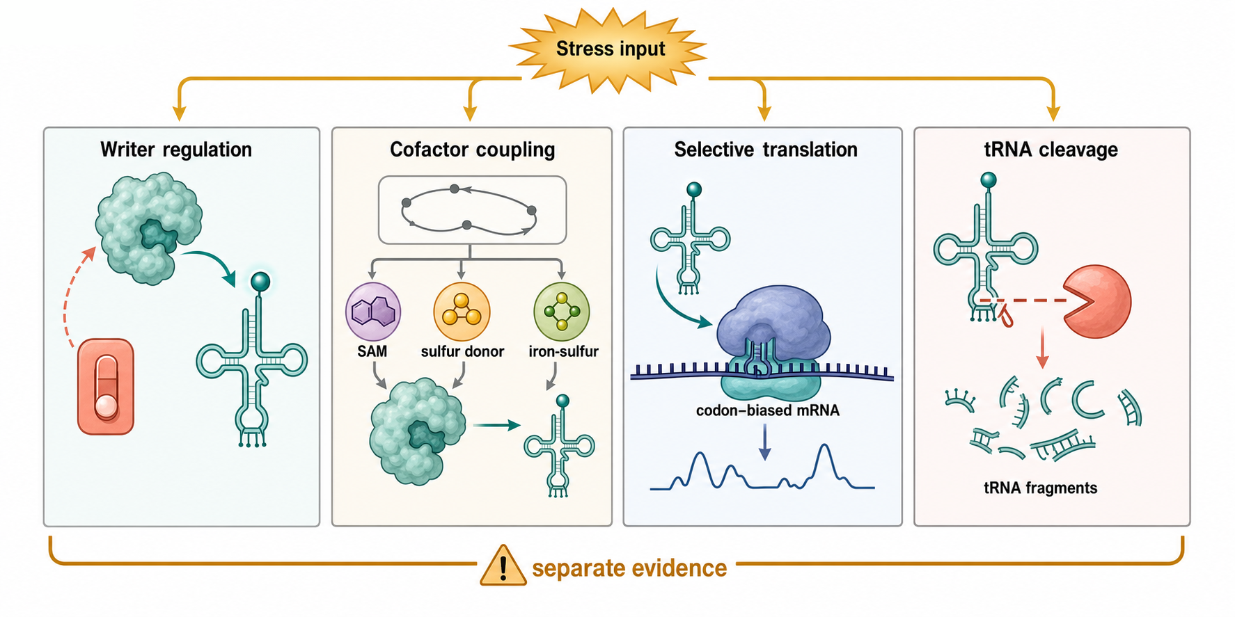
Figure 41.3. Four Routes of Stress Reprogramming. Four parallel panels illustrate distinct mechanisms by which stress can remodel tRNA-related gene expression: regulated writer activity or localization; metabolic coupling through cofactors such as S-adenosylmethionine, iron–sulfur clusters, and sulfur donors; selective translation of codon-biased stress-response transcripts that depend on specific modified tRNAs; and nuclease-driven tRNA cleavage that generates tRNA halves or shorter fragments. The figure emphasizes that these mechanisms are separable and require independent experimental evidence rather than being merged into a single epitranscriptomic stress narrative.
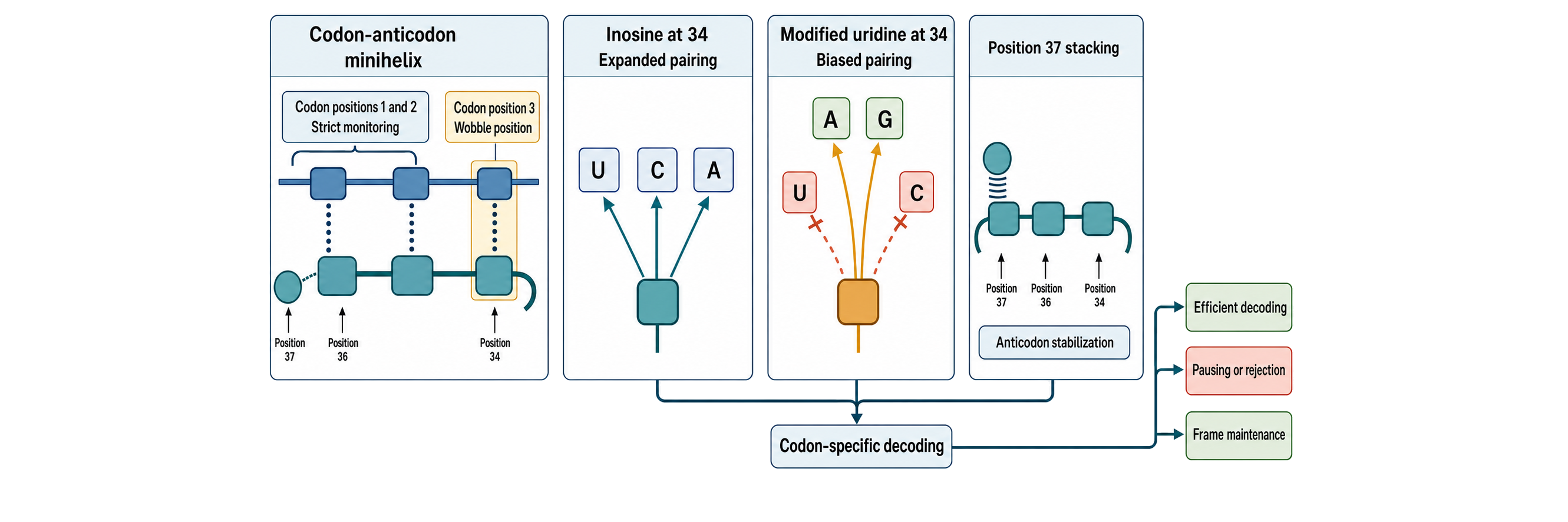
Figure 41.4. How Anticodon-Loop Modifications Expand, Restrict, and Stabilize Decoding. A codon–anticodon minihelix should contrast close monitoring at codon positions 1 and 2 with the chemically constrained wobble interaction between codon position 3 and tRNA position 34. A matched comparison should show inosine-34 expanding a permitted codon-ending set and a selected modified-uridine-34 chemistry narrowing or biasing the set, while avoiding the false rule that all U34 modifications act identically. A position-37 stacking module should show stabilization adjacent to the anticodon and suppression of slippage, followed by codon-specific outcomes such as efficient decoding, pausing or rejection, and frame maintenance.
A fourth mechanism is stress-induced tRNA cleavage. Under some stresses, mature tRNAs are cleaved in or near the anticodon loop to produce tRNA halves. Other cleavage events generate shorter tRNA-derived fragments. These fragments may inhibit translation initiation, displace RNA-binding proteins, enter Argonaute-associated pathways, or act through other protein interactions. However, cleavage is not automatically signaling. It can also be a damage response, a decay route, or a protective packaging response. A fragment-mediated stress model is strongest when a specific nuclease is identified, cleavage sites are mapped, fragment abundance is quantitatively linked to stress intensity, synthetic or endogenous fragments reproduce the phenotype at physiological concentrations, and blocking the fragment reverses the phenotype without broadly disrupting tRNA maturation.
Box 41.1. Checklist for tRNA Fragment Signaling Claims
Before accepting a functional role for a tRNA-derived fragment, the following questions should be answered:
- Are both the 5′ and 3′ ends of the fragment precisely mapped?
- Is the parent tRNA gene assignment unique, or is mapping ambiguity explicitly reported?
- Is the modification state of the fragment measured, not assumed from the parent tRNA?
- Is fragment abundance estimated to be at a physiological or otherwise relevant concentration?
- Is a specific nuclease or biogenesis pathway identified for this fragment?
- Is a direct effector—a protein, ribosome, or RNA target—known and tested?
- Does experimental perturbation specifically alter the fragment without broadly disrupting parent tRNA maturation, writer enzyme activity, or tRNA abundance?
- Is there a rescue experiment using a fragment-specific intervention, such as a blocking oligonucleotide or synthetic fragment at endogenous concentration?
The neurodevelopmental stress example from NSUN2 illustrates this logic. Aberrant tRNA methylation has been linked to cellular stress and neurodevelopmental disorder phenotypes, with hypomethylated tRNAs becoming more susceptible to cleavage in some models. The key conceptual point is that a modification enzyme can influence disease by preserving tRNA integrity, not only by changing codon decoding. A writer defect may increase tRNA fragmentation, induce stress pathways, and alter protein synthesis together. Causal dissection requires separating the enzyme’s catalytic role from any noncatalytic protein functions and from secondary stress responses.
41.5. tRNA fragments and signaling claims
tRNA-derived fragments are small RNAs whose sequences map to tRNAs or tRNA precursors. Major classes include 5′ tRNA halves, 3′ tRNA halves, shorter 5′ tRFs, 3′ CCA-containing fragments from mature tRNAs, 3′ trailer-derived fragments from precursors, and internal tRNA fragments. The names tRF and tiRNA are used inconsistently across the literature; in this chapter, tRNA fragment is the broad term, tRF is used for shorter mapped fragments, and tRNA half is used for longer cleavage products that commonly arise from anticodon-loop cleavage.
The first interpretive challenge is mapping. tRNA genes are repetitive, mature tRNAs contain modifications that impede reverse transcription, and many tRNAs share near-identical sequences. A small RNA read may map to multiple tRNA genes or isodecoders. A fragment with a CCA tail may come from a mature tRNA, whereas a 3′ trailer fragment points to precursor processing. But ligation bias, end chemistry, and reverse-transcription stops can distort apparent abundance. Demethylation treatments, specialized aligners, end-aware analysis, spike-ins, and orthogonal northern blot or mass-spectrometric evidence can improve confidence, but no single small-RNA pipeline makes all tRNA fragment calls reliable.
The second challenge is function. Some tRNA fragments are abundant, regulated, and associated with proteins. Some have reported roles in translation inhibition, transposon control, inheritance, immune signaling, apoptosis, cancer progression, or stress granule dynamics. Other fragments are probably decay products or protected remnants of abundant tRNAs. A functional fragment should meet higher standards than detectability. It should have defined ends, reproducible abundance, a plausible concentration, a known or testable effector, and perturbations that are specific to the fragment. If knocking down a tRNA-modifying enzyme changes both parent tRNA maturation and fragment abundance, the phenotype cannot automatically be assigned to the fragment.
The TRMT6/61A example shows both promise and complexity. TRMT6/61A installs m1A58 in tRNAs, and a study reported that TRMT6/61A-dependent methylation of tRNA-derived fragments regulates gene-silencing activity and the unfolded protein response in bladder cancer. This type of claim is mechanistically interesting because a modification on a fragment may alter small-RNA behavior. The evidence standard is correspondingly demanding: the modified fragment must be detected, the modification site and stoichiometry must be credible, the fragment must be distinguished from the parent tRNA, and the downstream gene-silencing activity must be separated from global effects of TRMT6/61A on tRNA maturation and translation.
Claims about tRNA fragments in immunity and mucosal biology should also be treated as graded evidence rather than as a single established pathway. Reviews summarize associations between tRNAs, tRFs, tiRNAs, and mucosal immune states, but association is not enough to establish a receptor, ligand, or regulatory circuit. A fragment may be an extracellular biomarker, a cell-intrinsic stress product, a passenger in a vesicle, or a direct immunomodulatory RNA. Direct immunological claims need dose-controlled exposure, contamination controls, end chemistry characterization, and evidence that innate immune receptors or RNA-binding proteins sense the fragment itself.
The safest summary is that tRNA fragments are an important and heterogeneous class of RNAs, not a single regulatory system. Some fragments are likely functional in specific contexts; many remain candidates. The field has moved beyond dismissing all tRNA fragments as degradation products, but it should also avoid treating every stress-enriched tRNA-derived read as a signaling RNA. The most useful chapter-level habit is to ask: Which parent tRNA? Which exact ends? Which modification state? Which nuclease or processing pathway? Which binding partner? Which phenotype? Which rescue?
41.6. Disease links and detection limits
Disease links for tRNA modifications fall into several categories. First, inherited mutations in tRNA genes or tRNA-modifying enzymes can cause loss of a specific modification, impaired tRNA stability or decoding, and tissue-selective pathology. Mitochondrial diseases are a major example because mitochondrial translation is sensitive to tRNA structure and decoding chemistry, and because energy-demanding tissues such as brain, muscle, and endocrine tissues are vulnerable to respiratory-chain dysfunction.
Second, writer dysregulation can contribute to cancer phenotypes. Cancer cells often change translation demand, stress tolerance, metabolism, and proliferation, all of which intersect with tRNA abundance and modification. Reviews describe altered tRNA modifications and writer expression in human cancers. Some primary studies connect specific marks or enzymes to cell-cycle progression, DNA-damage response, unfolded protein response, or gene-silencing activity. The evidence is strongest when the study measures the relevant tRNA modification, links it to translation or fragment activity, tests catalytic dependence, and validates disease relevance in models beyond a single cell line. It is weaker when the study only reports writer overexpression, prognostic correlation, or bulk RNA-seq association.
Third, neurological and developmental disorders can arise from tRNA modification defects. Neural tissue may be sensitive because neurons are long-lived, metabolically active, and dependent on precise translation and stress management. The NSUN2-linked work on aberrant tRNA methylation and neurodevelopmental phenotypes is a landmark example in the provided bibliography. The broader lesson is that tRNA modification defects can produce disease through proteostasis stress, altered translation, increased tRNA cleavage, or developmental vulnerability. The exact mechanism must be established for each gene and tissue.
Fourth, infectious disease and pathogen biology involve tRNA modifications. Bacteria and parasites use tRNA modification systems to adapt translation, stress tolerance, and virulence programs. Host-pathogen studies must distinguish pathogen tRNA modifications from host RNA responses and from antibiotic or drug-induced damage. A modification enzyme can be a possible antimicrobial target if it is essential or virulence-linked and sufficiently different from host enzymes, but target validation requires biochemical selectivity, organismal fitness evidence, resistance analysis, and toxicity assessment.
Detection limits are not an appendix to disease interpretation; they are part of the mechanism. tRNA modifications can block reverse transcription, causing apparent dropouts or truncations. Some library protocols underrecover mature tRNAs because structured and modified tRNAs resist adapter ligation and reverse transcription. Some modifications are isobaric or chemically similar and require careful mass-spectrometric separation. Antibody-based methods developed for mRNA modifications may cross-react, miss low-stoichiometry marks, or fail on highly structured tRNAs. Direct RNA nanopore sequencing can measure full-length tRNA features in promising ways, but signal interpretation depends on training data, modification mixtures, tRNA structure, basecaller assumptions, and validation against orthogonal measurements.
Disease biomarker claims for tRNA fragments face additional hazards. Blood, serum, urine, and extracellular vesicle samples contain RNases, abundant tRNAs, hemolysis artifacts, platelet or immune-cell contributions, and extraction biases. A fragment that discriminates cases and controls may still be a sample-processing marker, inflammation marker, cell-composition marker, or parent-tRNA abundance marker rather than a disease-specific regulatory molecule. Biomarker development therefore requires preanalytical controls, independent cohorts, absolute or calibrated quantification, fragment-end validation, and comparison with existing clinical variables.
Some references currently listed in the detection block of references.md are not tRNA-specific, including general RNA detection, circRNA, coronavirus detection, and glycoRNA-related items. They may be useful for broader detection-principle comparisons but should not be used as direct support for tRNA modification disease claims without additional verification. The current local bibliography does include direct tRNA modification and detection anchors for this first draft, including Suzuki 2021, Lucas et al. 2024, and Behrens et al. 2021; later final-release curation should add more method-comparison depth for tRNA-seq library bias, modification-aware alignment, LC-MS/MS quantification standards, and clinical cohort validation.
Experimental Foundations and Evidence
The most direct evidence for tRNA modification identity and abundance comes from mass spectrometry. Nucleoside-level liquid chromatography-mass spectrometry can identify and quantify modified nucleosides after RNA digestion, but it loses positional information unless combined with sequence-specific purification or fragment mapping. Oligonucleotide mass spectrometry can retain sequence context but is technically more demanding. Mass spectrometry also depends on standards, digestion completeness, chromatographic separation, and careful assignment of isobaric species.
Sequencing-based methods provide position and transcript context but are indirect unless the modification creates a predictable signature. Some marks cause reverse-transcription stops, misincorporations, deletions, or altered nanopore current. Other marks are silent under a given protocol. Enzymatic pretreatments can remove or convert certain marks, improving readthrough or generating diagnostic changes. However, tRNAs are difficult sequencing targets because they are short, structured, repetitive, heavily modified, and sometimes aminoacylated or end-modified. A high-confidence sequencing claim usually requires spike-ins, replicate consistency, modification-enzyme perturbation, and orthogonal validation.
Genetics links writers to phenotypes. Deleting, knocking down, overexpressing, or mutating a writer can show that an enzyme matters for growth, stress survival, translation, or disease behavior. But writer perturbation is not identical to modification perturbation. The enzyme may have several tRNA substrates, non-tRNA substrates, scaffolding roles, or indirect effects on metabolism. A catalytic-dead rescue, substrate-specific rescue, or direct restoration of a modification strengthens the causal interpretation.
Translation assays are essential for decoding claims. Ribosome profiling can show codon-specific ribosome occupancy or changes in translation efficiency, but ribosome occupancy can reflect initiation, elongation, quality control, mRNA structure, or decay. Reporter assays with codon substitutions can test whether a codon class is sensitive to a modification pathway. In vitro decoding assays can isolate the ribosome, tRNA, mRNA, and elongation factors, providing mechanistic clarity. The strongest studies combine cellular and biochemical approaches rather than relying on one method.
Fragment studies require end-specific and modification-aware validation. Northern blotting can validate fragment size and stress induction but may not resolve exact ends. Small-RNA sequencing can map ends but is biased by end chemistry and modification. Synthetic fragment transfection can test sufficiency but risks nonphysiological concentration, end chemistry mismatch, or innate immune activation. Endogenous perturbation, such as nuclease mutation or targeted blocking oligonucleotides, can test necessity but may disturb parent tRNA biology. Interpretation improves when multiple perturbations converge.
Biological Contexts Across Systems
In bacteria, tRNA modifications support rapid translation and environmental adaptation. Bacterial pathogens encounter nutrient limitation, oxidative stress, host immune pressures, antibiotics, and niche-specific temperatures. Modification defects can change stress survival or virulence traits, but the specific effect depends on the organism and modification pathway. A writer that is important in one bacterium may be redundant, conditionally required, or absent in another. Comparative bacterial interpretation should therefore specify species, growth condition, and host context.
In eukaryotic cytosol, tRNA modifications integrate translation, growth signaling, stress pathways, and development. Yeast models have been especially useful for identifying modification enzymes, synthetic genetic interactions, and tRNA decay pathways. Mammalian systems add tissue specificity, disease genetics, and complex stress signaling. A mammalian writer can affect proliferating cells differently from differentiated neurons, immune cells, or stem cells because translation demand and stress thresholds differ.
In mitochondria, the tRNA modification problem is compact but clinically sharp. The mitochondrial genome encodes a limited tRNA set, mitochondrial tRNAs often have noncanonical structures, and mitochondrial translation produces core oxidative phosphorylation proteins. Nuclear-encoded enzymes import into mitochondria to modify mitochondrial tRNAs. Defects can impair energy production and cause tissue-selective disease. Because mitochondrial tRNA mutations can directly alter writer recognition sites, disease mechanisms often combine genetic and epitranscriptomic logic.
In cancer, altered tRNA modification is best viewed as part of translational remodeling. Cancer cells alter metabolism, growth signaling, stress resistance, and codon-demand landscapes. Some tumors may depend on specific writer pathways to maintain translation of proliferation or stress-response proteins. Other observed changes may be byproducts of high growth rate, aneuploidy, tissue composition, or treatment exposure. The mechanistic question is not whether a writer is differentially expressed, but whether a defined modified tRNA or tRNA fragment changes a defined cancer-relevant process at physiological levels.
Technology, Computational, Clinical, and Engineering Links
Modification-aware tRNA sequencing is becoming a practical technology rather than a niche assay. Nanopore direct RNA sequencing has been applied to quantitative analysis of tRNA abundance and modifications, offering the possibility of reading native molecules without reverse transcription. The approach is powerful because tRNAs are highly modified and because reverse transcription biases have long limited tRNA analysis. Yet nanopore interpretation still requires careful model training and orthogonal validation. A current signal may reflect a specific modification, a nearby modification combination, RNA structure, or basecaller uncertainty.
Computational interpretation must handle multi-mapping and isodecoders. Many tRNAs differ by only one or a few nucleotides, and fragments can be too short to assign uniquely. Computational pipelines should report ambiguity rather than forcing a fragment onto one gene. Modification-aware basecalling, end-aware mapping, and reference sets that include mature CCA-added tRNAs, precursor sequences, and mitochondrial tRNAs can reduce false assignments. For disease studies, batch effects and sample composition should be modeled explicitly.
Clinical translation of tRNA modification biology has two main routes. One route is biomarker development: modified nucleosides, writer expression, tRNA fragment profiles, or mitochondrial tRNA defects may help classify disease states. The second route is therapeutic targeting: a pathogen or cancer dependency on a writer could be exploited, or a mitochondrial modification defect might be corrected indirectly through metabolic or gene-targeted strategies. Both routes remain early for many claims. A biomarker needs analytical validity and clinical utility. A target needs causal disease dependence, selectivity, and safety.
Synthetic biology and therapeutic RNA design also learn from tRNA modifications. Engineered translation systems, recoding strategies, and suppressor tRNAs depend on anticodon-loop chemistry and aminoacylation identity. Conversely, therapeutic mRNA production often focuses on mRNA modifications, but translation efficiency and innate immune activation also depend on the cellular tRNA pool that reads the therapeutic sequence. Codon optimization can fail if it ignores tissue-specific tRNA abundance, modification state, and stress-linked codon optimality effects. See for codon optimality and mRNA stability.
Box 41.2. Detection Pitfalls in tRNA Modification Studies
- Reverse-transcription arrest: heavily modified bases—including m1A, m3C, and some pseudouridines—can stall or terminate reverse transcriptase, producing apparent modification dropouts or truncated cDNA ladders that are misread as absence rather than presence.
- Ligation bias: structured and end-modified tRNAs are poor substrates for RNA ligase-based adapter ligation, causing systematic under-recovery of many mature tRNA species in small-RNA library protocols.
- Multi-mapping: many tRNA genes and isodecoders differ by only one or a few nucleotides; short tRNA fragments are often too ambiguous to assign to a single gene, and pipelines that force a unique assignment inflate false-positive fragment calls.
- Loss of positional information in mass spectrometry: nucleoside-level LC-MS/MS identifies and quantifies modified nucleosides accurately but loses sequence context; oligonucleotide mass spectrometry retains position information but is technically demanding and less widely validated.
- Nanopore signal ambiguity: differences in ionic current at modified positions can indicate a specific modification, a nearby modification combination, tRNA secondary structure, or basecaller uncertainty; reliable interpretation requires training data from orthogonally validated modification standards.
- Extracellular sample artifacts: blood, serum, urine, and extracellular vesicle preparations are subject to hemolysis artifacts, platelet and immune-cell RNA contributions, RNase activity during storage, and extraction biases that can alter apparent tRNA fragment profiles independently of any biological signal.
Recent Consensus
The current consensus is that tRNA modifications are integral components of mature tRNA identity, not decorative chemical noise. Anticodon-loop marks tune decoding and reading-frame maintenance; body marks support folding, stability, maturation, and resistance to inappropriate cleavage; mitochondrial tRNA marks are particularly disease-relevant; and stress can remodel tRNA modification, translation, and fragmentation. Reviews now treat tRNA modification as a major interface between RNA chemistry, metabolism, translation, stress biology, and disease.
The second consensus is methodological: tRNA modification biology requires orthogonal measurement. A modification claim is stronger when mass spectrometry, sequencing signatures, enzyme genetics, and biochemical mechanism agree. A disease claim is stronger when it links genotype or writer perturbation to modified tRNA stoichiometry, molecular function, cellular phenotype, and organismal disease. A fragment claim is stronger when exact fragment ends, parent tRNA, modification state, effector, and rescue are all established.
The third consensus is cautionary. Dynamic tRNA modification and tRNA fragments are real, but many broad regulatory claims remain context-dependent or incompletely proven. The field should avoid two errors: dismissing tRNA fragments as mere degradation products in all settings, and treating every fragment or writer correlation as a regulatory pathway. Evidence grading matters.
Open Questions, Controversies, Deprecated Models, and Common Misconceptions
Open questions:
- How many tRNA modifications are dynamically regulated on short time scales?
- How often modification changes are adaptive rather than passive outputs of metabolism?
- How does modification stoichiometry vary by tissue, cell state, organelle, and disease, and how do combinations of modifications on the same tRNA molecule create emergent effects? Most assays measure average modification levels across populations of molecules, so individual combinatorial modification states remain difficult to infer.
Controversies:
- The most important controversy around tRNA fragments concerns function. Some fragments have credible mechanisms and reproducible phenotypes; many others have only abundance changes. The field needs better endogenous perturbation tools that alter a fragment without changing parent tRNA maturation. It also needs standardized reporting of fragment ends, mapping ambiguity, and modification-aware library bias.
Common misconceptions:
- “A writer is up-regulated, so the corresponding modification causes the phenotype.” Writer expression is only a starting observation. The modified tRNA, modification stoichiometry, catalytic requirement, and downstream mechanism must be measured.
- “A tRNA fragment read proves a functional small RNA.” A read proves that a sequence was captured by a library. Function requires additional evidence.
- “Anticodon modifications only affect accuracy, and body modifications only affect stability.” These are useful tendencies, not absolute rules. Anticodon-loop marks can influence stability and cleavage; body marks can indirectly reshape decoding by changing the available mature tRNA pool.
Deprecated or weakened claims:
- A deprecated or at least oversimplified model is that tRNA modifications are static housekeeping marks. Some marks are constitutive and essential, but others respond to stress, metabolism, development, or disease. The opposite oversimplification is that every tRNA modification is a reversible regulatory switch analogous to a signaling phosphorylation site. Many modifications are installed during maturation and may be stable for the life of the tRNA.