Chapter 95. Nuclear RNA Bodies and Architectural RNPs
Scope Note
Nuclear RNA architecture is the study of how RNA molecules help organize the eukaryotic nucleus into bodies, compartments, transcription neighborhoods, and chromatin-associated states. This chapter owns the nucleolus, nuclear speckles, paraspeckles, repeat-rich nuclear domains, and architectural ribonucleoprotein assemblies together with evidence specific to those contexts. Chapter 58 owns general phase-separation physics and criteria, Chapter 105 owns cellular stress granules, processing bodies, germ granules, and related cytoplasmic RNP bodies, and Chapter 118 owns viral replication, assembly, and packaging material states.
Executive Summary
RNA contributes to nuclear organization in several separable ways. First, abundant nascent RNAs can mark active genes and recruit processing, export, or surveillance factors to local neighborhoods. Second, specific architectural RNAs can act as scaffolds that nucleate nuclear bodies by binding multiple proteins and by remaining near their transcription sites. Third, repetitive RNA sequence can create multivalent binding platforms, but repeat enrichment alone does not prove function because repeats also create mapping, hybridization, and perturbation artifacts. Fourth, RNA can influence chromatin by recruiting or excluding proteins, by changing local material properties, and by linking transcription to spatial genome organization. The strongest architectural-RNA claims therefore combine imaging, molecule-specific perturbation, protein and chromatin readouts, and rescue or synthetic recruitment experiments.
The nucleolus is the best-developed example of an RNA-centered nuclear structure. Ribosomal DNA transcription by RNA polymerase I produces long pre-rRNAs that are processed, modified, folded, and assembled with ribosomal proteins in spatially ordered nucleolar subcompartments. Nuclear speckles are enriched for pre-mRNA splicing and processing factors and are positioned near active transcription neighborhoods; their relationship to Alu RNAs, snRNAs, and nascent pre-mRNAs is functional but context-dependent. Paraspeckles are a clearer architectural lncRNA case: the long NEAT1 isoform provides an essential scaffold for paraspeckle assembly, binds paraspeckle proteins, and can control nuclear retention or stress-responsive gene-expression programs. Repeat RNAs and other nuclear lncRNAs broaden the architecture problem because they can assemble phase-separated domains, alter heterochromatin or euchromatin relationships, and contribute to disease states, but they demand especially strict evidence standards.
Concept Inventory
- Architectural RNA: an RNA molecule whose physical presence, sequence, structure, binding valency, transcription site, or RNP assembly state contributes to the formation, maintenance, positioning, or function of a nuclear structure. The term does not mean every RNA found in a nuclear body is structural. A passenger RNA can accumulate in a body because the body processes, stores, or surveils that RNA.
- Nuclear bodies: non-membrane-bound subnuclear structures such as nucleoli, speckles, paraspeckles, Cajal bodies, histone locus bodies, and stress-induced domains. Their boundaries are operationally detected by microscopy, enrichment of marker proteins or RNAs, and changes in molecular mobility. Nuclear bodies can be condensates, but not every nuclear body should be reduced to liquid-liquid phase separation.
- Architectural RNPs: RNA-protein assemblies in which RNA and protein jointly create localization, binding, material, or regulatory properties. The RNP unit may be a ribosome assembly intermediate, a spliceosomal snRNP, a lncRNA-protein scaffold, or a repeat-RNA-associated protein network.
- Chromatin compartments: spatially and functionally distinct chromosomal regions, often described as active A compartments, inactive B compartments, lamina-associated domains, nucleolus-associated domains, or finer transcriptional neighborhoods. RNA can be a marker, product, participant, or driver of such compartments; the distinction is central to interpretation.
- Nuclear-domain material state: the measured exchange, permeability, layering, mobility, and deformation behavior of a nuclear body. General demixing physics and criteria are defined in Chapter 58; this chapter asks how a specific RNA, RNP, transcription site, and chromatin context produce a nuclear structure and function.
What to Know Before Reading This Chapter
The reader should know that the nucleus is not a well-mixed bag of DNA, RNA, and proteins. Chromosomes fold, genes move relative to nuclear landmarks, and many RNA-processing events occur while transcripts are still being made. Multivalent RNA-protein interactions can concentrate molecules, but a visible nuclear body also depends on transcription, RNA processing, chromatin attachment, enzymatic reactions, and cell state. Chapter 58 provides the general physical vocabulary; this chapter applies it only where a nuclear-body mechanism requires it.
The chapter uses four running examples. The nucleolus illustrates how high-output rRNA transcription and pre-ribosome assembly generate a large RNA-centered nuclear body. Nuclear speckles illustrate how an RNA-processing compartment is connected to active genes without being a simple storage site. Paraspeckles illustrate an architectural long noncoding RNA, NEAT1, whose long isoform is required for body assembly. Repeat RNAs illustrate both real nuclear architecture and the dangers of overinterpreting repetitive sequence, because repeat-derived reads and probes can be difficult to assign to one locus or molecule.
95.1. RNA organization of nuclear bodies and chromatin compartments
RNA can organize nuclear space by three broad mechanisms. The first is production-driven organization. A transcribed locus creates a local concentration of nascent RNA, RNA polymerase, processing factors, and RNA-binding proteins. This mechanism explains why active genes often sit near speckles or other active neighborhoods without requiring a single dedicated scaffold RNA. The RNA is still important: removing nascent RNA or transcription can disperse local factors, but the RNA may be one part of a coupled transcription-processing system.
The second mechanism is scaffold-driven organization. A specific RNA remains near its transcription site or chromosomal region and contains multiple protein-binding regions, repeat tracts, structural modules, or processing signals. Such an RNA can recruit many proteins at once and create an RNP body. NEAT1 in paraspeckles is the clearest textbook case in mammalian cells, while pre-rRNA in the nucleolus is an abundant functional scaffold and substrate at the same time. Scaffold RNAs often work because they are long, locally retained, and multivalent. Length alone is not enough; the relevant domains must be connected to specific binding proteins, localization behavior, and functional readouts.
The third mechanism is nuclear-domain material-state modulation. RNA abundance, length, structure, processing, and binding valency can change component mobility, internal layering, permeability, and persistence. In a nuclear body, however, these measurements must be connected to the transcription site, RNA isoform or domain, bound proteins, and chromatin contacts. General concentration-dependent and polymer-physics behavior is treated in Chapter 58; the architectural question here is whether the candidate RNA is required for the observed nuclear organization and its biological output.
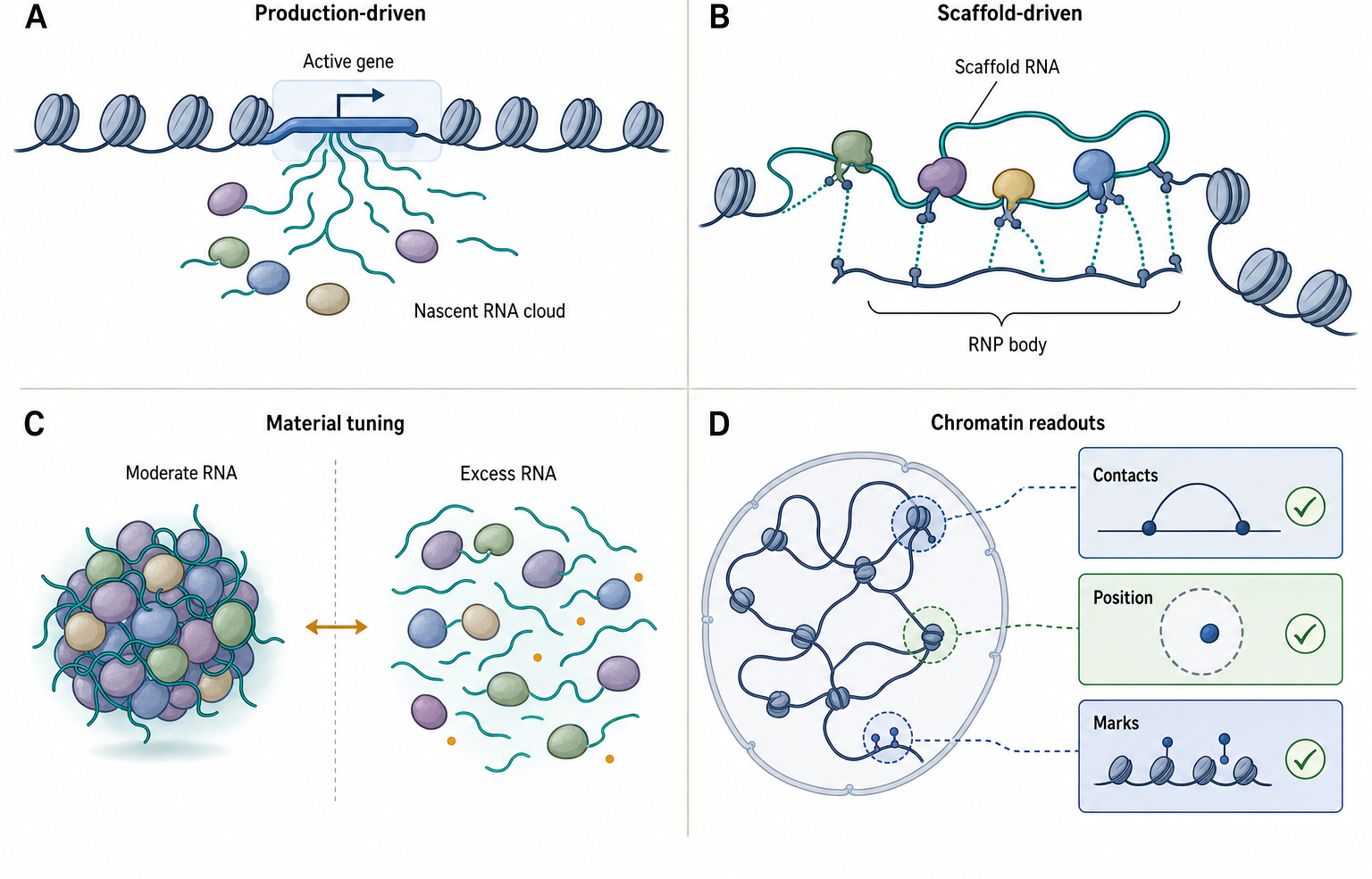
Figure 95.1. Mechanisms by Which RNA Organizes Nuclear Architecture. RNA architecture is a causal claim with multiple mechanisms, not a synonym for localization.
Chromatin compartments add another layer. RNAs can associate with active genes, Polycomb domains, lamina-associated domains, pericentromeric repeats, nucleolus-associated domains, and enhancer-promoter contacts. In some cases, RNA is a downstream product of compartment identity. In other cases, RNA helps recruit or stabilize compartment-specific proteins. The difference matters. If an RNA disappears after transcription inhibition and a nuclear domain changes, the simplest interpretation may be that global transcriptional state changed. A stronger claim requires molecule-specific perturbation, local rescue, and measurement of chromatin and transcriptional consequences. Recent work on RNA-dependent active subnuclear compartments and spatial multi-omics supports the idea that RNA can participate in subnuclear organization, but the local bibliography is still uneven for this chapter. Final bibliography item: add recent review anchors dedicated specifically to RNA-dependent nuclear architecture and RNA-chromatin compartment mechanisms.
Box 95.1. Organizer, Client, or Marker?
Architectural RNA claims are clearer when the RNA is assigned one of three working roles before the evidence is interpreted.
- Organizer: The RNA is needed to assemble, maintain, position, or tune a nuclear body or chromatin compartment. Evidence should include molecule-specific perturbation, structural readouts, and rescue or domain logic.
- Client: The RNA enters a nuclear body because the body processes, stores, edits, retains, exports, or surveils that RNA. Localization is real, but the RNA may not build the structure.
These roles can overlap. Nascent pre-rRNA is both substrate and organizer in the nucleolus, whereas many speckle-associated pre-mRNAs are better treated as clients or markers unless perturbation shows a structural requirement.
95.2. Nucleolar structure-function relationships and rRNA-centered organization
The nucleolus is the dominant nuclear body in many proliferating eukaryotic cells because ribosome production is one of the cell’s largest biosynthetic commitments. Its core RNA output is the long precursor rRNA transcribed from ribosomal DNA repeats by RNA polymerase I. In human cells, the major precursor contains sequences that will become 18S, 5.8S, and 28S rRNAs, separated by external and internal transcribed spacers. The precursor is not merely a message for later processing. It is the central substrate around which early ribosome assembly, modification, surveillance, and nucleolar spatial organization occur.
Nucleolar organization is usually described as three interrelated zones. Fibrillar centers contain ribosomal DNA and Pol I transcription machinery. Dense fibrillar components contain nascent pre-rRNA and early processing factors, including many small nucleolar RNPs. Granular components contain later pre-ribosomal particles as ribosomal proteins and assembly factors accumulate. This tripartite model is a simplification, but it gives a useful causal sequence: rDNA transcription creates pre-rRNA; pre-rRNA recruits modification and processing machinery; ribosomal proteins and assembly factors convert the transcript into export-competent subunits; failed intermediates are retained or degraded.
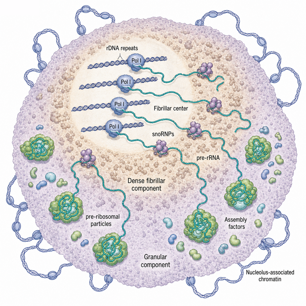
Figure 95.2. Nucleolar rRNA-Centered Organization. The nucleolus is organized by RNA synthesis, RNA maturation, RNP assembly, and nuclear positioning together.
rRNA organization is both linear and spatial. The pre-rRNA contains spacer regions that are removed, mature rRNA segments that fold into ribosomal cores, and chemical modification sites guided mostly by small nucleolar RNAs. Box C/D snoRNAs guide 2′-O-methylation, and box H/ACA snoRNAs guide pseudouridylation. These snoRNAs base-pair with pre-rRNA and bring enzymes to defined positions. The base-pairing interactions are architectural at a molecular scale because they arrange substrate and enzyme within a pre-ribosomal particle, but they are not usually called nuclear-body scaffolds. This distinction helps prevent a common overgeneralization: all RNAs in the nucleolus contribute to nucleolar function, but not all are equivalent as structural organizers.
The nucleolus also connects to whole-nucleus architecture. Nucleolus-associated domains are chromosomal regions that contact the nucleolar periphery and are often enriched for repressive chromatin features, inactive repeats, or particular gene classes. This association does not mean the nucleolus simply silences all nearby DNA. Nucleolar contacts can reflect nuclear positioning, genome organization, ribosome-biogenesis state, stress responses, and cell type. The nucleolus therefore links RNA production to chromatin organization: an extremely active RNA synthesis center can also create a spatial landmark for inactive or repeat-rich chromatin.
Evidence for nucleolar RNA architecture comes from microscopy, biochemical fractionation, pulse labeling of rRNA synthesis, rRNA processing assays, ribosome assembly studies, and perturbation of Pol I transcription or assembly factors. Strong mechanistic claims identify whether the perturbation affects rRNA transcription, pre-rRNA processing, ribosomal protein import, nucleolar material properties, or downstream stress signaling. For example, loss of a processing factor may fragment nucleoli because pre-rRNA maturation fails, not because the factor is a primary body scaffold. Conversely, direct depletion of pre-rRNA synthesis can rapidly reorganize nucleolar zones, indicating that ongoing rRNA production is a central organizing input.
The local reference list includes work on nucleolar URB1 and global nuclear organization, but it lacks several canonical nucleolus reviews and classic structure-function papers. Final bibliography item: add curated nucleolus reviews covering fibrillar centers, dense fibrillar components, granular components, nucleolus-associated domains, rDNA transcription, snoRNP-guided modification, and ribosome assembly quality control.
95.3. Nuclear speckles, Alu RNAs, snRNAs, and active-transcription neighborhoods
Nuclear speckles are irregular nuclear domains enriched in pre-mRNA splicing factors, serine-arginine-rich proteins, snRNP components, RNA export factors, and selected long noncoding or repeat-derived RNAs. They are sometimes called interchromatin granule clusters. A useful first definition is functional rather than morphological: speckles are hubs that concentrate factors used by many active genes, especially genes whose transcripts require splicing and processing. Speckles are not equivalent to spliceosomes. A spliceosome assembles on a specific intron; a speckle is a larger nuclear domain that stores, modifies, concentrates, or coordinates factors used in many RNA-processing events.
snRNAs connect speckles to splicing logic. U1, U2, U4, U5, and U6 small nuclear RNAs are core RNA components of spliceosomal snRNPs. These snRNAs recognize splice sites, help position catalytic RNA elements, and form dynamic RNPs with many proteins. Speckles contain snRNP-related factors, but a visible speckle is not simply a pile of completed spliceosomes. Much of the relevant biology is exchange: factors move between speckles, nascent transcripts, Cajal bodies, and nucleoplasm. The presence of snRNAs or snRNP proteins in a speckle therefore supports a processing-factor relationship, not necessarily active catalysis inside the speckle.
Box 95.2. Speckles Are Not Spliceosomes
A spliceosome is a transient RNP machine assembled on a particular intron. It uses U1, U2, U4, U5, and U6 snRNPs plus many proteins to recognize splice sites, rearrange RNA-RNA and RNA-protein contacts, catalyze two transesterification reactions, and release the ligated exons and intron product.
A nuclear speckle is larger and more collective. It contains many splicing and RNA-processing factors, but those factors exchange with nascent transcripts and other nuclear compartments. Speckle enrichment therefore supports the statement “this factor participates in a processing neighborhood” more directly than “splicing catalysis occurs inside this spot.” Some cotranscriptional processing may occur at speckle-proximal genes, yet the unit of catalysis remains the spliceosome assembled on an RNA substrate. The distinction matters when interpreting images: overlap between an snRNP marker and a speckle marker is not, by itself, a map of active intron removal.
Alu RNAs and Alu-derived sequence add another layer. Alu elements are primate SINE repeats derived from 7SL RNA ancestry, and many human transcripts contain Alu fragments in introns or untranslated regions. Alu-derived RNAs can bind RNA-processing proteins, influence nuclear localization, promote duplex formation when inverted repeats pair, and contribute to stress-responsive or disease-linked nuclear RNA states. Some studies connect Alu-rich RNAs and other repeat RNAs to speckle-proximal domains or chromatin neighborhoods. However, Alu sequence is highly repetitive, so localization and interaction claims require controls for probe specificity, read mapping, and indirect association with abundant pre-mRNAs.
Table 95.1. Major Nuclear RNA Architecture Examples. Different nuclear bodies use RNA in different causal roles, so one evidence rule cannot be transferred uncritically.
| Nuclear structure | Principal RNA classes | Main protein partners or activities | Architectural role | Strongest evidence | Key caveat |
|---|---|---|---|---|---|
| Nucleolus | Pre-rRNA, mature rRNA segments, snoRNAs, nucleolus-associated RNAs | Pol I machinery, snoRNPs, processing nucleases, ribosomal proteins, assembly factors | rRNA transcription and pre-ribosome assembly create layered fibrillar and granular zones | Perturbation of rRNA synthesis or processing changes nucleolar zones and ribosome assembly readouts | Morphology can change indirectly through stress signaling, ribosomal protein supply, or chromatin state |
| Nuclear speckle | snRNAs, nascent pre-mRNAs, MALAT1-context RNAs, Alu-containing RNAs | SR proteins, snRNP components, SON/SRRM2-type scaffolds, export and processing factors | Concentrates exchangeable RNA-processing factors near active, often intron-rich genes | Imaging and genome-proximity assays link speckles to active transcription neighborhoods | Speckles are not spliceosomes; proximity can be cause, consequence, or correlate of transcription |
| Paraspeckle | NEAT1_2, retained structured or edited client RNAs | NONO, SFPQ, PSPC1, FUS, RBM14, additional paraspeckle proteins | NEAT1_2 acts as an isoform-specific scaffold for ordered RNP-body assembly | NEAT1_2 loss, domain mapping, and rescue-style logic support scaffold necessity | Total NEAT1 expression does not prove NEAT1_2-dependent paraspeckle mechanism |
| Repeat-RNA focus | Satellite RNAs, short tandem repeat RNAs, SINE/LINE-derived RNAs, repeat-expanded disease RNAs | Repeat-binding RBPs, splicing factors, chromatin-associated proteins, stress-response proteins | Multivalent repeat sequence can seed nuclear foci, pathological assemblies, or chromatin-linked domains | Orthogonal imaging plus repeat-aware mapping and dose-relevant perturbation | Repetitive probes, ambiguous reads, and off-target antisense effects are frequent confounders |
| RNA-dependent chromatin compartment | Chromatin-associated lncRNAs, enhancer RNAs, repeat RNAs, nascent RNAs | Chromatin regulators, Polycomb-linked factors, lamina or nucleolar contact machinery, RBPs | RNA may recruit, exclude, or stabilize proteins that shape local chromatin position or state | Strongest support combines RNA perturbation with contacts, marks, accessibility, and transcription readouts | RNA can be a marker or product of compartment state rather than a driver |
| Active transcription neighborhood | Nascent pre-mRNAs, enhancer-associated RNAs, local noncoding transcripts | RNA polymerase II, elongation factors, splicing and export machinery, speckle-proximal factors | Local RNA production concentrates processing machinery and can position genes near nuclear bodies | Temporal imaging and acute perturbations can order transcription, RNA accumulation, and body proximity | Global transcription inhibition can collapse many processes and overstate RNA-specific causality |
The best current model treats speckles as active-transcription neighborhoods rather than as passive storage granules. Many highly expressed, long, intron-rich, or coordinately regulated genes are spatially close to speckles. Enhancer-promoter activity, transcription elongation, co-transcriptional splicing, and RNA export can all influence these contacts. Speckle proximity can be a cause, a consequence, or a correlate of gene activity. A gene may move toward a speckle because it is active; a speckle-proximal environment may improve processing efficiency; both may be driven by the same transcriptional program. Distinguishing these possibilities requires temporal data and perturbation designs.
Several experimental results have moved the field beyond static co-localization. Imaging can measure gene positions relative to speckles and nascent RNA transcription sites. Chromatin conformation and nuclear proximity methods can identify speckle-associated genomic regions. Acute perturbation of transcription, splicing factors, speckle proteins, or candidate RNAs can test whether speckle organization changes before or after transcriptional output. Still, the standards are demanding. If a factor is essential for splicing, its depletion will change thousands of RNAs and many nuclear features. A specific architectural claim should show that the structural effect is not merely a secondary consequence of global RNA-processing failure.
Nuclear speckles also illustrate a boundary case for architectural RNAs. The RNA species in or near speckles include nascent pre-mRNAs, splicing snRNAs, MALAT1 and other lncRNAs in some contexts, Alu-containing RNAs, and transcripts being processed or retained. Some may organize subdomains; others may be clients. The chapter therefore uses a conservative convention: call an RNA architectural only when perturbing that RNA, its relevant domains, or its production site changes speckle assembly, positioning, material behavior, or downstream processing in a way that can be rescued or otherwise separated from general transcriptional collapse.
The local bibliography for this subsection contains only partial anchors for U1 snRNAs and unrelated review material. Final bibliography item: add dedicated references for nuclear speckles, MALAT1, SON/SRRM2 speckle scaffolding, speckle-associated genome organization, Alu RNA nuclear localization, and speckle-proximal active chromatin.
95.4. Paraspeckles, NEAT1, repeat RNAs, and nuclear-domain organization
Paraspeckles are nuclear bodies built around the long noncoding RNA NEAT1. Mammalian NEAT1 is produced as at least two major isoforms. The shorter NEAT1_1 isoform can be abundant, but the long NEAT1_2 isoform is the essential scaffold for canonical paraspeckle assembly. NEAT1_2 remains near its transcription site, binds multiple paraspeckle proteins, and organizes a body with ordered internal architecture. This makes paraspeckles one of the strongest examples of an architectural lncRNA in mammalian nuclear biology.
The molecular logic is multivalent RNP assembly. NEAT1 contains regions that recruit proteins such as NONO, SFPQ, PSPC1, FUS, RBM14, and other paraspeckle-associated factors. Different NEAT1 domains contribute to initiation, elongation, shell organization, and protein recruitment. Yamazaki and colleagues showed that functional domains of NEAT1 architectural lncRNA can induce paraspeckle assembly through phase-separation-related mechanisms. This does not mean NEAT1 is a generic sticky polymer. Domain position, RNA processing, 3′-end formation, protein binding, and transcription-site retention all contribute to the final body.
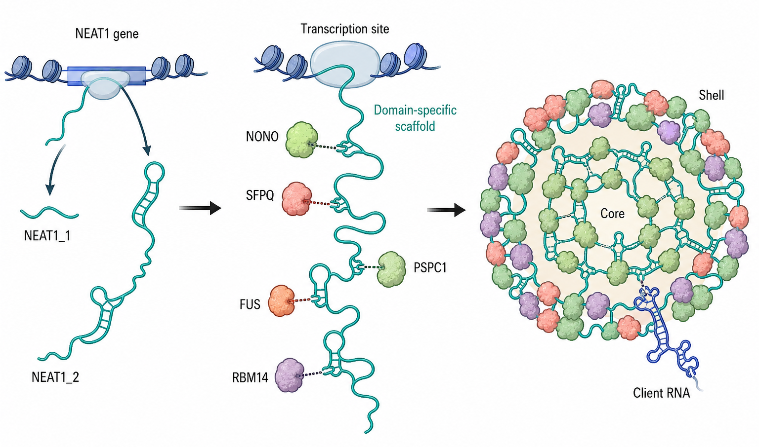
Figure 95.3. NEAT1_2 and Paraspeckle Assembly. Paraspeckles require isoform-specific, domain-specific RNA scaffold logic rather than generic lncRNA presence.
Paraspeckle functions are context-dependent. They can retain edited or structured RNAs in the nucleus, regulate availability of RNA-binding proteins, respond to stress or differentiation signals, and influence gene-expression programs. NEAT1 and paraspeckle proteins have been linked to cancer, viral infection, immune signaling, neuronal contexts, and developmental programs. The evidence is not uniform across contexts. NEAT1 induction can be a stress marker, a protective response, a pathological contributor, or an indirect consequence of altered transcription. Therefore, paraspeckle claims should specify cell type, stimulus, isoform, and readout.
Repeat RNAs broaden the paraspeckle lesson because repetitive sequence is naturally multivalent. Short tandem repeats, satellite repeats, SINEs, LINE fragments, endogenous retroviral sequences, and repeat-expanded disease RNAs can create many binding sites for RNA-binding proteins. Some repeat RNAs help define nuclear domains, while others form pathological foci that sequester proteins. Short tandem repeat-enriched architectural RNAs have been reviewed as components of nuclear bodies, and repeat-element RNAs have been reported to shape specific growth or plasticity circuits in recent cell-type and disease studies. These examples are important but must not be collapsed into one rule that repeat RNA equals architecture.
Material-state measurements refine but do not replace the paraspeckle mechanism. Exchange, mobility, permeability, deformation, or fusion-like behavior can describe how a paraspeckle behaves, but NEAT1_2 isoform identity, transcription-site retention, RNA-domain organization, and specific protein contacts explain where the body forms and how it is built. Conversely, failure to behave as a simple liquid does not make a layered or partially immobilized nuclear RNP unimportant. General criteria for phase behavior belong to Chapter 58.
Repeat RNAs raise special artifact concerns. Hybridization probes against repeat sequences can bind many loci or transcripts. Short sequencing reads from repeats can map to many genomic positions. Antisense oligonucleotides targeting repeat tracts can affect multiple RNAs. Overexpressed repeat RNA can create artificial foci because concentration is forced far above endogenous levels. A strong repeat-RNA architecture claim therefore needs orthogonal detection, locus or transcript specificity, dose-relevant perturbation, and ideally domain-specific rescue.
Box 95.3. Repeat-RNA Claims Need Repeat-Aware Controls
Repeat RNAs deserve both interest and skepticism. Repetition creates many potential binding sites for RNA-binding proteins, so repeat RNAs can plausibly seed foci, recruit chromatin factors, or sequester processing proteins. The same repetition also makes experiments hard to interpret.
Before accepting a repeat-RNA architecture claim, ask four questions. Detection: do independent probe sets, RNase controls, or orthogonal imaging methods identify the same RNA population? Assignment: can sequencing reads or in situ signals be attributed to a locus, family, transcript isoform, or repeat expansion rather than to a generic repeat class? Perturbation: does the intervention target the candidate RNA without broadly changing many related repeat RNAs or triggering stress? Rescue: can a physiological amount of the RNA, or a defined repeat/domain mutant, restore the architectural feature? A repeat-rich focus with no such controls may be a true biological structure, a toxic aggregate, or a technical artifact.
The local reference list includes NEAT1 paraspeckle work, a short tandem repeat architectural RNA review, and recent repeat-RNA primary papers. It lacks several foundational paraspeckle and NEAT1 discovery or review references. Final bibliography item: add core references for NEAT1_2 isoform biogenesis, paraspeckle protein architecture, RNA retention functions, and disease-context NEAT1 interpretation.
Experimental Foundations and Evidence
95.5. Context-specific imaging, perturbation, and causality for nuclear bodies
Architectural RNA claims begin with observation but cannot end there. The first evidence tier is localization: RNA FISH, live RNA tagging, spatial transcriptomics, or subcellular fractionation shows that an RNA is enriched in a nuclear body or chromatin region. This evidence is necessary for many claims, but it is weak by itself. Enrichment may reflect high expression, slow processing, nuclear retention, probe cross-reactivity, or the body acting on the RNA rather than the RNA organizing the body.
The second tier is molecular association. RNA-protein interaction mapping, proximity labeling, crosslinking, pull-downs, and colocalization with body markers identify candidate RNP partners. These methods must distinguish direct binding from co-residence in a dense compartment. Direct crosslinking, domain mutation, and in vitro reconstitution can strengthen the interpretation, but no single method is definitive. For repeat RNAs and abundant RNAs, background binding and indirect capture are major concerns.
The third tier is perturbation. Depleting the RNA, blocking its transcription, deleting a domain, changing its processing, or disrupting a binding site should change the nuclear body if the RNA is architectural. The design must separate the RNA molecule from the DNA locus and from global transcriptional effects. CRISPR deletion may remove regulatory DNA, transcription-factor binding sites, or chromatin elements. Transcriptional inhibition may collapse many nuclear processes at once. Antisense depletion may trigger RNase H cleavage, steric blocking, or off-target effects. A credible design uses multiple independent perturbations and interprets method-specific artifacts explicitly.
The fourth tier is rescue or synthetic sufficiency. Rescue can restore the RNA in a perturbation-resistant form, place the RNA at the endogenous locus, or express domain mutants that separate localization, protein binding, and function. Synthetic tethering can test whether recruiting an RNA domain to a locus or artificial platform is sufficient to recruit proteins or create a body. These experiments are powerful but can be misleading if expression level, copy number, or tethering geometry is nonphysiological. Sufficiency should therefore be paired with endogenous necessity.
Table 95.2. Method-Specific Caveats for Architectural RNAs. Method choice determines whether a study supports localization, association, necessity, sufficiency, or physiological function.
| Method | What it can support | Common artifact | Stronger control | Claim level enabled |
|---|---|---|---|---|
| RNA FISH | Fixed-cell enrichment of a defined RNA in a body, locus, or nuclear zone | Probe cross-reactivity, repeat binding, fixation effects, segmentation bias | Multiple probe sets, RNase controls, smFISH quantification, orthogonal marker imaging | Localization and spatial correlation |
| Live RNA imaging | Dynamics of RNA position, residence time, body contact, and response to perturbation | Tag arrays or aptamers can alter processing, export, binding, or expression level | Endogenous tagging, tag-position controls, matched untagged rescue, expression calibration | Dynamic localization and temporal ordering |
| RNA-protein capture | Candidate RBPs or RNP partners associated with an architectural RNA | Indirect capture from dense compartments, crosslinking bias, abundant-RNA background | Domain mutants, reciprocal assays, direct crosslink evidence, in vitro binding tests | Association; direct binding only with stronger evidence |
| CRISPR deletion | Necessity of a locus, promoter, RNA domain, or processing element for body formation | Deletes DNA regulatory elements or changes chromatin independently of RNA | Minimal domain edits, transcription-preserving mutations, allele-specific rescue | Necessity when DNA and RNA effects are separated |
| Antisense depletion | RNA-molecule requirement for maintaining a body or compartment | RNase H off-targets, steric effects, repeat-family targeting, stress responses | Independent ASOs, isoform-specific assays, RNA-level rescue, toxicity controls | Molecule-specific necessity |
| Transcription inhibition | Dependence of a body on ongoing RNA production or polymerase activity | Global collapse of transcription, processing, chromatin state, and stress pathways | Acute, titrated inhibition plus locus-specific perturbation and recovery kinetics | Production dependence; weak molecule specificity |
| Synthetic tethering | Sufficiency of an RNA domain to recruit proteins or nucleate a local assembly | Nonphysiological copy number, forced geometry, overexpression, tag-driven clustering | Endogenous-level expression, inactive-domain controls, endogenous necessity pairing | Sufficiency or domain activity under defined conditions |
| Spatial multi-omics | Joint maps of RNAs, proteins, chromatin contacts, and cell states in tissue or nuclei | Probe dropout, segmentation choices, batch effects, inferred rather than direct contacts | Probe validation, perturbation-linked samples, replicate tissue regions, orthogonal imaging | Integrated spatial correlation; causality only with perturbation |
Imaging standards deserve special attention. Conventional fluorescence microscopy can show colocalization, but diffraction-limited overlap does not prove molecular interaction. Super-resolution microscopy can resolve subdomains, but fixation, probe penetration, and antibody accessibility can distort architecture. Live imaging can measure dynamics, but tags can alter RNA processing or protein binding. Spatial molecular imaging and multiplexed methods can map many RNAs and proteins in fixed tissue, yet they still require probe validation and quantitative segmentation. The strongest imaging studies connect spatial maps to perturbation and functional readouts rather than relying on attractive pictures.
Chromatin standards are similarly strict. If an architectural RNA is proposed to organize a chromatin compartment, the study should measure changes in chromatin contacts, nuclear positioning, histone marks, accessibility, and transcription separately. These readouts can disagree. An RNA may change nuclear positioning without changing transcription, or change transcription without detectable compartment reorganization. Causality is strongest when an RNA perturbation changes a defined chromatin feature before secondary expression changes dominate, and when rescue restores the feature.
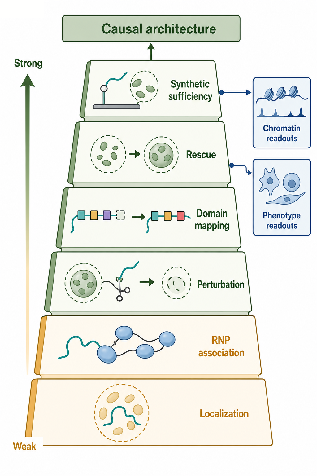
Figure 95.4. Evidence Ladder for Architectural RNA Causality. Causal architecture requires stronger evidence than colocalization or differential expression.
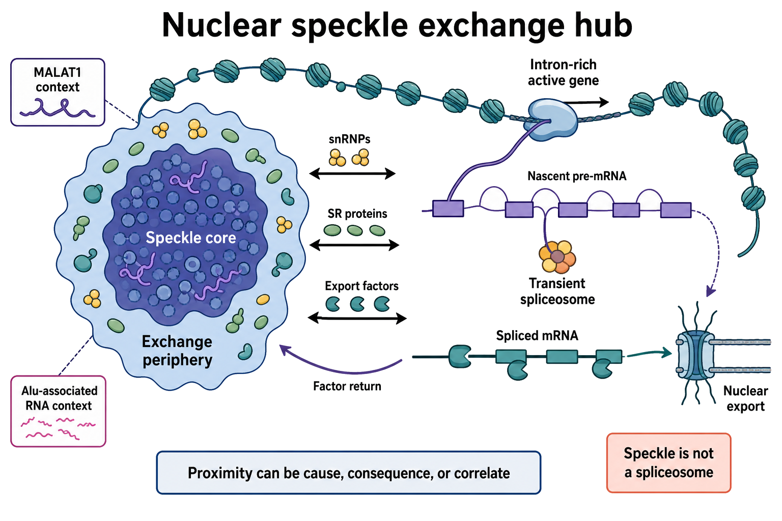
Figure 95.5. Nuclear Speckles as Exchange Hubs Near Active Transcription Neighborhoods. A speckle is an exchange hub in an active transcription neighborhood, not one giant assembled spliceosome; proximity can be cause, consequence, or correlate of gene activity.
One useful practical standard is an evidence ladder. A candidate architectural RNA is weakly supported by localization alone, moderately supported by localization plus specific RNP association, strongly supported by molecule-specific loss-of-function that changes body structure, and very strongly supported by rescue plus domain mapping or synthetic sufficiency. Claims about disease or physiology require an additional layer: the architectural change must explain a cellular, tissue, developmental, or clinical phenotype rather than merely accompany it.
Biological Contexts Across Systems
Nuclear RNA architecture changes with cell state. Proliferating cells often have prominent nucleoli because ribosome production is high. Differentiated cells can reshape nucleoli, speckles, and paraspeckles as transcriptional demand changes. Stress can repress Pol I transcription, reorganize nucleoli, induce NEAT1 and paraspeckles, alter splicing-factor distribution, or activate repeat transcription. Viral infection and innate immune activation can change nuclear RNA bodies because antiviral responses alter transcription, RNA retention, RNA editing, and RNA-binding-protein localization.
Cell-cycle state is another important qualifier. Nucleoli disassemble and reassemble around mitosis, many chromatin contacts are reorganized, and transcriptional output changes as cells move through division. A nuclear RNA body observed in an asynchronous culture may therefore represent a mixture of assembly states rather than one stable architecture. Nutrient availability, growth rate, DNA damage, and differentiation can also change whether an RNA is acting as a scaffold, a processing substrate, a retained product, or a stress-induced client. Architectural claims should specify whether the evidence comes from cycling cells, arrested cells, primary tissue, transformed cell lines, or acute stress models, because each context changes the expected baseline.
Developmental and tissue context also matter. A nuclear RNA body that is visible in cultured cancer cells may be smaller, absent, or differently organized in primary tissue. Recent reports of persistent nuclear RNAs in mouse brain and spatial multi-omics studies of cell-type-specific nuclear compartments underscore that nuclear RNA architecture can be long-lived or cell-type specific. These observations are provocative because they suggest that nuclear RNA is not always a transient processing intermediate. They also require careful interpretation: persistence, localization, and function are different claims.
Disease associations are abundant but uneven. Repeat-expanded RNAs can form nuclear foci in neurological and neuromuscular disease, sequester RNA-binding proteins, and disrupt splicing or RNA metabolism. NEAT1 and paraspeckles are associated with cancer, infection, inflammation, and neurobiology. Nucleolar size and rRNA synthesis are altered in many cancers and stress states. Speckle organization changes when splicing factors are mutated or transcriptional programs shift. In each case, the central question is whether the nuclear body change is causal, compensatory, or a marker of altered cell state.
Technology, Computational, Clinical, and Engineering Links
Architectural RNAs are useful experimental handles because they can be imaged, depleted, tethered, or engineered. RNA-guided recruitment systems can bring proteins or RNA domains to selected loci. Synthetic lncRNA scaffolds may eventually help organize nuclear reactions or regulate chromatin neighborhoods, although most such engineering remains exploratory. Live RNA tags and bright fluorescent RNA systems improve dynamic measurements but must be tested for effects on RNA maturation and localization.
Computational analysis faces several challenges. Nuclear-body RNAs are often lowly expressed, repetitive, long, alternatively processed, or retained in chromatin fractions. Short-read RNA-seq may undercount long retained isoforms, misassign repeats, or miss subcellular localization. Spatial and imaging datasets require segmentation choices that can change apparent body size and colocalization. Integrative analysis should combine transcript isoform models, repeat-aware mapping, chromatin contacts, protein localization, and perturbation time courses.
Clinical translation is mostly indirect at present. NEAT1 expression, nucleolar activity, repeat RNA foci, and speckle alterations can serve as biomarkers in some contexts, but biomarkers are not mechanisms. Therapeutic strategies that target architectural RNAs, such as antisense depletion of toxic repeat RNAs or modulation of NEAT1, must consider nuclear delivery, isoform specificity, off-target binding, and consequences for normal nuclear organization. The same multivalency that makes an RNA architectural can make selective targeting difficult.
Recent Consensus
The current consensus is that RNA is not merely cargo in the nucleus. Specific RNAs can help assemble nuclear bodies, and ongoing transcription can organize local nuclear neighborhoods. The nucleolus and paraspeckle provide strong examples, while speckles and repeat-rich domains show more mixed combinations of RNA clients, scaffold proteins, nascent transcription, and chromatin context. The field has moved away from simple co-localization claims toward perturbation, domain mapping, and physical models of multivalent RNP assembly.
Another consensus is that material-state language is useful only when it is joined to nuclear-body specificity. Exchange or demixing behavior does not identify the organizer. A mechanistic claim must connect a defined RNA isoform or domain to protein partners, transcription or chromatin position, body structure, and nuclear function; the general physical framework is supplied by Chapter 58.
Open Questions, Controversies, Deprecated Models, and Common Misconceptions
Open questions:
- How often RNA is a primary organizer rather than a product or client of nuclear architecture? Paraspeckles support primary RNA scaffolding. Speckles may depend more on protein scaffolds and transcriptional neighborhoods, with RNAs contributing in context-specific ways. Nucleoli depend deeply on rRNA production, but many nucleolar proteins and chromatin features also shape the body.
- What determines how repeat RNAs should be classified? Some repeat-derived RNAs are functional architectural components. Others are toxic gain-of-function molecules. Many are transcriptional byproducts or mapping artifacts. Repetitive sequence should raise both biological interest and evidentiary caution.
Common misconceptions:
- “A nuclear body is automatically a phase-separated droplet.” Nuclear bodies can have layered structures, chromatin attachments, active enzymatic reactions, arrested intermediates, and gel-like or solid-like states.
- “RNA localization proves RNA function.” Localization is a starting point. Architecture requires a causal test.
Deprecated or weakened claims:
- Broad claims that any lncRNA near chromatin recruits a specific chromatin regulator, or that any repeat-enriched RNA focus is a functional nuclear domain. The field now expects isoform resolution, repeat-aware mapping, direct perturbation, rescue, and functional specificity.