Chapter 156. Lipid Nanoparticles, Ionizable Lipids, Endosomal Trafficking, Endosomal Escape, and Biodistribution
Scope Note
This chapter explains lipid nanoparticles (LNPs) as delivery systems for RNA medicines, with emphasis on how composition, particle assembly, intracellular trafficking, endosomal escape, biodistribution, immune activation, and design workflows determine biological output. The focus is on RNA delivery rather than on antigen design, RNA editing chemistry, or clinical pharmacology in general; those topics are treated in neighboring chapters and are cross-referenced where they set the context for LNP use.
Executive Summary
LNPs are multicomponent colloidal particles that protect RNA during administration, concentrate RNA into cell-associated compartments, and create a chance for RNA to cross from endosomes into the cytosol. The most important point for interpreting LNP biology is that delivery is a sequence of conditional steps, not a single event. A particle must remain sufficiently intact during manufacture and storage, avoid premature clearance or destructive aggregation in biological fluids, encounter a permissive tissue and cell type, enter the cell, traffic through endosomal compartments, disrupt or reorganize enough membrane to release RNA into the cytosol, and do all of this without creating an unacceptable inflammatory, toxic, or immunogenic profile.
Modern RNA-LNPs typically contain four functional lipid classes: an ionizable lipid that binds RNA during acidic formulation and participates in endosomal membrane interactions after uptake; a helper phospholipid that affects bilayer packing and fusogenic behavior; cholesterol or a sterol component that tunes particle structure and membrane mechanics; and a polyethylene glycol lipid that controls particle size, colloidal stability, and circulation behavior. The exact formulation is a product-specific design variable. The same nominal molar ratio can produce different particles if mixing, buffer, total lipid concentration, flow rate, RNA concentration, pH, solvent removal, sterile filtration, or freezing conditions change. Manufacturing is therefore part of mechanism, not merely a downstream packaging step.
Endosomal escape remains the central mechanistic bottleneck. Uptake can be abundant while cytosolic RNA release remains rare. Ionizable lipids are designed to be relatively neutral at physiological pH and protonated in acidic endosomes, but protonation alone does not fully explain productive release. Current models emphasize lipid mixing, non-bilayer phases, membrane curvature stress, ion-pair formation with anionic endosomal lipids, transient pore or defect formation, and endosomal damage responses. The field agrees that escape is inefficient and context-dependent, but it does not yet have a universally predictive mechanism that links lipid structure, intracellular route, and RNA output across cells, cargos, and species.
Biodistribution reflects particle chemistry, route, dose, serum protein adsorption, tissue anatomy, cell state, and inflammatory history. Intravenous LNPs often show liver-biased delivery because liver sinusoids expose particles to hepatocytes and resident phagocytes and because serum proteins can promote hepatic uptake. Intramuscular and intradermal vaccine LNPs instead create local depots and drainage to lymph nodes, where antigen-presenting cells can take up RNA and initiate immune responses. Extrahepatic targeting is possible but remains harder than liver delivery because it requires overcoming vascular, extracellular, cellular, endosomal, and safety barriers simultaneously.
LNPs can also act as immune-active materials. The RNA cargo, ionizable lipid, PEG-lipid, impurities, route, dose, and tissue damage signals can all contribute to innate immune activation, complement activation, reactogenicity, and repeat-dose constraints. For vaccines, some inflammatory signaling may support immunogenicity; for chronic protein replacement, gene editing, or immune-cell programming, the same inflammatory signals may be liabilities. AI-guided lipid design and high-throughput screening can expand the chemical search space, but translation still depends on assays that measure the right endpoint, species models that predict human exposure, and manufacturing processes that preserve the intended particle state.
Concept Inventory
- Lipid nanoparticle: a nanoscale lipid-rich colloidal formulation used to carry nucleic acids or other cargos. In RNA delivery, an LNP is not a hollow bag in the simple liposome sense. Many RNA-LNPs contain an internal, heterogeneous lipid-RNA organization whose structure depends on formulation conditions and lipid chemistry.
- Ionizable lipid: a lipid with an acid-base group that changes protonation state across the pH range relevant to formulation, blood, tissues, and endosomes. Ionizable lipids are generally less cationic at physiological pH than permanently cationic lipids, which can reduce nonspecific toxicity, but they can become more positively charged in acidic environments.
- Helper lipid: a non-ionizable lipid, often a phospholipid such as distearoylphosphatidylcholine or a more fusogenic lipid, that contributes to particle structure, membrane packing, and interaction with biological membranes.
- Cholesterol or sterol component: a membrane-active lipid that modifies packing, fluidity, particle stability, and protein adsorption. Sterol identity and amount can affect expression and biodistribution.
- PEG-lipid: a lipid conjugated to polyethylene glycol. PEG-lipids reduce aggregation and help control particle size during formulation, but the PEG-lipid must be balanced because persistent surface PEG can reduce cellular uptake and anti-PEG immune responses can complicate repeat dosing.
- Endosomal escape: the process by which RNA leaves endocytic vesicles and reaches the cytosol. Endosomal escape is not equivalent to uptake, and it is not fully measured by fluorescence colocalization or total cellular RNA.
- Protein corona: the layer of adsorbed proteins that forms on a particle after contact with biological fluids. The corona can change uptake, biodistribution, complement activation, and immune recognition.
- Reactogenicity: short-term local or systemic inflammatory symptoms after administration, such as injection-site pain, fever, myalgia, or malaise. Reactogenicity is related to innate immune activation but is not a direct measurement of protective immunity or long-term toxicity.
What to Know Before Reading This Chapter
RNA drugs have a delivery problem because RNA is large, anionic, hydrophilic, nuclease-sensitive, and usually unable to cross intact plasma membranes by passive diffusion. A therapeutic mRNA must reach cytosolic ribosomes; an siRNA must reach Argonaute loading pathways; a guide RNA or editing guide must reach the compartment where its effector acts. The carrier therefore changes the biological identity of the RNA product. An mRNA sequence that translates strongly after electroporation may fail after systemic LNP dosing if the particle is cleared, trapped in endosomes, or delivered to the wrong cell type.
The chapter uses two running examples. The first is an intramuscular mRNA vaccine, where LNPs deliver antigen-encoding mRNA into local cells and antigen-presenting cells in a setting where controlled inflammation can be useful. The second is an intravenous RNA therapy aimed at hepatocytes, where liver delivery may be desirable but repeated exposure, innate immune activation, and off-target uptake by liver immune cells become major constraints. These examples show why the same delivery feature can be a virtue in one product and a liability in another.
The local reference set for this chapter includes recent review anchors on LNP chemistry, selective mRNA delivery, cardiovascular applications, and endosomal escape, plus recent papers on endosomal damage, internal mesophase structure, cellular barriers, and lipid-polymer hybrid vaccine particles (Eygeris et al. 2022; Soroudi et al. 2024; Zhao et al. 2024; Mrksich et al. 2024; Omo-Lamai et al. 2025; Yu et al. 2025; Johansson et al. 2025; Baghel et al. 2025). The references are adequate for a mechanistic draft but thin for product labels, regulatory guidance, classic lipid discovery history, anti-PEG clinical epidemiology, and complement-specific primary studies. Those gaps are marked explicitly rather than filled with unverified citations.
156.1. LNP composition and ionizable lipid design
An LNP for RNA delivery is best understood as a designed, metastable assembly of lipids and RNA rather than as a passive container. The defining component is usually the ionizable lipid. An ionizable lipid contains a head group that can accept protons, hydrophobic tails that partition into lipid-rich phases, and linkers or branching motifs that tune degradability, packing, and shape. During formulation at acidic pH, the ionizable lipid becomes sufficiently protonated to bind the negatively charged phosphate backbone of RNA. After administration at near-neutral extracellular pH, the same lipid is intended to be less charged, reducing nonspecific membrane disruption and serum protein binding compared with permanently cationic carriers. After endocytosis, acidification can again increase protonation and promote interactions with anionic endosomal lipids.
Ionizable lipid design therefore links chemistry to several biological checkpoints. The apparent pKa of the lipid, the pH at which half of the ionizable groups are protonated in a relevant environment, affects how much charge the particle carries during formulation, circulation, and endosomal maturation. A lipid with too much positive charge at physiological pH may bind serum proteins and cell membranes nonspecifically, increasing toxicity and clearance. A lipid that is too weakly protonated in endosomes may fail to interact strongly enough with endosomal membranes. The useful range is context-dependent because the effective pKa of a lipid in a particle can differ from a simple solution value, and the local endosomal membrane environment is not a uniform buffered flask.
Box 156.1. Apparent pKa Is Not a Delivery Score
Mechanism checklist. Apparent pKa helps describe when an ionizable lipid becomes protonated, but delivery depends on more than that number. Ask four questions before using pKa to interpret an LNP result. First, how was the apparent pKa measured: in isolated lipid, in a formulated particle, or with a surface probe that reports only part of the particle population? Second, what else changed with the lipid, such as tail branching, linker degradability, stereochemistry, helper lipid packing, particle size, or PEG exposure? Third, which cargo and endpoint were tested: mRNA expression, siRNA knockdown, editing, uptake, cytokines, or lipid distribution? Fourth, was the result reproduced across route, species, dose, and manufacturing conditions? A lipid can have a favorable apparent pKa and still fail because it clears too fast, damages membranes, traps RNA internally, activates immune pathways, or works only with one cargo class.
The hydrophobic domain also matters. Tail length, branching, unsaturation, ester or disulfide biodegradability, and stereochemistry influence particle packing, membrane curvature, lipid mixing, and metabolic clearance. Conical lipid shapes can favor non-bilayer arrangements that may destabilize endosomal membranes; cylindrical shapes can stabilize bilayers. Biodegradable linkages can reduce tissue persistence and toxicity, but rapid degradation before productive escape can reduce potency. A useful ionizable lipid must therefore balance RNA complexation, particle formation, endosomal activity, tolerability, and clearance.
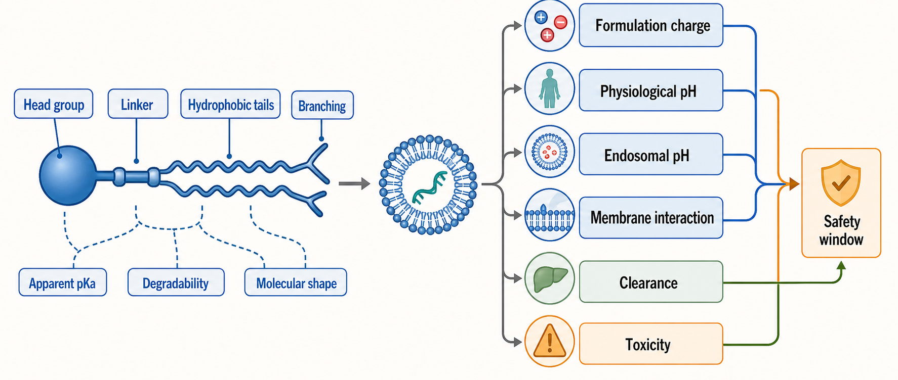
Figure 156.1. Ionizable lipid design variables and biological checkpoints. Ionizable lipid design links chemical structure to several delivery checkpoints. Charge state supports RNA complexation during acidic formulation and endosomal membrane interaction after uptake, whereas reduced charge at physiological pH can reduce nonspecific toxicity. Tail geometry, linkers, and degradability tune packing, non-bilayer propensity, persistence, and safety.
The historical contrast between permanently cationic lipids and modern ionizable lipids is pedagogically useful. Permanently cationic lipids can condense nucleic acids and interact with cell membranes, but persistent positive charge often creates toxicity, complement activation, and nonspecific biodistribution. Ionizable lipids were developed to decouple the formulation step from the in vivo exposure step: charge is useful when assembling the particle and when interacting with endosomal membranes, but persistent charge is undesirable in blood and extracellular tissue. This logic is treated as consensus in recent reviews of LNP chemistry and intracellular delivery (Eygeris et al. 2022; Zhao et al. 2024; Mrksich et al. 2024).
The same ionizable lipid may behave differently with different RNA cargos. An siRNA is short and relatively rigid compared with a long mRNA; a self-amplifying RNA can be much larger and more fragile than a conventional mRNA; a guide RNA may be delivered alone or with an encoded protein. RNA length, secondary structure, concentration, and buffer conditions change complexation and internal particle organization. A formulation optimized for one cargo cannot be assumed to transfer unchanged to another cargo. This is one reason platform language can be misleading: LNP delivery is modular, but it is not infinitely plug-and-play.
Evidence for ionizable lipid design comes from chemical libraries, formulation screens, in vitro expression or knockdown assays, animal biodistribution studies, structural and biophysical characterization, and clinical product performance. Each evidence class has a limitation. Cell culture screens can identify lipids that promote uptake or expression in a particular cell line, but cell lines often poorly model serum protein adsorption, tissue barriers, immune clearance, and human tolerability. Animal screens can reveal tissue delivery, but species differences in apolipoproteins, complement, innate immune tone, vascular fenestration, and endosomal biology can alter translation. Structural studies can show internal organization and phase behavior, but a static particle measurement does not by itself prove the intracellular escape mechanism. For this chapter, the design principles are supported by the chapter-local reviews and by recent work emphasizing internal self-assembled phases and escape barriers (Yu et al. 2025; Johansson et al. 2025).
Do not overgeneralize the term “ionizable lipid” into a single functional class. Two ionizable lipids can share a nominal pKa and still differ in tissue delivery, potency, toxicity, biodegradation, and reactogenicity. The term identifies a design strategy, not a guarantee of efficient or safe RNA delivery.
156.2. Helper lipids, cholesterol, PEG lipids, and assembly
The other lipid components are not inert fillers. Helper lipids, sterols, and PEG-lipids shape the particle before the first cell ever sees it. A typical RNA-LNP includes an ionizable lipid, a phospholipid helper, cholesterol or a related sterol, and a PEG-lipid. Changing the helper lipid can alter membrane packing, phase behavior, and fusogenic tendency. Changing cholesterol content can affect stiffness, internal organization, and protein interactions. Changing the PEG-lipid can shift particle size and stability, but also change uptake and repeat-dose immunology. These relationships are not independent, because a change in one lipid can change the organization and surface exposure of the others.
Helper phospholipids often supply structural support. Distearoylphosphatidylcholine is a common example in many formulations because it is saturated and bilayer-forming, but other helper lipids can be chosen to encourage membrane fusion, change phase transition properties, or influence tissue delivery. A helper lipid that improves endosomal membrane interaction could also reduce colloidal stability or increase inflammatory effects. The correct question is not whether a helper lipid is “better” in isolation, but whether the full formulation gives the intended potency, biodistribution, stability, and safety profile for a defined RNA cargo and route.
Cholesterol is often described as a stabilizer, but that word is too vague. In biological membranes, cholesterol changes lipid packing, fluidity, thickness, and the energy cost of curvature. In LNPs, cholesterol can influence particle morphology, RNA encapsulation, leakage, interactions with serum proteins, and endosomal behavior. Sterol analogs or modified sterols may shift delivery toward particular tissues in some systems, but the evidence must be interpreted carefully because sterol changes can also alter particle size, protein corona, and clearance. A single biodistribution result cannot identify the responsible physical mechanism without supporting formulation and biological assays.
PEG-lipids are amphiphiles with a lipid anchor and a hydrophilic polyethylene glycol chain. During rapid mixing, PEG-lipids help limit uncontrolled aggregation and narrow the size distribution. After injection, PEG on the particle surface can reduce nonspecific interactions and extend circulation in some contexts. However, PEG can also shield the particle from cellular uptake or endosomal interactions, so many LNP formulations use PEG-lipids with anchors that can desorb or redistribute over time. This behavior introduces a kinetic design variable: the surface that stabilizes the particle in a vial or syringe may not be the same surface that a cell encounters after dilution in tissue fluid or blood.
Table 156.1. Functional roles of major LNP components. Ionizable lipid, helper lipid, sterol, PEG-lipid, RNA cargo, buffer, and residual process variables serve different formulation and in vivo roles; component changes must be connected to critical attributes and biological tradeoffs.
| Component or variable | Formulation role | In vivo role | Common design tradeoff | Representative assay or critical quality attribute |
|---|---|---|---|---|
| Ionizable lipid | Becomes protonated during acidic mixing, complexes RNA, and drives particle assembly. | Reprotonates in acidic endosomes and can interact with anionic endosomal lipids during membrane remodeling. | Apparent pKa, head group, tail geometry, and degradability must balance potency, clearance, membrane damage, and toxicity. | Apparent pKa, encapsulation efficiency, particle potency, cytokine response, liver chemistry. |
| Helper phospholipid | Supports lipid packing, particle structure, and phase behavior. | Tunes membrane interaction, fusogenicity, and endosomal behavior. | Bilayer-stabilizing helpers can improve colloidal stability, whereas more fusogenic helpers may increase potency or inflammatory liability. | Lipid identity and ratio, particle size, polydispersity, expression or knockdown potency. |
| Cholesterol or sterol | Fills hydrophobic space and tunes morphology, stiffness, leakage, and internal organization. | Alters membrane mechanics, protein adsorption, uptake, and intracellular trafficking. | Sterol changes can improve expression or shift tissue exposure but may confound mechanism through size, corona, or clearance changes. | Sterol purity and ratio, leakage, morphology, protein-corona profile, functional expression. |
| PEG-lipid | Limits aggregation during rapid mixing and helps set particle size distribution. | Shields the surface, affects circulation and uptake, and may desorb or redistribute after dilution. | More persistent PEG can improve colloidal stability but reduce cell interaction and raise anti-PEG or repeat-dose concerns. | Particle size, polydispersity, aggregation, surface PEG behavior, anti-PEG or complement readout. |
| RNA cargo | Supplies the anionic polymer that templates lipid-RNA organization during assembly. | Defines the functional endpoint, such as mRNA translation, siRNA knockdown, or guide RNA activity. | Cargo length, structure, fragility, and concentration can make a formulation optimized for one RNA fail with another. | RNA integrity, encapsulation, residual free RNA, cargo-specific potency assay. |
| Buffer and process variables | pH, solvent dilution, concentration, charge ratio, flow ratio, and temperature control nucleation and growth. | Residual solvent, osmolality, aggregates, and freeze-thaw history can alter potency and tolerability. | Matching nominal lipid ratios is insufficient if mixing history, solvent removal, filtration, or storage differs. | Residual ethanol, pH, osmolality, sterility, endotoxin, size, polydispersity, lot potency. |
Assembly usually begins by mixing lipids dissolved in ethanol with RNA dissolved in an acidic aqueous buffer. Rapid dilution of ethanol and neutralization of charge drive lipid self-assembly and RNA encapsulation. Microfluidic or impingement mixing can control the time scale of nucleation and growth. Key manufacturing parameters include total lipid concentration, RNA concentration, nitrogen-to-phosphate or charge ratio, aqueous-to-organic flow ratio, total flow rate, pH, buffer identity, temperature, post-mixing dilution, ethanol removal, buffer exchange, concentration, sterile filtration, and fill-finish conditions. Each parameter can change particle size, polydispersity, encapsulation efficiency, residual solvent, surface composition, and potency.
Particle heterogeneity is a central issue. An LNP preparation is a population, not a single molecular structure. The population can contain particles of different sizes, different RNA loads, different lipid compositions, different surface PEG densities, and different internal arrangements. A potency assay averages over this population unless it is explicitly single-particle or fraction-specific. This matters because a small fraction of particles might carry most of the productive delivery activity, while another fraction might contribute disproportionately to inflammation or clearance. Recent reviews and biophysical studies emphasize that internal organization, including inverse or non-bilayer mesophase structures, can be relevant to escape and delivery (Yu et al. 2025; Mrksich et al. 2024).
Manufacturing controls are therefore biological controls. If two lots have similar total lipid molar ratios but different mixing histories, size distributions, residual ethanol, or freeze-thaw exposure, they may not be biologically equivalent. Regulatory chemistry, manufacturing, and controls assessments for RNA-LNPs must connect identity, purity, particle attributes, potency, stability, and safety. The chapter-local references support the mechanistic importance of assembly and composition, but dedicated regulatory sources and product-specific release specifications are not present in the local bibliography and should be added during later curation.
156.3. Cellular uptake and endosomal trafficking
Cellular uptake is the entry of LNP-associated material into a cell, usually through endocytic pathways. Uptake is necessary for most RNA-LNP activity, but it is not sufficient. A cell can contain large amounts of LNP-associated RNA while producing little protein or little RNA interference if the RNA remains trapped in endosomes or is routed to lysosomal degradation. This distinction is one of the most common interpretive traps in delivery biology.
LNPs can enter cells through several routes, including clathrin-mediated endocytosis, caveolae-associated uptake, macropinocytosis, phagocytosis by professional immune cells, and receptor-influenced pathways shaped by adsorbed serum proteins. The dominant route depends on particle properties, cell type, species, route of administration, dose, and the protein corona. Hepatocyte uptake after intravenous administration can involve serum protein adsorption and receptor-mediated internalization, whereas dendritic cells and macrophages can internalize particles by phagocytic and macropinocytic routes. Muscle cells at an intramuscular injection site see a different environment: extracellular matrix, local inflammation, lymphatic drainage, and resident immune cells all influence which cells internalize particles.
Endosomal trafficking is a maturation sequence. Early endosomes are mildly acidic sorting compartments. Some cargo recycles to the plasma membrane, some moves toward late endosomes and multivesicular bodies, and some is delivered to lysosomes. Along this route, luminal pH falls, lipid composition changes, Rab GTPase identity changes, and membrane curvature and intraluminal vesicles appear. The LNP encounters a changing series of membranes, not one generic “endosome.” Productive escape must occur before the RNA is degraded or trapped in compartments that do not communicate with the cytosol.
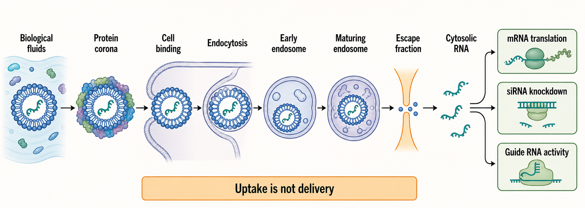
Figure 156.2. Uptake-to-expression trafficking cascade. Functional RNA delivery requires a sequence of conditional steps. LNPs contact biological fluids, adsorb proteins, bind cells, enter endocytic compartments, traffic through maturing endosomes, release a small fraction of RNA into the cytosol, and only then produce mRNA translation, siRNA knockdown, or guide RNA activity.
A causal trafficking sequence for an mRNA-LNP can be stated in steps. First, the particle contacts extracellular proteins and cell-surface glycocalyx components. Second, the particle binds directly or indirectly to uptake receptors or membrane domains. Third, the plasma membrane internalizes the particle into an endocytic vesicle. Fourth, the vesicle matures, acidifies, and exchanges membrane components. Fifth, the particle destabilizes, reorganizes, or mixes with the endosomal membrane. Sixth, some fraction of RNA crosses into the cytosol. Seventh, cytosolic mRNA is engaged by ribosomes, while cytosolic siRNA can enter RNA-induced silencing complex pathways. Failure at any step reduces functional output, and different assays measure different steps.
Fluorescent microscopy is useful but easy to overinterpret. A fluorescent lipid signal can mark lipid distribution without proving where RNA went. A fluorescent RNA label can change RNA behavior or remain fluorescent after partial degradation. Colocalization with endosomal markers can show association with compartments, but resolution limits and marker timing complicate interpretation. Reporter protein expression is closer to functional mRNA delivery, but it integrates uptake, escape, RNA stability, translation, protein folding, and protein half-life. For siRNA, target knockdown integrates cytosolic loading, target abundance, Argonaute activity, and downstream turnover. A rigorous study often needs multiple orthogonal assays.
Box 156.2. Evidence Ladder from Uptake to Cytosolic Function
A practical evidence ladder. Cell-associated fluorescence is the weakest delivery claim because particles can stick to membranes, extracellular matrix, dead cells, or phagocytes. Internalization assays are stronger, but they still do not show that RNA left the endocytic system. Endosomal colocalization, pH probes, and trafficking perturbations identify routes and compartments, yet they usually report opportunity for escape rather than escape itself. Membrane-damage reporters, lipid-mixing assays, split-enzyme systems, or cytosolic probes move closer to the escape event, but each has artifacts and detection thresholds. Functional readouts are usually most relevant: protein expression for mRNA, target knockdown for siRNA, editing for guide systems, or antigen presentation for vaccines. The strongest studies combine several rungs, normalize dose and particle attributes, and test whether the same conclusion survives a different assay class.
Endosomal maturation also changes immune signaling. Endosomes contain Toll-like receptors that sense nucleic acids, including TLR7 and TLR8 for single-stranded RNA in immune cells. Cytosolic leakage of RNA or endosomal damage can activate other stress and innate immune pathways. A formulation that increases escape may also increase endosomal damage signals. Recent work on limiting endosomal damage sensing highlights that productive escape and inflammatory sensing are mechanistically intertwined rather than completely separable (Omo-Lamai et al. 2025).
Do not treat high cellular uptake as proof of an effective LNP. The most relevant endpoint depends on the product: protein expression for mRNA, knockdown for siRNA, editing outcome for editor delivery, antigen presentation for vaccines, or cell-state programming for immunotherapy. Uptake assays belong at the beginning of the evidence chain, not at the end.
156.4. Endosomal escape and intracellular release
Endosomal escape is the central unsolved bottleneck in RNA-LNP delivery. The term means that intact or sufficiently functional RNA reaches the cytosol from an endosomal compartment. For mRNA, cytosolic release is required because ribosomes translate mRNA in the cytosol or on cytosolic faces of the endoplasmic reticulum. For siRNA, cytosolic release is required for loading into Argonaute-containing silencing complexes. For many RNA editing or CRISPR-related systems, escape must be coordinated with the location and timing of the effector molecule. A particle that is efficiently internalized but inefficiently escaped is biologically similar to a parcel delivered to the wrong locked room.
The traditional explanatory model begins with ionizable lipid protonation in acidifying endosomes. Protonated ionizable lipid can form ion pairs with anionic endosomal lipids such as phosphatidylserine or bis(monoacylglycero)phosphate. These interactions may promote lipid mixing, membrane curvature stress, and non-bilayer structures that create transient defects or destabilize the endosomal membrane. In this view, the ionizable lipid is not simply a proton sponge. It is a membrane-active chemical species whose charge state and shape change the local lipid phase behavior. Recent reviews on cationic and ionizable lipid evolution emphasize this shift from simple charge neutralization to membrane reorganization models (Mrksich et al. 2024; Zhao et al. 2024).
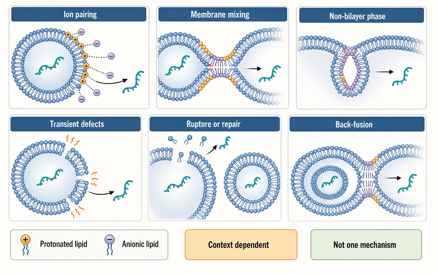
Figure 156.3. Competing but compatible endosomal escape models. Endosomal escape may involve several membrane events rather than one universal mechanism. Protonated ionizable lipids can interact with anionic endosomal lipids, driving lipid mixing, curvature stress, non-bilayer arrangements, defects, damage responses, or vesicle back-fusion depending on formulation and cell context.
Several mechanistic models coexist because the evidence is incomplete and context-dependent. One model emphasizes formation of inverted hexagonal or other non-bilayer phases that stress the endosomal membrane. Another emphasizes transient pores or membrane defects formed during lipid mixing. Another emphasizes rupture or damage of a subset of endosomes, followed by repair or inflammatory signaling. Another emphasizes back-fusion of intraluminal vesicles in multivesicular bodies. These models are not mutually exclusive. Different lipids, cargos, cell types, and endosomal compartments may use different combinations of events.
Escape efficiency is usually low. A large fraction of internalized RNA can be routed to lysosomes or remain trapped in vesicles. This low efficiency is one reason LNP dose, particle potency, and reactogenicity are tightly linked. Increasing dose can increase the absolute amount of escaped RNA, but it can also increase the number of particles that stimulate endosomal receptors, damage membranes, activate complement, or accumulate in non-target cells. Improving escape per particle is therefore a major design goal because it could reduce dose and inflammation, but better escape cannot be assumed to improve safety if escape is coupled to membrane damage.
The cargo itself affects escape measurement. mRNA expression can amplify a small amount of cytosolic RNA into a large protein signal if the mRNA is stable and efficiently translated. Conversely, a fragile mRNA can underreport escape if it degrades quickly after release. Self-amplifying RNA adds a further layer because replication can amplify cytosolic RNA after a threshold is crossed, but replicase expression and double-stranded RNA intermediates can change immune sensing. siRNA knockdown can persist after transient cytosolic entry because loaded Argonaute complexes are stable. Comparing escape across cargo classes requires careful normalization.
Direct evidence for escape is difficult. Researchers use endosomal pH probes, membrane damage reporters, galectin recruitment, split enzyme systems, fluorescence dequenching, electron microscopy, cryo-electron tomography, lipid mixing assays, subcellular fractionation, reporter translation, and genetic perturbation of trafficking pathways. Each method reports a related but imperfect feature. Galectin recruitment, for example, can indicate endosomal membrane damage that exposes luminal glycans to cytosolic galectins, but not every productive escape event must create a large galectin-positive rupture. Reporter expression shows functional delivery but cannot identify the exact membrane event. Biophysical assays reveal possible lipid phases but may not reproduce the crowded endosomal environment.
Recent chapter-local references are especially relevant here. Yu et al. 2025 focuses on internal self-assembled inverse mesophase structure and endosomal escape. Johansson et al. 2025 emphasizes cellular and biophysical barriers to RNA reaching the cytosol. Omo-Lamai et al. 2025 connects endosomal escape with damage sensing and inflammation. Together they support a cautious consensus: endosomal escape is real, inefficient, lipid-structure-dependent, and immunologically consequential, but no single assay or simple pKa rule currently predicts productive release across all systems.
Do not equate endosomal escape with lysosomal rupture as a universal event. Productive RNA release may occur through smaller, transient, or compartment-specific membrane defects. Also do not assume that more endosomal disruption is always better. For vaccines, some tissue stress may support immune priming; for repeat-dosed protein replacement or gene editing, excessive endosomal damage may narrow the therapeutic window.
156.5. Biodistribution, APC uptake, liver tropism, and extrahepatic delivery
Biodistribution describes where the administered formulation and its functional effects go in the body. For RNA-LNPs, biodistribution has several layers: where lipid components travel, where RNA remains intact, which cells internalize particles, which cells receive cytosolic RNA, and which tissues show protein expression, knockdown, editing, antigen presentation, toxicity, or inflammation. These layers can disagree. A tissue can contain lipid signal without productive RNA delivery; a small number of highly expressing cells can dominate a protein readout; immune-cell uptake can alter systemic cytokines without producing much therapeutic protein.
Intravenous LNP delivery often shows liver tropism. The liver receives a large fraction of cardiac output, has fenestrated sinusoids, contains professional phagocytes, and expresses receptors that can interact with serum-protein-coated particles. Hepatocytes are accessible from the sinusoidal side, and liver-resident macrophages and endothelial cells actively clear particles. Protein corona formation can promote or inhibit particular hepatic uptake routes. In some systems, apolipoprotein adsorption has been linked to hepatocyte delivery, although the chapter-local bibliography does not include the classic primary references needed for a detailed historical citation. The important mechanistic point is that liver tropism is not a magical property of lipid particles; it emerges from vascular anatomy, serum proteins, particle chemistry, and cell uptake machinery.
Liver delivery can be beneficial. Many siRNA and genome-editing strategies target liver-produced proteins. Hepatocytes can produce secreted therapeutic proteins from mRNA. The liver is also a major site for metabolism and immune surveillance, which can help clear biodegradable components. But liver delivery can be a liability when the desired target is immune cells, lung, heart, muscle, central nervous system, tumor, or local tissue. Unwanted hepatocyte or Kupffer-cell exposure can consume dose, create toxicity, or complicate interpretation of systemic biomarkers.
Table 156.2. Biodistribution endpoints are not interchangeable. Lipid distribution, RNA distribution, reporter expression, pharmacodynamic effect, immune activation, and toxicity are distinct endpoints; detecting one does not establish productive delivery or therapeutic activity.
| Endpoint | What it measures | What it does not prove | Common assay examples | Interpretation caveat |
|---|---|---|---|---|
| Lipid distribution | Location of labeled or quantified lipid components. | Intact particle delivery, RNA delivery, or cytosolic release. | Tagged lipid imaging, radiolabeling, lipid LC-MS, whole-organ fluorescence. | Lipid can persist, exchange, or separate from RNA; labels can change particle behavior. |
| RNA distribution | Tissue or cellular presence of RNA cargo or RNA-derived signal. | Functional cytosolic access or productive translation, knockdown, or editing. | qPCR, ddPCR, northern blotting, in situ hybridization, labeled RNA tracking. | Assays may detect degraded fragments, extracellular RNA, or endosome-trapped cargo. |
| Cellular uptake | Cell association or internalization of LNP-associated material. | Endosomal escape, intact RNA function, or therapeutic activity. | Flow cytometry, microscopy, tissue dissociation with fluorescent particles. | Sticky, phagocytic, or autofluorescent cells can dominate uptake without being productive target cells. |
| Reporter protein expression | Cells or tissues that translate an encoded reporter. | The exact uptake route, amount of escaped RNA, or absence of off-target exposure. | Luciferase imaging, fluorescent protein reporters, secreted reporter assays. | Protein output integrates RNA stability, translation efficiency, protein half-life, and detection sensitivity. |
| Pharmacodynamic effect | Downstream functional consequence of RNA delivery. | The upstream biodistribution path or all exposed off-target tissues. | Target knockdown, editing percentage, secreted protein, antigen-specific immune response, disease biomarker. | Delayed or systemic effects can obscure which cell type received cytosolic RNA. |
| Immune activation or toxicity | Inflammatory, complement, tissue-injury, or clinical safety response. | Therapeutic delivery or protective immunity. | Cytokine panels, complement assays, liver enzymes, histopathology, adverse-event monitoring. | Route, dose, schedule, species, and host immune state strongly affect interpretation. |
Antigen-presenting cell uptake is central for vaccines and immunotherapies. Dendritic cells, macrophages, and monocytes can internalize LNPs, translate antigen-encoding mRNA, process antigen, and present peptides on major histocompatibility complex molecules. They can also respond to RNA, lipid, damage, and cytokine signals. In an intramuscular mRNA vaccine, antigen expression may occur in muscle-resident cells and infiltrating immune cells, while antigen and inflammatory cues drain to lymph nodes. The desired outcome is not simply maximal expression in every cell; it is the right combination of antigen production, antigen presentation, innate immune context, and adaptive immune priming.
Extrahepatic targeting is a broad label for delivery outside the liver, but the barriers differ by tissue. Lung delivery must address pulmonary vascular and epithelial barriers and avoid excessive inflammatory toxicity. Spleen and lymph node delivery must distinguish among immune-cell subsets. Tumor delivery faces heterogeneous vasculature, high interstitial pressure, phagocytic cells, and immunosuppressive microenvironments. Muscle delivery can support local protein expression but may not generalize to systemic exposure. Central nervous system delivery must cross or bypass the blood-brain barrier. Eye and local tissue routes may reduce systemic exposure but introduce local tolerability and procedure constraints.
Strategies for extrahepatic delivery include changing ionizable lipid chemistry, adding charged or selective organ-targeting lipids, tuning particle size and PEG shedding, adding ligands, choosing local routes, and designing biodegradable or tissue-responsive lipids. Selective organ targeting by lipid composition is promising, but the field must distinguish true cell-specific cytosolic delivery from changes in uptake, inflammation, or reporter sensitivity. A formulation that shifts luciferase expression from liver to spleen in mice may not deliver the same cell type in humans, may not work with a larger RNA cargo, and may not be acceptable at a clinically relevant repeat-dose schedule.
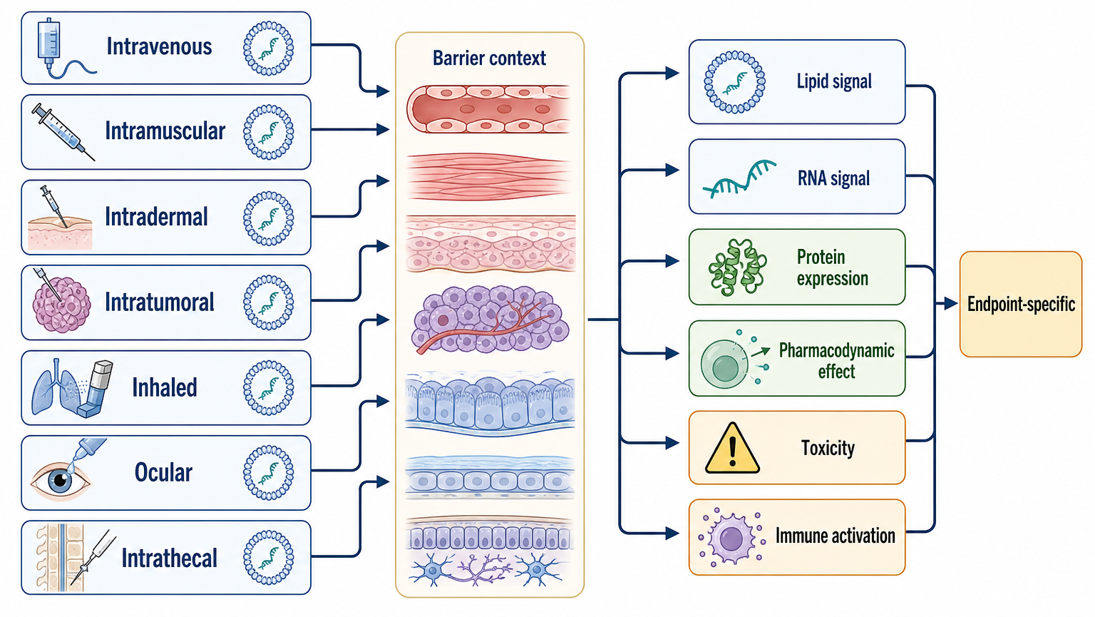
Figure 156.4. Biodistribution endpoints across routes. Biodistribution is endpoint-specific. Lipid signal, intact RNA, protein expression, pharmacodynamic effect, toxicity, and immune activation can map to different tissues or cell types, and each route imposes different anatomical and immunological barriers.
Route of administration is not a minor detail. Intravenous administration exposes particles immediately to blood proteins, complement, liver filtration, spleen, and vascular endothelium. Intramuscular administration creates a local depot, tissue injury, lymphatic drainage, and immune-cell recruitment. Intradermal or subcutaneous routes change local antigen-presenting cell access and lymphatic flow. Inhaled, intranasal, intratumoral, intrathecal, and ocular routes impose specialized mucus, extracellular matrix, epithelial, procedure-related, or safety barriers. Biodistribution claims should always specify route, dose, species, time point, assay, and whether the endpoint is lipid, RNA, protein, pharmacodynamic effect, or toxicity.
The chapter-local references support broad principles of selective intracellular delivery, cardiovascular applications, and physiological barriers (Soroudi et al. 2024; Zhao et al. 2024). They are not sufficient for a complete evidence map of every organ-targeting claim. Later curation should add product-specific studies, classic LNP-siRNA liver delivery papers, organ-selective lipid screens, and translational biodistribution datasets.
156.6. Protein corona, complement, reactogenicity, and repeat dosing
When an LNP enters blood or tissue fluid, it encounters proteins, lipoproteins, metabolites, extracellular matrix components, and immune sensors. Adsorbed proteins form a protein corona that can change the particle’s biological identity. The corona is not merely contamination; it is part of the in vivo surface. Opsonins can promote phagocytic uptake, apolipoproteins can alter receptor interactions, complement proteins can initiate inflammatory cascades, and albumin or other abundant proteins can mask or reshape interactions. The corona depends on species, disease state, route, dose, time after injection, and particle composition.
Complement activation is a major safety consideration for nanoparticle medicines. The complement system is a set of plasma and membrane proteins that can recognize foreign or altered surfaces, deposit opsonins, generate inflammatory anaphylatoxins, and promote clearance. LNPs can potentially activate complement through surface charge, PEG, impurities, aggregates, or adsorbed immune complexes. Complement activation can contribute to infusion reactions or pseudoallergic responses in susceptible settings. However, complement biology is route- and product-specific; an intramuscular vaccine reactogenicity profile should not be treated as identical to an intravenous infusion reaction risk.
Reactogenicity is the short-term inflammatory experience after administration. For an mRNA vaccine, fever, injection-site pain, fatigue, and myalgia reflect innate immune activation, tissue inflammation, cytokines, and local damage responses. Reactogenicity can coexist with strong immune priming, but it is not a precise surrogate for protective immunity. A highly reactogenic formulation is not automatically more effective, and a less reactogenic formulation is not automatically less immunogenic. For non-vaccine RNA therapies, reactogenicity is usually a liability because repeated or chronic administration demands tolerability.
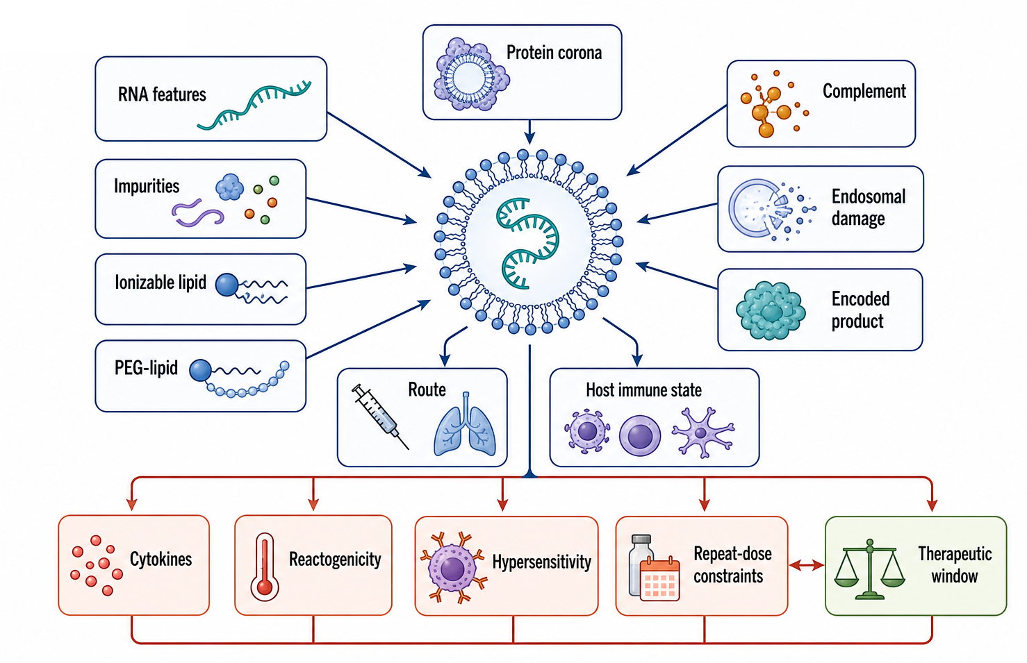
Figure 156.5. Immune activation pathways for RNA-LNP products. RNA-LNP immune effects arise from multiple sources. RNA sequence, modifications, impurities, ionizable lipids, PEG-lipids, protein corona, complement, endosomal damage, route, and host immune state combine to determine reactogenicity, hypersensitivity, repeat-dose constraints, and therapeutic window.
The RNA cargo and the LNP contribute separately and jointly to innate immune activation. RNA can activate endosomal Toll-like receptors, cytosolic RIG-I-like receptors, protein kinase R, oligoadenylate synthetase pathways, and other sensors depending on sequence, structure, modifications, impurities, and cellular context. LNPs can promote uptake into sensor-rich cells and can create endosomal damage signals. Ionizable lipids and helper components can alter inflammatory signaling even with non-immunostimulatory cargo. Double-stranded RNA impurities or uncapped RNA impurities can increase innate immune sensing. The product’s immune profile is therefore a combined property of RNA design, RNA purity, formulation, route, dose, and host state.
Anti-PEG responses illustrate why excipients can become immunological variables. PEG has long been used to improve colloidal stability and circulation properties, but some individuals have pre-existing anti-PEG antibodies or can develop anti-PEG responses after exposure. Anti-PEG antibodies can accelerate clearance, alter biodistribution, or contribute to hypersensitivity in some contexts. The local references note PEG-lipid design as a formulation issue, but they do not provide a complete clinical anti-PEG evidence base. A later citation pass should add dedicated reviews and primary studies on anti-PEG prevalence, assay variability, product-specific relevance, and repeat-dose outcomes.
Repeat dosing creates a different risk profile from one-time or two-dose vaccination. In a chronic mRNA protein-replacement product, a patient may need repeated exposure over months or years. Repeat dosing can reveal cumulative toxicity, adaptive immune responses to the encoded protein, antibodies against PEG or other excipients, complement changes, liver enzyme changes, and altered pharmacokinetics after the first dose. In gene editing, even a one-time dose can produce long-lived biological effects, so short-term tolerability is not enough. In cancer immunotherapy, inflammatory activity may be partly desired but must be controlled to avoid systemic toxicity.
Box 156.3. Repeat Dosing Rewrites the Safety Question
For a single or infrequent vaccine series, transient local inflammation may be acceptable if antigen presentation and adaptive immunity are strong. For chronic mRNA protein replacement, repeated genome-editing support, or recurrent immune-cell programming, the safety question changes. Each dose can meet a host that has already seen the RNA cargo, encoded protein, PEG-lipid, ionizable lipid metabolites, or tissue injury pattern. Repeat exposure can alter pharmacokinetics, biodistribution, complement activation, anti-PEG or anti-excipient responses, liver chemistry, cytokine tone, and adaptive immunity to the encoded product. A repeat-dose program therefore needs longitudinal evidence: pre-dose immune status, dose-by-dose clinical chemistry, cytokines, complement or hypersensitivity monitoring, product-specific pharmacodynamics, tissue injury markers, and retained potency after prior exposure. Acute tolerability is necessary, but it is not a substitute for schedule-specific safety.
Endosomal escape is tied to immunogenicity because membrane damage can be sensed. Omo-Lamai et al. 2025 is particularly relevant because it links inflammation triggered by LNP endosomal escape to damage sensing. This does not mean that all escape is bad. Rather, the therapeutic window depends on separating enough cytosolic RNA delivery from excessive endosomal disruption and downstream inflammation. The field is moving toward designs that improve productive escape per inflammatory signal, not simply toward maximum membrane disruption.
Safety assessment should distinguish acute reactogenicity, hypersensitivity, complement activation, cytokine release, liver toxicity, tissue-specific injury, adaptive immunity to the encoded product, adaptive immunity to excipients, and long-term pharmacology. These endpoints require different assays and time scales. A mouse cytokine panel, a human in vitro complement assay, a nonhuman-primate repeat-dose study, and a clinical adverse-event table answer different questions. Later chapters on pharmacokinetics, toxicology, and regulatory science treat these evidence standards in more detail.
156.7. AI-guided lipid design, screening, and translation barriers
LNP design is a high-dimensional optimization problem. Variables include ionizable lipid head group, linker, tail structure, stereochemistry, degradability, helper lipid identity, sterol identity, PEG-lipid anchor and chain length, molar ratios, RNA cargo, buffer, mixing method, route, dose, and target cell type. Traditional one-variable-at-a-time experimentation cannot efficiently explore this space. Combinatorial chemistry, high-throughput formulation, pooled screening, barcoded nanoparticles, automation, and machine learning are therefore attractive tools.
AI-guided lipid design can mean several different things. A model may predict which lipid structures are synthetically accessible, which formulations will be stable, which particles will express strongly in a cell type, which tissues will receive cargo in an animal, or which designs may reduce toxicity. These are different prediction tasks with different data requirements. A model trained on luciferase expression in mouse liver cannot be assumed to predict dendritic-cell delivery in human lymph nodes. A model trained on uptake cannot be assumed to predict endosomal escape. A model trained on one RNA length may not transfer to self-amplifying RNA. The input representation also matters: a lipid can be represented by chemical graph, descriptors, pKa estimates, tail features, or learned embeddings, while the formulation can be represented by molar ratios, particle measurements, and process variables.
High-throughput screens are powerful but can create false confidence. Reporter expression screens often favor formulations that express strongly in a convenient assay window, but they may miss delayed toxicity, complement activation, storage instability, or species-specific clearance. Pooled in vivo screens can compare many formulations, but barcode representation, dose competition, tissue dissociation bias, and cell sorting artifacts can distort rank order. Screens that measure RNA abundance may select uptake rather than cytosolic release. Screens that measure protein output may select for RNA stability and translation as well as delivery. The right screen must match the intended therapeutic endpoint.
Table 156.3. Screening endpoints and their failure modes. Uptake, reporter, knockdown, barcoded biodistribution, cytokine, complement, and stability screens answer different questions and can select misleading leads; each requires orthogonal follow-up tied to the intended product.
| Screen type | Useful question | Misleading interpretation | Minimum orthogonal follow-up | Translation risk |
|---|---|---|---|---|
| Uptake screen | Which formulations associate with or enter a chosen cell type. | High uptake means strong RNA delivery. | Functional expression or knockdown, endosomal localization, cell viability, and particle dose normalization. | Selects sticky or phagocytosed particles that remain endosome-trapped or inflammatory. |
| Reporter-expression screen | Which mRNA-LNPs produce detectable protein in a model system. | Reporter output is a pure measure of delivery. | RNA integrity, reporter half-life, cell-type mapping, dose response, and inflammatory readouts. | Overfits to reporter cargo, assay timing, mouse tissue, or translation efficiency. |
| siRNA knockdown screen | Which formulations deliver siRNA into Argonaute-accessible cytosol. | Knockdown potency will transfer to mRNA, guide RNA, or self-amplifying RNA. | Target rescue or orthogonal target assay, off-target assessment, cytokines, and tissue-level exposure. | Cargo-specific activity can mislead broader platform claims. |
| Barcoded in vivo biodistribution screen | How many formulations rank across tissues or sorted cells in one animal study. | Barcode abundance equals intact functional delivery. | Single-formulation validation with RNA, lipid, expression or pharmacodynamic endpoints, and histology. | Barcode competition, tissue dissociation bias, dose pooling, and species-specific uptake can distort rank order. |
| Cytokine or endosomal damage screen | Whether a formulation triggers acute innate signaling or membrane-damage markers. | Low signal in one panel means the product is broadly safe. | Complement, clinical chemistry, histology, repeat-dose testing, and human-relevant immune assays. | Misses delayed toxicity, adaptive immunity, or route-specific reactogenicity. |
| Complement or hypersensitivity assay | Whether the particle surface may trigger acute plasma immune cascades. | In vitro complement signal directly predicts clinical reaction frequency. | Human donor panels, species comparison, infusion-condition testing, and aggregate analysis. | Product route, dose, pre-existing antibodies, and assay format can change relevance. |
| Stability or CMC stress assay | Whether the formulation preserves critical quality attributes during storage or processing. | Stable size or encapsulation alone proves biological equivalence. | Potency after stress, RNA integrity, impurity testing, sterility, endotoxin, and lot comparability. | Discovery particles can fail scale-up, fill-finish, freezing, or shelf-life requirements. |
| AI-ranked virtual or active-learning screen | Which lipid structures or formulation variables deserve experimental priority. | A model prediction is validation of therapeutic delivery. | Prospective blinded testing with matched endpoint, negative controls, safety readouts, and process metadata. | Training data may encode uptake-only, mouse-only, proprietary, or publication-biased endpoints. |
Development programs also need stage-specific decision gates. A discovery campaign can tolerate rough particle analytics if the question is whether a lipid family has activity. Lead optimization must connect activity to particle attributes and early safety signals. Preclinical translation must test route, dose, species, biodistribution, immune activation, and repeat exposure. Chemistry, manufacturing, and controls development must show that the intended particle population can be made reproducibly and stored. Clinical entry requires a coherent bridge among mechanism, exposure, potency, safety, and manufacturing.
Table 156.4. Development-stage decision gates for RNA-LNP products. Discovery, lead optimization, preclinical translation, manufacturing scale-up, clinical entry, and repeat-dose development require different delivery, safety, and manufacturing evidence; success at one gate does not satisfy later gates.
| Stage | Delivery evidence | Safety evidence | Manufacturing evidence | Citation or data gap to close |
|---|---|---|---|---|
| Discovery | Activity of a lipid family in a defined cargo, cell model, route, or animal screen. | Basic cytotoxicity, acute cytokine signal, and obvious formulation intolerance. | Small-scale size, polydispersity, encapsulation, and RNA integrity. | Move beyond uptake or reporter signal to functional delivery and early inflammatory cost. |
| Lead optimization | Dose-response potency, target-cell mapping, cargo compatibility, and orthogonal escape or pharmacodynamic endpoints. | Cytokines, complement-relevant signals, membrane-damage markers, and early tolerability across candidate formulations. | Link composition and process variables to critical quality attributes and potency. | Connect mechanism claims to particle attributes rather than treating formulation as a black box. |
| Preclinical translation | Route-, dose-, species-, tissue-, and cell-type biodistribution with pharmacodynamic confirmation. | Clinical chemistry, histology, cytokines, complement, immunogenicity, and repeat-dose data when repeat use is intended. | Scalable process demonstration, storage stability, and stressed-lot potency. | Add product-specific, organ-selective, and translational biodistribution datasets. |
| CMC scale-up | Comparability of delivery after moving from discovery mixing to the intended manufacturing process. | Impurity, residual solvent, aggregate, endotoxin, bioburden, and filtration-related risk assessment. | Controlled mixing, solvent removal, buffer exchange, sterile filtration, fill-finish, and release specifications. | Add RNA-LNP CMC guidance and product-specific release-specification sources during later curation. |
| Clinical entry | Evidence bridge from mechanism, exposure, potency, and starting-dose rationale to the first patient or participant study. | Monitoring plan for reactogenicity, hypersensitivity, organ toxicity, cytokines, and product-specific pharmacology. | GMP lot release, validated potency assay, stability window, and chain-of-custody controls. | Add regulatory guidance, labels, and clinical pharmacology sources for approved or investigational products. |
| Repeat-dose or lifecycle development | Durability, redosing response, altered pharmacokinetics, and retained functional delivery after prior exposure. | Anti-PEG or excipient immunity, adaptive immunity to encoded product, cumulative toxicity, and complement changes. | Lot comparability, shelf-life extension, post-change bridging, and long-term stability. | Add anti-PEG, complement, and chronic-dose clinical evidence before making product-level generalizations. |
Translation barriers begin with scale. A formulation made in a small microfluidic device may not have the same mixing environment as a larger manufacturing process. Process scale changes residence time, mixing energy, temperature control, hold times, filtration stress, and batch-to-batch variability. The final product must meet specifications for particle size, polydispersity, encapsulation, lipid identity and purity, RNA integrity, residual solvent, endotoxin, bioburden, sterility, potency, and stability. A high-throughput discovery formulation may need substantial redesign before it becomes a manufacturable drug product.
Another barrier is model relevance. Mice are indispensable for discovery but imperfect for predicting human biodistribution, complement activation, cytokine responses, and dose scaling. Nonhuman primates can improve translational confidence for some endpoints, but they are expensive, limited in throughput, and still not humans. Human in vitro systems can model particular cell types but often lack tissue architecture, blood flow, immune context, and repeated exposure. Organoids, microphysiological systems, and humanized immune models may help, but each adds its own validation burden.
AI and screening also need negative data. A model cannot learn safety boundaries if failed formulations, toxic lipids, unstable particles, and weak delivery results are omitted. Publication bias and proprietary datasets are serious obstacles because many formulation outcomes remain internal to companies. Even when data are shared, batch metadata may be incomplete. For LNP design, a useful dataset should include lipid structures, formulation ratios, process parameters, particle characterization, RNA cargo properties, assay conditions, species, cell type, route, dose, time point, positive and negative controls, and uncertainty estimates.
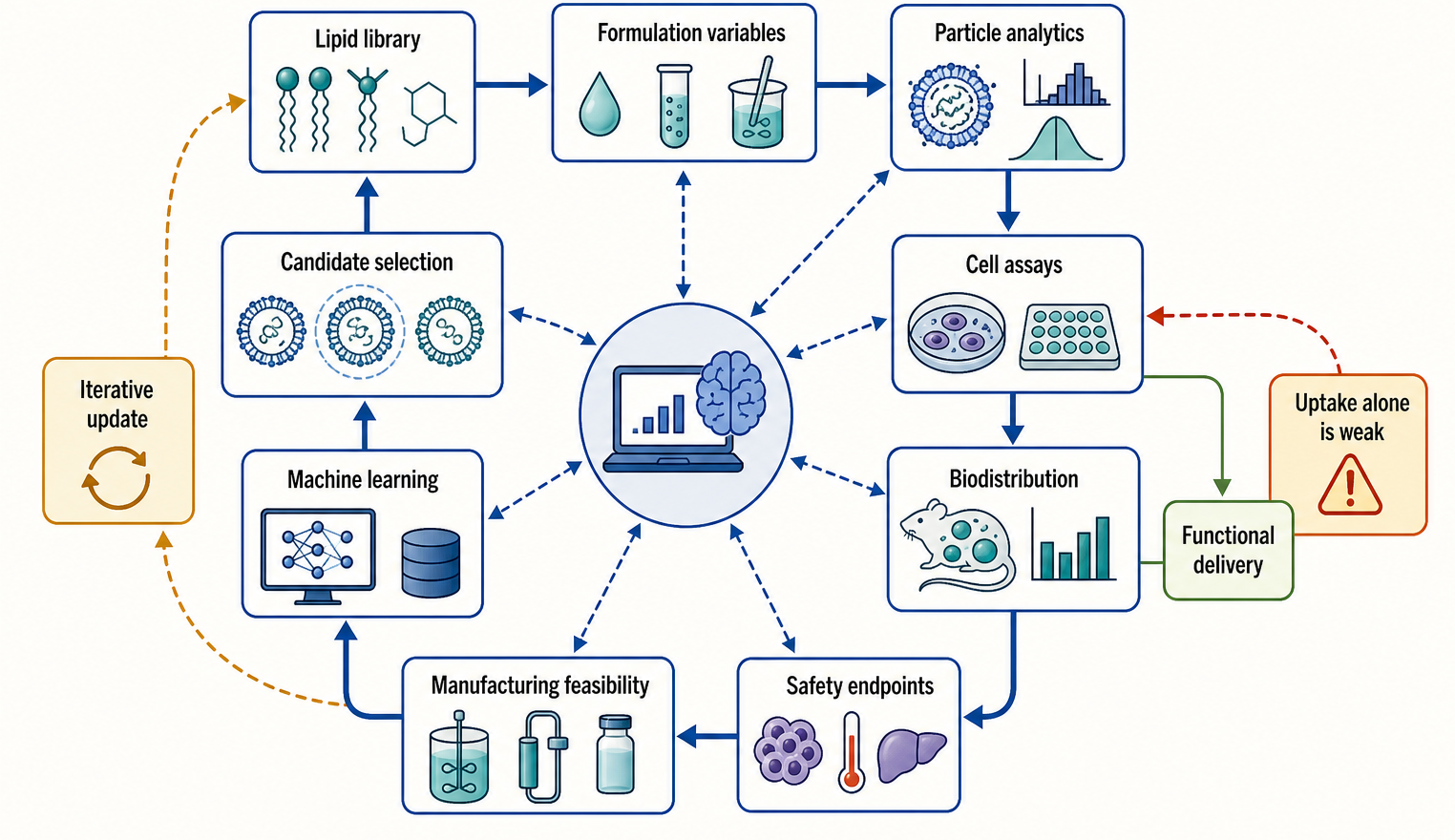
Figure 156.6. AI-guided LNP design loop. Data-driven LNP design is strongest when it integrates chemistry, formulation process variables, particle analytics, functional delivery assays, biodistribution, safety endpoints, and manufacturability. Models trained on weak endpoints, such as uptake alone, are not sufficient to validate therapeutic delivery.
Lipid-polymer hybrid nanoparticles and other hybrid systems expand the design space further. Polymer components can add mechanical stability, pH-responsive behavior, controlled release, or targeting features, but they also add manufacturing and safety complexity. Baghel et al. 2025 provides a chapter-local anchor for mRNA vaccine designs using lipid-polymer hybrid nanoparticles. The same evaluation principles apply: a hybrid carrier should be judged by functional delivery, immune profile, manufacturability, stability, and clinical feasibility, not by novelty of materials alone.
The consensus direction is not that AI will replace mechanistic delivery biology. Instead, computation can prioritize hypotheses, expose structure-activity relationships, design libraries, and integrate multi-endpoint data. Mechanistic experiments remain essential because the most important endpoints, endosomal escape, tissue-specific cytosolic delivery, inflammatory cost, and repeat-dose performance, are not directly observable from chemical structure alone. The best design programs combine chemistry, formulation science, cell biology, immunology, pharmacology, and manufacturing from the start.
Recent Consensus
Several points are well supported by the chapter-local references and by broad field consensus. First, RNA-LNPs are multicomponent systems whose biological behavior depends on the full formulation and process, not only on the ionizable lipid identity. Second, ionizable lipids improve the balance between RNA complexation and in vivo tolerability, but they do not eliminate toxicity or immune activation. Third, uptake and delivery are different endpoints; productive cytosolic access is the key functional barrier. Fourth, endosomal escape is inefficient, context-dependent, and incompletely understood. Fifth, biodistribution must be specified by route, species, cell type, and assay endpoint. Sixth, protein corona, complement, and innate immune sensing are not secondary details but determinants of efficacy and safety. Seventh, high-throughput and AI-guided design can accelerate discovery only when screens measure endpoints that translate to the intended product.
Open Questions, Controversies, Deprecated Models, and Common Misconceptions
Open questions:
- Which endosomal compartments contribute most to productive escape in different cell types?
- Can lipid structure predict escape without cell-specific empirical calibration?
- How can formulations increase productive cytosolic delivery while reducing endosomal damage signals?
- Which serum proteins drive beneficial versus harmful biodistribution in humans?
- Which animal models best predict anti-PEG responses, complement activation, and repeat-dose tolerability?
- How should potency assays be designed for complex RNA cargos such as self-amplifying RNA, circular RNA, editing systems, or multi-component cell programming products?
Common misconceptions:
- “Several misconceptions should be avoided.” An LNP is not simply a tiny lipid bubble around RNA. Ionizable lipids are not universally safe because they are neutral at physiological pH. Strong uptake is not strong delivery unless cytosolic function is demonstrated. More inflammation is not always better for vaccines and is usually worse for chronic therapeutics. Liver delivery is not an accident to ignore; it is a predictable consequence of anatomy and particle biology that may be useful or harmful. PEG is not immunologically invisible. AI-ranked lipids are not validated medicines until they pass formulation, mechanism, safety, manufacturing, and clinical evidence gates.
Deprecated or weakened claims:
- The proton sponge model, borrowed from some polymer delivery discussions, is too simple as a general explanation for LNP endosomal escape. Protonation matters, but current evidence favors membrane interaction, lipid mixing, phase behavior, and damage-response models rather than osmotic swelling alone. Similarly, “stealth” is an outdated shorthand for PEGylated particles. PEG can reduce some interactions while creating others, and biological fluids rapidly reshape the particle surface through corona formation.