Chapter 69. Termination, Ribosome Recycling, Recoding, and Nonstandard Translation Events
Scope Note
This chapter explains how ribosomes stop protein synthesis at stop codons, release completed polypeptides, recycle ribosomal subunits, and sometimes reinterpret the coding message through programmed frameshifting, stop-codon readthrough, selenocysteine insertion, or lineage-specific decoding. The emphasis is mechanistic: which molecules occupy the decoding center, how release and recycling differ among bacteria, archaea, eukaryotes, organelles, and viruses, how nonstandard events are measured, and how surveillance pathways distinguish useful recoding from translational failure.
Executive Summary
Translation termination is the decoding event in which a ribosome interprets UAA, UAG, or UGA not as an amino acid codon but as a signal to hydrolyze the ester bond between the completed polypeptide and the P-site transfer RNA. In bacteria, class I release factors RF1 and RF2 recognize overlapping sets of stop codons, and the GTPase RF3 helps release the class I factor after peptide release. In eukaryotic cytosol and archaea, a single class I factor, eRF1 or aRF1, recognizes all three standard stop codons, while eRF3 or a related GTPase accelerates termination. These factors are not tRNAs, but they mimic enough of the tRNA functional geometry to place a catalytic GGQ motif into the peptidyl transferase center. The stop codon is therefore decoded by a protein factor in the small-subunit decoding center, while peptide release is catalyzed in the large-subunit peptidyl transferase center.
Ribosome recycling is the separable process that disassembles the post-termination complex into ribosomal subunits, mRNA, and deacylated tRNA so that the components can begin another round of translation. Bacteria use ribosome recycling factor, EF-G, and initiation factor IF3; eukaryotes use ATP-binding cassette family factors such as ABCE1 together with release and initiation factors; organelles have specialized factors derived from bacterial ancestors but remodeled by organelle-specific rRNA and protein architecture. Recycling failures can leave ribosomes stalled at the end of open reading frames, reduce translational capacity, and feed into quality-control pathways.
Recoding is the controlled reinterpretation of an mRNA sequence by the translating ribosome. Programmed ribosomal frameshifting shifts the reading frame, most often by one nucleotide backward in many RNA viruses, when a slippery sequence and a downstream stimulatory RNA structure alter translocation or codon-anticodon realignment. Programmed stop-codon readthrough permits near-cognate tRNAs to decode a stop codon in a defined context, producing a C-terminally extended protein isoform. Selenocysteine insertion is a specialized UGA recoding event that requires a dedicated tRNA, a selenocysteine-specific elongation factor, and a selenocysteine insertion sequence element. Recoding is not simply translational error: its efficiency, sequence requirements, evolutionary conservation, and protein products can be biologically selected.
Viruses and organelles make extensive use of nonstandard translation events because compact genomes, unusual genetic codes, RNA structures, and host interaction pressures favor overlapping coding strategies. Retroviruses and many positive-strand RNA viruses use programmed frameshifting or readthrough to tune ratios of structural and enzymatic proteins. Mitochondria and chloroplasts use altered codon assignments, specialized ribosomes, and lineage-specific factors, so the same triplet can have different meaning depending on genetic compartment. These examples show why the genetic code is best treated as a cellular system of decoding rules rather than only a static codon table.
Surveillance pathways police the boundary between productive nonstandard translation and harmful decoding failure. Nonsense-mediated decay, no-go decay, nonstop decay, ribosome quality control, and innate immune surveillance respond to premature stop codons, ribosome stalls, aberrant readthrough, damaged mRNAs, viral RNAs, and collision-prone translation. Disease relevance includes inherited nonsense variants, ribosomopathies, neurodevelopmental and degenerative disorders linked to faulty surveillance, viral replication strategies that depend on recoding, and therapeutic attempts to modulate readthrough or recoding. The central caution is that ribosome footprints, reporters, or predicted RNA structures alone do not prove functional recoding; credible interpretation requires matched evidence for the mRNA signal, ribosomal event, protein product, efficiency, and biological consequence.
Concept Inventory
- Translation termination: the process by which a translating ribosome recognizes a stop codon in the A site and hydrolyzes the bond linking the nascent polypeptide to the P-site tRNA.
- Stop codon: UAA, UAG, or UGA in the standard code. Stop codons do not normally have cognate aminoacyl-tRNAs in the same way sense codons do; they are read primarily by release factors.
- Release factor: a protein factor that recognizes a stop codon or assists termination. Class I release factors directly promote peptide release, whereas class II release factors are GTPases that accelerate factor turnover.
- Post-termination complex: a ribosome still bound to mRNA and deacylated tRNA after the polypeptide has been released.
- Ribosome recycling: the factor-assisted dissociation of a post-termination complex into reusable subunits and ligands.
- Recoding: a programmed deviation from default triplet decoding that changes reading frame, stop-codon interpretation, or amino acid assignment at a defined site.
- Programmed ribosomal frameshifting: a recoding event in which the ribosome shifts into a different reading frame at a specific signal, generating a protein sequence not predicted from uninterrupted standard-frame translation.
- Stop-codon readthrough: incorporation of an amino acid at a stop codon, allowing translation to continue into a downstream extension.
- Selenocysteine: the selenium-containing amino acid inserted at selected UGA codons by a dedicated recoding system rather than by ordinary near-cognate suppression.
- Surveillance: cellular pathways that detect aberrant translation or mRNA features and trigger mRNA decay, nascent-chain degradation, ribosome rescue, stress signaling, or immune responses.
What to Know Before Reading This Chapter
This chapter assumes familiarity with the ribosome as a ribonucleoprotein machine with small and large subunits. The small subunit reads mRNA codons in the decoding center, while the large subunit contains the peptidyl transferase center that forms peptide bonds and later hydrolyzes the completed peptide from tRNA. The A site accepts incoming aminoacyl-tRNA or termination factors, the P site holds peptidyl-tRNA before peptide release, and the E site contains deacylated tRNA before exit.
The chapter also assumes the standard reading-frame concept. Once translation starts at an initiation codon, the ribosome normally reads mRNA in non-overlapping triplets. Shifting the frame by one nucleotide changes every downstream codon. Therefore a frameshift can create a different protein C terminus, fuse two open reading frames, or expose a premature stop codon. Chapters Chapter 66-Chapter 68 provide the decoding, initiation, and elongation background; the present chapter begins where elongation reaches the end of an open reading frame or encounters an mRNA signal that changes decoding behavior.
Two distinctions are essential. First, termination and recycling are coupled in cells but mechanistically distinct. Termination releases the polypeptide; recycling frees the ribosomal subunits and mRNA. Second, recoding and translational error are not synonyms. A near-cognate tRNA can accidentally decode a stop codon, and ribosomes can slip by chance, but programmed recoding uses sequence context, RNA structure, factor availability, and evolutionary selection to make a nonstandard event reproducible.
69.1. Termination factor recognition and stop-codon decoding
Translation termination begins when a stop codon enters the ribosomal A site. During elongation, most A-site triplets are decoded by aminoacyl-tRNAs delivered by elongation factors. Stop codons are different because cells generally do not maintain ordinary aminoacyl-tRNAs with anticodons that efficiently and accurately decode UAA, UAG, or UGA as standard amino acids. Instead, protein release factors inspect the A-site stop codon and trigger hydrolysis of the peptidyl-tRNA bond. Rodnina’s review of decoding and recoding emphasizes that termination is a decoding problem as much as a chemical-release problem: the ribosome must discriminate a true stop signal from near-cognate sense codons and from contexts where programmed recoding should compete with termination (Rodnina 2023, PMID 37159300).
Table 69.1. Nonstandard Translation Events and Evidence Standards. Summary of major nonstandard translation event classes, their required signals and products, what constitutes strong evidence, and the common experimental artifacts that confound interpretation.
| Event class | Core signal | Product | Strong evidence | Common artifact |
|---|---|---|---|---|
| Programmed -1 frameshifting | Slippery sequence plus RNA structure | Alternative-frame fusion protein | Mutational reporter, endogenous footprinting, recoded peptide, phenotype | RNA-structure mutation also changes protein sequence or RNA stability |
| Stop-codon readthrough | Weak stop context and downstream signal | C-terminally extended protein | Stop-context mutation, direct extension peptide, functional perturbation | Reporter overexpression or broad translational noise |
| Selenocysteine insertion | UGA plus Sec-tRNA and SECIS | Selenoprotein | SECIS dependence, Sec machinery dependence, selenopeptide or selenium-dependent activity | SECIS prediction without expression or protein evidence |
| Organellar reassignment | Compartment-specific tRNAs and release factors | Standard translation in that compartment | Genetic-code table, tRNA/factor repertoire, proteomics | Applying nuclear code to organellar transcripts |
| Viral recoding | Compact viral RNA signal | Regulated viral protein ratio | Infectious clone or replicon plus protein-ratio measurement | Recoding mutation changes overlapping RNA/protein constraints |
In bacteria, RF1 recognizes UAA and UAG, while RF2 recognizes UAA and UGA. This division explains why UAA is often the strongest bacterial stop codon: it can be recognized by both class I factors. Each bacterial class I release factor has domains that contact the stop codon in the decoding center and a universally important GGQ motif that reaches into the peptidyl transferase center. The letters GGQ refer to glycine-glycine-glutamine in the factor protein, not to an mRNA codon. The glutamine side chain and neighboring backbone geometry help position a water molecule for nucleophilic attack on the ester bond linking the nascent chain to the P-site tRNA. This reaction converts peptidyl-tRNA into a free polypeptide and deacylated tRNA.
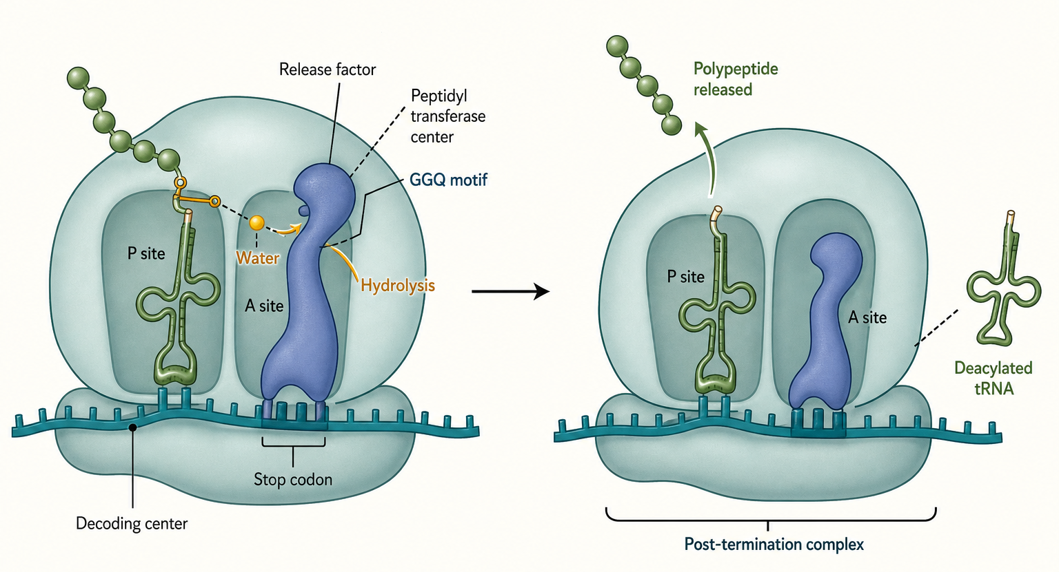
Figure 69.1. Stop-Codon Termination as a Two-Center Reaction. A stop codon in the A site is decoded by a release factor rather than by an aminoacyl-tRNA. The release factor contacts the small-subunit decoding center while its GGQ motif reaches the large-subunit peptidyl transferase center. Correct factor accommodation promotes water-mediated hydrolysis of peptidyl-tRNA, releasing the completed polypeptide and leaving a post-termination complex.
Eukaryotic cytosolic termination uses a different factor organization. eRF1 recognizes all three standard stop codons, and eRF3 is a GTPase that promotes efficient termination. Hellen’s review of eukaryotic termination and recycling describes eRF1 as a protein that functionally spans the decoding center and peptidyl transferase center, with stop-codon recognition and peptide release coordinated through conformational changes (Hellen 2018, PMID 29735640). Archaea use related logic with archaeal release factors. The important comparison is not that bacterial and eukaryotic factors are identical, but that both systems solve the same geometric problem: the A site must be occupied by a factor whose decoding-center interactions report “stop” while the factor’s catalytic motif activates hydrolysis in the large subunit.
Stop-codon context changes termination efficiency. Nucleotides immediately downstream of the stop codon, upstream coding sequence, mRNA structure, poly(A)-binding protein proximity in eukaryotes, and factor concentrations can influence whether termination is fast, slow, leaky, or paired with readthrough. A stop codon at the normal end of an open reading frame is usually embedded in an mRNP context that supports termination and later recycling. A premature termination codon far upstream of an exon junction complex or long 3′ untranslated region can be interpreted differently by surveillance pathways. This context dependence is why a codon table alone cannot predict all termination behavior.
The evidence base for termination is unusually broad. Biochemical reconstitution can measure peptide release from defined ribosomal complexes with purified factors. Cryo-electron microscopy and crystallography have visualized release factors in decoding and catalytic states. Ribosome profiling detects ribosome accumulation near stop codons and can reveal termination defects, but ribosome profiling requires careful interpretation because cycloheximide, harringtonine, lactimidomycin, chloramphenicol, nuclease digestion, and footprint assignment can create apparent peaks that are not direct kinetic measurements. Uematsu and Qian’s review of ribosome dynamics stresses that ribosome footprints are best interpreted as population snapshots shaped by initiation, elongation, termination, recycling, nuclease bias, and drug treatment rather than as simple dwell-time readouts (Uematsu and Qian 2025, PMID 40651611).
Boundary cases matter. Some organisms and organelles reassign stop codons, and some mRNAs encode intentional readthrough or selenocysteine insertion. In those cases, a triplet that looks like a stop codon in the standard code is not sufficient to infer termination. Conversely, not every low-level readthrough event has biological function. Translational accuracy has a measurable error distribution, and rare near-cognate suppression can occur without selection. The practical rule is to ask whether a stop-site event is reproducible, factor-dependent, conserved, protein-producing, and phenotypically meaningful.
69.2. Ribosome recycling systems
After peptide release, a ribosome remains bound to mRNA and usually retains a deacylated tRNA in the P site. This post-termination complex is not automatically ready for another round of translation. Ribosome recycling disassembles or remodels the complex so the small and large subunits, mRNA, and tRNA can be reused. Recycling is therefore an economy and quality-control step: without it, ribosomes would accumulate at stop codons, the pool of active subunits would fall, and downstream initiation or surveillance decisions would be distorted.
Bacterial recycling uses ribosome recycling factor (RRF), elongation factor G (EF-G), GTP hydrolysis, and initiation factor IF3. RRF structurally resembles a tRNA-like molecule in broad shape, but its function is not decoding. RRF binds the post-termination ribosome, EF-G promotes conformational changes using GTP hydrolysis, and the ribosomal subunits split. IF3 binds the small subunit and prevents immediate reassociation with the large subunit. The mRNA and deacylated tRNA then dissociate, leaving components available for initiation. This mechanism connects termination to the bacterial initiation cycle because IF3 also helps select proper start codon contexts and subunit states.
Eukaryotic recycling is organized around ABCE1, an ATP-binding cassette protein, together with release factors and initiation-related factors. ABCE1 binds post-termination or stalled ribosomal complexes and uses ATP-dependent conformational changes to split subunits. Hellen’s synthesis treats eukaryotic recycling as a staged process in which peptide release, factor accommodation, ABCE1 recruitment, ATP hydrolysis, and ligand release are coordinated rather than a single passive dissociation event (Hellen 2018, PMID 29735640). The same core distinction remains: termination releases the protein product, while recycling releases the ribosome from the mRNA-tRNA complex.
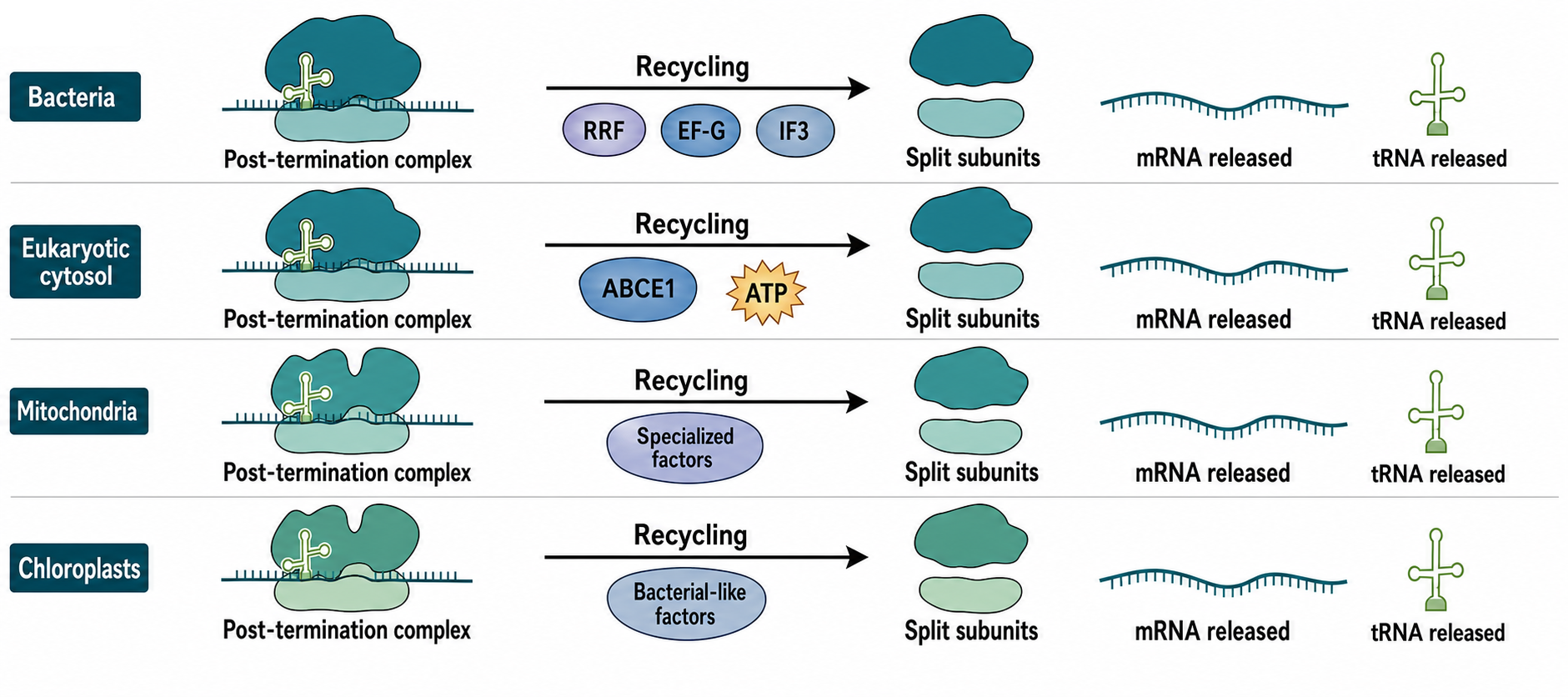
Figure 69.2. Ribosome Recycling Systems Across Compartments. Bacterial, eukaryotic cytosolic, mitochondrial, and chloroplast translation systems all recycle post-termination ribosomes, but the factor sets differ. Bacteria use RRF, EF-G, and IF3; eukaryotic cytosol uses ABCE1-centered ATP-dependent splitting; organelles use remodeled bacterial-like systems adapted to organelle-specific ribosomes.
Mitochondria and chloroplasts illustrate how recycling is conserved in purpose but divergent in implementation. These organelles descend from bacteria, yet their ribosomes have remodeled rRNA, organelle-specific proteins, altered mRNA features, and specialized biogenesis factors. Hillen and colleagues’ structural work on mitochondrial ribosome biogenesis and recycling provides evidence that GTPase-mediated remodeling of mitoribosomal complexes has organelle-specific architecture (Hillen et al. 2021, PMID 34135319). Chloroplast systems also use bacterial-like factors in an organellar environment, but the surrounding gene-expression system differs from free-living bacteria. Schmid and colleagues’ review of chloroplast ribosome biogenesis factors is useful background for how ribosome assembly and recycling factors become specialized in plastids (Schmid et al. 2024, PMID 37498958).
Recycling can be confused with rescue, but the terms should be separated. Recycling normally follows successful termination. Rescue occurs when a ribosome is stuck on a problematic mRNA, such as a nonstop mRNA without an in-frame stop codon, a chemically damaged message, a strong stall sequence, or a truncated transcript. Bacteria have trans-translation and alternative rescue factors; eukaryotes have ribosome quality-control and mRNA decay pathways that split stalled ribosomes and target nascent chains. Chapter 71 treats collisions and rescue in detail, but recycling factors in the present chapter should be understood as part of the same broader logic: ribosomal subunits are valuable, and cells invest energy to recover them from completed or failed translation events.
The evidence basis for recycling includes purified translation systems, factor-depletion genetics, ribosome profiling around stop codons, structural snapshots of post-termination complexes, and disease-associated factor perturbations. Each method has limits. In vitro reactions may lack mRNP context and competing initiation events. Structural states may be stabilized by mutations, antibiotics, nonhydrolyzable nucleotides, or sample preparation. Ribosome profiling can show stop-codon accumulation but cannot by itself distinguish slow peptide release from slow subunit splitting. A strong interpretation therefore triangulates factor dependence, biochemical activity, ribosome-state structure, and transcriptome-wide signatures.
Recycling defects can be selective. Turnbull and colleagues reported that the ABCF ATPase New1 resolves termination defects associated with specific tRNAArg and tRNALys isoacceptors in the P site, showing that the identity of the P-site tRNA can influence late translation events (Turnbull et al. 2024, PMID 39217469). This example is pedagogically important because it breaks the oversimplified view that termination depends only on the A-site stop codon. The P-site tRNA, nascent-chain context, factor repertoire, and ribosomal state can all affect the efficiency of the final steps of translation.
69.3. Programmed frameshifting and readthrough
Programmed ribosomal frameshifting is a recoding event in which the ribosome changes reading frame at a defined mRNA signal. The most familiar form is programmed -1 frameshifting, where the ribosome shifts one nucleotide backward relative to the original frame. A typical -1 frameshift signal contains a slippery sequence, where codon-anticodon pairing can realign in the new frame, followed by a downstream stimulatory RNA structure such as a pseudoknot or stem-loop. During translocation, pausing or mechanical resistance can increase the probability that tRNAs realign on overlapping codons. The result is a fusion protein or altered C-terminal sequence.
Frameshifting differs from random slippage because it is site-specific and often tuned to a useful efficiency. Retroviruses use programmed frameshifting to produce a controlled ratio of Gag structural proteins to Gag-Pol enzymatic proteins. Many positive-strand RNA viruses use frameshifting to expand coding capacity and regulate stoichiometry. Coronaviruses use a -1 frameshift to produce replicase polyproteins from overlapping open reading frames. If frameshifting is too low, enzymes are underproduced; if too high, structural-to-enzymatic ratios become abnormal. Viral dependence on frameshift efficiency makes recoding signals potential antiviral targets, but targeting them without harming host translation remains difficult.
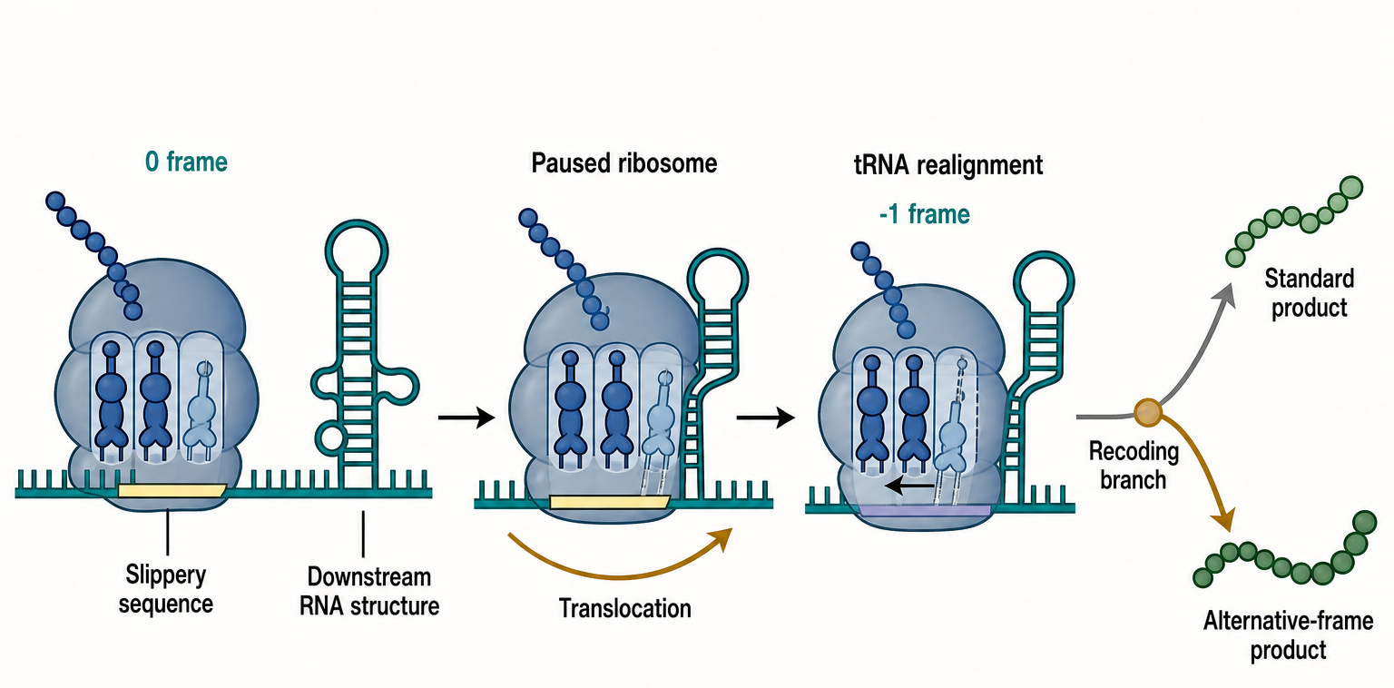
Figure 69.3. Programmed -1 Ribosomal Frameshifting. A programmed -1 frameshift signal contains a slippery sequence and a downstream stimulatory RNA structure. During translocation, ribosome pausing and codon-anticodon realignment shift the ribosome into the -1 reading frame, producing a downstream protein sequence different from standard-frame translation.
Programmed stop-codon readthrough is a different recoding strategy. Instead of shifting frame, a near-cognate tRNA incorporates an amino acid at a stop codon, and the ribosome continues translation into a downstream extension. Readthrough can create protein isoforms with new localization signals, regulatory motifs, membrane anchors, or interaction surfaces. Some viruses use readthrough to produce polymerase or capsid-associated proteins from compact genomes. In cellular genes, readthrough can be regulated by stop-codon identity, downstream nucleotides, RNA structures, tissue state, stress, and release-factor competition.
The mechanistic competition at a readthrough site is direct: release factors and near-cognate tRNAs compete for the A site. A strong stop context and abundant release factors favor termination. A weak stop context, favorable downstream sequence, modified tRNA availability, or stimulatory RNA element can allow amino acid incorporation. This competition links readthrough to both decoding fidelity and mRNA architecture. A UGA codon followed by specific downstream features may be read through more often than a UAA codon in a strong termination context, but context-specific measurements are necessary because codon identity alone is not predictive.
Reporter assays are common tools for measuring frameshifting and readthrough. A typical reporter places a candidate recoding signal between two coding modules so that downstream reporter activity depends on recoding. Reporters are powerful because they isolate signal sufficiency and allow mutational analysis, but they can mislead when the inserted sequence lacks native mRNP context, RNA structure, translation rate, or protein-product stability. Ribosome profiling can detect footprints in alternative reading frames or downstream of stop codons, but frame assignment depends on read length, P-site offset calibration, nuclease behavior, and mapping ambiguity. Proteomics can identify recoded peptides, but low abundance and shared peptide sequences limit sensitivity. The most credible cases combine reporter mutagenesis, endogenous perturbation, ribosome profiling or toeprinting, and direct protein evidence.
Box 69.1. Evidence Ladder for Endogenous Recoding
- Identify the candidate mRNA signal and default-frame product.
- Mutate the signal without disrupting unrelated protein or RNA constraints where possible.
- Measure recoding in a reporter and at the endogenous locus.
- Calibrate ribosome profiling or toeprinting for frame and position.
- Detect the recoded protein or peptide directly when feasible.
- Test whether changing recoding efficiency changes biological function.
Butcher and Jan’s review of tRNA mimicry in internal ribosome entry site-mediated translation and recoding highlights a broader principle: RNA structures can manipulate ribosome behavior by presenting surfaces or mechanical constraints that resemble normal translation ligands or alter their timing (Butcher and Jan 2016, PMID 27654067). This principle applies not only to frameshift stimulators but also to viral RNAs that recruit, position, or stall ribosomes. Rodnina’s decoding and recoding review similarly frames recoding as a kinetic branch point in the translation cycle, not as a violation of translation chemistry (Rodnina 2023, PMID 37159300).
Do not overgeneralize from viral examples. Viral genomes are compact and often evolve high recoding efficiency under strong stoichiometric constraints. Cellular readthrough and frameshifting events may be lower efficiency, condition-specific, or difficult to validate. Some apparent alternative-frame translation products can come from unannotated initiation, RNA editing, splicing, sequencing artifacts, or proteomic false discovery. The reader should treat recoding claims as mechanistic hypotheses until the mRNA signal, translational event, protein product, and function are linked.
69.4. Stop-codon recoding and selenocysteine
Selenocysteine is an amino acid structurally related to cysteine but containing selenium in place of sulfur. It is inserted into selected proteins at UGA codons, which are otherwise stop codons in the standard code. Selenocysteine insertion is therefore a stop-codon recoding event, but it is not ordinary readthrough. It requires a dedicated selenocysteine tRNA, specialized biosynthetic enzymes that convert a serine-loaded precursor tRNA into selenocysteyl-tRNA, a selenocysteine-specific elongation factor, and an RNA signal called a selenocysteine insertion sequence, usually abbreviated SECIS.
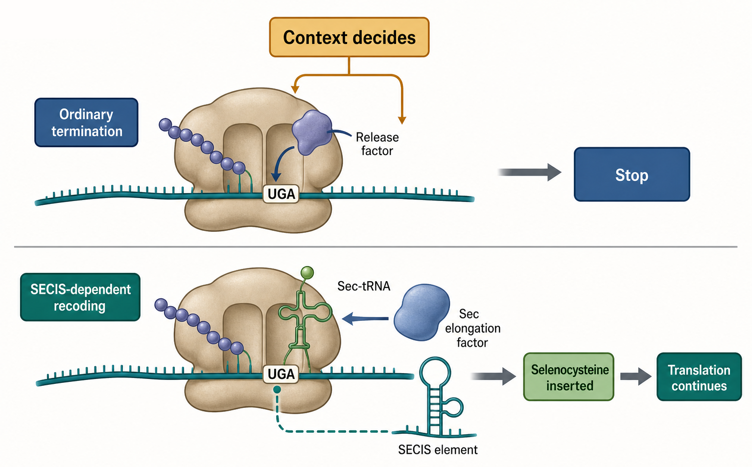
Figure 69.4. Selenocysteine Insertion Versus Ordinary UGA Termination. The same UGA triplet can lead to termination or selenocysteine insertion depending on context. Ordinary UGA termination recruits release factor, whereas a selenoprotein mRNA uses Sec-tRNA, a dedicated elongation factor, and a SECIS element to insert selenocysteine.
The location and architecture of SECIS elements differ among systems. In bacteria, SECIS elements are commonly near the UGA codon in the coding region. In eukaryotes, SECIS elements are usually in the 3′ untranslated region and require additional protein factors to communicate with the translating ribosome. The key mechanistic point is that UGA meaning is conditional. A UGA codon in a selenoprotein mRNA can recruit the selenocysteine insertion machinery, whereas a UGA codon in an ordinary termination context recruits release factors and stops translation. The codon is the same; the decoding environment differs.
Selenoproteins often participate in redox biology, thyroid hormone metabolism, antioxidant defense, protein folding, and selenium homeostasis. The selenium atom gives selenocysteine distinctive chemical reactivity, but that reactivity also imposes biosynthetic and regulatory costs. Organisms vary in selenoprotein repertoires: some lineages encode many selenoproteins, while others have reduced or lost the system. This variation makes selenocysteine a good example of code flexibility constrained by metabolism, ecology, and genome evolution.
Mukai and colleagues described natural facile recoding of selenocysteine, supporting the idea that selenocysteine decoding systems can be more flexible and diverse than a single textbook model suggests (Mukai et al. 2016, PMID 26991476). Vindry and colleagues used a CRISPR-Cas9 viral-like particle strategy targeting selenocysteine-tRNA genes to reduce UGA-selenocysteine recoding efficiency, illustrating how perturbing the dedicated tRNA can modulate Sec insertion in cells (Vindry et al. 2019, PMID 31212706). These primary studies support the chapter’s mechanistic framing, and a recent consensus review on eukaryotic and bacterial selenocysteine insertion machinery remains a final-reference-expansion item before final publication.
Stop-codon recoding also includes non-selenocysteine readthrough events that produce extended proteins. Pandit and colleagues reported termination codon readthrough of NNAT mRNA in a neuronal differentiation context, connecting readthrough to calcium-mediated differentiation biology (Pandit et al. 2023, PMID 37611826). That example should be read as a specific case, not as evidence that all readthrough is broadly regulatory. The relevant questions are which stop codon is involved, which amino acid is inserted, which downstream extension is produced, whether the event occurs at the endogenous locus, and whether perturbing the event changes function.
Selenocysteine claims are vulnerable to several artifacts. A UGA codon followed by conserved sequence does not prove Sec insertion; the mRNA may not be expressed, the SECIS may be inactive in a given organism, or annotation may have borrowed assumptions from another lineage. Conversely, proteomic absence of a selenopeptide does not prove absence of insertion because selenoproteins can be low abundance, condition-specific, or difficult to detect. Functional annotation should combine SECIS prediction, tRNA and factor repertoire, evolutionary conservation, expression, direct peptide evidence where possible, and selenium-dependent physiology.
Box 69.2. Do Not Annotate Stop Codons Without Context
- UAA, UAG, and UGA are stop codons in the standard nuclear code.
- UGA can encode selenocysteine in SECIS-dependent contexts.
- Organellar genetic codes can reassign triplets.
- Viral and cellular mRNAs can use programmed readthrough.
- Sequence-only annotation should record organism, compartment, transcript processing, and evidence level.
69.5. Nonstandard events in viruses and organelles
Viruses are especially rich in nonstandard translation events because viral genomes face pressure to encode many functions in limited nucleic acid length. Overlapping open reading frames, frameshifting, readthrough, leaky scanning, internal initiation, ribosome shunting, and structured RNA elements allow viruses to tune protein output from compact genomes. This chapter focuses on late elongation and termination-related events, while Chapter 67 and Chapter 70 treat viral initiation and IRES-driven regulation in more detail.
Positive-strand RNA viruses provide clear examples. Their genomic RNA can function as mRNA, replication template, and packaging substrate. Some viruses use readthrough to extend a capsid or replicase protein; others use -1 frameshifting to access polymerase domains. Coronavirus frameshifting occurs upstream of replicase open reading frame 1b and depends on a slippery sequence plus downstream RNA structure. Structural studies of coronavirus replication organelles by Wolff and colleagues and of SARS-CoV-2 NSP6 function by Ricciardi and colleagues address replication-organelle architecture rather than frameshifting directly, but they reinforce that viral translation products generated by nonstandard decoding operate inside specialized RNA replication environments (Wolff et al. 2020, PMID 32763915; Ricciardi et al. 2022, PMID 35551511). Den Boon and colleagues review positive-strand RNA virus replication organelles as structured cellular compartments that coordinate viral RNA synthesis, translation, and host interaction (den Boon et al. 2024, PMID 38724328).
Viral recoding signals must be interpreted within infection biology. A reporter may show that a viral sequence supports frameshifting, but the infection phenotype depends on viral protein ratios, polyprotein processing, RNA replication, immune sensing, and packaging. Conversely, a mutation that reduces viral replication may affect RNA structure, amino acid sequence, protein processing, or RNA stability in addition to frameshifting. Strong viral recoding studies therefore separate recoding efficiency from overlapping constraints by using compensatory mutations, synonymous controls, protein-ratio measurements, and infectious clone systems when available.
Organelles add a different kind of nonstandard translation. Mitochondria and chloroplasts have genetic systems derived from bacteria but remodeled by endosymbiosis. Mitochondrial genetic codes can reassign codons that are stops or sense codons in the standard code. Human mitochondria, for example, use a genetic code in which some assignments differ from nuclear translation. Plant organelles add extensive RNA processing and editing that can change codons before translation. Chloroplasts retain more bacterial-like translation features than mitochondria in many respects, but chloroplast gene expression is still coordinated with nuclear-encoded factors and photosynthetic physiology.
Organellar nonstandard events are not exceptions to molecular causality; they are consequences of altered decoder inventories. A codon is interpreted by the tRNAs, release factors, ribosomal RNA, ribosomal proteins, mRNA processing state, and accessory factors present in that compartment. If a mitochondrial genome lacks a tRNA or uses a modified tRNA with expanded pairing, codon assignment can shift. If an organelle release factor has altered specificity, stop-codon recognition can change. Robles and Quesada’s overview of plant organelle genetics provides broader context for the diversity of organellar gene expression systems (Robles and Quesada 2021, PMID 33672640).
The boundary between viral and organellar topics is useful for experimental design. Viral recoding often can be tested by infectious clones, replicons, and reporter constructs. Organellar recoding often requires genetic, biochemical, and comparative approaches because organellar transformation may be difficult, tissue-specific expression matters, and nuclear genes encode many organellar translation factors. In both cases, annotation by standard nuclear codon rules can be wrong. A sequence analyst must know the organism, compartment, transcript processing state, and translation system before assigning protein products.
69.6. Surveillance and disease relevance
Cells monitor translation because errors at termination, recycling, and recoding can produce truncated proteins, extended proteins, stalled ribosomes, depleted ribosome pools, or aberrant RNA-protein complexes. Surveillance is not one pathway but a collection of linked systems. Nonsense-mediated decay targets many mRNAs with premature termination codons. No-go decay responds to ribosomes stalled during elongation. Nonstop decay responds to mRNAs lacking proper stop codons. Ribosome quality control acts on stalled or collided ribosomes and can trigger nascent-chain ubiquitylation, mRNA cleavage, stress signaling, and subunit rescue. Alagar Boopathy and colleagues review proteostasis regulation through ribosome quality control and no-go decay, providing the main bridge from this chapter to Chapter 71 (Alagar Boopathy et al. 2023, PMID 37488089).
Premature termination codons are clinically important because many inherited and somatic variants create nonsense mutations. A premature stop codon can truncate a protein, reduce mRNA abundance through nonsense-mediated decay, or produce a dominant-negative fragment if the mRNA escapes decay. Therapeutic readthrough seeks to make ribosomes insert an amino acid at a premature stop codon, restoring some full-length protein. The concept is attractive but difficult. Readthrough drugs must be sufficiently selective, avoid widespread stop-codon suppression, produce a tolerable amino acid at the disease site, and generate enough functional protein without unacceptable toxicity. Disease benefit depends on the gene, mutation context, protein domain structure, tissue exposure, and surveillance response.
Surveillance also shapes viral infection. Viral RNAs can generate unusual translation patterns, double-stranded RNA intermediates, stalled ribosomes, or non-self RNA features. Innate immune sensors detect viral RNA in ways that intersect with translation status and RNA localization. Chen and colleagues review RNA virus surveillance from a One Health perspective, emphasizing that RNA virus detection spans molecular sensing, diagnostics, ecology, and public health (Chen et al. 2025, PMID 39681124). Kim and colleagues’ work on exogenous RNA surveillance by proton-sensing TRIM25 provides a recent primary example of RNA surveillance connected to antiviral signaling (Kim et al. 2025, PMID 40179174). These sources are not termination-specific, but they show why nonstandard viral translation cannot be separated from host RNA surveillance.
Ribosome heterogeneity and specialization may modulate termination and recoding, but this area requires caution. Different tissues, developmental stages, stress states, and organisms can vary in ribosomal protein paralogs, rRNA modifications, associated factors, and tRNA pools. Norris and colleagues review ribosome heterogeneity and specialization in development (Norris et al. 2021, PMID 33565275). Zhao and colleagues reported that 2′-O-methylation maintains ribosome structural and translation integrity, supporting the principle that rRNA modification state can affect ribosome function (Zhao et al. 2026, PMID 41928510). However, claims that a specialized ribosome selectively translates a specific recoding program need direct evidence, not only correlation between ribosome composition and gene expression.
Disease relevance extends beyond inherited nonsense variants. Ribosome recycling factor defects, mitochondrial translation defects, altered release-factor abundance, tRNA modification disorders, and viral recoding dependencies can all affect cell physiology. Protozoan parasites may have specialized ribosome features relevant to host-pathogen biology, as reviewed by Rodríguez-Almonacid and colleagues (Rodríguez-Almonacid et al. 2023, PMID 37108644). Ribosome dysfunction has been discussed in disease contexts such as osteoarthritis, though such links often involve broad translation stress rather than a single termination mechanism (van den Akker et al. 2022, PMID 34750309). The key interpretive discipline is to avoid assigning a disease phenotype to “recoding” unless the causal event has been measured.
Several artifacts recur in surveillance and disease studies. First, stop-codon readthrough reporters can overestimate therapeutic relevance because reporter proteins tolerate many C-terminal extensions and are expressed outside native chromatin and mRNP context. Second, ribosome profiling peaks at 3′ ends may reflect slow recycling, nuclease-protected scanning complexes, collided ribosomes, or library artifacts. Third, proteomic identification of extended proteins can be confounded by database search space and false discovery control. Fourth, viral recoding mutations can change RNA structure and protein sequence simultaneously. Each artifact is manageable, but only if the experiment is designed around the specific alternative explanation.
Recent Consensus
Current consensus treats termination as an active decoding reaction performed by release factors, not as passive ribosome fall-off at the end of an open reading frame. Stop codons are interpreted by factor-ribosome-mRNA interactions, and peptide release depends on precise communication between the decoding center and peptidyl transferase center. Bacterial, archaeal, eukaryotic, mitochondrial, and chloroplast systems differ in factor identity and details, but the core logic of stop recognition coupled to peptidyl-tRNA hydrolysis is conserved.
There is also broad agreement that recycling is mechanistically separable from termination and that recycling factors are essential for maintaining active ribosome pools. ABCE1-centered eukaryotic recycling and RRF/EF-G-centered bacterial recycling are now understood through a combination of structural and biochemical evidence, although many organism-specific details remain active research areas.
Recoding is accepted as a normal biological strategy in many viruses, organelles, and selected cellular genes. Programmed frameshifting, readthrough, and selenocysteine insertion are not rare curiosities; they are regulated decoding branch points. The consensus is also conservative: evidence for recoding must go beyond sequence motifs, and low-level translational noise should not be relabeled as function without endogenous evidence.
Open Questions, Controversies, Deprecated Models, and Common Misconceptions
Open questions:
- How many cellular readthrough and frameshifting events are functionally important under physiological conditions? High-throughput assays can nominate many candidates, but endogenous protein evidence and phenotypic tests lag behind reporter-based discovery.
- What determines how ribosome heterogeneity, rRNA modification, tRNA pools, and stress-regulated factor abundance quantitatively tune termination and recoding in tissues?
- How can researchers therapeutically modulate premature stop codons without causing broad translational infidelity?
Common misconceptions:
- “UGA always means stop.” UGA is a stop codon in the standard code, but it can encode selenocysteine in appropriate SECIS-dependent contexts and can have altered meaning in some organellar codes.
- “Ribosome profiling alone proves recoding.” Ribosome profiling is a powerful discovery method, but credible recoding claims require controls for mapping, nuclease bias, drug effects, alternative initiation, RNA processing, and protein stability.
- “Recycling is just the last part of termination.” Recycling uses distinct factors and energy inputs and can fail independently after peptide release.