Chapter 120. Retroviral and Retrotransposon Replication Life Cycles
Scope Note
This chapter owns complete retroviral and retrotransposon replication life cycles. For retroviruses it follows entry, capsid remodeling, reverse transcription, nuclear access, integration, proviral transcription, RNA processing and export, genome selection, assembly, budding, maturation, latency, and reactivation. For LTR retrotransposons, LINEs, and SINEs it follows transcription, translation or access to helper proteins, ribonucleoprotein or virus-like-particle assembly, reverse transcription, nuclear access, insertion, and re-expression of the new copy. Comparative reverse-transcriptase and RNA-polymerase folds, kinetics, specificity, fidelity, inhibitor classes, and resistance biochemistry belong to Chapter 23; RNA-virus replication complexes and population dynamics belong to Chapter 116.
Executive Summary
Retroviruses and retrotransposons share an RNA-to-DNA insertion strategy but place it in different life cycles. Retroviruses enter a new cell in a particle, reverse-transcribe the packaged genome, integrate, express the provirus, select full-length RNA for translation or packaging, assemble, bud, and mature. LTR retrotransposons carry out a related particle-associated cycle within one cell. LINEs assemble RNA-protein particles and perform target-primed reverse transcription at chromosomal DNA, while SINEs obtain helper proteins from an autonomous non-LTR system.
The retroviral life cycle begins when a virion delivers two copies of a single-stranded, positive-sense RNA genome into the host cell cytoplasm. Reverse transcription converts this RNA into a double-stranded DNA copy with long terminal repeats (LTRs) at each end, using a host tRNA as primer for minus-strand synthesis and a purine-rich RNA fragment (the polypurine tract, PPT) as primer for plus-strand synthesis. The process requires two template switches, producing LTRs that contain promoter and polyadenylation signals for the integrated provirus. The resulting pre-integration complex traffics to the nucleus, where integrase catalyzes the insertion of the viral DNA into the host chromosome. Once integrated, the provirus can be transcribed by host RNA polymerase II, producing both full-length genomic RNA and, through alternative splicing, the subgenomic mRNAs that encode viral proteins.
LTR retrotransposons package their RNA with Gag-like and Pol proteins in an intracellular virus-like particle, complete tRNA-primed reverse transcription there, and deliver the resulting DNA to integrase. LINE-1 follows a different order: the RNA is translated, ORF1p and ORF2p associate preferentially with the source RNA, the ribonucleoprotein gains nuclear access, and ORF2p couples target cleavage to cDNA synthesis. A SINE RNA lacks its own protein machinery and must enter a compatible LINE-derived ribonucleoprotein pathway. Classification and copy catalogs belong to Chapter 16; the cycle steps and their regulation belong here.
Replication is regulated at specific molecular steps. Capsid-directed restriction can block productive uncoating, SAMHD1 can reduce the nucleotide supply for reverse transcription, APOBEC3 proteins can damage nascent viral DNA, and tetherin can block release of completed particles. These factors are included only where they reveal or directly control a replication step; their broader immune and evolutionary consequences belong to Chapter 99. After integration, a provirus can enter latency, a reversible state in which the DNA persists but productive viral transcription and particle production stop. Reactivation re-enters the replication program by restoring transcriptional competence.
Life-cycle regulation is step-specific. Entry receptors and envelope activation govern cell access; capsid state governs productive reverse transcription and nuclear transport; template sequence and primer placement govern DNA synthesis; integration targeting affects proviral context; transcription and RNA export determine whether full-length RNA is available; packaging, budding, and proteolytic maturation determine whether the next particle is infectious. Restriction factors are retained only where they block one of these steps. Biotechnology exploits the same logic by preserving genome transfer while separating or deleting functions required for renewed autonomous replication.
Concept Inventory
- Reverse transcriptase (RT): the RNA-dependent DNA polymerase used at the RNA-to-DNA step of retroviral and retrotransposon cycles. This chapter follows its primers, template transfers, RNase H coordination, and products in cycle order; comparative folds, kinetics, fidelity, inhibitor binding, and resistance biochemistry belong to Chapter 23.
- RNase H: the reverse-transcriptase-associated activity that cleaves RNA in RNA-DNA hybrids, removes the template in a controlled pattern, preserves the polypurine-tract primer, and later removes RNA primers.
- Long terminal repeat (LTR): the repeated sequence at each end of the integrated provirus and of the linear double-stranded DNA product of reverse transcription. LTRs are generated by the two template-switching events during reverse transcription and are organized into U3 (unique 3′), R (repeat), and U5 (unique 5′) regions. The U3 region contains the promoter and enhancer elements for transcription by RNA polymerase II; the R region defines the transcript start and polyadenylation sites; U5 contributes to integration and transcription. LTRs are not exclusive to retroviruses — they also define LTR retrotransposons — but they are not present in non-LTR retrotransposons or in non-retroviral mobile elements.
- Integrase (IN): the enzyme that processes retroviral or LTR-retrotransposon DNA ends and catalyzes concerted strand transfer into target DNA; host repair then completes the insertion and target-site duplication.
- Provirus: the integrated, double-stranded DNA copy of the retroviral genome inserted into the host chromosome. The provirus is flanked by LTRs and serves as the template for host RNA polymerase II transcription. Once integrated, the provirus is replicated along with the host genome during cell division unless it is silenced by epigenetic mechanisms. The integrated provirus is the basis for retroviral latency, for the persistence of endogenous retroviruses in germline DNA, and for the permanent genetic modification achieved by lentiviral vectors.
- Retroviral genome dimerization: the non-covalent association of two copies of the full-length genomic RNA in the virion. Dimerization is initiated by base pairing between complementary sequences in the dimerization initiation site (DIS), a stem-loop structure near the 5′ end of the genome. The dimeric RNA is selectively packaged through interaction between the nucleocapsid domain of the Gag polyprotein and the psi packaging signal. Dimerization is functionally important because it enables recombination during reverse transcription (the reverse transcriptase can switch between the two co-packaged RNA templates) and because it may contribute to genome integrity and selective packaging.
- tRNA primer: the host transfer RNA that serves as the primer for minus-strand DNA synthesis during retroviral reverse transcription. Different retroviruses use different tRNA species: HIV-1 uses tRNA(Lys3), Moloney murine leukemia virus uses tRNA(Pro), and Rous sarcoma virus uses tRNA(Trp). The 3′ terminal 18 nucleotides of the tRNA base-pair with the primer binding site (PBS) near the 5′ end of the viral genomic RNA. The tRNA is packaged into virions during assembly and is positioned on the PBS by the nucleocapsid domain of Gag. The use of a host tRNA as primer means that reverse transcription cannot initiate until the virion has incorporated the correct tRNA species during assembly in the producer cell.
- Polypurine tract (PPT): a purine-rich, approximately 15-nucleotide RNA segment of the retroviral genome that resists RNase H digestion and serves as the primer for plus-strand DNA synthesis. After minus-strand synthesis and RNase H digestion of the genomic RNA, the PPT RNA remains base-paired to the minus-strand DNA. The 3′ end of the PPT RNA is extended by the DNA polymerase activity of reverse transcriptase to initiate plus-strand synthesis. HIV-1 and most retroviruses have a single PPT near the 3′ end of the genome, but some lentiviruses have a central PPT that creates a central DNA flap during reverse transcription, a structural feature proposed to facilitate nuclear import of the pre-integration complex.
- Minus-strand strong-stop DNA: the initial product of reverse transcription, a short (approximately 180 nucleotides in HIV-1) minus-strand DNA that is complementary to the U5 and R regions at the 5′ end of the genomic RNA. After its synthesis primed from the tRNA at the PBS, RNase H removes the RNA template from the RNA-DNA hybrid, exposing single-stranded minus-strand strong-stop DNA. This DNA must then transfer to the 3′ end of the same or the co-packaged genomic RNA, where it anneals through complementary R sequences. This is the first template switch, and it is essential for continuing minus-strand elongation.
- Plus-strand strong-stop DNA: the short plus-strand DNA initiated from the PPT primer, extending through the U3, R, and U5 regions and into the 3′ end of the tRNA primer. After RNase H removes the tRNA primer, the plus-strand strong-stop DNA can anneal through its PBS-complementary sequence to the PBS-complementary sequence at the 3′ end of the minus-strand DNA. This second template switch circularizes the DNA intermediate and allows completion of both strands, yielding a linear double-stranded DNA with LTRs at both ends.
- Template switch in retroviral reverse transcription: an ordered transfer of a growing DNA end between template positions. Two terminal transfers are obligatory for LTR generation, whereas internal switches between co-packaged RNAs generate recombinant or rearranged DNA products. Population consequences belong to Chapter 116.
- Target-primed reverse transcription (TPRT): the integration-coupled reverse transcription mechanism used by non-LTR retrotransposons, most notably LINE-1. The L1 ORF2 protein contains an endonuclease domain that nicks one strand of chromosomal DNA, typically at a loose consensus sequence (5′-TTTT/A-3′ in mammals). The liberated 3′ hydroxyl of the nicked DNA serves as the primer for reverse transcription, with the L1 RNA as template. A second nick on the opposite strand generates the primer for second-strand synthesis. TPRT is fundamentally different from retroviral reverse transcription because reverse transcription occurs at the site of integration, the integration site itself primes DNA synthesis, and the resulting insertion is often 5′-truncated and variably inverted. The absence of an LTR-integration mechanism explains why non-LTR retrotransposon insertions lack LTRs.
- LINE-1 (L1) replication system: ORF1p binds and chaperones L1 RNA, while ORF2p supplies the endonuclease and reverse-transcriptase activities that initiate target-primed reverse transcription. Copy classification, abundance, and host consequences belong to Chapter 16 and Chapter 99.
- ORF2p: the LINE-1 protein that couples target-DNA cleavage to RNA-templated DNA synthesis. Its endonuclease creates the primer, and its reverse-transcriptase region copies the associated RNA at the insertion site.
- LTR-retrotransposon replication: intracellular particles support tRNA-primed reverse transcription, LTR-generating strand transfers, and integrase-mediated insertion without requiring an infectious extracellular phase. Family classification belongs to Chapter 16.
- Non-LTR retrotransposon replication: an RNA-protein cycle that lacks LTR-generating transfers and usually couples target cleavage to reverse transcription. Autonomous LINEs encode the helper machinery; SINEs and processed RNAs can use it in trans. Classification belongs to Chapter 16.
- APOBEC3: the family of apolipoprotein B mRNA-editing enzyme catalytic polypeptide-like 3 cytidine deaminases that restrict retroviruses, retrotransposons, and some DNA viruses. APOBEC3G (A3G), the prototypical family member, is packaged into HIV-1 virions in the absence of the viral Vif protein and deaminates cytidine residues to uridine on minus-strand DNA during reverse transcription. The resulting G-to-A hypermutation on the plus strand produces non-functional proviruses and introduces premature stop codons. Different APOBEC3 family members have different target preferences (A3G favors 5′-CC, A3F favors 5′-TC) and different specificities for retroviruses versus retrotransposons. HIV-1 counteracts APOBEC3 proteins through its Vif protein, which recruits a cullin-RING ubiquitin ligase to target APOBEC3 for proteasomal degradation. The APOBEC3-Vif interaction is species-specific, contributing to the host-range restriction of retroviruses. Some APOBEC3 family members, particularly APOBEC3A and APOBEC3B, can restrict LINE-1 retrotransposition, and APOBEC3-mediated mutagenesis contributes to the mutational signatures observed in many human cancers.
- TRIM5alpha: a capsid-lattice restriction factor that can derail productive uncoating, reverse transcription, and nuclear entry. Species-specific recognition is included only to explain replication permissiveness; broader immune and evolutionary consequences belong to Chapter 99.
- Tetherin (also known as BST2: an interferon-inducible type II transmembrane protein that restricts the release of enveloped viruses, including retroviruses, by physically tethering nascent virions to the plasma membrane. Tetherin has an unusual topology with an N-terminal cytoplasmic tail, a transmembrane domain, an extracellular coiled-coil domain, and a C-terminal GPI anchor, allowing both ends to be inserted into either the cell membrane or the viral envelope. HIV-1 counteracts tetherin through its Vpu protein, which sequesters tetherin in intracellular compartments and targets it for degradation. HIV-2 and SIVs that lack Vpu use their Env or Nef proteins to counteract tetherin. Tetherin also restricts other enveloped viruses including filoviruses (Ebola, Marburg), arenaviruses (Lassa), and herpesviruses (Kaposi’s sarcoma-associated herpesvirus), and its broad antiviral spectrum reflects a general, lipid-anchored tethering mechanism that is not sequence-specific.
- SAMHD1 (sterile alpha motif and HD domain-containing protein 1): a deoxynucleoside triphosphate triphosphohydrolase that restricts retroviral replication in non-dividing cells by depleting the intracellular dNTP pool to levels below those required for efficient reverse transcription. SAMHD1 is most active in resting CD4-positive T cells, dendritic cells, macrophages, and monocytes, which are natural targets of HIV-1 infection. HIV-2 and certain SIV lineages encode Vpx, an accessory protein that targets SAMHD1 for proteasomal degradation by recruiting it to the CRL4-DCAF1 E3 ubiquitin ligase complex, thereby restoring the dNTP pool and enabling reverse transcription in myeloid cells. HIV-1 lacks Vpx and is restricted by SAMHD1 in myeloid cells, which is one factor contributing to the different pathogenesis of HIV-1 and HIV-2. SAMHD1 mutations cause Aicardi-Goutieres syndrome, a congenital type I interferonopathy characterized by chronic innate immune activation, linking SAMHD1 to nucleic acid homeostasis and autoimmunity.
- Latency in the context of retroviral infection: the reversible, non-productive state of an integrated provirus that is transcriptionally silent but can be reactivated to produce infectious virus. HIV-1 latency is established primarily in resting, memory CD4-positive T cells, where the provirus integrates into transcriptionally active chromatin regions but becomes silenced as the cell returns to a quiescent state. Latency is maintained by multiple mechanisms: the absence of host transcription factors such as NF-kappaB and NFAT in resting T cells, epigenetic silencing through histone deacetylation and DNA methylation, transcriptional interference from neighboring host genes, and the sequestration of the viral transactivator Tat in an inactive state. The latent reservoir has an extremely long half-life (estimated at approximately 44 months) and is maintained by clonal proliferation of latently infected cells. Latency is the primary obstacle to curing HIV-1 infection, because antiretroviral therapy blocks new infection but does not eliminate cells that already harbor an integrated provirus, and interruption of therapy inevitably leads to viral rebound from the latent reservoir.
- Lentiviral vector: a replication-incompetent gene delivery system derived from HIV-1 or related lentiviruses. Lentiviral vectors are produced by transient transfection of producer cells with separate plasmids encoding: (1) the transfer vector containing the gene of interest flanked by LTRs and the packaging signal; (2) the Gag-Pol polyprotein for structural and enzymatic functions; (3) the Rev protein for nuclear export of unspliced vector RNA; and (4) a heterologous envelope glycoprotein, typically vesicular stomatitis virus G protein (VSV-G), for broad tropism and stability. Third-generation lentiviral vector systems further separate Gag-Pol into separate gag and pol expression cassettes and delete the tat gene, with transgene transcription driven by a heterologous promoter. Lentiviral vectors can transduce non-dividing cells, integrate stably into the host genome, and mediate long-term transgene expression, making them ideal for ex vivo gene therapy of hematopoietic stem cells and for manufacturing chimeric antigen receptor (CAR) T cells. Self-inactivating (SIN) vectors, in which a deletion in the 3′ LTR U3 region removes the viral promoter upon integration, reduce the risk of insertional oncogenesis. The principal safety concern is insertional mutagenesis, as leukemias occurred in early SCID-X1 gene therapy trials using gamma-retroviral vectors that integrated preferentially near transcriptional start sites. Lentiviral vectors have an integration preference that overlaps with LEDGF/p75-mediated tethering to actively transcribed gene bodies, providing a relatively safer integration profile.
What to Know Before Reading This Chapter
The reader should understand the fundamentals of retroviral reverse transcription as a defining biochemical pathway: the conversion of a single-stranded RNA genome into a double-stranded DNA provirus by the viral reverse transcriptase. This is distinct from RNA-dependent RNA synthesis (Chapter 116) and from DNA-dependent DNA synthesis (Chapter 20, Chapter 21). The chapter assumes that the reader knows the Central Dogma (DNA to RNA to protein) and the standard DNA replication and transcription mechanisms that retroviruses violate.
The reader should be comfortable with the idea that the retroviral genome is not a static information store but a dynamic intermediate that serves as mRNA for translation, as a template for reverse transcription, and as the packaged genetic material in virions. The same RNA molecule fulfills all three roles at different times and in different cellular compartments. This functional versatility of retroviral RNA distinguishes retroviruses from most other virus families.
The reader needs only the mechanistic classification used in this chapter: retroviruses and LTR retrotransposons complete reverse transcription before integrase-mediated insertion, whereas non-LTR retrotransposons couple reverse transcription to a target-DNA nick. Full object classification, repeat-family terminology, RNA-output annotation, and multimapping belong to Chapter 16.
Several topics are intentionally separated. Comparative polymerase enzymology, specificity, fidelity, inhibitor classes, and resistance biochemistry belong to Chapter 23. RNA-virus replication complexes, quasispecies, selection, and population dynamics belong to Chapter 116. Telomerase is covered in Chapter 100, and detailed RNA structural and condensation principles in Chapter 117 and Chapter 118.
120.1. Retroviral RNA genome replication and reverse-transcription cycles
The retroviral cycle begins when envelope proteins engage cellular receptors and trigger fusion, delivering a capsid that contains two noncovalently linked copies of positive-sense genomic RNA, nucleocapsid, reverse transcriptase, integrase, protease, and a packaged tRNA primer. Entry does not immediately release a naked ribonucleoprotein. Capsid remodeling, reverse transcription, trafficking, and nuclear access are coupled, and the timing of uncoating remains virus- and cell-dependent. The dimeric genome provides both an intact template and an alternative template for recombination.
Only a small set of genome features must be carried forward to understand replication. Gag supplies structural and nucleocapsid functions needed for assembly, RNA selection, and particle formation. Pol supplies protease, reverse transcriptase with RNase H, and integrase. Env enables entry but is not required for the intracellular replication of LTR retrotransposons. HIV-1 regulatory proteins such as Tat and Rev connect proviral transcription and unspliced-RNA export to renewed genome production, while Vif and Vpu counter host blocks discussed only where they intercept replication. Detailed genome and RNA-output annotation belongs with viral genome-strategy and RNA-structure chapters rather than forming an object catalog here.
Reverse transcription converts the single-stranded RNA genome into a linear double-stranded DNA with LTRs at both ends. The process is catalyzed entirely by the reverse transcriptase enzyme and requires no host DNA polymerases. The reaction can be divided into seven steps, each with well-characterized biochemical requirements.
Step one: Initiation of minus-strand DNA synthesis. A host tRNA(Lys3) packaged in the virion base-pairs with the primer binding site (PBS), an 18-nucleotide sequence near the 5′ end of the genomic RNA. The reverse transcriptase extends the tRNA 3′ end, synthesizing minus-strand DNA complementary to the U5 and R regions. This short product (approximately 180 nucleotides in HIV-1) is called minus-strand strong-stop DNA and is the first discrete intermediate of reverse transcription.
Step two: First template switch (minus-strand transfer). The RNase H activity of reverse transcriptase degrades the RNA strand of the RNA-DNA hybrid, exposing the single-stranded minus-strand strong-stop DNA. The R sequence at the 3′ end of the minus-strand strong-stop DNA is complementary to the R sequence at the 3′ end of the viral genomic RNA. Base pairing between these R sequences transfers the minus-strand strong-stop DNA from the 5′ end to the 3′ end of the genomic RNA. This inter-molecular or intra-molecular template switch is the first of two obligate transfers and is essential for continuing minus-strand DNA synthesis. Mechanistically, it resembles copy-choice recombination and contributes to the high recombination rate of retroviruses.
Step three: Minus-strand elongation. The reverse transcriptase continues extending the minus-strand DNA toward the 5′ end of the genomic RNA. Concurrently, the RNase H domain degrades the genomic RNA as the polymerase reads through it. The RNase H cleavages are not random: specific cleavages define the boundaries of the polypurine tract, leaving the PPT RNA intact and base-paired to the minus-strand DNA. The RNase H also removes the tRNA primer from the 5′ end of the minus-strand DNA, exposing the PBS-complementary sequence.
Step four: Initiation of plus-strand DNA synthesis. The intact PPT RNA serves as the primer for plus-strand DNA synthesis. The reverse transcriptase extends the 3′ end of the PPT, replicating the U3, R, and U5 regions and copying the first 18 nucleotides of the tRNA primer, which was not removed by RNase H before PPT-primed synthesis began. The resulting product is plus-strand strong-stop DNA. Some retroviruses use a central PPT to create a second plus-strand initiation site, generating a central DNA flap during reverse transcription.
Step five: Removal of the tRNA primer. The RNase H domain now cleaves at the junction between the tRNA primer and the U5 region of the plus-strand strong-stop DNA, removing the tRNA primer and exposing a single-stranded PBS sequence at the 3′ end of the plus-strand strong-stop DNA.
Step six: Second template switch (plus-strand transfer). The exposed PBS sequence on the plus-strand strong-stop DNA is complementary to the PBS sequence at the 3′ end of the minus-strand DNA. Base pairing between these PBS sequences circularizes the intermediate. The reverse transcriptase extends both the plus-strand and minus-strand DNA ends in a reciprocal, strand-displacement synthesis that produces the complete linear, double-stranded viral DNA with LTRs at both ends.
Step seven: Completion. DNA synthesis concludes with fully duplicated LTRs. The LTR is composed of U3 (unique to the 3′ end of the viral RNA, but copied to both ends during reverse transcription), R (repeat, present at both ends of the RNA), and U5 (unique to the 5′ end). After integration, the U3 region of the 5′ LTR serves as the viral promoter for RNA polymerase II transcription.
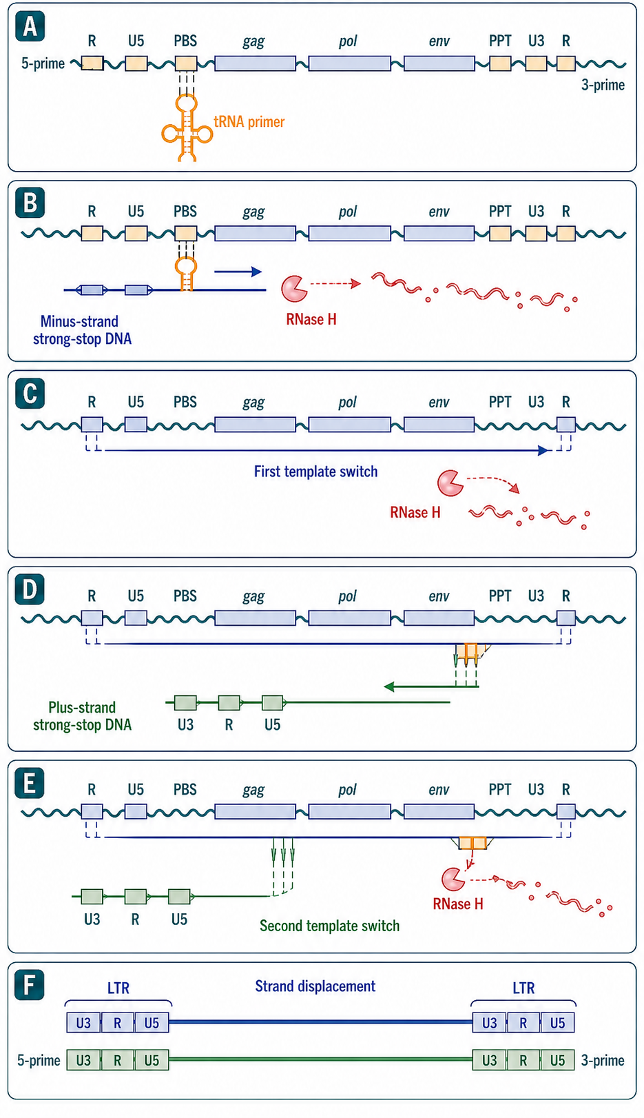
Figure 120.1. Stepwise retroviral reverse transcription and LTR generation. The two complete LTRs are constructed by ordered strand transfers rather than copied directly from two pre-existing RNA LTRs.
The resulting linear DNA remains associated with viral and host proteins in a pre-integration complex. Nuclear entry and chromatin engagement place the DNA at an integration site, where integrase and host repair create the provirus. Reverse-transcriptase structure, kinetics, fidelity, inhibitor binding, and resistance biochemistry are treated in Chapter 23; the life-cycle question here is whether each ordered intermediate reaches the next step.
“Reverse transcriptase only exists in retroviruses.” In fact, reverse transcriptases are widespread in biology. Telomerase is a specialized reverse transcriptase (Chapter 100). LINE-1 and other non-LTR retrotransposons encode reverse transcriptases (this chapter, Section 120.2). Group II introns encode reverse transcriptases that mediate their own mobility. Prokaryotic retrons and defense-associated reverse transcriptases use reverse transcription for diverse functions (Chapter 114). Hepatitis B virus, a DNA virus that replicates through an RNA intermediate, encodes a reverse transcriptase. The discovery of reverse transcriptase was revolutionary because it identified the enzyme that retroviruses use, but the evolutionary and functional distribution of reverse transcriptases is far broader.
“LTRs are always retroviral.” LTRs are found in LTR retrotransposons, which are not infectious retroviruses, and in endogenous retroviruses, which are embedded in host genomes. The presence of LTRs simply indicates that the element was generated by a retrovirus-like reverse transcription and integration mechanism; it does not by itself distinguish an infectious retrovirus from an intracellular retrotransposon from a fossilized germline insertion.
Integration begins the expression half of the cycle. Host RNA polymerase II transcribes the provirus, and RNA processing generates different coding outputs while full-length unspliced RNA is retained for Gag and Gag-Pol production and for packaging. Nuclear export must therefore distinguish RNA destined for translation or packaging from fully processed transcripts. At the plasma membrane, Gag concentrates full-length RNA and viral proteins, assembly drives budding, and protease cleavage remodels the immature particle into an infectious virion. Entry into a new cell then repeats the cycle.
120.2. LINE, SINE, and LTR retrotransposon replication cycles
Retrotransposon classification and RNA-output annotation are treated in Chapter 16. The mechanistic distinction needed here is between LTR elements, which generate DNA in a particle and then use integrase, and non-LTR elements, which couple a target-DNA nick to reverse transcription at the insertion site. LTR retrotransposons use Gag-like and Pol proteins to assemble intracellular particles, prime reverse transcription with a tRNA, perform the same obligatory strand transfers that generate terminal repeats, and deliver the completed DNA to integrase. Their lack of an infectious extracellular phase does not change the core RNA-to-DNA mechanism.
Ty1 in yeast is the most thoroughly characterized LTR retrotransposon. Ty1 RNA is transcribed by RNA polymerase II from LTR promoters, exported to the cytoplasm, and translated into Gag and Gag-Pol (the latter through a +1 ribosomal frameshift). Gag assembles into virus-like particles in which reverse transcription occurs. A mature Ty1 virus-like particle contains the full-length double-stranded DNA product. The DNA is then imported into the nucleus and integrated, preferentially near tRNA genes and other RNA polymerase III-transcribed loci, a targeting achieved in part through interaction between the Ty1 integrase and subunits of RNA polymerase III. This integration targeting is a useful reminder that integration is not random: different retroelements and retroviruses exhibit different integration site preferences determined by the interactions between their integrase proteins and host factors.
Non-LTR retrotransposons use target-primed reverse transcription (TPRT), a fundamentally different mechanism in which DNA cleavage and reverse transcription are coupled at the site of integration. The LINE-1 (L1) element is the paradigm. Full-length L1 RNA is a bicistronic transcript. ORF1p is an approximately 40-kilodalton protein with RNA-binding and nucleic acid chaperone activity that multimerizes and forms ribonucleoprotein particles with L1 RNA. ORF2p is an approximately 150-kilodalton protein with an N-terminal endonuclease domain and a C-terminal reverse transcriptase domain. The two proteins bind preferentially to the L1 RNA that encoded them (cis-preference), a property that limits the retrotransposition of other cellular mRNAs but is not absolute.
In TPRT, the L1 ribonucleoprotein complex reaches the nucleus (after nuclear envelope breakdown during mitosis, or potentially through active nuclear import in some contexts). The ORF2p endonuclease domain nicks one strand of chromosomal DNA, preferentially at a loose consensus sequence (5′-TTTT/A-3′ in mammals; the slash marks the cleavage site). The liberated 3′ hydroxyl of the nicked DNA serves as the primer for reverse transcription, with the L1 RNA as template. In the simplest model, ORF2p reverse transcriptase extends the chromosomal 3′ end, copying the L1 RNA poly(A) tail first. Second-strand cleavage and synthesis are less well understood but may involve the same endonuclease domain nicking the opposite strand and the reverse transcriptase performing a second round of DNA synthesis, or host DNA repair activities filling in the second strand. The resulting insertion is characterized by a variable-length poly(A) tail at the 3′ end (from the L1 RNA poly(A) sequence), short target-site duplications (because the endonuclease makes staggered nicks), frequent 5′ truncation (because reverse transcription stalls before reaching the L1 RNA 5′ end), and occasional 5′ inversions (because of template-switching events during TPRT). The absence of LTRs is a direct consequence of the TPRT mechanism, which does not employ integrase or generate terminal repeats.
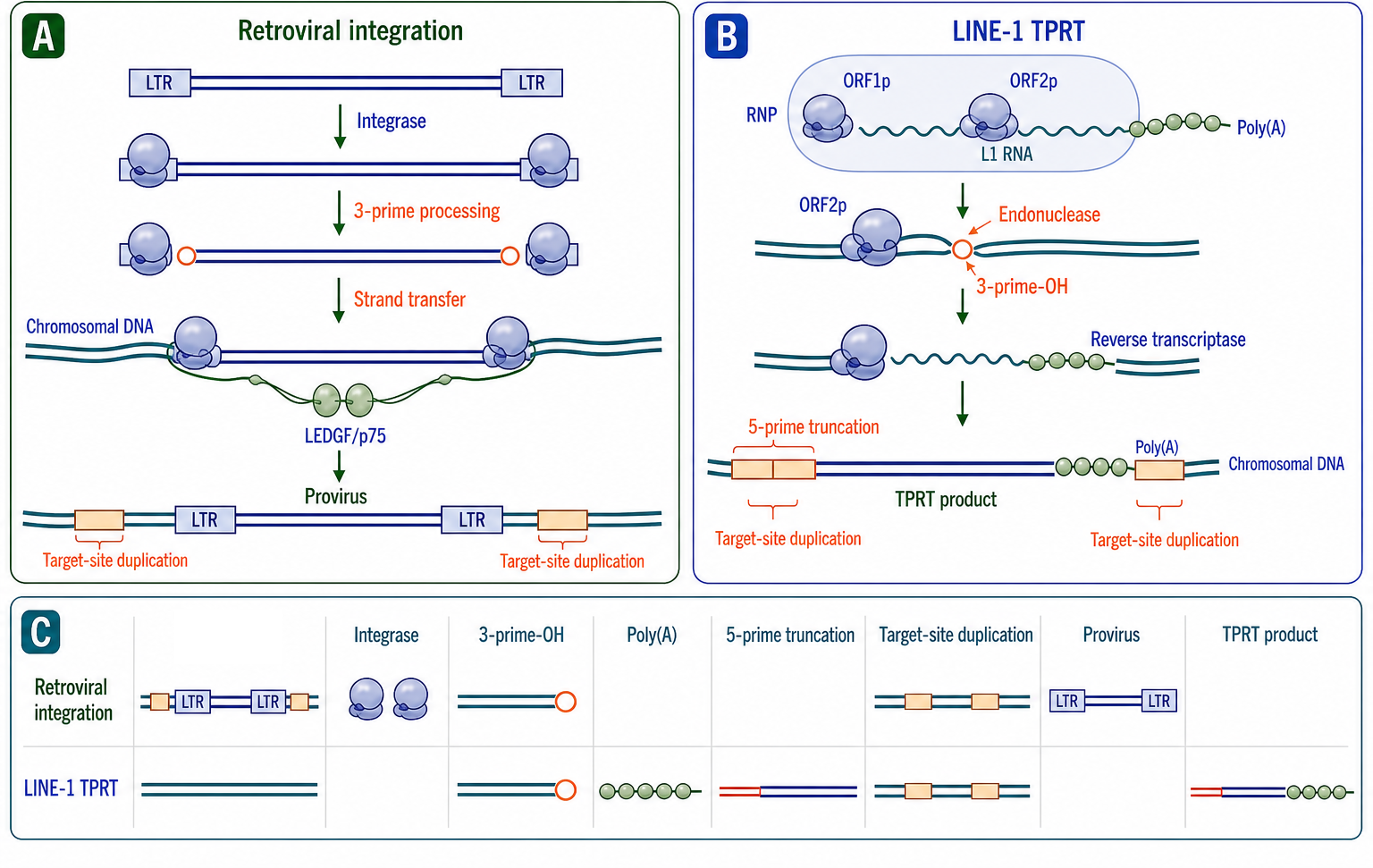
Figure 120.2. Target-primed reverse transcription and product signatures. Non-LTR reverse transcription occurs at the insertion site and therefore couples DNA cleavage, synthesis, and repair.
Non-autonomous RNAs can access the L1 machinery without encoding ORF1p or ORF2p. The replication question is substrate recognition: an RNA must enter an ORF2p-containing ribonucleoprotein and present a compatible 3′ region for target-primed reverse transcription. This route can mobilize SINE RNAs or generate processed pseudogenes from cellular messenger RNAs. Their classification and abundance belong to Chapter 16; mechanistically, their insertions share the poly(A)-linked and target-site features produced by ORF2p-dependent target-primed reverse transcription.
Target-primed reverse transcription explains why non-LTR insertions are often incomplete. Reverse transcriptase begins at the RNA 3′ end and may fail before reaching the 5′ end, producing truncation. Second-strand cleavage and synthesis remain less fully resolved than first-strand initiation. Target-site duplication records staggered cleavage and repair, while occasional inversion records template rearrangement during insertion. These product features are mechanistic evidence, not merely annotation labels.
A new insertion completes a replication cycle only if it retains or acquires the sequence needed for later expression. A full-length LINE insertion can generate a new source RNA; a 5′-truncated copy usually cannot support autonomous cycling. A SINE insertion can become a new source if its internal promoter and 3′ helper-recognition features remain functional. An LTR-retrotransposon insertion can re-enter its cycle through LTR-driven transcription and virus-like-particle formation. Host chromatin, RNA processing, translation, particle or ribonucleoprotein assembly, and nuclear access regulate these steps directly, while their broader consequences belong to Chapter 99.
120.3. Packaging, dimerization, recombination, reverse transcription, and integration
Retroviral RNA packaging is a highly selective process that ensures the encapsidation of two copies of full-length, unspliced genomic RNA into each assembling virion, while excluding spliced viral mRNAs and cellular RNAs. The selectivity is mediated by the packaging signal (psi, Ψ), an RNA element located in the 5′ untranslated region of the genome, between the primer binding site and the gag start codon. In HIV-1, the psi signal encompasses four stem-loop structures (SL1 through SL4) that are recognized with high affinity by the nucleocapsid domain of the Gag polyprotein, which contains two CCHC zinc-finger motifs.
Genome dimerization is initiated by base pairing between palindromic sequences in the dimerization initiation site (DIS), a hexanucleotide loop in SL1. The DIS loop sequence is GCGCGC in most HIV-1 group M strains, and intermolecular base pairing between DIS loops of two genomic RNAs initiates dimer formation. This kissing-loop interaction can mature into a more stable extended duplex through a structural rearrangement that involves the nucleocapsid chaperone activity. Dimerization and packaging are coupled: the nucleocapsid domain of Gag recognizes structural features of the dimeric psi element, and Gag binding stabilizes the dimeric conformation. The selective packaging of the dimeric RNA genome means that each virion carries two RNA copies that are genetically distinct if the producer cell was infected with more than one virus, or that are identical if the producer cell carried a single provirus.
During reverse transcription, the polymerase-nascent-DNA complex can switch between the two co-packaged RNA templates. Internal copy-choice switching can generate a mosaic DNA product, while intramolecular switching can create deletions or duplications. The mechanistic variables are template proximity within the capsid, sequence complementarity, RNA damage or structure, RNase H cleavage, and polymerase pausing. The population consequences of recombination, including selection and resistance evolution, belong to Chapter 116.
Integration of the linear double-stranded viral DNA into the host chromosome is catalyzed by integrase in the context of the pre-integration complex (PIC), a large ribonucleoprotein structure that forms in the cytoplasm after reverse transcription and traffics to the nucleus. The PIC contains the viral DNA, integrase (as a tetramer bound to the viral DNA ends), additional copies of reverse transcriptase, the viral matrix and Vpr proteins, and several host proteins including LEDGF/p75 (also called PSIP1), which tethers the PIC to host chromatin.
The two-step integration reaction begins with 3′ processing: integrase hydrolyzes the phosphodiester bond after the conserved CA dinucleotide at each 3′ end of the viral DNA, removing the terminal GT dinucleotide and exposing a 3′ hydroxyl group. The processed viral DNA ends remain bound to integrase in a stable synaptic complex. After nuclear entry and engagement with host chromatin, the strand transfer reaction proceeds: the 3′ hydroxyl groups of the processed viral DNA attack the host DNA backbone in a staggered, transesterification reaction. The two viral DNA strands attack opposite strands of the host DNA, separated by a staggered cleavage that is five base pairs for HIV-1 (four to six base pairs depending on the retrovirus). The result is a gapped integration intermediate in which the viral DNA is covalently joined to host DNA at the 3′ ends, but the 5′ ends of the viral DNA are not yet joined and the short overhangs left by the staggered cleavage are not yet filled. Host DNA repair factors, including components of the non-homologous end joining and base excision repair pathways, complete the integration by removing the unpaired viral 5′ dinucleotides, filling the single-strand gaps, and ligating the 5′ ends to host DNA. The integrated provirus is flanked by a target-site duplication of the staggered cleavage length (five base pairs for HIV-1).
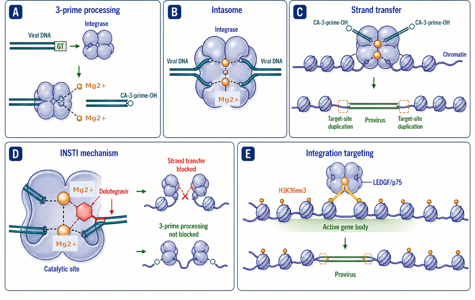
Figure 120.3. Packaging, dimerization, recombination, and integration as one replication chain. Packaging determines the templates available to reverse transcriptase, and recombination changes the DNA substrate before integration.
Integration is not random. Different retroviruses integrate with different genomic preferences. HIV-1 and lentiviruses integrate preferentially within the bodies of actively transcribed genes, a targeting mediated by the interaction of integrase with LEDGF/p75, which itself binds histone H3 trimethylated at lysine 36 (H3K36me3), a mark of transcriptional elongation. Gamma-retroviruses such as Moloney murine leukemia virus integrate preferentially near transcriptional start sites and CpG islands, a preference mediated by the interaction of their integrase with BET family proteins (bromodomain and extraterminal domain proteins, particularly BRD2, BRD3, and BRD4). These distinct integration preferences have important consequences for vector safety: gamma-retroviral integration near promoters contributed to insertional oncogenesis in early gene therapy trials, whereas the lentiviral preference for intragenic integration is generally considered safer, though it is not risk-free. The term “random integration” should therefore be avoided; more accurate language is “site preference” or “integration bias.”
The misconception “integration is random” is as misleading as the opposite misconception that integration only happens in actively transcribed regions. Different retroviruses integrate with different, probabilistically specified genomic preferences, and the preference is determined by the specific integrase-host factor interaction. No retrovirus integrates entirely randomly, but neither does any retrovirus integrate exclusively into a single class of genomic sites. The integration landscape is a probability distribution over the genome, not a deterministic targeting code. Integration can occur into transcriptionally silent regions, into heterochromatin, and into repetitive DNA, but at frequencies lower than predicted by random distribution.
120.4. Replication restriction, latency, and reactivation
Host factors are included here when they directly expose or control a replication step. A restriction phenotype is mechanistically informative only when the blocked intermediate is identified: capsid remodeling before reverse transcription, nucleotide supply during DNA synthesis, nascent-DNA editing, integration, or particle release. Broader immune signaling, host evolution, and disease consequences belong to Chapter 99.
APOBEC3G illustrates restriction coupled to reverse transcription. When packaged into a particle, APOBEC3G can encounter the minus-strand DNA intermediate and deaminate cytidine, yielding G-to-A changes in the completed plus strand and often a nonfunctional provirus. HIV-1 Vif prevents this block by recruiting a ubiquitin-ligase complex that depletes APOBEC3G before packaging. The decisive replication readouts are APOBEC3 incorporation, reverse-transcription products, mutation context, and infectivity, rather than a general interferon signature.
TRIM5alpha recognizes an assembled capsid lattice rather than a free capsid monomer. Its restriction can accelerate or misdirect capsid remodeling and reduce productive reverse transcription and nuclear entry. Species-specific capsid recognition is biologically important, but the replication mechanism is tested by capsid binding, uncoating kinetics, early and late DNA products, and nuclear entry. Signaling functions of TRIM5alpha are outside this section’s primary scope.
Tetherin/BST2 acts at the opposite end of the cycle by retaining nascent enveloped particles at the cell surface. Its dual membrane anchoring makes the physical release step the relevant intermediate, and HIV-1 Vpu restores release by reducing effective tetherin at the budding site. Particle-associated RNA, extracellular particle number, and infectivity distinguish a release block from a defect in genome synthesis or packaging.
SAMHD1 restricts reverse transcription by lowering the available deoxynucleoside triphosphate pool, especially in nondividing cells. HIV-2 and some simian immunodeficiency viruses package Vpx, which promotes SAMHD1 degradation and restores a more permissive nucleotide supply. Measuring nucleotide concentration together with strong-stop, intermediate, and full-length DNA products distinguishes substrate limitation from later defects in integration or transcription.
Reverse-transcription intermediates can also become innate immune ligands, but sensor pathways and disease consequences are treated in Chapter 99, Chapter 108, and Chapter 109. The replication-relevant point is that the abundance, location, and lifetime of RNA-DNA hybrids and viral DNA intermediates reflect capsid integrity, synthesis rate, nuclease access, and completion of integration.
Latency is a reversible replication state of an integrated, potentially replication-competent provirus. Viral DNA persists while productive transcription, genome-RNA accumulation, protein production, and particle formation fall below the threshold for continued spread. Latency can arise from integration-site context, insufficient transcription-factor activity, restricted P-TEFb availability, chromatin repression at the long terminal repeat, stochastic Tat feedback, and transcriptional interference. These mechanisms can coexist in the same reservoir.
Reactivation is the transition back into productive replication. It begins when promoter initiation and elongation resume, Tat positive feedback becomes self-sustaining, full-length RNA accumulates, and export and translation restore structural and enzymatic proteins. Detecting one induced transcript is not enough to demonstrate replication-competent reactivation: the chain should be followed through multiply spliced and full-length RNAs, protein production, particle release, and infectivity. Conversely, failure to reactivate in one assay does not prove irreversible silencing because stimulus, cell state, integration site, and proviral defects all constrain the response.
Latency must also be separated from defective proviruses. Many integrated copies cannot complete replication even if transcription is induced. Intactness assays, near-full-length sequencing, quantitative outgrowth, and rebound-competent lineage analysis answer different questions. A transcription-positive but replication-defective provirus can inflate reservoir estimates, while a deeply latent intact provirus can be missed by a single stimulation.
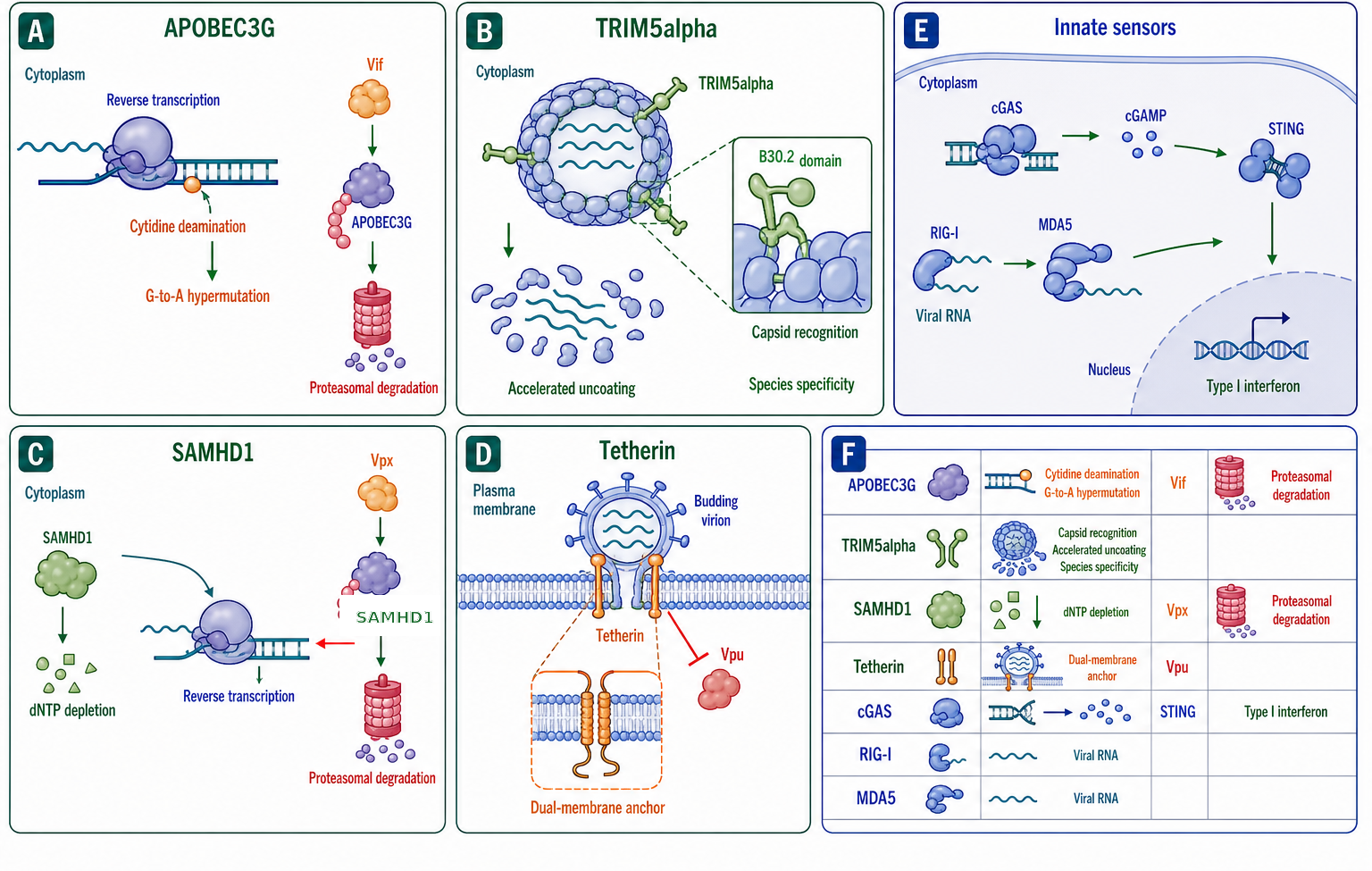
Figure 120.4. Complete retroviral cycle with restriction, latency, and reactivation. A complete cycle contains ordered checkpoints; host factors are informative when assigned to a blocked intermediate, and latency is a reversible branch after integration rather than loss of the genome.
Latency places an integrated genome outside productive transcription, assembly, and spread until reactivation re-enters the expression half of the cycle. A cycle-resolved analysis therefore distinguishes proviral persistence, induced RNA, protein production, particle release, and renewed infectivity rather than treating them as one readout.
120.5. Replication-derived genome change and biotechnology
Replication leaves diagnostic changes in DNA. Integrase acts on both viral DNA ends and attacks target phosphodiester bonds at staggered positions; host repair fills the gaps, generating short target-site duplications. The completed provirus carries long terminal repeats created during reverse transcription. Target-primed reverse transcription instead begins at a target nick and commonly produces a 3′ poly(A) tract, target-site duplication, 5′ truncation, and occasional inversion. These features distinguish mechanisms even after the original RNA and proteins are gone.
Genome change can occur before insertion. Copy-choice recombination combines sequence from the two packaged RNAs, while nonhomologous template switching can delete, duplicate, or rearrange segments. RNase H cleavage and primer selection determine long-terminal-repeat boundaries. In target-primed reverse transcription, incomplete first-strand synthesis and second-strand processing determine truncation and inversion. Thus the inserted product records both templated synthesis and failures or switches during replication.
Integration-site preference is another replication-derived feature. Lentiviral integrase complexes are biased toward actively transcribed gene bodies through host tethering factors such as LEDGF/p75, whereas other retroviral genera and retrotransposons use different host interactions or sequence preferences. These are probabilistic biases, not deterministic targeting codes. For biotechnology, integration profiles must be measured in the relevant cell type because chromatin state and host-factor availability alter the distribution.
Table 120.1. Mechanistic signatures of integrated DNA products. The integrated DNA retains evidence of primer choice, template transfers, cleavage geometry, incomplete synthesis, and repair.
| Mechanistic feature | Retrovirus | LTR retrotransposon | Non-LTR target-primed reverse transcription |
|---|---|---|---|
| Site of cDNA synthesis | Incoming particle or capsid-associated complex | Intracellular virus-like particle | Chromosomal target site |
| First primer | Packaged host tRNA | Packaged host tRNA | Target-DNA 3′ hydroxyl |
| Plus-strand initiation | Polypurine-tract RNA | Polypurine-tract RNA | Second-strand mechanism incompletely resolved |
| Terminal transfers | Two obligatory transfers generate LTRs | Two obligatory transfers generate LTRs | No paired LTR-generating transfers |
| Insertion catalyst | Integrase | Integrase | ORF2p endonuclease and reverse transcriptase |
| Product signature | LTR-flanked provirus and short target-site duplication | LTR-flanked insertion and target-site duplication | Poly(A), frequent 5′ truncation, target-site duplication, occasional inversion |
| Frequent incomplete product | Unintegrated or transfer-defective DNA | Particle or integration-defective DNA | Truncated, inverted, or incomplete second-strand insertion |
Table 120.1. Mechanistic signatures of integrated DNA products
| Mechanistic feature | Retrovirus | LTR retrotransposon | Non-LTR target-primed reverse transcription |
|---|---|---|---|
| Site of cDNA synthesis | Incoming particle or capsid-associated complex | Intracellular virus-like particle | Chromosomal target site |
| First primer | Packaged host tRNA | Packaged host tRNA | Target-DNA 3′ hydroxyl created by endonuclease |
| Plus-strand primer | Polypurine-tract RNA | Polypurine-tract RNA | Second-strand initiation is less fully resolved and can involve target cleavage and repair |
| Obligatory template transfers | R-mediated minus-strand transfer and PBS-mediated plus-strand transfer | Same terminal strand-transfer logic | No LTR-generating pair of transfers; internal switching can generate inversion or rearrangement |
| Insertion enzyme | Integrase after completed linear DNA synthesis | Integrase after completed linear DNA synthesis | ORF2p endonuclease and reverse transcriptase coupled at target DNA |
| Characteristic product | LTR-flanked provirus plus short target-site duplication | LTR-flanked intracellular element plus target-site duplication | Poly(A)-tailed insertion, frequent 5′ truncation, target-site duplication, and occasional inversion |
| Mechanistic failure signature | Incomplete strong-stop transfer, deletions, unintegrated linear or circular DNA | Incomplete particle synthesis or integration | Truncation, inversion, endonuclease-independent junction, or incomplete second strand |
Step-specific perturbations diagnose the life cycle when they are paired with intermediate measurements. An entry block prevents capsid delivery; a reverse-transcription block reduces ordered DNA products; an integration block accumulates unintegrated DNA; an export or packaging block separates intracellular full-length RNA from particle-associated RNA; and a maturation block produces released but poorly infectious particles. Comparative inhibitor classes, binding, and resistance biochemistry belong to Chapter 23.
Lentiviral vector engineering separates the cis-acting genome from trans-acting production functions. The transfer RNA retains the long terminal repeats, packaging signal, and transgene cassette, while Gag-Pol, Rev, and envelope functions are supplied from separate constructs in producer cells. This partition reduces the probability that one packaged genome can complete autonomous replication. A self-inactivating vector carries a deletion in the 3′ long-terminal-repeat U3 region that is copied to the 5′ end during reverse transcription, reducing promoter activity in the integrated vector. This design is a direct application of the strand-transfer mechanism that duplicates terminal sequences.
Vector safety depends on replication logic. Sequence overlap among production constructs can permit recombination, so modern systems minimize homology and distribute essential functions. Residual plasmid DNA, replication-competent recombinant particles, vector copy number, integration-site distribution, and mobilization by helper viruses require distinct assays. Pseudotyping changes entry tropism but does not alter the reverse-transcription and integration machinery carried by the vector genome. Clinical manufacturing and disease-specific applications belong to Chapter 157.
Target-primed reverse transcription can also be reconstructed as an insertion tool or exploited in reporter assays. An intron-interrupted reporter becomes active only after transcription, splicing, reverse transcription, and reinsertion, coupling signal to a completed cycle. Mutating ORF2p endonuclease separates canonical target-primed insertion from endonuclease-independent routes, while reverse-transcriptase mutation tests DNA synthesis. Long-read junction analysis then verifies target-site duplication, poly(A) tract, truncation, inversion, and transduction of flanking sequence.
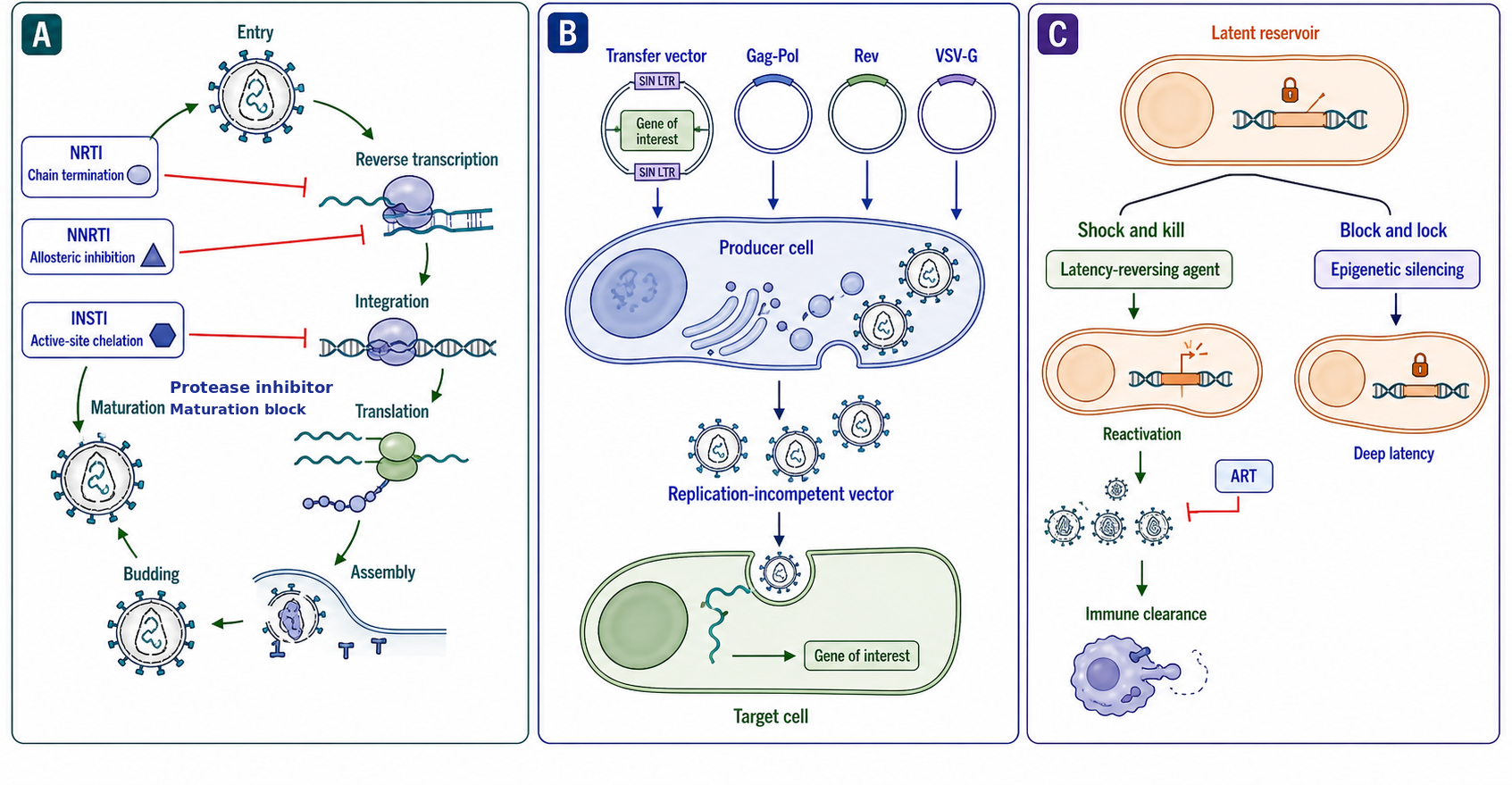
Figure 120.5. Replication logic of a self-inactivating lentiviral vector. Vector engineering preserves genome transfer while distributing or deleting functions required for renewed replication.
Biotechnology therefore depends on preserving some replication steps while disabling others. A transfer vector must be packaged, reverse-transcribed, and integrated efficiently, yet it must not encode a complete autonomous cycle. A retrotransposition reporter must require every step being assayed and exclude expression from the input plasmid. Mechanism-specific controls make these systems useful both as technologies and as experiments on replication.
Experimental Foundations and Evidence Standards
Evidence for retroviral reverse transcription comes from complementary reconstitution and cycle-resolved experiments. Purified components establish primer and RNase H requirements, while infection time courses identify minus-strand strong-stop DNA, transferred intermediates, plus-strand products, completed LTR-containing DNA, and integrated provirus. Structural and kinetic comparison of isolated reverse transcriptases belongs to Chapter 23.
The evidence for integration comes from in vitro assays in which purified integrase, synthetic viral DNA end substrates, and a target DNA plasmid produce integration products detectable by PCR or gel shift. Crystal structures of the prototype foamy virus integrase in complex with viral DNA ends (the intasome) were followed by structures of the HIV-1, Rous sarcoma virus, and mouse mammary tumor virus intasomes, revealing both the conserved catalytic core and the family-specific quaternary structures. Cryo-electron tomography of HIV-1 particles has visualized the capsid core structure. LEDGF/p75 was identified as the HIV-1 integration targeting factor through biochemical pull-down, genetic knockout, and chromatin immunoprecipitation followed by sequencing (ChIP-seq).
Evidence for L1 retrotransposition comes from cultured-cell assays in which an intron-interrupted reporter becomes active only after transcription, splicing, reverse transcription, reinsertion, and expression from the new copy. These assays demonstrate cis preference and the requirements for ORF1p and ORF2p. Junction sequencing tests the expected target-site duplication, poly(A), truncation, and inversion signatures. Biochemical reconstitution with purified ORF2p, RNA, and target DNA confirms the coupling of nicking and reverse transcription.
Evidence for replication restriction comes from step-resolved perturbations: capsid binding and uncoating for TRIM5alpha, nucleotide pools and DNA intermediates for SAMHD1, packaged deaminase and mutation context for APOBEC3, and cell-associated versus released particles for tetherin. Evidence for latency combines intact proviral sequence, transcriptional inducibility, protein and particle production, quantitative outgrowth, and rebound-competent lineage analysis. No one assay captures all latent, intact, and replication-competent proviruses.
Principal interpretive hazards include mistaking input or plasmid DNA for a reverse-transcription product, treating an expressed reporter as evidence of completed reinsertion, confusing APOBEC3 mutation signatures with polymerase or amplification error, and equating transcriptional induction with replication-competent reactivation. DNase controls, intron-interrupted reporters, junction sequencing, mutation-context analysis, and infectivity assays address different hazards and should not be substituted for one another.
Biological Contexts and Cross-Chapter Boundaries
This chapter sits at the intersection of RNA virology and genome replication. Comparative reverse-transcriptase and RdRP folds, kinetics, specificity, fidelity, inhibitor classes, and resistance biochemistry belong to Chapter 23, while replication-complex and population dynamics belong to Chapter 116. Here enzyme behavior is introduced only at the life-cycle step where it changes an intermediate or transition.
RNA structural features that govern packaging and dimerization connect to Chapter 117, and the biophysics of RNA condensation and particle assembly connect to Chapter 118. This chapter owns how those states determine which genome enters reverse transcription and how two templates enable recombination.
Host restriction is retained only when it blocks a defined replication step. Innate sensing, host regulation, evolutionary consequences, and disease synthesis belong to Chapter 99, Chapter 108, and Chapter 109. Repeat classification, RNA-output annotation, and multimapping belong to Chapter 16.
Vector manufacturing and clinical applications belong to Chapter 157, and drug pharmacology belongs to Chapter 160. This chapter provides the cycle map: which cis-acting sequence is retained, which trans-acting protein is supplied, and which intermediate demonstrates that a step occurred.
The boundary is life cycle versus comparison or consequence. This chapter follows how each retroviral or retrotransposon cycle begins, proceeds, pauses, restarts, and produces a new heritable copy or infectious particle. It does not compare polymerase families, derive inhibitor resistance, catalog repeat objects, or synthesize host regulatory, immune, evolutionary, and disease consequences.
Recent Consensus
The ordered retroviral cycle from entry and capsid remodeling through reverse transcription, integration, proviral expression, genome packaging, budding, and maturation is established in outline, while coupling among capsid state, DNA synthesis, and nuclear entry remains active research. The two obligatory reverse-transcription transfers and integrase-mediated insertion are established. LTR-retrotransposon particle-associated cycles and the first-strand target-primed mechanism of non-LTR elements are established, while non-LTR second-strand completion is less resolved.
The first-strand mechanism of target-primed reverse transcription is established structurally and biochemically, while second-strand cleavage, synthesis, and repair remain less completely resolved. The role of LEDGF/p75 in biasing HIV-1 integration toward actively transcribed gene bodies is established. APOBEC3, TRIM5alpha, tetherin, and SAMHD1 each restrict a distinguishable replication step, and viral countermeasures can restore that step.
Latency is a reversible replication state rather than an absence of proviral DNA, and many integrated proviruses are defective rather than latent. Reactivation must be measured beyond transcriptional induction when the claim is renewed replication. In biotechnology, replication-defective vector design depends on separating cis-acting genome signals from trans-supplied proteins and on copying a self-inactivating U3 deletion during reverse transcription.
Open Questions, Controversies, Deprecated Models, and Common Misconceptions
Open questions:
- How does the HIV capsid core uncoat to release the reverse transcription complex and the pre-integration complex? Competing models remain, and the role of host factors in regulating uncoating is incompletely understood.
- How does the pre-integration complex traverse the nuclear pore, and does nuclear entry require an intact capsid? Recent cryo-electron tomography studies have sharpened this question. Whether the central DNA flap created by central polypurine-tract priming in lentiviruses is truly required for nuclear import, or whether it serves another function, remains debated.
- Which molecular combinations of integration site, chromatin, transcription-factor availability, Tat feedback, and cell state establish reversible latency rather than deep silencing or defective proviral expression?
- Which measurements best connect transcriptional reactivation to full-length genome RNA, particle production, and replication competence across heterogeneous latent proviruses?
- How are second-strand cleavage, synthesis, and repair coordinated during target-primed reverse transcription, and which host factors determine truncation or inversion?
Controversies:
- Models of capsid uncoating and nuclear entry differ in how much of reverse transcription occurs within an intact or remodeled capsid and where the pre-integration complex becomes fully exposed.
- Models of non-LTR second-strand synthesis differ in the timing of the second nick, the polymerase responsible for second-strand completion, and the contribution of host repair pathways.
Common misconceptions:
- “Reverse transcriptase is only for HIV.” Reverse transcriptases are widespread and ancient.
- “LTRs are always retroviral.” LTRs are also found in LTR retrotransposons and endogenous retroviruses.
- “Integration is random.” Different retroelements exhibit distinct, factor-mediated integration preferences.
- “Detecting induced viral RNA proves latency reversal.” RNA induction does not establish that an intact provirus completed protein production, particle formation, and renewed infectivity.
- “A retrotransposition reporter measures transcription.” A well-designed reporter becomes active only after RNA production, splicing, reverse transcription, integration, and expression from the new copy.