Chapter 28. Alternative Splicing, Cryptic Splicing, Microexons, and Regulated Isoform Choice
Scope Note
Alternative splicing is the regulated production of more than one mature RNA isoform from a single precursor RNA. It works on the same spliceosome chemistry described in Chapter 27, but it asks a different biological question: among several possible splice-site choices, which exon-intron pattern is selected in a given cell, developmental state, stress condition, disease state, or species? This chapter covers the major modes of alternative splicing, the sequence elements and RNA-binding proteins that bias splice-site choice, the distinction between regulated isoforms and cryptic splicing errors, the special biology of microexons, the measurement problem created by transcript isoforms, and the evidence required to claim that a splicing change has a function.
The chapter treats alternative splicing as an RNA regulatory layer rather than as a catalog of all known isoforms. It emphasizes mechanistic grammar, evidence limits, and interpretation. Detailed spliceosome assembly, trans-splicing chemistry, spliced-leader systems, and the core mechanism of recursive splicing belong in Chapter 27; this chapter retains the recursive-splicing boundary needed to interpret isoform choice. Transcript annotation and genome browsers belong in Chapter 18; 3′ end choice and alternative polyadenylation belong in Chapter 29; nonsense-mediated decay belongs in Chapter 35; neuronal local translation and RNA granules are treated in Chapters 74-76; therapeutic oligonucleotides and RNA medicines are treated in later therapeutic chapters.
Executive Summary
Alternative splicing expands RNA output by changing which exons and splice sites are joined into a mature transcript. Common event types include cassette-exon inclusion or skipping, use of alternative 5′ or 3′ splice sites, mutually exclusive exon choice, intron retention, alternative first or last exons coupled to promoter or polyadenylation decisions, and complex combinations of several events in one transcript. These choices can alter coding sequence, protein domains, intrinsically disordered regions, localization signals, untranslated regions, translation efficiency, RNA stability, and sensitivity to nonsense-mediated decay.
Recursive splicing is a related but distinct operation in which one long intron is removed through successive intramolecular splicing reactions at internal recursive sites. It is normally a processing route within one precursor, not trans-splicing between separate RNAs and not automatically an alternative mature-isoform choice. Recursive sites can nevertheless influence isoform output when a transient recursive exon becomes included or when recursive and direct intron removal compete.
Splice-site choice is not determined by the core GU-AG splice-site dinucleotides alone. The spliceosome reads a larger regulatory environment that includes splice-site strength, branch-point and polypyrimidine-tract quality, exon and intron length, RNA secondary structure, transcription elongation kinetics, chromatin-associated context, and many cis-regulatory sequence elements. Cis-regulatory elements are RNA sequences in the same pre-mRNA molecule, such as exonic splicing enhancers, exonic splicing silencers, intronic splicing enhancers, and intronic splicing silencers. RNA-binding proteins bind these elements and bias spliceosome assembly, often through position-dependent effects rather than a single universal activating or repressing role.
Cryptic splicing occurs when the spliceosome uses a splice site that is normally silent, weak, repressed, or newly created by mutation. A pseudoexon is an intronic segment that resembles an exon enough to be recognized under certain genetic or regulatory conditions. A poison exon is a regulated exon whose inclusion introduces a premature termination codon or otherwise produces an unstable or unproductive transcript. These categories overlap but are not synonyms. Cryptic exons and pseudoexons are often pathological or stress-induced, whereas poison exons can be normal feedback devices for controlling RNA-binding protein abundance.
Microexons are very short exons, often only a few to a few tens of nucleotides. Because their length gives the spliceosome less exon definition surface, many microexons require specialized enhancer architecture and tissue-specific RNA-binding proteins. Neuronal and developmental programs are especially enriched for regulated microexon inclusion, where even a few amino acids can tune protein interaction surfaces, trafficking motifs, and synaptic functions. Microexon biology is therefore a useful antidote to a common misconception: exon length does not predict functional importance.
Isoform quantification is difficult because reads are usually shorter than full transcripts, many genes produce related isoforms that share exons, and event-level changes do not automatically identify the full molecules present in the cell. Short-read RNA-seq is powerful for detecting splice junctions and estimating percent spliced in, but transcript-level isoform reconstruction is model-dependent. Long-read RNA-seq improves full-length isoform discovery and phasing, yet still faces biases from RNA integrity, reverse transcription, amplification, coverage, error correction, and quantification depth.
Functional interpretation requires more than a statistically significant splice-junction change. A strong claim usually needs orthogonal detection, perturbation of the causal regulator or cis element, rescue or isoform-specific manipulation, and a phenotype that tracks the isoform rather than only the gene. Therapeutic splice switching is strongest when the target event is mechanistically tied to disease, the relevant tissue is reachable, and off-target effects can be monitored. Direct examples include SMN2 splicing modulation in spinal muscular atrophy, classic beta-globin splice-correction models, and current splice-switching oligonucleotide design platforms.
Concept Inventory
- Alternative splicing: regulated production of multiple RNA isoforms from one precursor RNA by using different splice sites or exon-intron patterns. It is not equivalent to transcriptional initiation, RNA editing, or alternative polyadenylation, although these processes can combine with splicing to produce transcript diversity.
- Splice isoform: a transcript molecule distinguished by its exon-exon junctions or retained introns. A protein isoform is a translated product, so a splice isoform should not be assumed to produce a stable protein without translation and proteomic evidence.
- Cassette exon: an exon that can be included or skipped between two flanking constitutive exons. It is also called an exon-skipping event.
- Alternative splice-site event: uses different 5′ donor sites or 3′ acceptor sites, changing exon boundaries and sometimes shifting reading frame or protein motif sequence.
- Intron retention: the presence of an intron in an otherwise processed transcript. Retained introns can support nuclear detention, regulated decay, translated sequence insertion, or unproductive splicing depending on context.
- Recursive splicing: stepwise removal of one long intron through internal splice sites that create transient processing intermediates. Recursive splicing is usually intramolecular cis-splicing; it is not synonymous with alternative splicing or trans-splicing.
- Cis-splicing element: a sequence or structural feature within the pre-mRNA that affects splice-site choice on the same RNA molecule. Enhancer and silencer labels are context-dependent.
- RBP combinatorial control: regulation produced by multiple RNA-binding proteins acting together with splice-site strength, RNA structure, transcription kinetics, and local RNA sequence.
- Cryptic splicing: splice-site use that is normally hidden or rare but becomes detectable after mutation, altered RBP concentration, stress, repeat activation, or failure of repression.
- Pseudoexon: an intronic segment with exon-like signals that can be mistakenly or conditionally included in mature RNA.
- Poison exon: an exon whose inclusion reduces productive protein output, often by introducing a premature termination codon and triggering nonsense-mediated decay.
- Microexon: a very short exon. Different studies use different length cutoffs, so the definition should state the cutoff used. In this chapter, the term means short exons whose recognition and functional consequences are strongly affected by their small length.
- Percent spliced in: the estimated fraction of transcripts from a locus or event that include a given exon or splice junction. PSI is an event-level statistic, not a direct count of full-length transcript molecules.
What to Know Before Reading This Chapter
Splicing removes introns from a precursor messenger RNA and joins exons into a mature RNA. The core spliceosome recognizes a 5′ splice site at the upstream exon-intron boundary, a branch point and polypyrimidine tract near the 3′ end of the intron, and a 3′ splice site at the downstream intron-exon boundary. Chapter 27 explains the chemistry: two transesterification reactions remove the intron as a lariat and ligate the exons. Alternative splicing uses the same chemistry but changes which splice sites are chosen.
The word “isoform” must be used precisely. A transcript isoform is an RNA molecule with a specific arrangement of exons, retained introns, untranslated regions, and ends. A protein isoform is a polypeptide translated from a coding transcript. Many RNA isoforms are never translated, are rapidly degraded, or differ only in untranslated regions. Therefore, the existence of an RNA isoform does not prove the existence of a protein isoform. This distinction is especially important when a transcript contains a premature termination codon, a long retained intron, or a poison exon that triggers nonsense-mediated decay.
Splicing decisions happen on a physical RNA molecule while the RNA is being transcribed, folded, bound by proteins, processed at its 5′ and 3′ ends, and packaged into a ribonucleoprotein particle. A splice site is therefore not just a short sequence. Its use depends on splice-site consensus, distance from neighboring sites, RNA structure, nearby enhancers and silencers, the concentration and activity of RNA-binding proteins, transcription elongation rate, and competition among spliceosome assembly routes. The same sequence can be included in one cell type and skipped in another.
The running examples in this chapter are metazoan pre-mRNAs, especially neuronal genes and disease-associated human genes. However, alternative splicing is not exclusively a nervous-system phenomenon and not exclusively a human phenomenon. Many eukaryotes use regulated splicing; plants, fungi, animals, and protists differ in intron architecture, regulatory proteins, and the relative importance of intron retention, cassette exons, and other event types. The chapter uses mammalian examples because the reference set and disease literature are strongest there.
28.1. Alternative splicing modes and isoform consequences
Alternative splicing modes are recurring patterns by which a precursor RNA can be processed into different exon-intron arrangements. The simplest and most commonly taught mode is a cassette exon. Imagine three exons arranged as exon 1, exon 2, and exon 3. In one mature RNA, all three exons are joined. In another mature RNA, exon 2 is skipped and exon 1 is joined directly to exon 3. This event can add or remove a protein segment, alter reading frame, insert a stop codon, or change an untranslated region depending on where the exon lies.
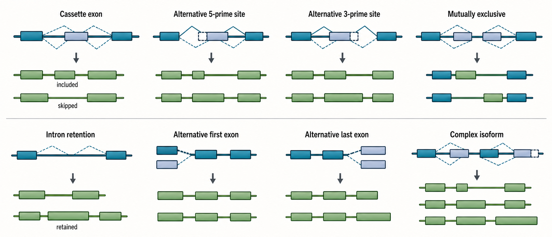
Figure 28.1. Alternative Splicing Event Modes. Major alternative splicing event types alter different parts of a transcript. Cassette exons, alternative splice sites, mutually exclusive exons, and intron retention are splice-site choice events, whereas alternative first and last exons often couple splicing to promoter choice or 3′ end formation.
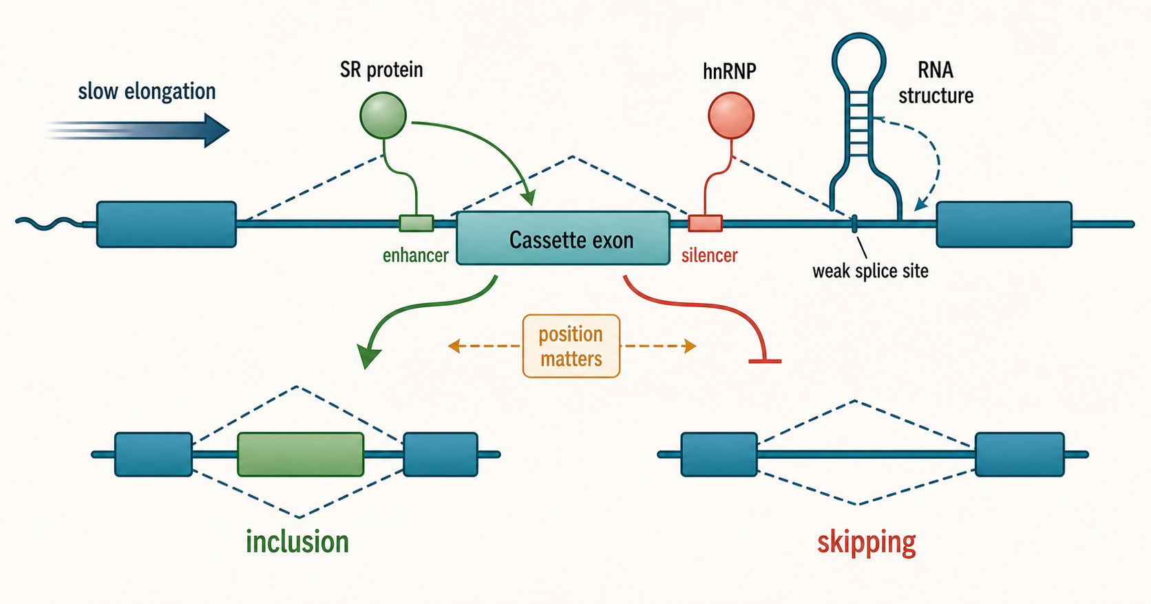
Figure 28.2. Combinatorial RBP Control Around a Cassette Exon. Alternative splicing integrates local splice-site strength, cis-regulatory elements, RNA structure, and RNA-binding proteins. The same RBP family can activate or repress depending on binding position, competing factors, and the architecture of the regulated exon.
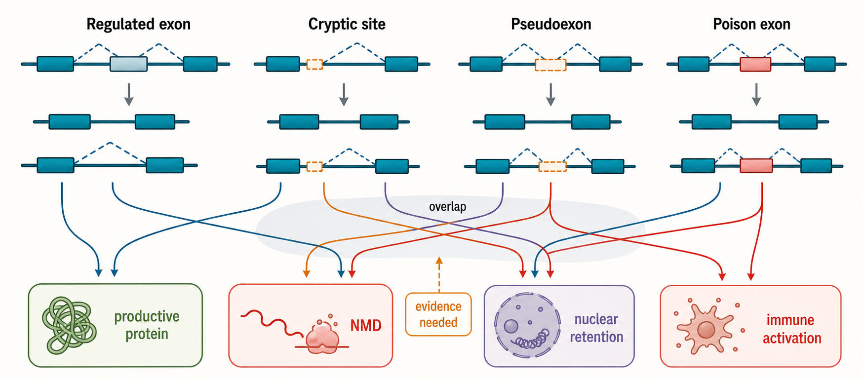
Figure 28.3. Cryptic Exons, Pseudoexons, and Poison Exons. Cryptic splicing, pseudoexon inclusion, and poison-exon inclusion describe different properties of abnormal or regulated exon use. A cryptic event is normally hidden or rare, a pseudoexon originates from an intronic exon-like segment, and a poison exon reduces productive output through premature termination and nonsense-mediated decay; these categories overlap and require explicit evidence to distinguish.
Alternative 5′ splice-site and alternative 3′ splice-site events change exon boundaries. In an alternative 5′ splice-site event, two donor sites compete at the upstream side of an intron. In an alternative 3′ splice-site event, two acceptor sites compete at the downstream side. These events can be small, changing only a few codons, or large enough to remodel a protein domain. They are particularly important because they can create subtle amino-acid substitutions at domain edges, preserve most of a protein, but alter a short motif that controls localization, phosphorylation, protein-protein interaction, or degradation.
Table 28.1. Alternative Splicing Modes and Consequences. Summary of the major alternative splicing event types, their molecular effects, and the most frequent interpretive errors associated with each.
| Event type | Minimal RNA change | Common coding consequence | Common noncoding consequence | Main detection strategy | Frequent overinterpretation |
|---|---|---|---|---|---|
| Cassette exon | Single exon included or skipped between flanking constitutive exons | Protein domain insertion or deletion | UTR exon alters regulatory elements or stability | Junction-spanning reads; PSI estimation | Equating exon skipping with loss-of-function |
| Alternative 5′ splice site | Upstream donor site shifted | Coding extension or truncation at exon edge | 5′ UTR length change affects translation | Junction-count comparison at competing donors | Assuming small coding changes are always inconsequential |
| Alternative 3′ splice site | Downstream acceptor site shifted | Residues added or removed at exon start | 3′ UTR boundary changes RBP and miRNA sites | Junction-count comparison at competing acceptors | Overlooking frameshift risk from non-triplet boundary shifts |
| Mutually exclusive exons | One of two adjacent exons included, the other excluded | Related protein variants with distinct domain surfaces | Differential RBP-binding sites in exon bodies | Paired junction reads; minigene reporter | Treating any anti-correlated exon pair as mutually exclusive without structural evidence |
| Intron retention | Intron sequence present in mature RNA | Premature stop codon if in-frame; novel peptide if translated | Nuclear detention signal; altered RNA half-life | Intronic read coverage relative to flanking exons | Assuming all retained introns are splicing errors |
| Alternative first exon | Different promoter drives distinct first exon | Distinct N-terminal peptide or signal sequence | Different 5′ UTR alters translational regulation | TSS mapping; promoter-proximal junction reads | Conflating promoter switch with spliceosome-mediated regulation |
| Alternative last exon | 3′ end choice changes terminal exon | Truncated or extended C-terminal domain | 3′ UTR changes miRNA targeting and mRNA stability | PolyA-seq; 3′-end junction reads | Conflating alternative polyadenylation with alternative splicing |
| Complex multi-event isoform | Several exon and splice-site changes in one transcript | Remodeled protein with multiple domain differences | Multiple UTR and RBP-binding site changes | Long-read RNA-seq for phasing | Inferring complex isoform from individual short-read events without phasing evidence |
Mutually exclusive exons are alternatives in which one exon is included while another nearby exon is skipped. The phrase should not be used loosely for any pair of exons with opposite changes. True mutually exclusive regulation implies that the mature transcript generally contains one choice but not both. Mechanisms can include steric constraints, RNA structure, splice-site incompatibility, exon length constraints, or RBP-mediated repression of one choice while another is activated. Mutually exclusive events are common in genes whose protein products need related but functionally distinct modules.
Intron retention is the inclusion of an intron in a mature or partially mature RNA. Retained introns can have several fates. Some retained-intron transcripts stay in the nucleus and are later spliced or degraded. Some are exported and translated, adding amino-acid sequence or changing the reading frame. Many introduce premature termination codons and become substrates for nonsense-mediated decay. Intron retention is therefore not automatically a splicing failure. In developmental and stress contexts, retained introns can regulate timing, storage, and dose of gene expression. The evidence requirement is to show the RNA species, its location, and its consequence, rather than assuming that all retained introns are either mistakes or regulatory programs.
Recursive splicing solves a different problem: excision of a very long intron in several stages. In the classic zero-nucleotide ratchet-point model, an upstream exon is first joined to an internal 3′ splice site. That junction recreates a 5′ splice site, allowing the remaining downstream intron segment to be removed in a later reaction. Work in Drosophila identified conserved ratchet points and the lariat intermediates expected from sequential removal, while vertebrate studies showed that many recursive sites are coupled to a short recursive exon whose inclusion or skipping depends on competition among reconstituted splice sites. The mature product can therefore look identical to a product made by direct removal of the whole intron even though its processing history differed.
Recursive splicing belongs near alternative-splicing biology only at this boundary. A recursive exon can enter a mature isoform when splice-site competition changes, and recursive versus direct removal can vary by cell type or transcriptional context. Most detected recursive junctions, however, are short-lived intermediates rather than stable alternative transcripts. Evidence should combine recursive junctions with nascent or total RNA, expected read-density progression, lariat or branch evidence, and site perturbation where possible. Reverse-transcription template switching, ambiguous mapping, cryptic promoters, ordinary exon skipping, and low-level splice noise can each mimic part of the signature. Chapter 27 develops the chemistry and lineage comparison; this chapter asks only whether a recursive route changes stable isoform abundance or function.
Alternative first and last exons are boundary cases that connect splicing to transcription initiation and 3′ end formation. If a gene uses different promoters, the first exon can change before the spliceosome acts. If a gene uses different polyadenylation sites, the last exon and 3′ untranslated region can change through cleavage and polyadenylation choices. These are still transcript isoform differences, but the causal mechanism may not be splicing alone. Chapter 29 treats alternative polyadenylation in detail, and Chapter 18 explains why transcript models must represent promoter, splicing, and 3′ end choices together.
The consequences of alternative splicing depend on molecular position. A coding exon can add or remove a structured domain, a short linear motif, a disordered segment, a transmembrane helix, or a catalytic residue. A UTR exon can affect RNA localization, translation, stability, microRNA targeting, or RBP binding without changing the encoded protein. An event that introduces a premature termination codon can reduce productive protein output by nonsense-mediated decay, which is discussed in Chapter 35. A splice change can also alter RNA structure or RNP packaging in ways that affect export and localization.
CD44 provides a useful example of context-dependent exon choice. CD44 encodes a cell-surface glycoprotein with variable exons whose inclusion is associated with cell adhesion, migration, epithelial-mesenchymal transitions, immune states, and cancer contexts. The CD44 literature illustrates why splicing should be interpreted as a regulated network output rather than as a single on-off switch. Multiple RBPs can influence CD44 variable exons, and the same exon pattern can have different implications depending on cell type and signaling state.
Do not overgeneralize isoform number into functional complexity. A gene may produce many annotated RNA isoforms, but only a subset may be abundant, stable, exported, translated, conserved, or phenotypically relevant. Conversely, a single short exon can have a major effect if it changes an interaction surface or creates a decay-triggering stop codon. The correct unit of interpretation is not “how many isoforms exist” but which RNA molecules are produced, under what conditions, by which mechanisms, and with what evidence for consequence.
28.2. Cis-regulatory elements and RBP combinatorial control
The spliceosome must recognize short and imperfect signals within long pre-mRNAs. In mammals, the GU at many 5′ splice sites and the AG at many 3′ splice sites are necessary but not sufficient. A functional intron also depends on a broader 5′ splice-site motif, a branch point, a polypyrimidine tract, and surrounding sequence. Many potential splice sites exist by chance in introns. Regulatory elements and RBPs help the cell choose among them.
Cis-regulatory elements are features on the same RNA molecule that affect splicing. Exonic splicing enhancers are exonic sequences that promote use of nearby splice sites. Exonic splicing silencers repress use of nearby splice sites. Intronic splicing enhancers and intronic splicing silencers have analogous functions from introns. These labels are useful but incomplete. The same short motif can act differently depending on its distance from the regulated exon, whether it is upstream or downstream, which proteins bind it, whether the RNA folds to expose or hide it, and which other motifs are nearby.
RNA-binding proteins are trans-acting factors because they are encoded elsewhere and can bind many RNA targets. SR proteins, named for serine-arginine-rich domains, often promote exon recognition by binding enhancers and recruiting or stabilizing spliceosome components. Heterogeneous nuclear ribonucleoproteins, or hnRNP proteins, often repress nearby sites by blocking access, looping RNA, or competing with enhancer-bound factors. This SR-versus-hnRNP contrast is pedagogically useful, but it is too simple as a rule. Many RBPs activate in one position and repress in another. The outcome depends on the local assembly.
Position-dependent regulation can be understood causally. If an RBP binds upstream of an alternative exon and blocks a polypyrimidine tract, the 3′ splice site may be repressed because U2AF or related factors cannot bind efficiently. If an RBP binds downstream of a weak exon and recruits spliceosome components or stabilizes exon definition across the exon, the exon may be included more often. If two RBPs bind nearby, one can displace the other, cooperate with it, remodel the RNA, or recruit a cofactor. The splicing decision is an integrated result of these local physical interactions.
RNA structure adds another layer. A stem-loop can hide a splice site, bring distant elements into proximity, or create a binding platform for an RBP. Long-range intronic base pairing can enforce mutually exclusive exon choices by making simultaneous inclusion physically unfavorable. RNA structure in splicing is not simply a stable fold drawn in isolation; nascent RNA folds co-transcriptionally, is remodeled by helicases and RBPs, and competes with spliceosome assembly. Chapter 25 gives the broader co-transcriptional context, and Chapter 54 treats in-cell RNA folding more deeply.
Transcription elongation can influence splicing because splice sites emerge from RNA polymerase II in time. A slower polymerase can give a weak upstream exon more time to be recognized before a stronger downstream competitor appears. A faster polymerase can favor skipping in some kinetic models. Chromatin marks and adaptor proteins can also correlate with splicing outcomes. These mechanisms should not be collapsed into a single rule that “slow elongation means inclusion.” The outcome depends on exon architecture, regulator concentration, and whether the event is kinetically limited.
Combinatorial control is well illustrated by tissue-specific programs. RBFOX proteins bind UGCAUG-like motifs and are prominent regulators in neuronal and muscle splicing. PTBP1 often represses neuronal exons in non-neuronal cells and changes during neuronal differentiation. MBNL and CELF families are central in developmental and disease-linked muscle splicing programs. QKI, NOVA, and many hnRNP proteins contribute to context-specific networks. Recent work on Snap23 microexon regulation by MBNL, QKI, and RBFOX2 emphasizes that a single microexon can integrate several RBPs in a tissue-specific manner.
Table 28.2. Regulators and Evidence Needed for Direct Control. Splicing regulators and genomic features that influence splice-site choice, with the evidence required to claim direct causality rather than association.
| Regulator or feature | Mechanism class | Evidence that supports involvement | Evidence needed for direct causality | Common caveat |
|---|---|---|---|---|
| SR proteins | Exon enhancer binding; spliceosome component recruitment | CLIP binding to exon; increased inclusion after overexpression | Mutation of SR-binding ESE abolishes inclusion; rescue with wild-type but not RNA-binding-defective SR variant | SR proteins regulate hundreds of exons; indirect effects are common |
| hnRNP proteins | Silencer binding; splice-site blocking; RNA looping | CLIP binding near silencer; decreased inclusion after hnRNP knockdown | Silencer mutation phenocopies knockdown; binding-defective hnRNP fails to rescue | Repression or activation depends on binding position relative to the regulated exon |
| RBFOX-family proteins | UGCAUG intronic enhancer binding; position-dependent activation or repression | CLIP at UGCAUG sites; tissue-correlated inclusion changes | Motif mutation reduces inclusion; RBFOX knockdown phenocopied by cis-element deletion | Activation versus repression is position-dependent; upstream often represses |
| PTBP-family proteins | Polypyrimidine-tract blocking; silencer binding | CLIP near 3′ splice sites; inclusion increase after PTBP1 depletion | Silencer motif mutation rescues inclusion in PTBP-high cells | PTBP1 and PTBP2 have overlapping but distinct target sets |
| MBNL-family proteins | Structured YGCY-motif binding; competition with CELF proteins | CLIP at YGCY motifs; MBNL knockdown reproduces myotonic dystrophy-like splicing pattern | Motif mutation or MBNL overexpression rescues event; isoform-specific rescue confirmed | CUG or CCUG repeat expansion sequesters MBNL in disease contexts |
| NOVA proteins | YCAY intronic cluster binding; neuronal exon activation | CLIP at YCAY clusters; inclusion reduction in NOVA knockout neurons | Motif mutation disrupts inclusion; NOVA re-expression rescues in neurons | Primarily neuronal expression; multiple clustered YCAY motifs required |
| QKI | ACUAAY-containing motif binding; roles in splicing and mRNA stability | CLIP data; QKI knockdown splicing changes in muscle and brain | Motif mutation reduces effect; contribution of specific QKI isoforms tested | QKI also regulates mRNA export and stability, not splicing alone |
| RNA secondary structure | Hiding splice site; bringing elements into proximity; enforcing mutual exclusion | DMS-MaPseq or SHAPE reactivity change at regulated site; structure-disrupting mutation shifts splicing | Compensatory mutations restore structure and function; helicase perturbation phenocopies structural disruption | In-cell structure differs from in vitro; cotranscriptional folding dynamics are not captured by most assays |
| Transcription elongation | Kinetic model: slower polymerase allows upstream exon more time to assemble spliceosome before downstream competitor | Pol II ChIP speed correlates with splicing outcome; slow-elongation mutant shifts inclusion | Calibrated Pol II speed perturbation changes predicted event; control exons unaffected | Effect size is event-dependent; not all splice events are kinetically limited |
| Chromatin-associated context | Exon marking by H3K36me3 and related marks; adaptor protein recruitment | ChIP-seq correlation of marks with inclusion; adaptor protein pulldown from chromatin | Loss of mark or adaptor shifts splicing; cis-element control experiment separates chromatin from sequence effects | Chromatin and elongation effects are mechanistically intertwined and hard to separate |
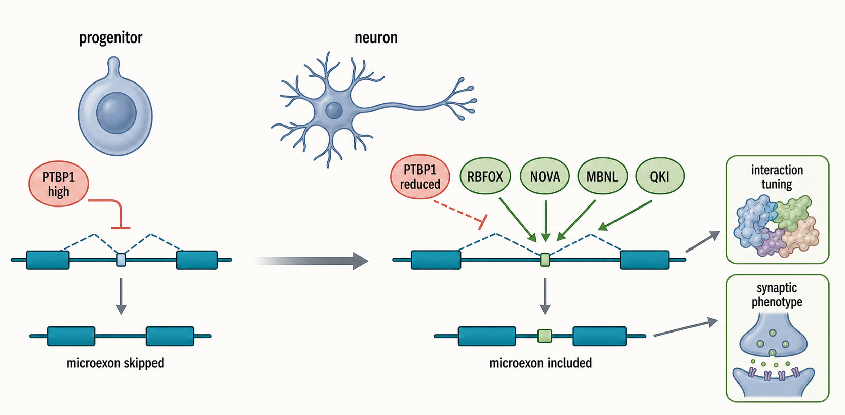
Figure 28.4. Neuronal Microexon Program. Neuronal differentiation changes RBP abundance and activity, shifting from a PTBP1-high progenitor state that represses neuronal exons toward inclusion of selected microexons driven by RBFOX, NOVA, MBNL, and QKI. Even a few inserted amino acids encoded by a microexon can tune a protein interaction surface or localization behavior at the synapse.
The strongest maps of RBP control combine binding and perturbation. CLIP-seq family methods can identify where an RBP contacts RNA in cells. RNA-seq after RBP knockdown, knockout, overexpression, or acute degradation can identify splicing changes. Motif enrichment can suggest direct regulation. But none of these alone proves direct causal control. Binding without a splicing change may be nonfunctional or redundant. A splicing change after RBP perturbation may be indirect. A motif may be present but inaccessible. Stronger evidence combines binding at the relevant location, directionally consistent perturbation, reporter or endogenous cis-element mutation, and rescue.
28.3. Cryptic splicing, pseudoexons, and poison exons
Cryptic splicing is the use of splice sites that are normally hidden, inefficient, or repressed. The term “cryptic” is relational: a site is cryptic relative to a cell type, condition, reference annotation, or wild-type genotype. A cryptic splice site may be present in the genome all along, but not used detectably because it is weak, blocked by an RBP, embedded in unfavorable RNA structure, too close to another site, or eliminated by surveillance. A point mutation can strengthen such a site or weaken the normal site enough that the cryptic site wins the competition.
Pseudoexons are intronic sequences that resemble exons. A pseudoexon typically has candidate splice sites, a length compatible with exon definition, and sometimes enhancer-like motifs. Under normal conditions, pseudoexons are usually repressed or too weak to be included. Disease-associated variants can activate a pseudoexon by creating a new splice site, strengthening an enhancer, disrupting a silencer, or changing RNA structure. Regulatory changes can also reveal pseudoexons without a DNA variant at the pseudoexon itself.
Poison exons are different in logic. A poison exon is included in a transcript to reduce productive gene output. The usual mechanism is introduction of a premature termination codon that causes nonsense-mediated decay. This is a negative-feedback strategy for some splicing regulators: when the regulator is abundant, it promotes inclusion of a poison exon in its own transcript or in transcripts of related regulators, reducing productive protein synthesis. Poison exons can also shape developmental transitions. A poison exon is therefore not necessarily a pathological error; it can be a normal regulatory device.
The three categories can overlap. A pseudoexon can be cryptically activated by mutation and behave as a poison exon if it introduces a premature termination codon. A normally regulated poison exon might be called cryptic in a cell type where it is usually silent. For clarity, this chapter uses cryptic splicing for abnormal or normally hidden splice-site use, pseudoexon for an intronic exon-like sequence, and poison exon for an exon whose inclusion lowers productive output. The evidence should specify which property is being claimed.
Table 28.3. Cryptic Splicing Terminology. Definitions, typical causes, and validation requirements for the overlapping categories of aberrant and regulated splicing that deviate from annotated canonical isoforms.
| Term | Definition | Typical cause | Productive or unproductive consequence | Validation assay | Boundary case |
|---|---|---|---|---|---|
| Cryptic splice site | A splice site present in the genome but normally silent, weak, or repressed | Point mutation strengthening site; RBP loss; cellular stress | Depends on reading frame and position; often unproductive | RT-PCR or RNA-seq showing novel junction; minigene assay with candidate variant | A site used at constitutively low level may not be functionally cryptic |
| Pseudoexon | An intronic segment resembling an exon that can be conditionally included | Deep intronic variant; regulatory RBP loss; transposon element carrying exon-like signals | Usually unproductive; introduces premature stop codon if in-frame | Minigene assay; patient-derived RNA-seq; orthogonal RT-PCR in relevant tissue | Some pseudoexons overlap conditionally regulated alternative exons |
| Poison exon | A regulated exon whose inclusion reduces productive transcript output | Normal RBP autoregulatory feedback; developmental program; dysregulation in disease | Unproductive by design; triggers NMD or creates non-functional truncation | NMD inhibitor or UPF1 knockdown enriches isoform; isoform-specific RT-PCR | Normally regulatory poison exons can become pathological if the regulatory switch fails |
| NMD-sensitive isoform | A transcript isoform containing a premature termination codon more than approximately 50-55 nt upstream of the last exon junction complex | PTC-creating exon inclusion; frameshift; stop-codon readthrough failure | Unproductive; degraded by NMD machinery | Cycloheximide treatment or UPF1 knockdown increases isoform abundance | Not every PTC-containing isoform is efficiently degraded by NMD |
| Retained intron | A transcript in which one or more introns remain unspliced in the exported or cytoplasmic RNA fraction | Weak splice sites; nuclear detention signal; developmental or stress regulation | Variable: may be nuclear-retained, later spliced, translated, or an NMD substrate | Intronic read coverage; strand-specific RT-PCR spanning the intron-exon boundary | Regulatory retained-intron RNAs can be stable, exported, and functionally important |
| Splice noise | Low-level aberrant splicing lacking regulatory function | Intrinsic spliceosome error rate; weak competing sites; sequencing library or alignment artifact | Usually unproductive at physiological abundance | Absent enrichment in matched conditions; event fails minigene test; not reproducible | Becomes biologically relevant if surveillance fails or basal noise level is elevated by stress |
Cryptic splicing is an important mechanism in human genetic disease. A variant that appears intronic and distant from canonical splice sites can still create a new exon or activate a latent splice site. Standard exon-focused variant interpretation can miss these events. RNA sequencing of patient-derived cells, minigene assays, and targeted RT-PCR can reveal abnormal junctions. Clinical RNA-seq for Mendelian diagnostics is increasingly used to connect variants to aberrant splicing, expression outliers, and allele-specific effects. However, the assayed tissue matters: a disease-relevant splice event in brain, retina, muscle, or lung may be absent from blood or fibroblasts.
Cryptic splicing also appears in repeat-rich and transposon-derived regions. Many introns contain Alu elements, LINE elements, endogenous retrovirus fragments, and other repeats that carry splice-like signals. These sequences are usually suppressed by combinations of weak splice-site architecture, RNA structure, chromatin context, RNA surveillance, and RBPs. hnRNPM repression of LINE-associated cryptic splicing provides a recent example in which loss of repression can expose repeat-linked double-stranded RNA signals and promote interferon responses. This example links splicing fidelity to innate immune activation, a topic expanded in Chapter 108.
Not every novel junction is meaningful cryptic splicing. Low-level splice noise exists because spliceosome recognition is probabilistic, sequencing libraries can contain artifacts, and alignment across repeats or paralogous regions can create false junctions. A cryptic event should be supported by adequate read depth, plausible splice signals, reproducibility, strand and annotation checks, and preferably orthogonal validation. If the claim is disease causality, the evidence should show that the event occurs in relevant cells, changes RNA or protein output in a plausible way, and segregates or recurs consistently with the phenotype.
The boundary between regulated unproductive splicing and surveillance is especially important. Alternative splicing coupled to nonsense-mediated decay can buffer RBP abundance and reshape regulatory networks. But detection of an NMD-sensitive isoform after translation inhibition or NMD-factor knockdown does not prove that the isoform is normally abundant or functional. Such experiments enrich unstable RNAs by design. They are useful for discovering hidden isoforms, but interpretation must separate existence, regulatory use, and physiological consequence.
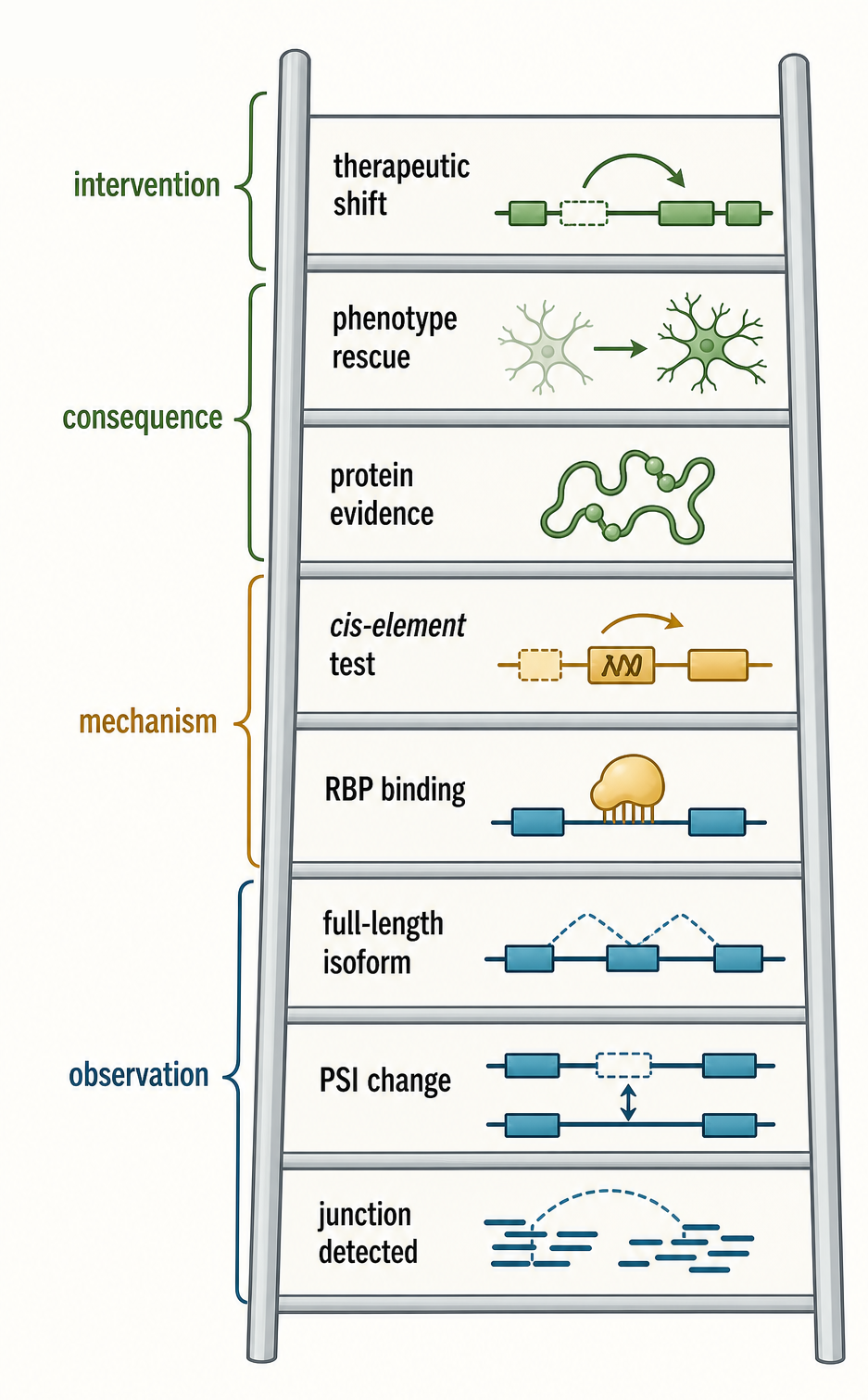
Figure 28.5. Isoform Evidence Ladder. Different assays answer different splicing questions. A statistically significant junction change is a starting observation; functional and therapeutic claims require convergent evidence across RNA detection, mechanism, molecular consequence, and phenotype.
Box 28.1. Common Misconceptions in Alternative Splicing
- An RNA isoform does not automatically produce a protein isoform; many splice isoforms are nonproductive, unstable, untranslated, or differ only in RNA regulatory regions.
- PSI is an event-level estimate, not a count of full-length transcript molecules; a high PSI change does not identify the complete isoform.
- Long-read RNA-seq improves isoform phasing but does not remove biases from RNA integrity, amplification, coverage depth, error correction, or quantification.
- Microexons can be strongly functional despite encoding only a few amino acids, because short peptide inserts can alter interaction surfaces, localization motifs, and developmental programs.
- Poison exons can be normal regulatory elements that deliberately reduce productive protein output through NMD-coupled feedback; they are not always pathological errors.
- Cryptic splicing is not always caused by a DNA variant; altered RBP concentrations, cellular stress, repeat-element activation, and surveillance failure can all expose cryptic sites.
- Intron retention is not always failed splicing; retained introns can support nuclear detention, regulated decay, or developmental timing in a controlled manner.
28.4. Microexons and neuronal/developmental programs
Microexons are short exons whose small size creates both a recognition problem and a functional opportunity. Different studies use different cutoffs, such as exons of 3-27 nucleotides or other short ranges, so authors must state their definition. The key biological point is not the exact cutoff but the fact that the exon is short enough to challenge ordinary exon definition and to encode a very small peptide segment if translated.
For the spliceosome, a microexon offers limited space for exon-bound factors. A typical exon can contain several enhancers, silencers, and binding sites. A microexon may have only a few nucleotides between splice sites. Recognition therefore often depends on strong flanking intronic elements, specialized RBPs, favorable spacing, and local RNA architecture. Because very short exons are easy to miss in annotation and short-read analysis, microexons also create a measurement problem.
For proteins, a microexon can encode one to several amino acids. That sounds small, but many protein interactions depend on short motifs, flexible loops, linker lengths, charge patches, and domain-interface geometry. A few amino acids can change whether a protein binds a partner, localizes to a synapse, enters a signaling complex, or responds to phosphorylation. Microexons are especially common in genes encoding regulators of neuronal connectivity, cytoskeletal dynamics, vesicle trafficking, small GTPase regulation, and synaptic organization.
Neuronal microexon programs illustrate developmental timing. During neurogenesis, progenitor cells and differentiating neurons express different RBP combinations. PTBP1 is high in many non-neuronal or progenitor contexts and tends to repress many neuronal exon programs. RBFOX, NOVA, MBNL, QKI, and other RBPs contribute to inclusion of neuronal exons and microexons in differentiated states. The result is not a single “neuronal splicing factor” but a coordinated shift in multiple regulators, splice-site choices, transcript ends, localization signals, and translation programs.
Microexons are also implicated in disease. Autism spectrum disorder studies have connected misregulated neuronal microexon programs to synaptic and neurodevelopmental phenotypes, although each event requires separate mechanistic validation. A recent example links mis-splicing of a neuronal microexon in CPEB4 to altered protein behavior and aggregation in autism spectrum disorder context. The point is not that every microexon is disease-causing, but that short exons can alter proteins in ways large enough to matter.
Microexons are not limited to the nervous system. Muscle, immune, cancer, and developmental contexts can show short-exon regulation. Snap23 microexon regulation by MBNL, QKI, and RBFOX2 in tissue-specific and striated muscle disease contexts shows how short exons can integrate splicing regulators outside a narrow brain-only frame. Colorectal cancer studies linking RBFOX2 and PTBP1 to alternative microexon splicing emphasize that tumor contexts can reuse or disrupt developmental splicing logic.
Single-cell and single-neuron studies add another layer. Bulk tissue averages can hide splicing differences among cell types or neuronal subtypes. Single-cell RNA-seq can separate cell populations, but many single-cell protocols capture only transcript ends and have sparse coverage across internal junctions. Recent single-neuron work on regulated microexon splicing and synaptic function points toward cell-resolved interpretation, but such data must be evaluated with attention to coverage, dropout, amplification bias, and validation outside the sequencing assay.
A common misconception is that a microexon should be ignored because it is too short to matter or too difficult to quantify. The opposite mistake is to assume that every conserved microexon has a major phenotype. A measured microexon event becomes biologically interpretable when the field can connect regulated inclusion to a specific RNA isoform, a protein or RNA consequence, the RBP or cis element that controls the event, and a cellular or organismal phenotype.
28.5. Quantification and isoform interpretation
Splicing quantification begins with the question being measured. Event-level analysis asks whether a particular exon, splice site, or intron is used more or less often. Transcript-level analysis asks which full-length isoform molecules exist and how abundant each is. Gene-level expression asks how much total RNA derives from a gene. These are related but not interchangeable. A gene can show no total expression change while switching isoforms. A cassette exon can change PSI without changing the abundance of the dominant full-length transcript much. A transcript isoform can be assembled computationally even when direct evidence for its full-length molecule is weak.
Percent spliced in, or PSI, is a common event-level measure. For a cassette exon, PSI estimates the fraction of transcripts including the exon among transcripts that include or skip it. Conceptually, inclusion reads support exon 1-exon 2 and exon 2-exon 3 junctions, while skipping reads support exon 1-exon 3. In practice, read length, mappability, fragment bias, multimapping, low expression, alternative neighboring events, and annotation choice affect the estimate. PSI is useful, but it is not a direct molecule count.
Short-read RNA-seq remains central because it is scalable, quantitative, and compatible with many perturbation designs. It can discover novel junctions, quantify known events, and compare conditions. Transcript assembly tools such as Cufflinks helped establish RNA-seq as a way to infer unannotated transcripts and isoform switching during differentiation. Modern methods have improved, but the core ambiguity remains: short reads often do not span enough junctions to phase distant events on the same molecule.
Long-read RNA-seq addresses that phasing problem by sequencing longer cDNA or direct RNA molecules. It can reveal full-length isoforms, combinations of distant exons, retained introns, alternative starts and ends, and complex splice patterns that short reads split into separate events. Recent systematic assessment of long-read RNA-seq methods emphasizes both the value and the biases of long-read approaches for transcript identification and quantification. Long reads are not automatically definitive: coverage, RNA integrity, template switching, internal priming, incomplete reverse transcription, platform error profiles, and analysis pipelines still matter.
Table 28.4. Isoform Quantification Methods. Comparison of methods for measuring RNA splice isoforms, including their primary outputs, technical strengths and weaknesses, best-use contexts, and the orthogonal validation each requires.
| Method | Primary output | Strengths | Weaknesses | Best use | Required orthogonal validation |
|---|---|---|---|---|---|
| RT-PCR | Band size indicating isoform; semi-quantitative ratio | Low cost; high sensitivity; directly reveals junction identity | Low throughput; not genome-wide; gel resolution is limited for similar-size isoforms | Validating known events; testing minigene constructs; clinical diagnostics | Sanger sequencing of amplicon to confirm junction identity |
| qRT-PCR junction assay | Quantitative isoform ratio at a single locus | Precise ratio across many samples; adaptable to high throughput | Junction-spanning primer design is challenging; one event at a time | Robust quantification of a single validated splice event across many conditions | RT-PCR confirmation of amplicon identity before assay design |
| Bulk short-read RNA-seq | Junction reads; event-level PSI; gene expression counts | Genome-wide; scalable; well-benchmarked quantification pipelines | Short reads cannot phase distant events; isoform reconstruction is model-dependent | Discovery and quantification of splice junctions across many conditions | RT-PCR or targeted long-read for high-priority events |
| Event-level PSI analysis | Per-event inclusion fraction genome-wide | Interpretable statistic; integrates skipping and inclusion junction types | Does not report full-length isoform molecules; sensitive to annotation choice | Comparing specific events systematically across conditions or disease cohorts | Isoform-level validation by long-read sequencing or RT-PCR |
| Transcript assembly | Full-length isoform models inferred from read coverage | Generates isoform-resolution hypotheses; integrates all junction types | Model-dependent; low-abundance isoforms are often fragmented or misassembled | Generating isoform catalogs for further experimental testing | Long-read sequencing to confirm or refute assembled models |
| Targeted long-read sequencing | Full-length reads at enriched loci | Phasing of distant exon combinations; high depth at targeted loci | Amplification biases; capture efficiency varies; limited to pre-selected targets | Validating complex isoforms at specific loci after discovery by short-read data | Gene-level quantification by short-read RNA-seq for context |
| Whole-transcriptome long-read RNA-seq | Full-length isoform sequences genome-wide | Phased isoforms without pre-selection; no amplification in direct approaches | Lower throughput per cost; error rates require correction pipelines; RNA integrity critical | Transcriptome-wide isoform discovery and complex junction phasing | Short-read RNA-seq for quantification; RT-PCR for validation of key events |
| Direct RNA sequencing | Native RNA sequence with modification signals | Retains RNA modifications; no reverse transcription step; no amplification bias | High error rate; lower throughput; limited availability and coverage depth | Studying isoforms alongside RNA modification patterns (e.g., m6A sites) | Complementary cDNA-based long-read sequencing for higher depth |
| Single-cell RNA-seq | Gene expression per cell; limited internal isoform information | Cell-type resolution; large number of cells; gene expression per population | 3′ or 5′ biased; sparse junction coverage; poor isoform phasing | Cell-type composition analysis and gene-level expression per cell type | Full-length or targeted single-cell long-read assay for isoform questions |
| CLIP-seq integration | RBP binding sites near splice events | Directly links protein occupancy to RNA contact site near regulated event | Binding does not prove functional regulation; crosslinking and capture efficiency vary | Identifying candidate regulated exons downstream of a specific RBP | Perturbation RNA-seq after RBP knockdown; cis-element mutation in minigene |
| Proteomics for isoform-specific peptides | Isoform-specific protein evidence by mass spectrometry | Direct protein-level confirmation of isoform existence | Peptides may be shared, too short, post-translationally modified, or too low abundance | Confirming protein expression of a high-priority isoform with a unique peptide | RNA-level isoform data for peptide prediction; antibody-based validation when possible |
Single-cell RNA-seq adds cell identity but usually sacrifices isoform completeness. Many high-throughput single-cell assays sequence only the 3′ or 5′ end of transcripts. These protocols are excellent for cell-type composition and gene expression but weak for internal splicing unless paired with specialized full-length, targeted, or long-read approaches. When a study claims cell-type-specific isoform choice, the reader should ask whether reads actually span the diagnostic junctions and whether the isoform can be distinguished from related transcripts.
Isoform interpretation also requires annotation discipline. Genome annotations are incomplete and versioned. A “novel” isoform may be novel relative to one annotation release but present in another database, tissue atlas, or long-read dataset. Conversely, an annotated isoform may be supported by weak historical evidence and absent in the tested cell type. Chapter 18 explains transcript models and annotation versioning; this chapter emphasizes that splicing analysis must report annotation source, genome build, and whether novel junctions were allowed.
Differential splicing is not the same as differential transcript function. A statistically significant change in PSI can arise from a large sample size and a small effect. A large PSI change in a lowly expressed gene may produce little molecular consequence. A splice event may occur in a minor cell population whose abundance changes between samples, creating apparent differential splicing without a change inside any cell type. Tumor samples, inflamed tissues, and developing organs are especially vulnerable to this composition effect. Proper interpretation may require matched cell-type markers, single-cell data, purified populations, or deconvolution.
Protein-level validation is often the bottleneck. If a splice event changes coding sequence, the study should ask whether the predicted protein isoform is produced, stable, and localized correctly. Mass spectrometry can detect isoform-specific peptides when the peptide is unique, abundant, and technically observable, but many splice-derived peptides are too short, shared, modified, or low abundance. Antibodies may not distinguish isoforms. Reporter assays and tagged constructs can help, but overexpression may create nonphysiological localization or interactions. Therefore, RNA-level evidence should not be silently converted into protein-level claims.
28.6. Functional validation, therapy, and evidence limits
Functional validation asks whether a splicing difference causes a biological outcome. The minimal observation is an RNA change, such as a junction difference in RNA-seq or RT-PCR. A stronger claim connects the RNA change to a molecular consequence, such as altered protein domain, NMD, RNA localization, or translation. A still stronger claim shows that changing the isoform changes the phenotype while holding total gene expression and other confounders as constant as possible.
Table 28.5. Evidence Levels for Functional Splicing Claims. Hierarchy of evidence types used to support claims about regulated splicing, ordered from initial observation to functional and therapeutic demonstration.
| Evidence level | What it shows | What it does not show | Example control | Claim strength |
|---|---|---|---|---|
| Junction detection | A splice junction exists in the sample | Abundance, regulation, or biological function | Annotation check; strand verification; spike-in read depth control | Existence of isoform only |
| Differential PSI | An event changes between conditions or genotypes | Full-length isoform identity; protein consequence | Matched cell-type composition; biological replicates; event-level controls | Association; not mechanism |
| Full-length isoform assignment | A complete transcript molecule with specific exon combination exists | Functional significance of that isoform | Long-read validation; multiple independent molecules supporting the model | Isoform existence; not function |
| RBP binding | An RBP contacts RNA near the regulated event | Whether binding is functional or redundant | Negative CLIP control; binding-dead RBP mutant as specificity control | Candidate regulator identification |
| RBP perturbation | RBP loss or gain shifts the splice event | Directness; whether effect is on one RNA or many indirect targets | Rescue with wild-type RBP; unrelated RBP perturbation as negative control | Regulatory involvement; not direct causality |
| Cis-element mutation | A sequence motif is required for the regulatory effect | Whether motif acts in native chromatin context; long-range effects | Compensatory mutation; multiple motif positions tested in reporter | Direct cis-element requirement demonstrated |
| Endogenous genome editing | A variant or element is required at the native genomic locus | Organismal phenotype; compensatory mechanisms in vivo | Synonymous edit control; isogenic cell comparison | Strong causal evidence at RNA level |
| Isoform-specific knockdown or rescue | One isoform mediates the downstream outcome | Off-target effects when isoforms share sequence | Isoform-specific ASO or construct; rescue isoform versus unrelated null construct | Causal isoform assignment |
| Protein isoform detection | Predicted protein isoform is produced in cells | Relative abundance compared with other isoforms; localization; activity | Isoform-specific antibody or unique mass spectrometry peptide | Protein isoform existence |
| Phenotype rescue | Restoring the isoform reverses the phenotype | Mechanism; whether rescue specificity is confirmed | Rescue with unrelated isoform as negative control; dose-matching | Strong functional evidence |
| Therapeutic splice modulation | The splice event can be pharmacologically shifted toward a desired outcome | Clinical efficacy; safety; in vivo target specificity | Scrambled ASO control; off-target isoform monitoring; dose-response | Preclinical or clinical intervention evidence |
Cis-element tests are valuable because they separate local RNA grammar from global regulator effects. In a minigene reporter, a genomic segment containing the regulated exon and flanking intronic sequence is cloned into a reporter transcript. Mutating an enhancer, silencer, splice site, or branch-point candidate can show whether that element affects splicing in the tested cells. The limitation is that a minigene may omit long-range intronic elements, chromatin context, transcription kinetics, or native RNA abundance. Endogenous genome editing of the same element is stronger when feasible.
RBP perturbation tests identify trans-acting regulators. Knockdown, knockout, overexpression, or acute depletion of an RBP can shift splicing. Rescue with wild-type or binding-defective RBP variants can test specificity. CLIP-seq can show binding near the regulated event. The strongest argument combines RBP binding, loss-of-function effect, rescue, and cis-element dependence. Even then, RBPs often regulate hundreds of RNAs, so a phenotype after RBP perturbation may reflect many events. Isoform-specific rescue or knockdown is needed to assign a phenotype to one splice event.
Isoform-specific manipulation is becoming more precise. Antisense oligonucleotides can block splice sites or regulatory elements to promote exon skipping, exon inclusion, or pseudoexon suppression. CRISPR-based RNA targeting and Cas13 systems can knock down selected isoforms when unique sequence exists. Genome editing can repair or recreate splice-altering variants. These tools are powerful, but target specificity is constrained by shared exons, overlapping isoforms, nuclear accessibility, delivery, RNA turnover, and off-target hybridization or cleavage.
Therapeutic splice switching is most convincing when the disease mechanism is clearly tied to an event. If a variant activates a poison-like pseudoexon that disrupts a protein, blocking that pseudoexon can be a rational strategy. If an exon-skipping strategy restores a reading frame, the therapeutic goal is to produce a shorter but functional protein. If a developmental splicing program is globally altered, therapy is harder because many targets may contribute and the correct adult versus developmental isoform balance may be tissue-specific. Clinical and translational examples anchor this principle without replacing the later therapeutics chapters: nusinersen redirects SMN2 splicing with clinical benefit in infantile-onset spinal muscular atrophy, risdiplam illustrates small-molecule SMN2 splicing modulation, beta-globin IVS2-654 studies show antisense correction from cell models to mice, and modern SSO platform work links RNA diagnosis to therapeutic design.
The evidence standard for a splice-switching intervention is stricter than “the junction moved.” A convincing program should define the aberrant or therapeutic junction, show dose-responsive correction in a relevant biological system, measure the intended RNA and protein or functional consequence, and test whether related transcripts or hybridization-compatible off-targets are perturbed. ASO therapy reviews, variant-eligibility consensus guidelines, and SSO off-target studies all converge on this logic: sequence complementarity gives a rational starting point, but tissue exposure, chemistry, disease stage, assay context, and transcriptome-wide specificity determine whether the splice change is interpretable as therapy.
Box 28.2. How to Evaluate a Disease-Associated Pseudoexon
- Assess whether the candidate variant is likely to create or strengthen splice signals, such as a new 5′ or 3′ splice site, branch point, or exonic splicing enhancer.
- Confirm that the pseudoexon is detected in disease-relevant tissue or a justified cellular proxy, because splice events can be highly tissue-specific and absent from blood or fibroblasts.
- Verify the event by RT-PCR or targeted sequencing independent of the discovery RNA-seq data, checking junction identity by Sanger sequencing.
- Determine whether pseudoexon inclusion alters RNA stability, introduces a premature termination codon, disrupts protein reading frame, or triggers nonsense-mediated decay.
- Test whether correction of the event, for example by pseudoexon suppression with an antisense oligonucleotide, rescues molecular or cellular phenotype in a relevant model.
- Confirm that normal isoforms and related gene transcripts are not disrupted by the proposed therapeutic intervention before advancing toward clinical application.
Clinical RNA-seq expands diagnosis by detecting aberrant splicing in patient samples. The evidence limit is tissue and context. A blood sample may not express the disease gene. Cultured fibroblasts can alter splicing through culture stress. NMD can hide pathogenic transcripts unless decay is inhibited, but inhibition can reveal many nonphysiological isoforms. Diagnostic interpretation therefore needs gene expression, tissue relevance, variant data, familial segregation, and targeted validation.
Cancer and inflammatory disease studies often report isoform switches, but causal interpretation is difficult. Splicing changes can be drivers, passengers, markers of cell composition, consequences of signaling, or responses to stress. CD44 variable exon inclusion in cancer and immune contexts, DDX17-regulated PXN-AS1 isoforms in hepatocellular carcinoma, bacterial toxin-induced alternative splicing in infection responses, and arthritis-associated splicing changes all illustrate biologically interesting links. Each case still requires event-specific validation before the isoform is called causal.
The most important evidence limit is that “splicing change” is not one evidence class. A junction read, a PSI estimate, a long-read isoform, a minigene result, an RBP-binding peak, a patient RNA outlier, a proteomic peptide, and a therapeutic rescue answer different questions. A robust chapter, paper, or clinical report should state which question each method answers and which question remains open. This is the same evidence discipline introduced in Chapter 5, applied specifically to isoform biology.
Box 28.3. Clinical and Therapeutic Evidence Limits for Splice Switching
- Splice-switching therapy evidence starts with a defined RNA event and should progress through junction correction, dose response, protein or RNA-fate consequence, tissue relevance, off-target assessment, and clinical or disease-model outcome.
- SMN2 splicing modulation, beta-globin IVS2-654 splice correction, and current SSO platform examples show why a therapy claim needs both molecular correction and context-specific functional evidence.
- Chapter 28 should use these examples to teach evidence standards; detailed ASO chemistry, delivery, regulatory logic, and product-specific clinical interpretation belong in the later therapeutics chapters.
Recent Consensus
The current consensus is that alternative splicing is a major eukaryotic regulatory layer, but its biological meaning is event-specific. Regulated splice choices can alter protein sequence, RNA fate, translation, localization, or decay. Many tissue-specific and developmental programs are reproducible and mechanistically linked to RBPs. Microexons, especially in neuronal and developmental contexts, can have disproportionate functional effects despite their small size. Cryptic splicing and pseudoexon activation are important in genetic disease, cancer, repeat biology, and stress responses.
Recursive splicing is an established route for stepwise removal of selected long introns, especially well supported in Drosophila and a subset of vertebrate genes. Its prevalence, kinetics, and contribution to mature isoform choice remain context-dependent. A recursive intermediate should not be counted automatically as a stable alternative isoform, and recursive splicing should not be conflated with trans-splicing between distinct precursor molecules.
The field also agrees that measurement and interpretation remain limiting. Short-read RNA-seq is strong for junctions and event-level changes but weak for complete isoform molecules. Long-read RNA-seq improves isoform discovery but needs careful benchmarking and quantification. Functional claims require perturbation and rescue, not only differential splicing. Clinical or therapeutic claims require tissue-relevant evidence, target specificity, and phenotype-level validation.
Open Questions, Controversies, Deprecated Models, and Common Misconceptions
Open questions:
- How many low-abundance isoforms are functional?
- How often does cell-type composition explain apparent disease-associated splicing?
- How do RNA structure and transcription kinetics combine quantitatively with RBP binding?
- How many microexon changes are causal in neurodevelopmental disorders rather than correlated markers?
- How should full-length isoforms be defined and benchmarked across short-read, long-read, single-cell, and proteomic platforms?
Common misconceptions:
- “Every alternative RNA isoform produces a protein isoform.” Many splice isoforms are nonproductive, unstable, untranslated, or differ only in RNA regulatory regions.
- “Cryptic splicing is always caused by mutation.” Mutation is one route, but altered RBP levels, stress, repeats, RNA structure, and surveillance failure can also expose cryptic sites.
- “Poison exons are errors.” Poison exons can be regulated feedback elements that deliberately reduce productive protein output.
- “Microexons are too short to matter.” Short peptide inserts can alter interaction surfaces, localization motifs, and developmental programs.
- “PSI equals full-length isoform abundance.” PSI is an event-level estimate and may not identify complete transcript molecules.
- “Long-read RNA-seq solves all isoform problems.” Long-read methods improve phasing but still have coverage, error, library, and quantification biases.
- “Recursive splicing is a form of trans-splicing or always creates a new mature isoform.” Recursive splicing usually removes segments of one long intron from one precursor; its intermediates can disappear from the final RNA.
Deprecated or weakened claims:
- Alternative splicing should not be described simply as a way to maximize protein number.
- SR proteins should not be treated as universal activators, and hnRNP proteins should not be treated as universal repressors.
- Intron retention should not be treated automatically as splicing failure.
- Novel splice junctions should not be treated automatically as functional.
- Annotated transcript isoforms should not be treated automatically as expressed in a sample.
- RNA isoforms should not be treated automatically as protein isoforms.