Chapter 135. Ribosome Profiling, Translation Measurement, and Single-Cell Translatomics
Scope Note
This chapter explains how translation is measured at transcriptome scale, with ribosome profiling as the central positional method. The scope includes nuclease-protected ribosome footprints, library construction, footprint assignment, initiation and elongation profiling, pausing and collision measurements, termination and recycling readouts, open reading frame discovery, microproteins, noncanonical translation, single-cell and spatial translatomics, and peptide-level validation. The chapter treats ribosome profiling as a biochemical and computational inference chain: a footprint can be highly informative about ribosome occupancy, reading frame, and translation state, but a footprint is not by itself a direct measurement of protein abundance, protein function, or translation rate.
Executive Summary
Ribosome profiling, usually abbreviated Ribo-seq, sequences short RNA fragments protected from nuclease digestion by ribosomes. A translating ribosome covers a reproducible segment of messenger RNA, and nuclease treatment removes much of the unprotected RNA. The protected fragment can then be purified, converted to a sequencing library, aligned to a genome or transcriptome, and assigned to a ribosomal position such as the peptidyl-tRNA site, or P site. Genuine translating ribosomes often produce footprints with characteristic lengths, enrichment in coding regions, start and stop codon structure, and three-nucleotide periodicity. These properties make Ribo-seq one of the strongest transcriptome-wide approaches for detecting translated regions and comparing ribosome occupancy across conditions.
Ribo-seq is not a direct census of protein production. The number of footprints over an open reading frame reflects initiation, elongation, pausing, termination, ribosome recycling, nuclease accessibility, fragment recovery, mappability, RNA abundance, library bias, and analysis choices. Translation efficiency, commonly calculated as Ribo-seq signal normalized by matched RNA-seq abundance, is an operational comparison rather than a pure molecular rate constant. A transcript can show high ribosome density because initiation is frequent, because elongation is slow, because ribosomes are stalled, or because technical recovery is biased. Protein abundance further depends on completion of translation, protein folding, secretion, processing, degradation, and detectability.
Modern translation measurement is a family of complementary methods. Standard monosome Ribo-seq maps ribosome occupancy at high resolution. Initiation profiling enriches or infers ribosomes at start sites. Elongation and pausing analyses use codon-resolved footprints, time courses, perturbations, and metagene patterns. Disome and collision profiling enrich footprints from adjacent ribosomes and connect translation measurement to ribosome quality control. Termination and recycling studies inspect footprints near stop codons, downstream regions, release-factor perturbations, and recycling-factor perturbations. ORF discovery combines periodicity, initiation evidence, coding potential, conservation, perturbation, and peptide detection to distinguish translated ORFs from scanning, regulatory association, or artifacts.
Single-cell and spatial translatomics are important but still technically constrained. True single-cell, codon-resolution footprint sequencing is difficult because ribosome-protected fragments are short, scarce, and easily lost during digestion and library construction. Related approaches measure cell-type-specific ribosome association, nascent protein synthesis, spatial RNA localization, proximity to ribosomes, or peptide products. These methods answer valuable questions, but they must be named by the property measured. A cell-type-specific ribosome-associated RNA profile is not the same as a single-cell P-site map, and RNA localization is not the same as local translation.
Concept Inventory
- Ribosome profiling: a sequencing-based footprinting method for measuring where ribosomes occupy RNA. The key molecular object is the ribosome-protected fragment, or RPF: a piece of RNA that remains after nuclease digestion because a ribosome or ribosome-containing complex protected it. A canonical cytosolic eukaryotic monosome footprint is often near 28 to 30 nucleotides, but footprint length is not universal. Bacterial ribosomes, organellar ribosomes, initiating ribosomes, terminating ribosomes, collided ribosomes, nuclease choices, salt conditions, inhibitor treatments, and size-selection windows can all shift the protected fragment distribution.
- Translation efficiency: a ratio-like comparison between ribosome occupancy and RNA abundance. In common practice, investigators compute translation efficiency by comparing normalized Ribo-seq reads over a feature with normalized RNA-seq reads from the same feature. This comparison helps identify transcripts whose ribosome occupancy changes beyond transcript abundance, but the metric does not isolate initiation rate or protein output by itself. High translation efficiency can reflect more initiation, slower elongation, stronger ribosome stalling, isoform changes, or library-specific bias.
- P-site assignment: the computational placement of each footprint at the ribosomal peptidyl-transfer RNA site. A-site assignment places the footprint at the aminoacyl-transfer RNA decoding site. Because the sequenced fragment begins and ends outside the ribosomal active sites, the offset from a read end to the P site or A site must be calibrated, usually separately for each footprint length. Accurate site assignment is necessary for reading-frame analysis, codon-level pausing, start-site inference, and stop-codon profiling.
- Initiation profiling: Ribo-seq-related strategies that enrich or infer ribosomes at start sites. Elongation profiling studies how ribosomes move through coding sequences. Pausing means local slowing or accumulation of ribosomes, while collision means a physical encounter between adjacent ribosomes when a trailing ribosome catches a slower leading ribosome. Noncanonical translation describes translation outside the annotated principal coding sequence, including upstream ORFs, overlapping ORFs, alternative-frame ORFs, ORFs in annotated noncoding RNAs, circular RNA-derived ORFs, and viral recoding products. A microprotein is a small protein product from a short ORF; the term should be reserved for cases with protein-product evidence or strong convergent functional evidence, not merely any short ORF with a few footprints.
What to Know Before Reading This Chapter
The first prerequisite is the physical organization of translation. A ribosome reads an mRNA in codons, usually moving in three-nucleotide steps. During elongation, the A site accepts an aminoacyl-tRNA, the P site holds the tRNA linked to the growing peptide, and the E site holds the departing deacylated tRNA. Translocation moves the mRNA-tRNA complex by one codon. Ribo-seq works because a ribosome bound to an RNA protects a reproducible segment of that RNA from nuclease digestion, allowing the position of the ribosome to be inferred from the protected sequence.
The second prerequisite is that a sequencing read is an experimental survivor, not a pristine molecular event. A footprint has passed through harvest, optional inhibitor treatment, lysis, nuclease digestion, ribosome purification, fragment isolation, ribosomal RNA depletion, adapter ligation, reverse transcription, amplification, sequencing, alignment, duplicate handling, and offset assignment. Each step can distort the final distribution. Strong Ribo-seq interpretation therefore depends on visible quality-control evidence rather than faith in the method name.
The third prerequisite is the distinction between RNA abundance, ribosome occupancy, translation rate, and protein abundance. RNA-seq measures transcript abundance or recoverability. Ribo-seq measures protected ribosome footprints. Polysome profiling separates RNAs by the number of ribosomes associated with them. Translating ribosome affinity purification, or TRAP, enriches ribosome-associated RNAs from tagged cells or compartments. Nascent protein labeling measures new protein synthesis over a pulse. Mass spectrometry detects peptides or proteins that survive extraction, digestion, chromatographic separation, ionization, and database search. These measurements overlap but do not report the same biological property.
As a running example, consider a mammalian stress-responsive mRNA with a short upstream ORF in the transcript leader. RNA-seq may show little change in mRNA abundance after stress. Standard Ribo-seq may show increased footprints on the upstream ORF and decreased footprints on the main coding sequence. Initiation profiling may reveal a stress-dependent start site in the leader. Reporter mutagenesis may show that disrupting the upstream start codon restores main-ORF translation. Mass spectrometry may fail to detect the short upstream peptide because it is unstable or poorly ionized. The combined evidence can still support upstream-ORF-mediated translational regulation, while the evidence for a stable microprotein product remains weaker.
135.1. Ribo-seq design and nuclease footprints
Ribo-seq begins with a simple protection principle: a ribosome-bound segment of RNA is less accessible to nuclease than adjacent exposed RNA. The biological interpretation, however, depends on how that protection snapshot is made. A typical experiment chooses a biological state, rapidly harvests cells or tissue, optionally uses translation inhibitors, lyses material under conditions intended to preserve ribosome-mRNA complexes, digests exposed RNA, purifies ribosome-protected fragments, selects a size range, prepares a sequencing library, and maps the resulting reads. Each stage asks a design question. Does the experiment need the least perturbed snapshot possible, the highest start-site enrichment, the cleanest monosome fraction, the best recovery from low input, or the strongest collision enrichment?
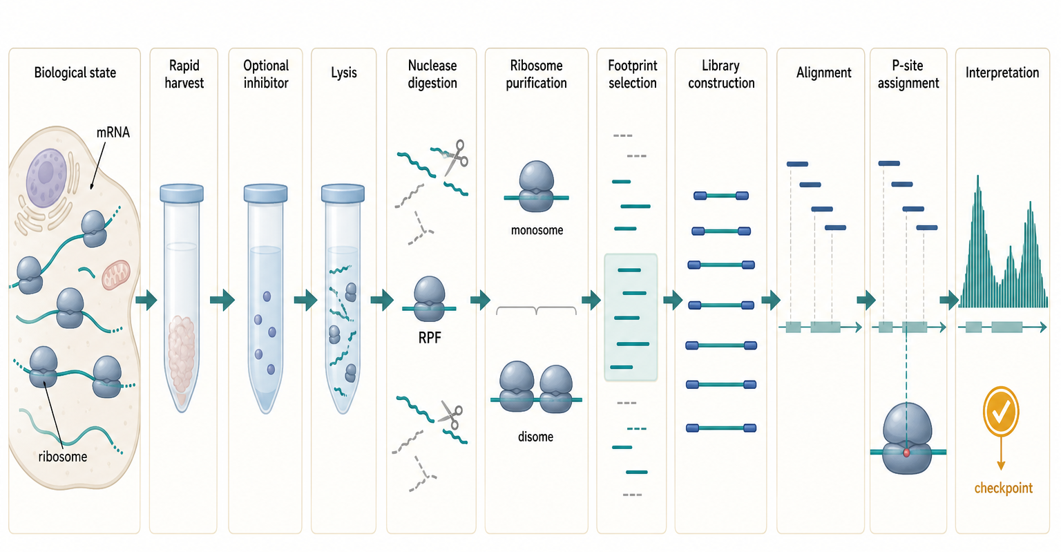
Figure 135.1. From translating ribosome to sequencing footprint. Ribosome profiling converts nuclease-protected ribosome footprints into a map of ribosome occupancy. Each experimental step shapes which fragments survive and what biological claim the dataset can support.
The first design decision is harvest. Translation can change within seconds during heat shock, nutrient withdrawal, hypoxia, immune activation, drug exposure, viral infection, and mechanical disruption. Pretreating cells with an elongation inhibitor can freeze many ribosomes in place before lysis, but inhibitors can also create artificial pauses, alter start-site distributions, or trigger stress responses. Harvesting without pretreatment may reduce drug artifacts, but ribosomes can run off, initiate, or stall during handling if lysis is not fast and cold enough. There is no universally correct choice; the appropriate design depends on whether the study prioritizes physiological minimal perturbation, positional reproducibility, or enrichment of a specific translation state.
The second design decision is nuclease digestion. RNase I, micrococcal nuclease, and other nucleases do not cut all RNA contexts equally. Digestion efficiency depends on buffer composition, temperature, secondary structure, RNA-binding proteins, ribosome state, and nuclease concentration. Under-digestion leaves long fragments that blur positional resolution and may include RNA-protein complexes unrelated to ribosomes. Over-digestion can erode footprints, bias fragment ends, or reduce library complexity. Ribosomal RNA is abundant and often generates protected or stable fragments that must be depleted before sequencing or removed during analysis. A convincing Ribo-seq study reports the footprint length distribution and shows that the selected fragments behave like ribosome footprints rather than random degradation products.
Footprint length is both a biochemical fact and a diagnostic. In many eukaryotic cytosolic monosome datasets, a dominant class near 28 to 30 nucleotides is expected. Bacterial footprints are often different because bacterial ribosomes, mRNA architecture, nuclease conditions, and translation inhibitors differ. Mitochondrial and chloroplast ribosomes can produce still different protected lengths because organellar ribosomes have distinct protein extensions, rRNA architecture, genetic codes, transcript leaders, and translation factors. Initiating ribosomes, elongating ribosomes, terminating ribosomes, stalled ribosomes, collided ribosomes, and ribosome-associated quality-control complexes may protect different fragments. The correct rule is not “all footprints must be 28 nucleotides”; the rule is that the chosen footprint class must be justified by organism, ribosome state, method, and quality-control behavior.
The strongest early evidence that fragments represent translating ribosomes is three-nucleotide periodicity over coding regions. Because elongating ribosomes move codon by codon, P-site-assigned footprints should preferentially occupy one reading frame relative to a translated ORF. Coding-region enrichment, start-codon accumulation, stop-codon structure, and depletion from introns or untranslated regions are additional diagnostics. These signals are not all-or-nothing. A sample with many short ORFs, strong stress, organellar translation, viral RNAs, or noncanonical translation can have unusual patterns. Nevertheless, a dataset with no coherent footprint length, weak coding enrichment, little periodicity, and high ribosomal RNA contamination cannot support fine codon-resolution claims.
The experimental sample also determines what translation states are visible. Monosome Ribo-seq isolates individual ribosomes and is the standard approach for many ORF and translation-efficiency studies. Disome or trisome profiling selects longer fragments protected by adjacent ribosomes and is designed to enrich collision or queueing states. Polysome-associated profiling can preserve broader ribosome loading information but usually loses single-ribosome resolution. Ribosome-nascent-chain complexes, membrane-associated ribosomes, mitochondrial ribosomes, and viral replication-compartment ribosomes may require specialized extraction. A footprint map is therefore never just a property of the transcriptome; it is a property of the transcriptome under a specific biochemical sampling procedure.
Table 135.1. Ribo-seq design choices and interpretation. Ribo-seq design choices determine whether a dataset supports ORF discovery, differential translation, codon-level pausing, collision profiling, or termination analysis.
| Design choice | Measured or enriched property | Major strengths | Common artifacts | Essential diagnostics |
|---|---|---|---|---|
| Harvest without pretreatment | Untreated ribosome occupancy at rapid lysis | Minimizes drug-induced pauses and stress responses | Run-off, continued initiation, or handling-induced stalling | Fast cold lysis, coherent footprint lengths, periodicity, start/stop metagene profiles |
| Inhibitor pretreatment | Drug-arrested ribosome positions or enriched translation states | Improves positional preservation and can enrich starts or elongating ribosomes | Drug-specific start peaks, elongation stalls, redistribution during treatment | Treated/untreated comparison, dose and time control, expected metagene shift |
| Nuclease choice | Footprint boundaries and end chemistry shaped by digestion | Tunes protected fragment recovery for the organism and ribosome state | Sequence bias, under-digestion, over-digestion, non-ribosomal protected fragments | Digestion titration, fragment length histogram, coding enrichment, frame periodicity |
| Monosome selection | Individual ribosome-protected fragments | Supports ORF discovery, translation-efficiency analysis, and P-site maps | Loss of collided or polysome states; contamination by non-ribosomal RNPs | Fractionation trace, monosome-size fragments, length-specific periodicity |
| Disome selection | Longer fragments protected by adjacent ribosomes | Enriches candidate collision, queueing, and stall contexts | Dense normal translation mistaken for collision; size-window and ligation bias | Disome size window, monosome comparison, stall motif dependence, QC-factor response |
| Organellar enrichment | Mitochondrial or chloroplast ribosome footprints | Separates organellar translation from cytosolic background | Cytosolic contamination, noncanonical footprint lengths, genetic-code mismatch | Organelle mapping fraction, organelle-specific length classes, code-aware annotation |
| Ribosomal RNA depletion | Non-rRNA footprint library complexity | Recovers sequencing depth for informative mRNA or viral footprints | Residual rRNA/tRNA domination; depletion bias against selected fragments | rRNA/tRNA mapping fractions, depletion method, unique-molecule complexity |
| Footprint size selection | Chosen monosome, initiation, disome, or organellar length classes | Matches the library to the biological state under study | Excludes meaningful footprint classes or mixes incompatible ribosome states | Per-length abundance, periodicity, offset calibration, separate state analysis |
The main misconception is that nuclease protection is automatically equivalent to productive translation. Ribosomes that are initiating, scanning, stalled, collided, terminating, rescued, or nonproductively associated can all protect RNA. Other RNA-protein complexes can also generate protected fragments if the library does not isolate ribosomes well. The evidence for productive elongation is strongest when footprint size, ribosome fractionation, reading-frame periodicity, ORF boundaries, perturbation response, and matched controls converge.
Box 135.1. What a Ribosome Footprint Can Prove
Ribo-seq begins with a physical observation: a fragment of RNA survived nuclease digestion because a ribosome or ribosome-containing complex protected it. That observation is most persuasive when the fragment has an expected length class, maps to a plausible translated feature, shows three-nucleotide periodicity, and agrees with start or stop structure. Even then, the immediate claim is ribosome occupancy. Productive elongation requires evidence that the footprints move in frame through an open reading frame rather than reflecting scanning, initiation arrest, termination, collision, rescue, decay, or non-ribosomal protection. Protein-output claims require another step: completion of translation, product stability, and detection or functional evidence. A useful reading habit is to ask, “Which link in the chain is being measured: protection, occupancy, elongation, peptide production, or protein function?”
135.2. Library construction and footprint assignment
After nuclease digestion, the experiment becomes a small-RNA library construction problem with unusually strong interpretive consequences. Footprints must have ends compatible with adapter ligation or cDNA synthesis. Enzymatic end repair can change recovery. Ligases prefer some end sequences and structures over others. Reverse transcriptases stop at some modifications or structured regions. PCR amplification can create duplicate reads. Size selection can enrich the desired footprint class or accidentally exclude biologically meaningful variants. Unique molecular identifiers can help identify amplification duplicates, but they do not remove all ligation or reverse-transcription bias. A read count is therefore an endpoint of both biology and library chemistry.
The library design should match the question. For differential translation across genes, consistent recovery and replicate concordance are more important than single-nucleotide boundary precision. For codon-level pausing, footprint-size classes and offset calibration are critical. For ORF discovery, short ORFs, leader regions, and alternative reading frames must be retained rather than filtered away by coding-sequence-only analysis. For organellar or viral translation, the reference sequence and genetic code must match the system. For low-input material, duplicate handling and contamination assessment become central because a few molecules can dominate apparent signal.
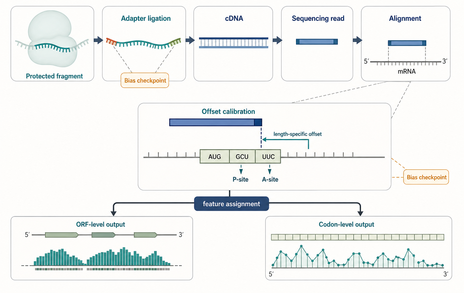
Figure 135.2. Library construction and footprint assignment as an inference chain. A ribosome footprint becomes a biological observation only after library construction and computational assignment. Biases introduced at each step determine whether gene-level, ORF-level, or codon-level claims are defensible.
Alignment is not a mechanical afterthought. Ribosome footprints are short, so they can map ambiguously to paralogs, repeats, pseudogenes, ribosomal RNA, transfer RNA, mitochondrial genes, chloroplast genes, viral genomes, and shared exons among transcript isoforms. Mapping to a genome captures splice junction information only if the aligner and annotation are configured correctly. Mapping to a transcriptome simplifies exon structure but can hide genomic ambiguity and isoform sharing. For many mammalian genes, a footprint assigned to a coding exon cannot distinguish all transcript isoforms. For ORF discovery, annotation incompleteness is not a nuisance; it is part of the scientific problem.
Footprint assignment asks where the ribosome was when the footprint was protected. The sequenced fragment is offset from the ribosomal active sites. Many analysis pipelines estimate a length-specific offset from the read end to the P site by using annotated start codons or known coding regions and choosing the offset that maximizes frame coherence. The A site can then be inferred relative to the P site. This calibration is reliable only when the footprints in that length class are dominated by the ribosome state being modeled. A mixed class containing initiation, elongation, termination, and collision-derived fragments may require separate treatment. Offset errors of one or two nucleotides can change the apparent codon pause, reading frame, or start-site call.
Quality control should be reported at library, alignment, and biological levels. Library diagnostics include footprint length distribution, adapter contamination, ribosomal RNA contamination, duplicate burden, mapping rate, and replicate concordance. Alignment diagnostics include the fraction of reads in coding sequences, untranslated regions, introns, noncoding RNAs, organellar transcripts, and repetitive regions. Translation diagnostics include three-nucleotide periodicity, start and stop metagene profiles, P-site offset calibration, and coding-region coverage. Biological diagnostics include matched RNA-seq, known positive controls, negative controls, perturbation response, and comparison to independent protein or reporter assays where appropriate.
Table 135.2. Library and computational quality controls. Quality-control diagnostics are part of the evidence. They determine whether a Ribo-seq dataset can support gene-level, ORF-level, or codon-level conclusions.
| Analysis step | Diagnostic | Failure mode detected | Interpretation affected | Minimum reporting expectation |
|---|---|---|---|---|
| Length distribution | Fragment-size histogram by sample and retained window | Under-digestion, over-digestion, wrong size cut, mixed footprint states | ORF discovery, P-site assignment, codon-level claims | Show per-sample length plots and selected length classes |
| rRNA/tRNA contamination | Fraction of reads mapping to rRNA, tRNA, and abundant stable RNAs | Failed depletion or non-informative library complexity | Depth, differential translation, rare ORF detection | Report prefilter and postfilter fractions plus depletion strategy |
| Mapping categories | CDS, UTR, intron, ncRNA, organellar, viral, repetitive, and unmapped fractions | Reference mismatch, annotation gaps, non-ribosomal fragments | Coding enrichment, organellar claims, viral or noncanonical ORF calls | Provide category breakdown and reference annotation version |
| Duplicate burden | PCR duplicate rate, UMI collapse, and unique molecule count | Low input or amplification-dominated libraries | Quantitative occupancy, differential translation, replicate reliability | State deduplication method, UMI use, and library complexity |
| Periodicity | Reading-frame enrichment after length-specific assignment | Degradation, offset error, sparse coverage, non-elongating fragments | Translated ORF calls and codon-resolution interpretation | Report periodicity by footprint length and feature class |
| P-site offset | Offset from read end to inferred ribosomal P site | Mislocalized pauses, start sites, stop signals, or ORF frames | Codon pausing, initiation sites, stop-region profiles, smORFs | List offsets by footprint length and calibration method |
| Start/stop metagene | Averaged signal around annotated start and stop codons | Inhibitor artifacts, run-off, annotation mismatch, poor calibration | Initiation enrichment, termination pileups, global QC | Show metagene profiles for the relevant treatment and annotation set |
| Matched RNA-seq | Paired RNA abundance from the same condition and feature model | TE confounded by RNA abundance, isoform shifts, or RNA stability | Translation-efficiency and differential-translation claims | Include matched biological replicates and replicate-aware modeling |
| Replicate concordance | Correlation, PCA, dispersion, and shared differential effects | Batch effects, unstable digestion, library failure, outlier samples | All quantitative comparisons and condition effects | Report replicate number, concordance metrics, and outlier handling |
Translation efficiency is a useful but overloaded output of Ribo-seq analysis. The common gene-level version compares normalized Ribo-seq signal in a coding region with normalized RNA-seq signal for the corresponding transcript or gene. This can identify cases where ribosome occupancy changes without comparable RNA abundance changes. The ratio is especially useful in stress responses, viral infection, RNA modification studies, codon-usage studies, developmental transitions, and perturbations of RNA-binding proteins or translation factors. But the ratio cannot tell whether the change came from initiation, elongation, stalling, termination, recycling, isoform choice, or technical bias without additional evidence.
Box 135.2. Translation Efficiency Is Not a Rate Constant
In most Ribo-seq studies, translation efficiency means normalized footprint signal divided by matched RNA-seq signal for the same gene, transcript, or ORF. This ratio is useful because it asks whether ribosome occupancy changes more than RNA abundance. It is not, by itself, the initiation rate, elongation rate, or number of proteins made per mRNA per minute. A higher ratio can result from more initiation, slower elongation, ribosome pausing, improved footprint recovery, altered isoform use, RNA-seq bias, or depletion of untranslated RNA molecules. A lower ratio can reflect reduced initiation, faster elongation, ribosome run-off, RNA-seq normalization effects, or changed transcript models. Mechanistic interpretation therefore requires matched replicates, feature-aware modeling, footprint diagnostics, and, when possible, perturbation or protein-level validation.
Statistical modeling should respect the two-assay design. A strong differential translation analysis usually models RNA-seq and Ribo-seq counts from biological replicates and tests whether the condition effect differs between assays. Simple ratios without replicate-aware uncertainty can overstate differences, especially for low-count genes. ORF-level analysis benefits from periodicity-aware models rather than simple aggregate counts. Codon-level analysis requires length-specific offsets, local sequence bias controls, and awareness that adjacent codons are not independent because one ribosome protects a multi-codon segment.
The principal computational misconception is that a pipeline output is a biological claim. A called translated ORF, a high translation-efficiency value, or a codon-pause score is a hypothesis supported to a degree by the data and model. The defensible claim depends on diagnostics, thresholds, replicates, annotation assumptions, and orthogonal validation. A transparent Ribo-seq analysis should state what was filtered, how offsets were chosen, how ambiguous reads were handled, which feature model was used, and what evidence level each output supports.
135.3. Initiation, elongation, pausing, and collision profiling
Translation initiation is the process by which a ribosome is recruited to an mRNA, identifies a start codon or start region, and commits to elongation. In many eukaryotic contexts, initiation is highly regulated through cap recognition, scanning, initiation factors, upstream ORFs, RNA structure, internal initiation elements, and stress pathways. In bacteria, initiation depends on bacterial initiation factors, start codon context, Shine-Dalgarno interactions or leaderless initiation, mRNA structure, and coupling to transcription. Initiation profiling aims to locate the sites where ribosomes enter ORFs and to infer how start-site choice changes across conditions.
Drug-based initiation profiling uses inhibitors that enrich ribosomes at start sites or alter the distribution of initiating and elongating ribosomes. Some approaches trap initiating ribosomes before they enter productive elongation. Others allow elongating ribosomes to run off while initiation remains blocked, or compare treated and untreated profiles. These designs can reveal annotated AUG starts, near-cognate starts, upstream ORFs, internal starts, and alternative starts in viruses or stress conditions. Their weakness is the same as their strength: perturbing translation can create patterns that do not exist in untreated cells. Start-site evidence is strongest when the peak is in frame with downstream footprints, is reproducible, responds as expected to initiation perturbation, and is supported by sequence context or mutagenesis.
Elongation profiling asks how ribosomes move after initiation. A ribosome does not glide at a perfectly uniform speed. Elongation can be slowed by limited charged tRNAs, rare or slowly decoded codons, amino acid starvation, nascent peptide interactions with the ribosome exit tunnel, mRNA secondary structure, RNA modifications, polybasic peptide sequences, damaged RNA, regulatory arrest peptides, programmed frameshift signals, and collisions. Ribo-seq can reveal local footprint enrichment, but a footprint pileup is not automatically a pause. It may also reflect nuclease preference, ligation bias, drug-induced arrest, mapping ambiguity, high upstream initiation, or a transcript isoform that was not modeled.
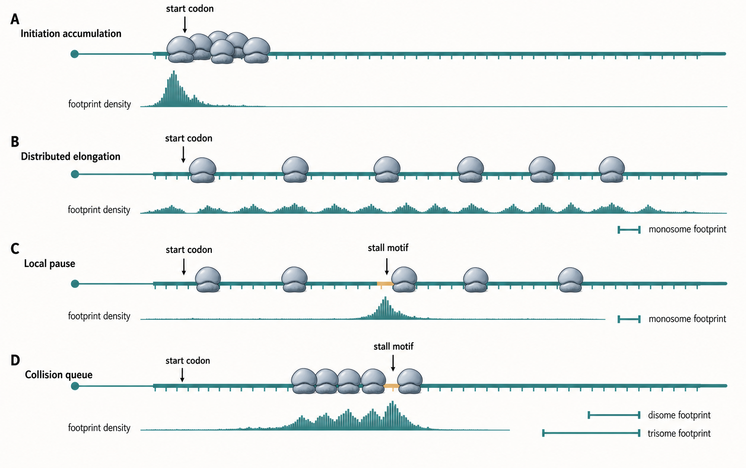
Figure 135.3. Initiation, elongation, pausing, and collision signatures. Footprint enrichment can reflect initiation, elongation, pausing, or ribosome collision. The correct interpretation depends on footprint class, perturbation design, frame assignment, and orthogonal evidence.
Codon-level pause interpretation requires the most caution in the chapter. The ribosome footprint covers multiple codons, and the assigned P site or A site is an inference. Codon identity, amino acid identity, tRNA abundance, nascent peptide charge, local mRNA structure, GC content, and ribosome context are correlated. A heat map of pauses by codon can be dominated by library bias or drug-specific effects if the analysis is not controlled. Stronger evidence comes from matched untreated samples, time-resolved run-off, perturbation of tRNA supply or stall motifs, reporter mutagenesis, conservation of the pause motif, and connection to a downstream consequence such as quality-control recruitment or altered protein output.
Collision profiling enriches a different physical state. When a leading ribosome stalls or slows, a trailing ribosome can catch up. Adjacent ribosomes can protect longer fragments, often called disome or trisome footprints depending on the selected size class and interpretation. These profiles can identify queueing behind polybasic tracts, rare-codon clusters, damaged mRNA, no-go decay substrates, frameshift elements, regulatory arrest peptides, and stress-induced stalls. Collision profiling is especially important because collided ribosomes can recruit ribosome-associated quality-control factors and stress-signaling pathways. The method therefore connects RNA measurement to proteostasis and quality-control biology.
Not every disome-sized footprint is a pathological collision. Highly translated mRNAs naturally contain many ribosomes, and some close spacing can occur without quality-control activation. Different organisms and compartments also have different ribosome spacing constraints. The strongest collision claim combines disome enrichment, monosome comparison, stall motif dependence, perturbation of quality-control factors, and evidence that the event changes RNA decay, nascent-chain fate, stress signaling, or ribosome rescue.
The running upstream-ORF example illustrates why initiation, elongation, and collision should be separated. A stress condition might increase initiation at the upstream ORF. If ribosomes then stall on that upstream ORF, footprints may accumulate locally and reduce scanning or reinitiation at the main ORF. If initiation remains high while elongation slows, collided ribosomes may appear upstream of the stall. Standard Ribo-seq may show all three features as read enrichment in the leader, but initiation profiling, disome profiling, reporter mutation, and time-resolved analysis are needed to decide which mechanism is dominant.
135.4. Termination and recycling measurements
Translation termination begins when a stop codon enters the ribosomal A site. Release factors recognize the stop codon, stimulate hydrolysis of the peptidyl-tRNA bond, and release the completed polypeptide. Ribosome recycling then separates ribosomal subunits or resets the ribosome for another round of translation, using organism-specific factors. Termination and recycling are often treated as the end of translation, but they are regulated steps that influence readthrough, ribosome queues, mRNA surveillance, protein C-terminal extensions, and rescue pathways.
Ribo-seq can detect termination-related signatures because footprints often show structure near stop codons. A normal coding-region profile may decline after the stop codon, while termination defects, release-factor limitation, stop-codon readthrough, recycling defects, or rescue pathway perturbations can create excess footprints at stop codons or in downstream regions. Metagene analysis around annotated stops can reveal global changes, while gene-level analysis can identify transcripts with unusually high stop-proximal density. In bacteria and organelles, rescue systems for nonstop or stalled ribosomes can create distinctive patterns. In eukaryotes, termination also intersects with nonsense-mediated decay, readthrough, upstream ORFs, downstream ORFs, and RNA quality-control pathways.
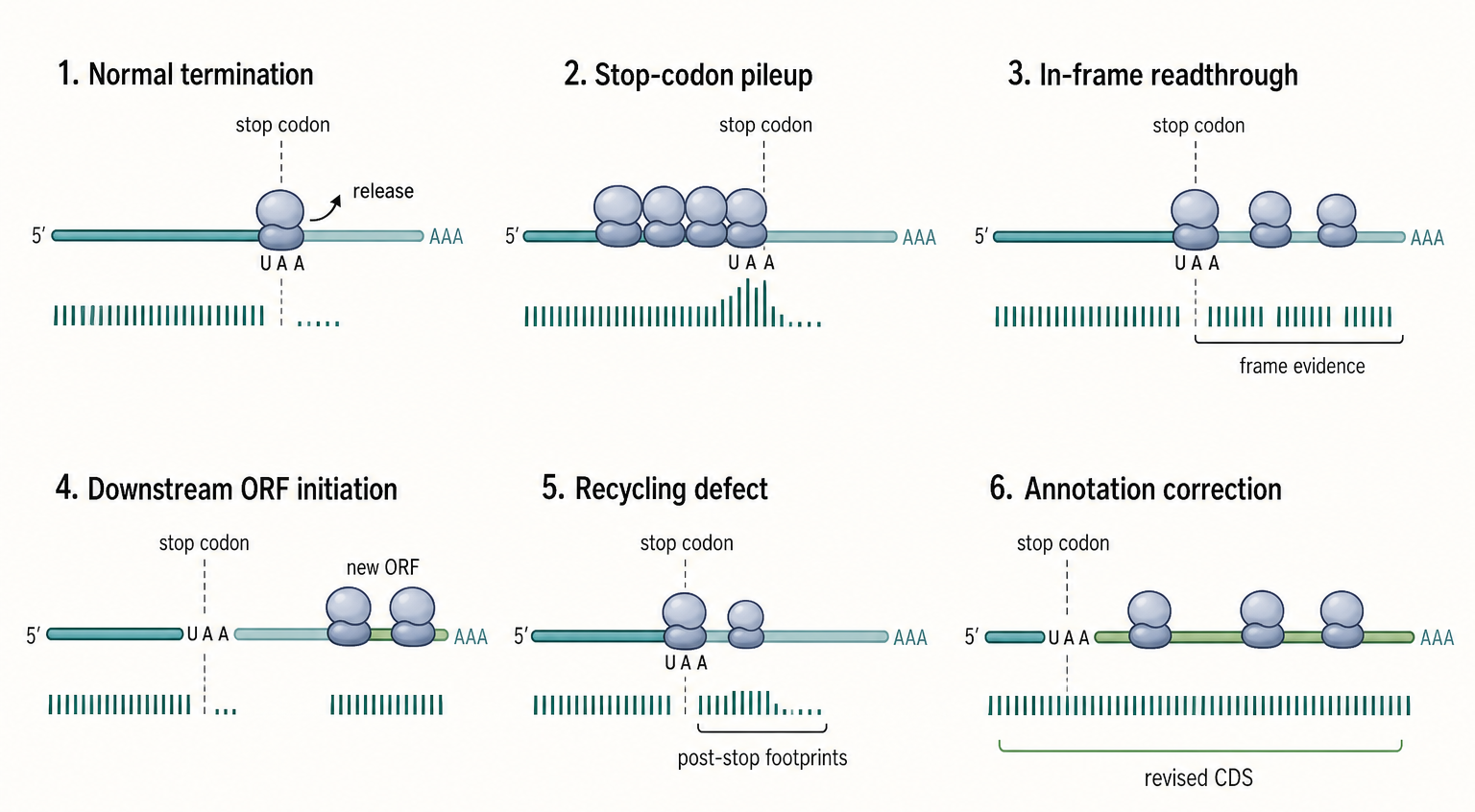
Figure 135.4. Stop-codon and post-stop signatures. Downstream or stop-proximal footprints are not a single phenomenon. Stop-region interpretation requires frame information, transcript annotation, factor perturbation, and protein-level evidence when readthrough is proposed.
The main challenge is that downstream footprints have multiple explanations. A transcript annotation may have the wrong end, an unannotated coding extension, an overlapping ORF, a downstream ORF, an alternative isoform, or a retained intron. Ribosomes may read through a stop codon, reinitiate downstream, scan after termination, or protect an RNA fragment through a non-ribosomal complex. RNA decay intermediates can also complicate interpretation. Therefore, a claim of defective termination or recycling should not rest only on downstream read density. It should use stop-codon frame information, transcript annotation review, matched RNA-seq, release-factor or recycling-factor perturbation, peptide evidence for readthrough when possible, and genetic tests of the stop codon or downstream region.
Termination profiling can also be used to study programmed readthrough and recoding. Some viral and cellular messages use cis-acting RNA structures, stop-codon contexts, or trans factors to promote readthrough. Ribo-seq can identify candidate readthrough by footprints extending beyond annotated stops in frame, but the evidence is stronger when C-terminally extended peptides are detected or when mutating the stop context changes the downstream signal. Programmed frameshifting adds another layer because footprints may shift reading frame near a slippery sequence or structured element. Frame-shifted footprints require careful offset assignment and cannot be interpreted from aggregate read density alone.
Recycling measurements are more indirect. If ribosome recycling is impaired, ribosomes may accumulate after peptide release or remain associated near stop codons. In bacteria, recycling defects can influence ribosome availability and translation of downstream genes in operons. In eukaryotes, recycling can affect reinitiation after upstream ORFs and interactions with mRNA surveillance. Perturbations of recycling factors, release factors, rescue factors, or quality-control proteins can therefore be combined with Ribo-seq to infer which step is affected. The boundary case is that perturbing these factors can also alter initiation, elongation, mRNA stability, and stress signaling, so interpretation must be system-wide.
Termination and recycling are also important for therapeutic and synthetic RNA design. An engineered mRNA must terminate efficiently and avoid unintended readthrough, cryptic downstream ORFs, or ribosome queues that could reduce protein output or generate unexpected peptides. Ribo-seq can inspect stop-codon context, untranslated-region footprints, and ribosome accumulation near engineered features, while proteomics and immunodetection test the protein product.
Table 135.3. Stop-region footprint interpretations. Footprints near stop codons can reflect termination, readthrough, recycling, rescue, reinitiation, annotation error, or RNA decay. Mechanistic claims require discriminating evidence.
| Observed pattern | Possible interpretation | Alternative explanation | Strengthening evidence | Caution |
|---|---|---|---|---|
| Stop-codon pileup | Slow termination, release-factor limitation, or recycling delay | Normal stop dwell, inhibitor artifact, nuclease bias, wrong stop annotation | Frame-specific stop metagene, release/recycling perturbation, matched RNA-seq | Stop enrichment alone does not prove readthrough |
| Downstream in-frame reads | Stop-codon readthrough or C-terminal coding extension | Unannotated isoform, downstream ORF, overlapping CDS, mapping ambiguity | Same-frame periodicity, stop-context mutation, peptide spanning extension | Review transcript models before assigning recoding |
| Downstream out-of-frame reads | Programmed frameshift, reinitiation, or rescue-associated footprinting | Offset error, decay fragment, repetitive mapping, non-ribosomal protection | Slippery motif or structure, reporter test, shifted peptide or factor response | Aggregate downstream density is not frame evidence |
| 3′ UTR footprints | Downstream ORF, reinitiation, readthrough, or ribosome-associated RNA | RNA-binding protein footprint, decay intermediate, unmodeled coding exon | Initiation peak, periodicity, start mutation, peptide or reporter support | UTR signal is not automatically productive translation |
| Factor-perturbation response | Release, recycling, rescue, or quality-control step is involved | Indirect initiation, elongation, mRNA stability, or stress effects | Specific rescue, orthogonal factor assay, matched RNA-seq, global controls | Perturbed translation factors are often pleiotropic |
| Operon or polycistronic context | Coupled downstream translation or reinitiation after upstream stop | Adjacent gene annotation error, RNA processing, overlapping CDS | Gene-specific frame assignment, operon model, upstream stop or RBS mutation | Do not impose monocistronic assumptions on bacteria, viruses, or organelles |
| Organellar stop-region signatures | Organelle-specific termination, recoding, or rescue pathway | Genetic-code mismatch, cytosolic contamination, unusual footprint size | Organelle enrichment, code-aware annotation, length-specific offsets, factor perturbation | Cytosolic offsets and stop rules may be wrong |
The common misconception is that translation ends cleanly at the first annotated stop codon in every transcript. Biology is less tidy. Alternative isoforms, readthrough, overlapping ORFs, upstream ORF reinitiation, viral recoding, organellar genetic codes, RNA damage, and incomplete annotations all complicate the stop boundary. The correct evidence question is whether the data support normal termination, programmed recoding, defective release, defective recycling, or annotation revision in the specific system.
135.5. ORF discovery, microproteins, and noncanonical translation
Open reading frame discovery by Ribo-seq asks whether a segment of RNA is translated, not merely whether it could encode a peptide. A potential ORF is any nucleotide interval with a start-compatible codon, an in-frame coding region, and a stop codon or other termination boundary. Genome annotations historically favored longer conserved protein-coding sequences, so short ORFs, overlapping ORFs, upstream ORFs, alternative starts, lineage-specific ORFs, viral ORFs, and ORFs in annotated noncoding RNAs were often missed or classified inconsistently. Ribo-seq changed this landscape by providing transcriptome-wide evidence of ribosomes moving in frame across many previously unannotated regions.
The first evidence filter is periodicity. A translated ORF should show footprints preferentially assigned to one reading frame relative to the proposed ORF. The second filter is boundary coherence: footprints should begin near a plausible initiation site and decline near a plausible termination site, allowing for biological exceptions such as readthrough or overlapping ORFs. The third filter is initiation evidence, such as a start-site peak from initiation profiling, a favorable start codon context, perturbation of initiation, or loss of downstream footprints when the proposed start is mutated. The fourth filter is independence from annotation artifacts, such as misassigned UTRs, retained introns, pseudogene mapping, repetitive sequence, or unmodeled isoforms.
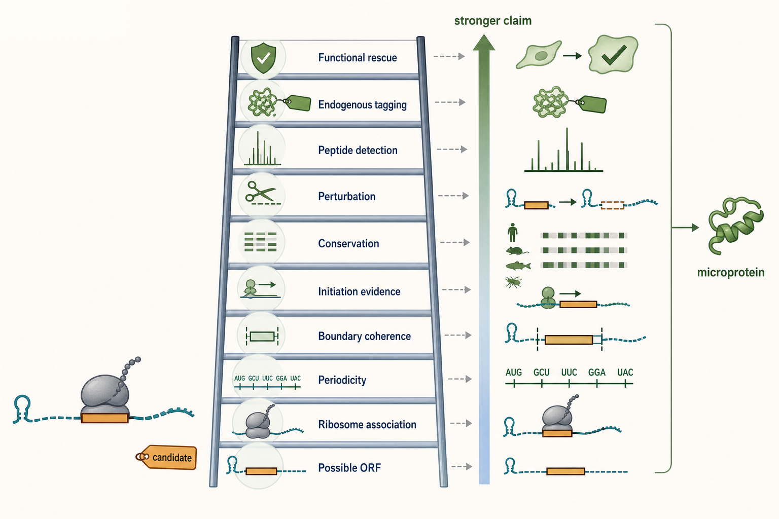
Figure 135.5. Evidence ladder for noncanonical ORFs and microproteins. Noncanonical translation is a graded claim. Periodic footprints can identify translated candidates, but stable microprotein annotation requires convergent protein-level or functional evidence.
Conservation can strengthen ORF claims, but it should be used carefully. Deep amino acid conservation supports protein-coding function for many canonical genes, but it is not required for all real translation. Young ORFs, species-specific microproteins, viral accessory ORFs, immune-induced ORFs, stress-specific ORFs, and some upstream ORFs may lack deep conservation. Conversely, nucleotide conservation in a UTR can reflect RNA structure, RNA-binding protein motifs, splice regulation, or other functions unrelated to protein coding. The best conservation evidence distinguishes amino acid constraint, reading-frame preservation, start/stop conservation, and the possibility of overlapping RNA-level constraints.
Microproteins require a higher evidence bar than translated ORFs. A small ORF may be translated yet produce a peptide that is unstable, rapidly degraded, retained in the ribosome, or nonfunctional. A microprotein claim means that a peptide product exists as a biologically meaningful protein-like molecule. Stronger evidence includes peptide detection by mass spectrometry, targeted proteomics, endogenous epitope tagging, antibody detection, ribosome-nascent-chain capture, CRISPR editing of the start codon, rescue with a peptide-coding construct, phenotypic effects that require the coding frame, and conservation of the amino acid sequence. Failure to detect a peptide does not prove absence, especially for short or low-abundance products, but it limits the strength of a protein-product claim.
Noncanonical translation can be regulatory even when no stable protein accumulates. Upstream ORFs often regulate downstream main-ORF translation through scanning, reinitiation, leaky scanning, ribosome stalling, and stress-dependent changes in initiation-factor activity. Overlapping ORFs can regulate ribosome traffic or produce alternative peptides. Internal ORFs may arise from alternative initiation, proteolytic processing logic, or viral strategies. Circular RNAs and annotated long noncoding RNAs can sometimes carry translated ORFs, but ribosome association alone does not erase their noncoding or regulatory roles. Some RNAs may be dual-function molecules, while others may show ribosome engagement without stable protein production.
ORF discovery pipelines must control false positives. Short ORFs are numerous by chance, so the background search space is large. A few reads can produce apparent periodicity in a tiny ORF. Ambiguous mapping can project canonical coding reads onto paralogous or pseudogene loci. Ribosome scanning in leaders can generate footprints without productive elongation. RNA-binding proteins and structured RNAs can protect fragments. Translation inhibitors can create artificial start peaks. Large custom proteomics databases can inflate false discovery unless search-space expansion is handled carefully. A responsible ORF catalog should grade evidence rather than sorting every candidate into a binary translated or not translated category.
Table 135.4. Evidence grades for ORF and microprotein claims. ORF discovery should be graded by evidence level. A footprint-supported translated ORF and a functional microprotein are related but distinct claims.
| Claim level | Minimum evidence | Stronger evidence | Common false positives | Appropriate wording |
|---|---|---|---|---|
| Possible ORF | Start-compatible codon, in-frame coding interval, and plausible stop boundary | Transcript support, conserved start/stop, coding-potential signal | Random short ORFs, retained introns, UTR annotation errors | “contains a candidate ORF” |
| Ribosome-associated candidate | Ribo-seq reads overlap the candidate interval | Reproducible occupancy, ribosome fraction enrichment, coding-region bias | Scanning ribosomes, RNP protection, RNA decay fragments, ambiguous mapping | “ribosome-associated ORF candidate” |
| Translated ORF | In-frame periodic footprints with coherent boundaries | Initiation peak, start-codon perturbation, amino acid constraint, independent dataset | Sparse-read periodicity, offset error, inhibitor peak, paralogous mapping | “evidence supports translation of this ORF” |
| Regulated translated ORF | Condition-dependent footprint change beyond matched RNA-seq change | Replicate-aware TE model, reporter or genome edit, mechanism-linked perturbation | Isoform shift, RNA stability change, batch effect, local stalling | “regulated ribosome occupancy or translation, mechanism specified” |
| Peptide-supported microprotein | ORF evidence plus peptide, tag, antibody, or nascent-chain product evidence | Targeted proteomics, synthetic peptide match, endogenous tagging, condition-specific detection | Expanded-database FDR, shared peptide, contaminant, tag-driven stabilization | “peptide-supported microprotein candidate” |
| Functional microprotein | Product evidence plus phenotype tied to the coding frame | Start/stop editing, rescue by coding construct, interaction or localization evidence | RNA-level function, off-target edit, overexpression artifact, peptide-independent effect | “functional microprotein in the tested system” |
Four ORF-related claims should remain separate: an RNA contains a possible ORF, an ORF is occupied by ribosomes, an ORF is productively translated, and an ORF produces a functional protein. These claims are related but not equivalent. Ribo-seq is powerful for the second and often the third claim when periodicity and boundaries are strong. Protein-level methods and genetics are needed for the fourth claim.
135.6. Single-cell, spatial, and peptide-level validation
Bulk Ribo-seq averages translation over all cells in a sample. This is often useful, but it can hide cell-state specificity. A tissue sample may contain neurons, glia, endothelial cells, immune cells, and dividing progenitors. A tumor may contain malignant clones, stromal cells, infiltrating lymphocytes, hypoxic regions, and necrotic areas. A developmental embryo may contain rapidly changing lineages. If only one population strongly translates a transcript, the bulk signal may be diluted. If two populations regulate translation in opposite directions, the average may suggest no change. Single-cell and spatial translatomics seek to assign translation-related measurements to cell identity, position, or microenvironment.
True single-cell Ribo-seq is technically harder than single-cell RNA-seq. A single cell contains limited RNA, and only a subset is being translated at any moment. Footprints are short and require nuclease digestion, ribosome preservation, adapter ligation, and depletion of abundant contaminants. The digestion and purification steps that make Ribo-seq positional also cause losses that are difficult to tolerate at single-cell input. Short reads can be hard to assign to isoforms or small ORFs. As a result, many methods called single-cell translatomics do not produce a full codon-resolution footprint map from each individual cell. They instead measure ribosome-associated RNA, nascent protein synthesis, reporter translation, proximity to ribosomes, or paired RNA and protein features.
Cell-type-specific ribosome capture is an important intermediate strategy. Translating ribosome affinity purification uses tagged ribosomal proteins expressed in a defined cell type to enrich ribosome-associated RNAs from complex tissue. Related approaches use immunoprecipitation, proximity labeling, or sorting to enrich translating mRNAs from selected cells. These methods can preserve biological context better than dissociating cells, and they are valuable for neurons, glia, immune cells, and rare tissue populations. Their limitation is resolution: they usually report which RNAs associate with ribosomes in a cell type, not the precise P-site positions, pausing sites, or noncanonical ORF frames in each cell.
Spatial translation measurement adds position. Spatial RNA technologies can map where transcripts are located, and imaging can detect nascent protein synthesis through puromycin analogs, amino acid analogs, SunTag-like reporters, or other labeling strategies. However, RNA localization is not local translation. A localized mRNA may be stored, repressed, transported, degraded, or translated only after stimulation. A nascent-protein signal can show where translation is active but may not identify the transcript being translated. Spatial translatomics is strongest when RNA identity, ribosome association, nascent-chain signal, local perturbation, and peptide or reporter evidence point to the same conclusion.
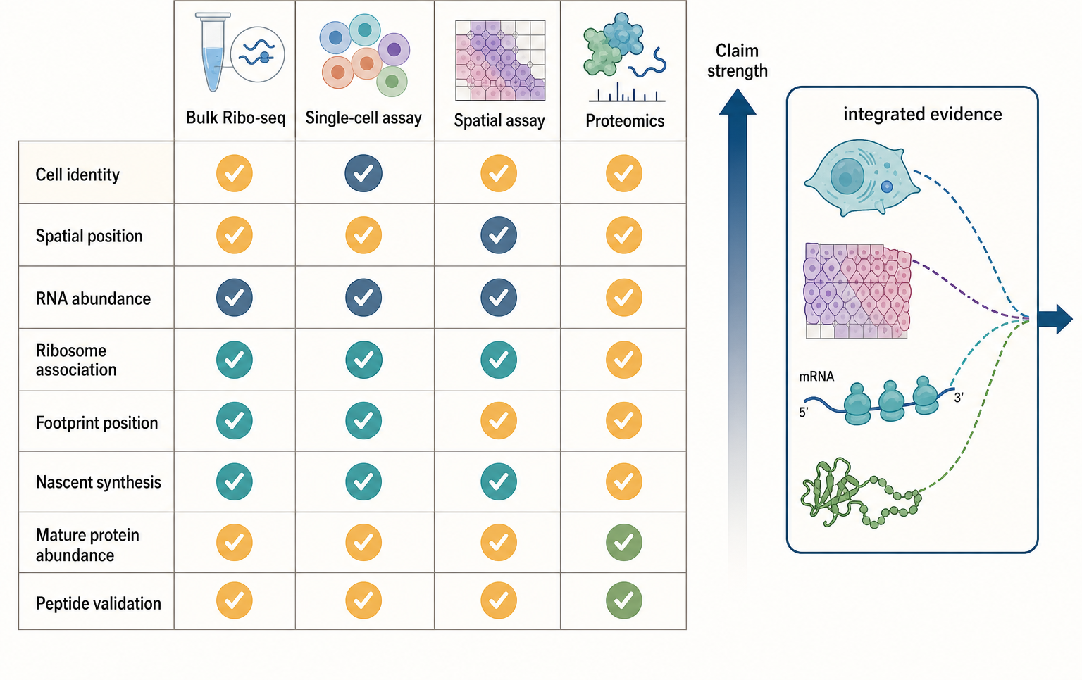
Figure 135.6. Integrating single-cell, spatial, and peptide-level translation evidence. Cell-resolved and spatial translation claims are strongest when orthogonal measurements connect RNA identity, location, ribosome association, nascent synthesis, protein abundance, and peptide evidence.
Peptide-level validation connects translation measurements to protein products. Shotgun mass spectrometry can detect peptides from some predicted ORFs, but microproteins and noncanonical peptides are difficult. Small proteins may yield few tryptic peptides, contain hydrophobic segments, be rapidly degraded, occur only in rare cells, or be expressed only under a narrow condition. Database choice is decisive. If a predicted ORF is missing from the search database, its peptide cannot be assigned. If the database includes every possible small ORF, false discovery control becomes harder. Targeted proteomics, synthetic peptide standards, endogenous tagging, immunodetection, and genetic rescue can improve confidence.
Single-cell proteomics and multimodal assays complement translation measurements but do not replace them. Protein abundance in a cell reflects translation, degradation, secretion, dilution by growth, and measurement sensitivity. Antibody-derived tags or other protein readouts in single-cell assays can show protein abundance for selected targets, but they do not reveal ribosome positions. Conversely, Ribo-seq can show translation of an ORF whose protein is too unstable to detect. Integrating RNA-seq, Ribo-seq, nascent labeling, spatial imaging, proteomics, and perturbation is the most reliable way to connect transcript regulation to protein phenotype.
The frontier is not only technical sensitivity but also vocabulary. A method should state whether it measures footprint position, ribosome association, nascent synthesis, mature protein abundance, peptide evidence, or computational inference. Calling all of these “translation” can obscure the evidence chain. For example, a cell-type-specific TRAP experiment can identify mRNAs associated with ribosomes in Purkinje cells, but it does not prove codon-specific pausing in a single Purkinje cell. A spatial RNA map can show dendritic localization of an mRNA, but it does not prove dendritic peptide synthesis. Clear naming prevents overinterpretation.
Box 135.3. Name the Measured Property in Cell-Resolved Translatomics
When evaluating a single-cell or spatial translatomics claim, first name the measured property. A true single-cell footprinting experiment should assign ribosome-protected fragments, ideally with frame and position information, to individual cells. A ribosome-capture experiment measures RNAs associated with tagged or enriched ribosomes from a cell type or compartment. A nascent-labeling experiment measures new protein synthesis over a pulse but may not identify the source transcript. A spatial RNA map measures transcript position, not translation. A single-cell protein assay measures accumulated protein abundance, which also depends on degradation, secretion, and dilution. Peptide detection supports product existence but is usually sparse. These readouts can support strong biology when combined, but they should not be reported as interchangeable measurements.
Recent Consensus
First, Ribo-seq is a high-resolution ribosome occupancy method. Its strongest claims concern translated-region detection, reading-frame structure, relative ribosome occupancy, and positional changes under well-controlled comparisons. It should not be described as a direct protein abundance assay.
Second, quality-control diagnostics are core evidence. Footprint length distributions, ribosomal RNA contamination, mapping rates, coding-region enrichment, three-nucleotide periodicity, start and stop metagene profiles, replicate concordance, offset calibration, and matched RNA-seq determine whether a dataset supports gene-level, ORF-level, codon-level, or translation-state claims.
Third, initiation, elongation, pausing, collision, termination, and recycling are different biological steps that require different experimental designs. Standard monosome profiling can suggest many events, but specialized profiling, perturbation, and orthogonal validation are usually needed for mechanistic assignment.
Fourth, noncanonical ORF discovery is best handled as evidence grading. Periodic footprints outside annotated coding sequences can be strong evidence for translation. Stable microprotein and functional protein claims require peptide, genetic, conservation, perturbation, or rescue evidence.
Fifth, single-cell and spatial translatomics are emerging rather than solved equivalents of bulk Ribo-seq. Current approaches are valuable when they state what they measure and when they combine occupancy, location, nascent synthesis, protein detection, and perturbation appropriately.
Open Questions, Controversies, Deprecated Models, and Common Misconceptions
Open questions:
- How well can steady-state footprint density be converted into elongation rate? Footprint density is influenced by initiation, elongation, pausing, termination, nuclease recovery, and mappability. Time-resolved run-off, perturbation, and kinetic modeling improve inference, but simple read pileups should not be interpreted as direct dwell-time measurements without controls.
- What is the biological meaning of pervasive noncanonical translation? Some short ORFs encode functional microproteins. Others regulate downstream translation without producing stable proteins. Some annotated long noncoding RNAs may be dual-function transcripts. Many ribosome-associated regions may represent scanning, abortive initiation, decay intermediates, or technical artifacts. The field increasingly treats noncanonical translation as a graded phenomenon rather than a single annotation category.
- Single-cell translation measurement remains unsettled because the desired measurement is difficult to obtain. The field needs clearer distinctions among true single-cell footprinting, low-input bulk footprinting, cell-type-specific ribosome capture, nascent labeling, protein abundance assays, and computational inference. The strongest future methods will likely combine molecular capture with cell identity, spatial position, and validation rather than relying on one assay.
Common misconceptions:
- “Ribo-seq reads over an RNA do not prove abundant protein.” A translation-efficiency ratio is not a universal intrinsic property of a transcript. Inhibitor-treated profiles are not neutral snapshots unless drug effects are controlled. A footprint pileup is not automatically a regulated pause. A disome-sized fragment is not automatically a harmful collision. A noncanonical ORF with sparse footprints is not automatically a microprotein gene. Absence of peptide evidence is not definitive absence of translation, but it does lower confidence in claims about stable protein products.