Chapter 48. m6A, m6Am, and Related Methylations
Scope Note
This chapter treats N6-methyladenosine (m6A), N6,2′-O-dimethyladenosine (m6Am), and related adenosine methylations as marks, enzyme substrates, reader signals, and experimental claims. Chapter 46 supplies evidence rules; Chapter 47 compares methyltransferase folds, cofactors, catalytic cycles, substrate recognition, kinetics, demethylation, and other modification-enzyme families; this chapter owns the m6A/m6Am pathway contexts and consequences. Chapter 49 covers other marks, Chapter 50 editing, Chapter 132 detection, and Chapter 153 therapeutic mRNA chemistry.
Executive Summary
m6A is a methyl group on the N6 atom of adenosine. In mRNA and many long noncoding RNAs, m6A is usually discussed as an internal base modification enriched in particular sequence and transcript contexts, often near stop codons, 3′ untranslated regions, long internal exons, or regulated regions, depending on cell type and assay. m6Am is chemically related but topologically distinct: the adenosine is methylated at N6 and the ribose is methylated at the 2′-O position, most commonly at the first transcribed nucleotide immediately after an m7G cap. Confusing m6A with m6Am can misassign enzymes, detection signals, and biological mechanisms. Reviews of m6A and m6Am chemistry and methyltransferase structures emphasize that chemical identity, RNA position, and enzyme context must be specified together.
The canonical internal m6A writer is a nuclear methyltransferase system built around METTL3 and METTL14, with WTAP and additional adaptor or regulatory proteins helping control localization, substrate selection, and transcript-region preference. METTL3 is generally treated as the catalytic core, whereas METTL14 supports RNA binding and complex architecture in the current structural model. Other methyltransferases broaden the adenosine-methylation landscape: METTL16 modifies particular structured RNAs and participates in S-adenosylmethionine-related regulation; PCIF1/CAPAM installs m6Am at cap-adjacent adenosines; and METTL4 can methylate adenosines in small nuclear RNA contexts such as U2 snRNA. final reference item notes: add direct landmark discovery and structural primary papers for METTL3/METTL14, METTL16, and PCIF1/CAPAM during the bibliography expansion.
Reader proteins convert some m6A marks into outcomes. YTH-domain proteins recognize methylated adenosine through an aromatic binding pocket and connect modified RNA to decay, translation, localization, splicing, or nuclear export depending on family member and context. YTHDF proteins are often described in relation to cytoplasmic mRNA fate, especially decay and translation coupling, whereas YTHDC proteins are often described in nuclear processing or export contexts. Other proteins may be indirect or context-dependent effectors: some bind altered RNA structures exposed by m6A, some associate with modified RNPs, and some show modification-dependent phenotypes without being simple pocket-based readers. A reader claim therefore needs modified-versus-unmodified binding evidence and a downstream effect, not only co-enrichment with an m6A-marked transcript.
Erasers make the field more dynamic but also more difficult to interpret. FTO and ALKBH5 are Fe(II)- and 2-oxoglutarate-dependent oxygenases linked to demethylation of adenosine methylations, with substrate preferences and cellular outputs that depend on compartment, RNA class, and assay. FTO has been connected to both m6A and m6Am biology in different contexts; ALKBH5 is strongly associated with nuclear RNA metabolism and fertility-related phenotypes in the broader literature. Local references support disease examples involving FTO-mediated mRNA stability in pancreatic cancer, but direct mechanistic demethylase discovery papers remain final reference item items for this chapter.
m6A biology spans ordinary mRNA metabolism, noncoding RNA function, viral infection, immunity, and therapeutic RNA design. In mRNA, m6A can influence stability, translation, splicing, nuclear export, phase behavior, and stress responses. In enhancer RNAs, m6A has been implicated in transcriptional condensate formation and gene activation. In U2 snRNA, METTL4-dependent m6Am affects pre-mRNA splicing. In viral and antiviral contexts, methylation can influence innate immune recognition, viral replication, RNA stability, and host response, but the direction of effect is virus- and system-specific. In therapeutic mRNA, cap structure, cap-adjacent methylation, nucleoside substitution, codon design, purification, and delivery all interact with innate sensing and translation; m6A and m6Am should not be reduced to a single “immune evasion” label.
The largest interpretive hazard is detection. Antibody enrichment, crosslinking-based site mapping, enzymatic or chemical methods, mass spectrometry, and direct RNA sequencing measure different features. m6A and m6Am can be difficult to separate in some workflows, and cap-adjacent marks are especially vulnerable to errors caused by fragmentation, incomplete cap-aware annotation, and transcript start-site uncertainty. Stoichiometry is often unknown: an enriched region may represent a small modified subpopulation, multiple nearby candidate adenosines, or an isoform-specific event. Current consensus is that m6A and m6Am are real and biologically important, but individual site, enzyme, reader, disease, and therapeutic claims must be scaled to the method and validated with orthogonal evidence.
Concept Inventory
- M6A: an adenosine whose base carries a methyl group on the exocyclic N6 amino group. The methyl group does not change adenosine into a different Watson-Crick base in the same way that adenosine-to-inosine editing changes pairing interpretation, but it changes local shape, hydration, stacking, protein recognition, and sometimes RNA structure. In many sequence contexts, m6A is read by proteins rather than by direct Watson-Crick recoding.
- M6Am: an adenosine that carries both N6 base methylation and ribose 2′-O-methylation. In mRNA, the term usually refers to a cap-adjacent first nucleotide: m7GpppAm can be further N6-methylated to m7Gpppm6Am. This topology makes m6Am part of cap biology as well as adenosine methylation biology. Its interpretation depends on transcription start-site annotation, cap status, and first-nucleotide identity.
- Internal m6A: m6A located within the body of an RNA molecule rather than at the first transcribed nucleotide next to the cap. Internal m6A on mRNA is often enriched in DRACH-like motifs, where D means A/G/U, R means A/G, and H means A/C/U, but motif presence is not sufficient for methylation. RNA structure, transcript region, transcriptional timing, writer complex recruitment, and competing RNA-binding proteins influence which motif instances are modified.
- Writer: an enzyme or enzyme complex that installs the mark. The canonical mRNA m6A writer complex includes METTL3, METTL14, WTAP, and accessory factors. A reader is a protein or complex that preferentially recognizes the modified state and changes RNA fate. A YTH-domain protein is the clearest reader class for m6A. An eraser is an enzyme that removes or reverses a methyl mark; FTO and ALKBH5 are the central examples in mammalian adenosine methylation biology, with substrate specificity still requiring context-aware interpretation.
- Stoichiometry: the fraction of RNA molecules carrying a mark at a specified site. A transcript with an m6A peak is not necessarily fully methylated. A biologically meaningful m6A site can have low, medium, or high occupancy, but the downstream model must match the occupancy. A low-occupancy site may still matter if it marks a special RNA subpopulation, but that subpopulation must be demonstrated rather than assumed.
- Topology: where the mark sits in the RNA molecule and in the RNA life cycle. A methylated adenosine in a pre-mRNA intron, a mature mRNA 3′ untranslated region, an enhancer RNA, a circRNA junction-proximal region, a viral genome, a U2 snRNA, and a therapeutic mRNA cap have different enzyme access, reader exposure, turnover, and detection constraints.
What to Know Before Reading This Chapter
The reader should understand RNA polarity and cap structure. Eukaryotic mRNAs are often capped at the 5′ end with an inverted 7-methylguanosine linked by a triphosphate bridge. The first and second transcribed nucleotides can carry ribose 2′-O-methylation, producing cap1 and cap2 structures. When the first nucleotide is adenosine and also N6-methylated, the cap-adjacent nucleotide is called m6Am. This is chemically related to internal m6A but belongs to the cap-proximal topology introduced in Chapter 26.
The reader should also separate transcript abundance from modification fraction. If writer depletion lowers a transcript’s abundance, then fewer reads over an m6A peak may reflect loss of RNA rather than loss of methylation per molecule. If a stress treatment changes isoform usage, a methylation peak may appear or disappear because the region is present in different transcript isoforms. Chapter 46 introduced the need for input normalization, stoichiometry, and orthogonal validation.
Finally, the reader should remember that RNA methylation can be causal, correlative, or secondary. A cancer sample with high METTL3 expression and high global m6A may reveal a real disease association, but it does not by itself identify direct methylated target RNAs, the relevant reader, or a safe therapeutic strategy. This chapter uses disease examples to teach mechanisms and evidence limits rather than to claim that every association is actionable.
48.1. Chemistry, Topology, and Vocabulary of m6A and m6Am
The chemical distinction between m6A and m6Am is small in notation but large in interpretation. m6A is a base methylation: a methyl group is attached to the N6 atom of adenosine. m6Am adds the same N6 base methylation to an adenosine whose ribose is already methylated at the 2′-O position. Internal m6A can occur in many transcript regions. Cap-adjacent m6Am is tied to the first transcribed nucleotide, cap maturation, and transcription start-site choice. A sequencing experiment that enriches methylated adenosines without cap-aware resolution can therefore mix mechanistically different molecules.
Table 48.1. Evidence ladder for m6A and m6Am claims. Each row describes the standard of evidence appropriate to a distinct claim type, the most common overstatement, and the controls needed to avoid it.
| Claim type | Minimum evidence | Stronger evidence | Common overclaim | Useful controls |
|---|---|---|---|---|
| Regional m6A candidate | Antibody enrichment peak in MeRIP-seq with input normalization | Concordant signal across two independent antibodies | Treating enrichment peak as exact site or fully occupied mark | IgG negative control, spike-in RNA, input library |
| Exact m6A site | Crosslinking mutation or truncation signature near candidate adenosine | Confirmation by chemical or enzymatic site-resolution method | Reporting crosslinking artifact as a definitive site call | Unmodified RNA standard, writer-knockdown comparison |
| m6Am cap-adjacent site | Decapping or cap-proximal enrichment signal at annotated TSS | Cap-aware first-nucleotide sequencing with confirmed TSS | Treating cap-adjacent peak as internal m6A | Alternative TSS annotation, cap-intact vs. cleaved comparison |
| Modification stoichiometry | Quantitative estimate from spike-in-calibrated method | Site-level calibration with known-occupancy standards and replicates | Reporting stoichiometry from enrichment ratio alone | Spike-in standard, matched input control |
| Writer substrate | Loss of regional signal after writer knockdown | Site-level methylation loss plus catalytic-dead rescue | Equating writer-knockdown phenotype with direct methylation | Catalytic mutant control, transcript abundance unchanged |
| Eraser substrate | Signal increase after eraser knockdown | Site-level gain and in vitro biochemical demethylation | Assuming any FTO or ALKBH5 effect reflects direct demethylation | Orthogonal site method, RNA abundance normalization |
| Reader mechanism | Binding preference for modified over unmodified RNA in vitro | Pocket mutation abolishes binding and downstream phenotype | Co-IP with m6A-marked RNA taken as reader evidence | m6A-mutant RNA, YTH pocket mutant, dose-matched unmodified control |
| Disease mechanism | Altered regulator expression associated with disease state | Causal chain from methylation site to patient-relevant phenotype | Treating regulator expression as functional methylation evidence | Isogenic cell line, site-mutant rescue, patient cohort validation |
| Therapeutic design effect | Changed translation or immunogenicity in matched cell assay | In vivo pharmacology with matched constructs differing by single variable | Generalizing one modification effect across all mRNA designs | Modified vs. unmodified matched mRNA, delivery-matched comparison |
Table 48.2. Detection methods and what they can infer. Different m6A and m6Am detection strategies access different levels of information, and no single method provides chemical identity, site resolution, and stoichiometry together.
| Method class | Measures directly | Usually does not measure alone | Special hazard for m6A/m6Am | Best use |
|---|---|---|---|---|
| Antibody enrichment (MeRIP-seq, m6A-seq) | Regional m6A-enriched RNA fragments | Exact site identity or modification fraction | Cross-reactivity with m6Am, m1A, or unrelated epitopes | Transcriptome-wide discovery of candidate m6A regions |
| Crosslinking-assisted site mapping (miCLIP) | Near-nucleotide crosslink mutation or truncation signature | Modification fraction or chemical identity | Crosslinking artifacts can mimic site-diagnostic signatures | Localizing candidate sites to near-nucleotide resolution |
| Chemical or enzymatic site-resolution (MAZTER, m6ACE-seq) | Site-level modification-dependent read-through or cut | Relative stoichiometry without spike-in calibration | Sequence-context dependency can bias coverage of some sites | Confirming specific sites with single-nucleotide precision |
| LC-MS/MS nucleoside quantification | Modified nucleoside identity and bulk abundance | Transcript or site of origin | Co-elution of m6A and m6Am if chromatographic separation is incomplete | Confirming chemical identity and quantifying global modification level |
| Cap-aware m6Am method (m6Am-seq) | First-nucleotide m6Am relative to transcription start site | Internal m6A positions | Incomplete decapping or TSS uncertainty confounds the signal | Mapping cap-adjacent m6Am and distinguishing it from internal m6A |
| Direct RNA sequencing (nanopore) | Native RNA ionic current signal reflecting base modification | Precise modification identity without benchmarked models | Signal overlap between m6A, m6Am, and neighboring sequence context | Isoform-resolved modification profiling when validated against standards |
| Genetic writer or eraser perturbation | Modification-dependent signal changes after regulator depletion | Chemical identity or site-level stoichiometry directly | Indirect effects from RNA abundance, cell-state, or off-target activity | Validating candidate sites by confirming signal loss or gain |
The topology of internal m6A is often described by enrichment patterns: many mammalian mRNA maps show signal near stop codons, in long exons, and in 3′ untranslated regions. These patterns are useful summaries, but they are not chemical rules. A DRACH motif can remain unmethylated, and a methylated site can behave differently depending on local RNA structure, RNA-binding proteins, and transcript isoform. Internal m6A can also occur in pre-mRNA and noncoding RNAs, so a genomic coordinate must be interpreted through transcript annotation and RNA processing state.
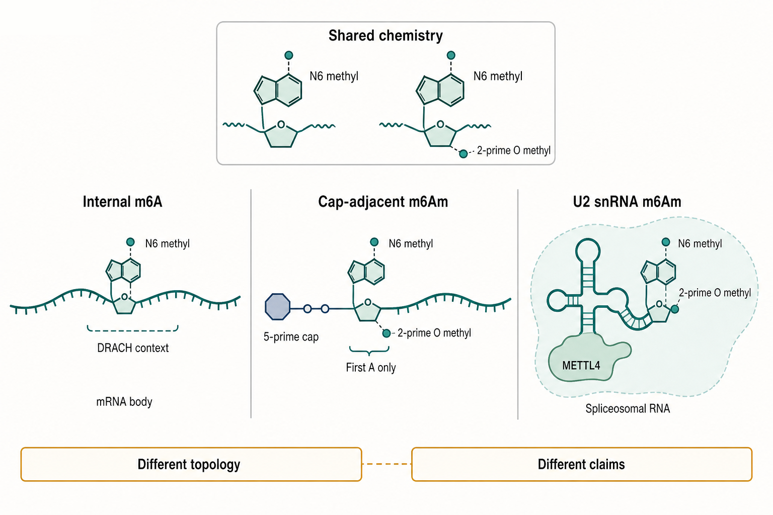
Figure 48.1. Chemistry and topology of m6A and m6Am. m6A and m6Am share N6 methylation but differ in topology and biological context. Internal m6A can occur within mRNA and noncoding RNA bodies, often enriched near stop codons, long exons, and 3′ untranslated regions in DRACH-like sequence contexts. mRNA m6Am is usually cap-adjacent and depends on first-nucleotide identity and cap-aware annotation; only transcripts beginning with adenosine are eligible. U2 snRNA m6Am, installed by METTL4, illustrates that the same chemical abbreviation can appear in a different RNP machine with different downstream consequences.
Cap-adjacent m6Am has a different denominator. Only transcripts that begin with adenosine can receive adenosine m6Am at the first nucleotide, and the site must be interpreted relative to the cap rather than relative to an internal transcript coordinate. Alternative transcription start sites can change whether a transcript is eligible for m6Am without changing the coding sequence. This feature makes m6Am claims sensitive to start-site mapping, cap capture, and sample preparation. Reviews of m6Am biology emphasize disease and regulatory interest, but also the need to avoid treating m6Am as simply “m6A near the 5′ end”.
The physical effect of N6 methylation is context-dependent. The methyl group can alter local base stacking, change how water and proteins contact the base, and destabilize some RNA duplex contexts by favoring conformations less compatible with canonical pairing. In single-stranded regions, it can create a recognizable hydrophobic feature for reader proteins. In structured RNAs, methylation can expose or hide binding sites by shifting local structure. These direct chemical effects are usually smaller than the full cellular phenotypes attributed to m6A; the cellular outcomes often require readers, RNA decay machinery, translation factors, or nuclear processing complexes.
Related adenosine methylations complicate vocabulary. Some small nuclear RNAs and other noncoding RNAs carry m6A-like or m6Am-like marks installed by enzymes outside the canonical mRNA writer complex. METTL4-dependent m6Am in U2 snRNA is one experimentally supported example, and the consequence is linked to spliceosomal function rather than ordinary mRNA turnover. This example illustrates why a chapter on “m6A biology” should not silently restrict the term to polyadenylated mRNA.
The practical vocabulary rule is to name chemical identity and topology together. “m6A in the LATS1 mRNA 3′ region” is a different claim from “m6Am at the first transcribed adenosine of a capped mRNA” and from “m6Am in U2 snRNA.” Each claim implies different eligible enzymes, assays, controls, and mechanisms. When the site is unknown, the text should say “m6A-enriched region” or “methylated adenosine signal” rather than upgrading the observation to a precise modified nucleotide.
48.2. Writer Complexes and Substrate Selection
The best-established internal mRNA m6A writer is not a lone enzyme choosing sites by motif alone. It is a nuclear complex centered on METTL3 and METTL14, with WTAP and additional proteins contributing to complex localization, RNA recruitment, and substrate selection. Structural reviews describe METTL3 as the catalytic methyltransferase component and METTL14 as an RNA-binding and structural partner that helps shape the active complex. The methyl donor is S-adenosylmethionine, often abbreviated SAM, which supplies the methyl group transferred to adenosine.
Substrate selection begins with sequence but does not end there. DRACH motifs are enriched among many internal m6A sites, yet the transcriptome contains far more DRACH motifs than methylated sites. A useful mechanistic model includes motif availability, RNA secondary structure, transcriptional timing, splice-site and exon architecture, local RNP composition, and recruitment by adaptor proteins. RNA polymerase II transcription and pre-mRNA processing can create windows during which some sites are exposed to the writer complex before mature mRNP assembly hides or exports the RNA.
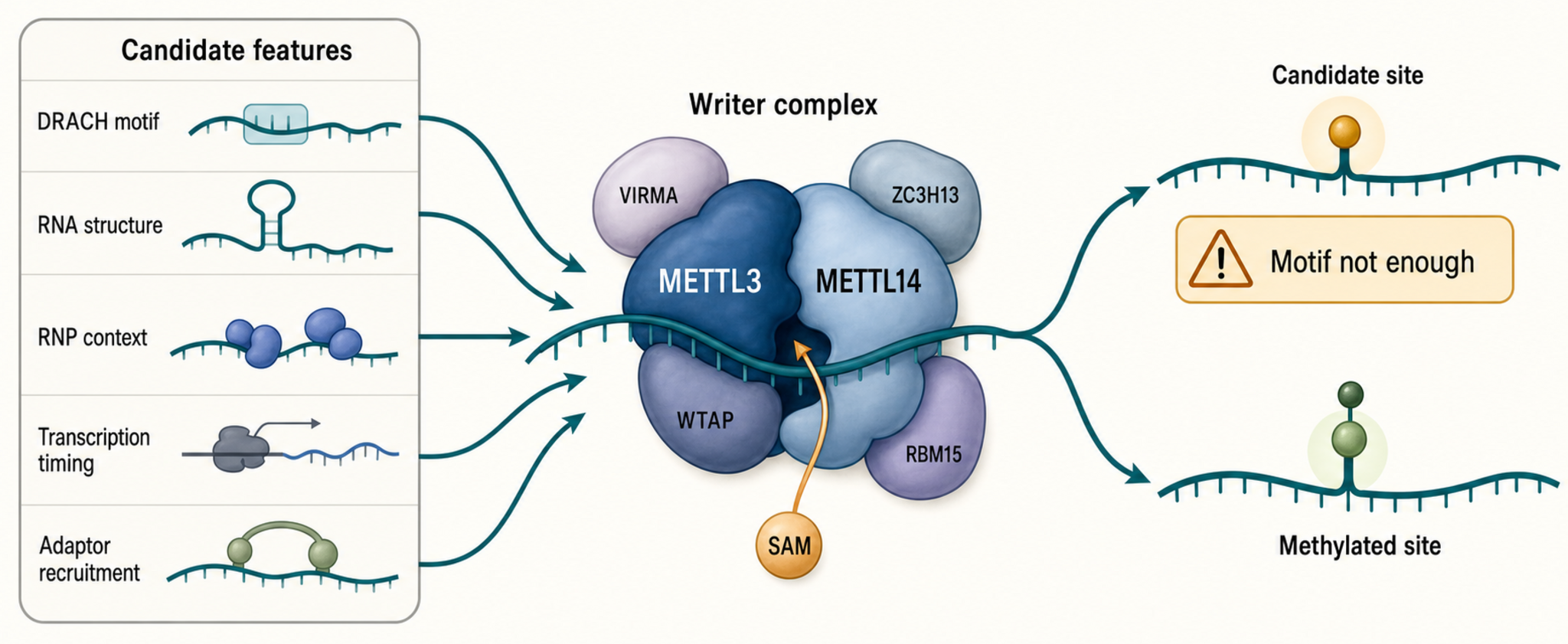
Figure 48.2. Writer-complex substrate selection. The canonical internal mRNA m6A writer system chooses substrates through a combination of sequence context, local RNA structure, RNP composition, transcriptional timing, and adaptor-mediated recruitment. METTL3 is the catalytic core and METTL14 supports RNA binding and complex architecture, while accessory factors including WTAP, VIRMA, ZC3H13, and RBM15 influence localization, region preference, and RNA recruitment. A DRACH motif is a candidate feature that defines which adenosines are eligible, not a site call; the transcriptome contains far more motif instances than methylated sites.
Accessory proteins help explain why a generic motif becomes a specific site in a specific cell. WTAP supports nuclear speckle-associated writer activity and complex organization. VIRMA, ZC3H13, RBM15/RBM15B, HAKAI, and other factors are described in the broader m6A literature as regulators of writer localization, region preference, or recruitment to selected RNAs. final reference item notes: add verified direct sources for each accessory factor before making factor-specific claims beyond this consensus summary. The general principle is already supported by writer-complex reviews: substrate selection is combinatorial rather than a one-protein one-motif rule.
METTL16 shows why “writer” is a family term, not a synonym for the METTL3 complex. METTL16 modifies selected structured RNAs and has been linked in the broader literature to regulation of SAM synthetase transcripts and U6 snRNA methylation. Its substrates depend strongly on RNA structure and cellular metabolic context. final reference item notes: add direct METTL16 primary references and structural sources during the next bibliography pass. For this chapter, METTL16 is important as a boundary case: not every m6A writer uses the same recruitment logic or transcript-region distribution.
PCIF1/CAPAM is the major cap-adjacent m6Am writer in mammalian mRNA. It is associated with the phosphorylated C-terminal domain of RNA polymerase II and acts in a cap-proximal context, which helps explain why m6Am is coupled to transcription initiation and cap maturation rather than distributed like internal m6A. Reviews of m6Am regulation treat PCIF1/CAPAM as central to m6Am installation and to emerging disease questions. final reference item notes: add direct PCIF1/CAPAM discovery and structure papers.
METTL4 adds another boundary case. In human cells, METTL4 can catalyze m6Am methylation in U2 snRNA, and perturbation affects pre-mRNA splicing. This result matters pedagogically because it separates chemical naming from RNA class. A mark called m6Am is not automatically a cap-adjacent mRNA mark. The substrate can be a spliceosomal snRNA, and the phenotype can be splicing rather than mRNA translation or decay.
Writer perturbation experiments require careful interpretation. Knocking down METTL3 may reduce internal m6A, but it can also alter cell proliferation, stress, differentiation, transcription, and RNA abundance. A strong writer-substrate claim is supported by site-level methylation loss, catalytic-dead controls, rescue, unchanged transcript abundance where possible, and a plausible recruitment mechanism. A strong writer-function claim further connects methylation loss to reader binding or RNA fate, ideally by mutating the candidate adenosine without changing unrelated features of the RNA.
48.3. Erasers, Readers, and Effector Mechanisms
The eraser concept asks whether a methyl mark can be removed from RNA after installation. FTO and ALKBH5 are the principal mammalian proteins discussed as demethylases for adenosine methylations. They belong to the Fe(II)- and 2-oxoglutarate-dependent dioxygenase family, which uses oxidative chemistry to reverse alkylated nucleobase modifications. The challenge is not whether these enzymes can influence methylation signals; the challenge is specifying which RNA substrate, which methylated adenosine form, which compartment, and which downstream pathway are involved.
FTO illustrates context dependence. Some studies connect FTO to internal m6A, others to cap-adjacent m6Am, and many disease papers report changes in RNA stability or translation after FTO perturbation. A pancreatic cancer example links FTO to NEDD4 mRNA stability and gemcitabine resistance through a PTEN/PI3K/AKT pathway model. That example is useful as a disease mechanism case, but it should not be generalized to all cancers or all FTO substrates. The exact methylated site, RNA class, and directness of the effect remain central to evidence grading.
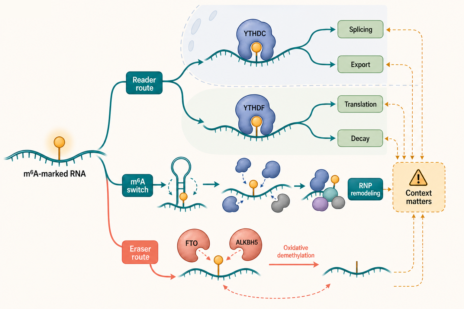
Figure 48.3. Reader, eraser, and effector routes. m6A can influence RNA fate through direct readers, altered RNP composition, and demethylation pathways, with the dominant output determined by reader identity, cellular compartment, RNA class, and site occupancy. Cytoplasmic YTHDF proteins can link modified mRNAs to decay or translation, while nuclear YTHDC proteins are associated with splicing and export. FTO and ALKBH5 can reverse adenosine methylations in an oxidative chemistry mechanism, but their substrate preferences and compartmental activities require context-aware interpretation before assigning a specific demethylase role.
Readers provide the clearest route from a chemical mark to a molecular outcome. YTH-domain proteins contain a pocket that preferentially accommodates m6A compared with unmodified adenosine. YTHDF proteins, including YTHDF1, YTHDF2, and YTHDF3, are cytoplasmic readers often connected to mRNA decay, translation, and cooperative RNA fate decisions. YTHDF2 is commonly associated with recruitment of decay machinery and has attracted interest as an antitumor target in immunological contexts. The details are still context-specific: the same reader family can participate in multiple outputs depending on RNA, cell state, and protein partners.
YTHDC proteins emphasize nuclear and processing-linked outcomes. YTHDC1 has been associated in the broader literature with splicing, nuclear export, and chromatin-adjacent RNA regulation; YTHDC2 has specialized roles in germline and translation-associated contexts. final reference item notes: add direct YTHDC primary sources before expanding these mechanisms. The general lesson is that “reader” does not mean one universal output. A methylated site can be linked to decay in one transcript class, altered splicing in another, and localization or translation in another.
Not every effector is a canonical YTH reader. Some RNA-binding proteins may prefer the structural state created by m6A rather than the methylated base itself. This is sometimes described as an m6A switch: methylation changes local RNA structure, and an RNA-binding protein binds a sequence or motif that becomes exposed. Other proteins can associate with m6A-marked RNAs through RNP complexes without directly reading the methyl group. Reviews of RNA-binding proteins and effectors emphasize that binding, recruitment, scaffolding, and downstream enzymatic activity should be separated analytically.
Effector mechanisms usually fall into several categories. In decay, reader binding can recruit deadenylation, decapping, or exonucleolytic pathways. In translation, reader or writer interactions can alter initiation efficiency, ribosome loading, or stress-dependent translation. In splicing, methylation can change splice-site choice by recruiting or excluding nuclear proteins or by modifying snRNA function. In localization, reader-containing RNPs can traffic transcripts to compartments or granules. In phase behavior, multivalent reader-RNA interactions can promote condensate-like assemblies, although such claims require careful concentration, reversibility, and artifact controls.
The strongest effector evidence follows the mark through the pathway: writer creates a site; site mapping and stoichiometry support the modification; reader binds the modified form; mutation of the site or reader pocket disrupts binding; and the predicted RNA fate changes without broad confounding. Many published studies contain only part of this chain. This does not make them useless, but it should shape the wording. “Consistent with an m6A-reader mechanism” is weaker and often more accurate than “m6A directly causes decay.”
48.4. mRNA, ncRNA, Viral RNA, and Therapeutic RNA Contexts
In mRNA, internal m6A can affect several stages of the RNA life cycle. Nuclear m6A can influence processing, export, or surveillance. Cytoplasmic m6A can alter translation, storage, decay, or localization. Because these processes are coupled, a measured change in protein output may arise from altered mRNA abundance, ribosome recruitment, decay rate, localization, or stress-state composition. A clear mRNA mechanism should state which step is measured and how the experiment distinguishes it from neighboring steps.
Enhancer RNAs provide a noncoding example. Enhancer RNAs are transcripts produced from active enhancer regions and can participate in transcriptional regulation. A primary study reported that m6A methylation on enhancer RNAs facilitates transcriptional condensate formation and gene activation. This is not a license to say all enhancer RNAs work through m6A or that all condensate claims are settled. It is a concrete case in which RNA methylation, noncoding transcription, and transcriptional activation were linked experimentally.
Other noncoding RNAs broaden the substrate field. Long noncoding RNAs can carry m6A marks that affect stability, localization, or protein interactions. Circular RNAs can be detected and functionally studied with specialized assays, and reviews of circRNA biology emphasize that detection and annotation require caution because back-splice junctions, linear RNA contamination, and RNase treatment can mislead interpretation. For circRNAs, an m6A claim must show both circular RNA identity and modification evidence; one does not validate the other.
Small nuclear RNA methylation connects adenosine methylation to the spliceosome. METTL4-dependent m6Am in U2 snRNA affects pre-mRNA splicing. This example creates a cross-chapter bridge to Chapter 27, where snRNAs are treated as structured spliceosomal components rather than mRNA-like transcripts. The same chemical label can therefore participate in different machines: a cap-adjacent mark on mRNA, an internal-like or specialized mark on snRNA, and a regionally enriched mark on long RNAs.
Viral RNA contexts are biologically important and difficult to generalize. Viral genomes and transcripts can be methylated by host machinery, viral factors, or both, and methylation can influence replication, RNA stability, translation, packaging, or immune sensing. Innate antiviral immunity includes sensors that discriminate RNA by features such as double-strandedness, 5′ end chemistry, cap status, nucleotide composition, and modification state. Whether m6A promotes or restricts a particular virus depends on the viral life cycle, cell type, timing, and which host proteins are available. Oncolytic viral therapy adds another layer because viral replication and immune activation can be intentionally exploited.
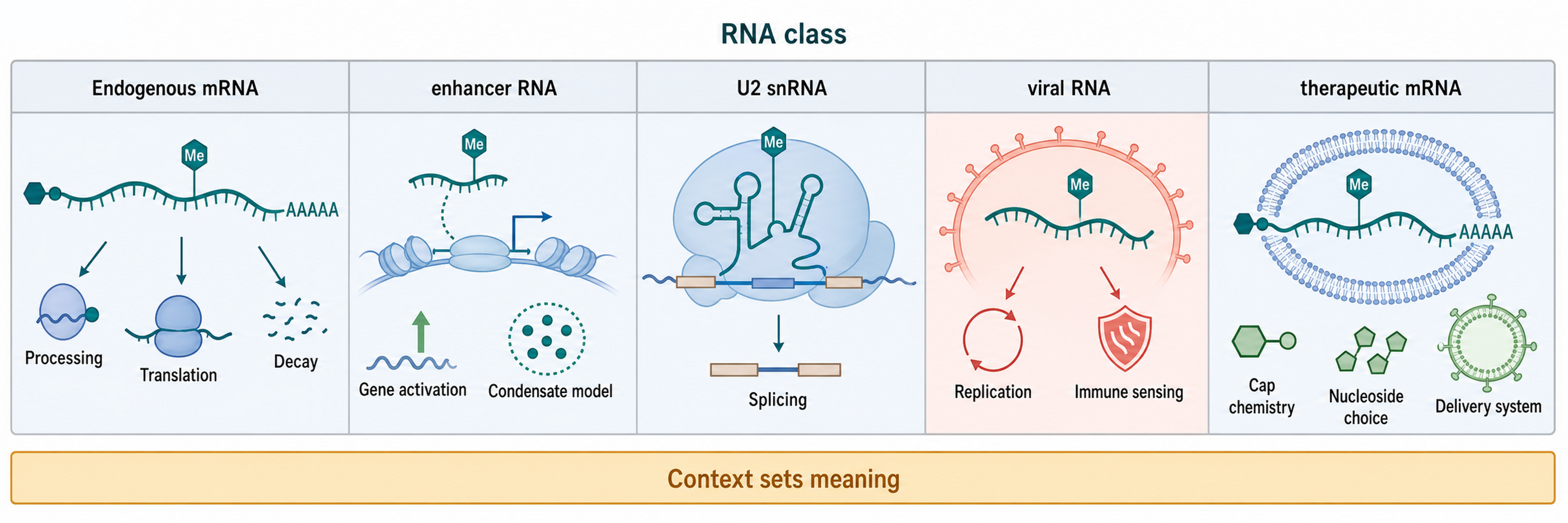
Figure 48.4. RNA-class context matrix. RNA class changes the meaning of an adenosine methylation claim, and the same chemical mark can participate in mechanistically distinct processes depending on transcript identity and cellular location. Endogenous mRNA m6A can affect nuclear processing, translation, decay, or stress-response compartmentalization; enhancer RNA m6A has been linked to transcriptional condensate formation and gene activation; U2 snRNA m6Am affects pre-mRNA splicing; viral RNA methylation influences replication, stability, and innate immune sensing in virus- and cell-type-specific ways; and therapeutic mRNA performance reflects the entire design and delivery system rather than any single methylation mark.
Therapeutic mRNA is related but not identical to endogenous mRNA modification biology. Engineered mRNAs may include optimized caps, cap1 structures, modified nucleosides such as N1-methylpseudouridine, purified products that remove double-stranded RNA contaminants, optimized untranslated regions, codon choices, and delivery systems such as lipid nanoparticles or engineered exosomes. m6Am and cap-adjacent methylation may influence stability and innate immune recognition, but therapeutic performance reflects the whole design and delivery system. Chapter 153 treats synthetic nucleoside choices and vaccine immunobiology in depth.
The recurring lesson across contexts is that RNA class controls mechanism. m6A on a cytoplasmic mRNA may recruit decay machinery. m6A on an enhancer RNA may affect transcription-associated assemblies. m6Am on U2 snRNA may affect splicing. Methylation on viral RNA may alter host-pathogen conflict. Cap-adjacent methylation on therapeutic mRNA may interact with innate sensing and translation. A useful chapter-level claim names the RNA class before naming the methylation effect.
48.5. Development, Stress, Immunity, and Disease Links
m6A is repeatedly linked to development because development depends on timed changes in RNA production, translation, and decay. A methylation system that changes RNA lifetime or translation can reshape cell-fate transitions without changing DNA sequence. The same logic applies to gametogenesis, embryogenesis, stem-cell differentiation, tissue regeneration, and reproduction-related disease contexts, where reviews describe broad involvement of m6A regulators but vary in the directness of evidence. The strongest developmental claims identify specific cell types, target RNAs, methylation sites, and reader-dependent outputs.
Stress responses create dynamic RNA states. Heat shock, oxidative stress, nutrient stress, DNA damage, hypoxia, and infection can change writer, eraser, and reader localization or activity. Stress granules and other RNP assemblies can concentrate mRNAs and RNA-binding proteins, making it tempting to interpret any m6A change as a phase-separation mechanism. That interpretation needs direct evidence: altered methylation, modified-RNA binding, assembly behavior, and functional consequences should be measured separately.
Innate immunity is a particularly important boundary area. Host receptors can detect RNA features that indicate infection or mislocalization, including uncapped or incorrectly capped 5′ ends, double-stranded RNA, triphosphorylated RNA, and unusual modification patterns. Methylations can reduce or reshape sensing in some contexts, but viral evolution, host restriction, and therapeutic design all use different logic. A modified therapeutic mRNA designed to lower innate activation is not equivalent to a viral RNA that uses host methylation to evade detection. Reviews of antiviral immunity and mRNA vaccine technology support a cautious, feature-specific view.
Cancer studies often report altered expression of m6A regulators, altered global m6A levels, or methylation-dependent changes in oncogenic and tumor-suppressive transcripts. For example, METTL3-mediated m6A of LATS1 has been linked to breast cancer tumorigenesis and glycolysis. FTO has been connected to pancreatic cancer drug resistance through NEDD4 mRNA stability. Reviews and primary studies also discuss endometrial, thyroid, prostate, and oral cancer contexts. These examples show biological reach, but they should be graded by whether they demonstrate direct methylation sites, reader mechanisms, patient relevance, and therapeutic window.
Cardiovascular disease provides a noncancer disease context. Reviews link m6A machinery to cardiac hypertrophy, heart failure, vascular biology, ischemic injury, and metabolic stress, while emphasizing that causal pathways can differ by cell type and model. Cardiomyocytes, endothelial cells, fibroblasts, immune cells, and vascular smooth muscle cells have different RNA programs, so a bulk tissue m6A change may reflect cell composition as well as molecular regulation.
Aging-related disease and reproductive disease reviews expand the association map. These areas are useful for hypothesis generation, but association density can exceed mechanism density. The chapter’s evidence rule is therefore conservative: disease association becomes mechanistic evidence only when the study links regulator perturbation to site-level methylation, RNA fate, cell phenotype, and disease-relevant models. Without that chain, the result is a candidate pathway.
48.6. Detection, Stoichiometry, and Controversy
m6A detection began at scale with antibody enrichment approaches such as m6A-seq or MeRIP-seq, which enrich RNA fragments containing methylated adenosine. These methods are powerful for discovery but usually provide regional peaks rather than exact sites or stoichiometry. Fragment length, antibody specificity, RNA abundance, sequence composition, and input normalization all shape the signal. Reviews of m6A detection emphasize that antibody enrichment should be interpreted as candidate regional evidence unless supported by higher-resolution or orthogonal methods.
Site-resolution methods improve localization but introduce their own assumptions. Crosslinking-based approaches can create mutation or truncation signatures near antibody-bound m6A. Enzymatic and chemical strategies can exploit sequence or modification sensitivity. m6Am-focused methods require cap-aware enrichment, decapping logic, or first-nucleotide resolution. final reference item notes: add verified miCLIP, m6A-CLIP, MAZTER, m6ACE-seq, m6Am-seq, and direct RNA m6A/m6Am method papers. Until those sources are added, this chapter names method classes but avoids detailed protocol claims.
Mass spectrometry is central for chemical identity and abundance. LC-MS/MS can quantify nucleosides and distinguish some related marks when standards and chromatography are adequate. Its weakness is localization: bulk digestion of poly(A) RNA can show that m6A or m6Am exists in the purified fraction, but not which transcript or site carried it. Purity is crucial because rRNA, tRNA, snRNA, and degraded fragments can contribute modified nucleosides. Cap-adjacent m6Am also requires workflows that preserve or specifically analyze cap-proximal structures.
Direct RNA sequencing is attractive because native RNA passes through the sensor without reverse transcription, but modification calls remain model-dependent. A signal shift can reflect modified bases, neighboring sequence, structure, damage, motor behavior, or training-set bias. Chapter 132 treats these technologies in detail. For Chapter 48, the key interpretive point is that direct RNA calls should be benchmarked against modified and unmodified standards, writer or eraser perturbations, and independent site methods before being treated as definitive m6A or m6Am maps.
Stoichiometry remains the hardest quantity for many biological arguments. A transcript can have a strong enrichment peak because it is abundant, because a small fraction is highly enriched, because multiple nearby adenosines contribute, or because a subset of isoforms carries the mark. Quantitative site-level assays, spike-ins, calibration standards, and matched input are needed to estimate modification fraction. When stoichiometry is unknown, mechanistic models should avoid language implying that all copies of the RNA are modified.
Controversy in m6A biology is therefore not mainly about whether m6A exists. It concerns site confidence, modification fraction, enzyme specificity, reader directness, and causality in complex phenotypes. Some disease papers overinterpret regulator expression as methylation mechanism. Some viral studies report opposite effects because viruses and cell systems differ. Some proposed readers may be indirect effectors. Some cap-adjacent signals may be confused with internal m6A if the assay does not resolve topology. These disagreements are productive when they lead to better chemistry-aware experiments.
Box 48.1. Do not confuse motif, peak, site, and function
- A DRACH motif is a sequence feature identifying adenosines eligible for methylation; its presence is not evidence that methylation occurs.
- An enrichment peak from antibody-based methods is regional candidate evidence for one or more nearby methylated adenosines, not a site assignment.
- A site call assigns modification to a specific nucleotide and requires crosslinking, chemical, or enzymatic resolution beyond the peak.
- Stoichiometry estimates the fraction of RNA molecules carrying the mark at the specified site; enrichment strength is not a reliable proxy.
- Function requires perturbation of the modification or its reader and a measured change in downstream RNA fate or cellular behavior, not only co-enrichment.
Box 48.2. Disease association grading
Grade m6A and m6Am disease claims from weakest to strongest:
- Regulator expression change in disease tissue or cell line (weakest association, many confounders).
- Global methylation level change measured by LC-MS/MS or antibody enrichment.
- Candidate target site identified in a disease-relevant transcript.
- Methylation-dependent change in RNA fate (stability, translation, splicing) at the candidate site.
- Reader or eraser mechanism linking the methylation site to the disease-relevant output.
- Disease-relevant phenotype rescued by site mutation or reader perturbation.
- Therapeutic window or patient-cohort validation of the causal model (strongest evidence).
Box 48.3. Cap-adjacent m6Am checklist
Before claiming a cap-adjacent m6Am site, confirm each of the following:
- Is the transcription start site mapped at nucleotide resolution for this transcript and cell type?
- Is the first transcribed nucleotide adenosine, making it eligible for m6Am?
- Is cap status preserved in the RNA preparation, or is it explicitly modeled after cap removal?
- Does the assay distinguish m6Am from internal m6A (e.g., cap-aware enrichment, decapping control)?
- Is ribose 2′-O methylation confirmed or assumed from cap1 chemistry?
- Are alternative transcription start sites and isoforms considered, since a shifted TSS changes first-nucleotide identity?
Recent Consensus
Current consensus supports m6A as a major regulatory mark in mammalian mRNA and noncoding RNA biology, with a canonical METTL3-METTL14-centered writer complex, YTH-domain readers, and context-dependent erasers and effectors. m6Am is now treated as a related but distinct cap-adjacent mark with its own writer logic, detection constraints, and disease questions. The field also agrees that RNA class and topology matter: m6A in mRNA, m6A in enhancer RNA, m6Am near the cap, and METTL4-dependent m6Am in U2 snRNA are not interchangeable mechanisms.
The consensus is also methodological. Antibody maps are useful discovery tools but not final site-resolved quantitative maps. Writer, eraser, and reader labels require biochemical and cellular evidence. Disease associations are important but need causal chains. Therapeutic RNA design must integrate cap chemistry, nucleoside choice, purification, sequence design, and delivery rather than treating any one methylation as the whole mechanism.
Open Questions, Controversies, Deprecated Models, and Common Misconceptions
Open questions:
- How much of the m6A map is highly occupied in each cell type?
- How rapidly does methylation change on existing RNA molecules?
- How is substrate selection encoded by transcription and RNP assembly?
- How often do non-YTH proteins act as direct readers?
- Which disease associations identify tractable targets rather than state markers?
- How can m6Am biology be quantified reliably when start-site annotation and cap-aware quantification remain limiting?
Common misconceptions:
- “The term m6A is synonymous with all RNA methylation.” M6A is one modification class; m6Am and other methyl marks have distinct chemistry, topology, enzymes, detection constraints, and biological consequences.
- “The term m6Am means simply internal m6A near the 5′ end.” M6Am is a cap-adjacent mark whose position and cap context change its interpretation.
- “A DRACH motif is evidence of methylation.” A motif is a candidate sequence context, not direct chemical evidence.
- “A writer knockdown phenotype is automatically methylation-dependent.” Writer proteins can have multiple substrates, scaffolding roles, indirect effects, and perturbation artifacts.
- “A reader protein bound to an RNA is automatically reading m6A.” Binding evidence must be connected to modification dependence and functional output.
- “A global increase in m6A in a tumor identifies the causal transcript.” Causal disease claims require transcript-specific mechanism and perturbation evidence.
- “A therapeutic mRNA containing modified nucleosides is the same biological object as an endogenous mRNA carrying site-specific m6A.” Therapeutic RNAs are engineered products whose modification chemistry, stoichiometry, sequence context, purification, and delivery differ from endogenous site-specific m6A regulation.