Chapter 150. Antisense Oligonucleotides, RNase H Gapmers, Splice-Switching Oligos, and Steric Blockers
Scope Note
This chapter explains antisense oligonucleotides as sequence-programmed drugs and experimental reagents that act by binding RNA. The chapter emphasizes four mechanistic classes: RNase H-recruiting gapmers that reduce RNA abundance, splice-switching oligonucleotides that redirect pre-mRNA splicing, poison-exon or pseudoexon blockers that restore productive isoforms, and steric blockers that mask RNA motifs without intentionally cleaving the target. The chapter connects sequence design, chemistry, tissue exposure, toxicity, pharmacodynamic biomarkers, approved medicines, individualized therapeutics, and regulatory evidence expectations.
Executive Summary
Antisense oligonucleotides are short synthetic nucleic acids, usually 12-30 nucleotides long, designed to bind a complementary RNA by Watson-Crick base pairing. Binding is not the therapeutic mechanism by itself. The mechanism depends on the oligonucleotide chemistry, target position, cellular compartment, and proteins recruited or blocked by the RNA-ASO duplex. A gapmer contains a central DNA-like region flanked by chemically modified wings. The wings increase affinity and nuclease resistance, while the central gap supports recognition by RNase H1, an endogenous endoribonuclease that cleaves RNA strands in RNA-DNA hybrids. Gapmers are therefore knockdown agents for nuclear or cytoplasmic transcripts, including mRNAs, long noncoding RNAs, repeat-containing RNAs, and some viral RNAs. Their strengths are catalytic turnover and broad cellular distribution after some routes of dosing; their liabilities include hybridization-dependent off-target cleavage, sequence- and chemistry-dependent protein binding, hepatotoxicity, nephrotoxicity, thrombocytopenia, and inflammatory signals.
Splice-switching oligonucleotides are designed not to recruit RNase H. They use chemistries such as PMO, 2′-O-methyl phosphorothioate, or 2′-MOE phosphorothioate that form stable RNA duplexes but do not present a DNA-like gap. When a splice-switching oligonucleotide masks a splice site, branch point, exonic splicing enhancer, intronic splicing silencer, or other cis-regulatory element, the spliceosome and splicing regulatory proteins perceive a different pre-mRNA. This can skip an exon, include an otherwise skipped exon, suppress a poison exon, prevent pseudoexon recognition, or shift an isoform ratio. The same physical idea underlies many steric blockers: the oligonucleotide occupies an RNA motif so that a protein, another RNA, a ribosome, or a structural rearrangement cannot use that motif.
Clinical antisense design is therefore a problem in mechanism matching. A disease caused by toxic gain of RNA or excess protein may fit gapmer knockdown if partial reduction is tolerated. A disease caused by an exon-disrupting mutation may fit splice switching if skipping restores an open reading frame or if inclusion restores a functional isoform. A private deep-intronic variant may fit individualized pseudoexon suppression if the aberrant exon is clearly causal and the corrected transcript is measurable. Pharmacology and delivery constrain all of these choices. Unconjugated phosphorothioate ASOs distribute well to liver, kidney, spleen, and some other tissues after systemic dosing but poorly cross the blood-brain barrier; intrathecal dosing can expose the central nervous system; skeletal and cardiac muscle remain difficult, especially for large neutral backbones such as PMO unless dose, conjugation, or chemistry is optimized. Approved drugs and n-of-1 cases demonstrate that antisense mechanisms can be clinically decisive, but they also show that sequence specificity does not eliminate the need for careful toxicology, biomarkers, and regulatory judgment.
Concept Inventory
- Antisense oligonucleotide (ASO): a synthetic oligonucleotide designed to bind a complementary RNA sequence. In this chapter, ASO is the umbrella term for gapmers, splice-switching oligonucleotides, steric blockers, and some allele-selective oligonucleotides.
- Gapmer: an ASO with a central DNA-like gap and modified flanking wings. The central gap allows RNase H1 to cleave the RNA strand of the ASO-RNA heteroduplex. The wings usually contain high-affinity modifications such as 2′-MOE, LNA, or cEt to increase potency and stability.
- RNase H1: a cellular endoribonuclease that recognizes RNA-DNA hybrids and cleaves the RNA strand. Therapeutic gapmers exploit RNase H1 rather than encoding an exogenous nuclease.
- Splice-switching oligonucleotide (SSO): an ASO that changes pre-mRNA splicing by blocking access to splice sites or splicing regulatory elements without intentionally causing RNase H-mediated RNA cleavage.
- Steric blocker: an oligonucleotide whose primary mechanism is physical occlusion of an RNA feature. A steric blocker may block a splice factor, RNA-binding protein, ribosome, miRNA seed interaction, RNA-RNA contact, or RNA structure transition.
- Poison exon: an exon whose inclusion introduces a premature termination codon or destabilizing feature that reduces productive protein expression, often through nonsense-mediated decay. Poison exons can be regulated physiological switches or disease-relevant mis-splicing events.
- Pseudoexon: an intronic sequence that is aberrantly recognized as an exon, often after a variant strengthens a cryptic splice site, creates a splicing enhancer, weakens a silencer, or changes local RNA structure.
- Pharmacodynamic biomarker: a measured change that demonstrates target engagement or downstream biological response, such as reduced target RNA, altered exon inclusion, restored protein, lowered toxic protein, or a disease-relevant fluid biomarker.
- Boundary cases: some ASOs have mixed mechanisms. A chemically modified ASO without a canonical gap may still reduce RNA through noncanonical mechanisms or toxicity-associated RNA loss, as emphasized by Hori et al. (2019). Conversely, a gapmer can have steric effects before cleavage or in cell compartments where RNase H1 access is limiting. Mechanistic classification is a working model that must be tested for each sequence, chemistry, cell type, and target.
What to Know Before Reading This Chapter
The reader should understand that RNA molecules are directional polymers with sequence-specific base-pairing potential, that most eukaryotic protein-coding transcripts are processed from pre-mRNAs by spliceosome-mediated intron removal, and that RNA abundance can be changed by either reducing synthesis, changing processing, or accelerating decay. This chapter builds from Chapter 149, which covers backbone and sugar chemistry. The central point here is that chemistry is not only a stabilizing decoration; chemistry determines whether a bound ASO recruits RNase H1, resists nucleases, binds plasma and cell-surface proteins, enters tissues, activates immune receptors, or remains a passive steric blocker.
The chapter also assumes the evidence standards from Chapter 5. A decrease in RNA after ASO treatment is not automatically proof of direct RNase H cleavage. A change in an RT-PCR splice band is not automatically proof that a therapeutic protein is restored. A patient-level biomarker improvement is not automatically proof of clinical benefit. Strong antisense evidence connects sequence complementarity, dose response, chemistry, intracellular target exposure, on-target RNA change, protein or pathway correction, disease-relevant phenotype, and safety monitoring.
150.1. Gapmer design and RNase H recruitment
Antisense Design Logic: Matching Chemistry, Target Site, and Mechanism
An antisense oligonucleotide is a programmable binding reagent before it is a drug. The designer chooses a target RNA region, selects an oligonucleotide length, chooses a backbone and sugar chemistry, and then tests whether hybridization produces the intended biological effect. The same target sequence can behave differently when bound by a DNA-like gapmer, a PMO, an LNA-rich mixmer, or a 2′-MOE steric blocker. The design question is therefore not simply “where can the ASO bind?” but “what should happen after the ASO binds?”
The most basic design variable is complementarity. A fully complementary ASO can form a stable RNA-ASO duplex, but perfect complementarity is not always desirable. Allele-selective gapmers may deliberately place a single-nucleotide variant near the center of the ASO so that RNase H-dependent cleavage favors the mutant allele over the wild-type allele. The discrimination is limited because many ASO chemistries bind strongly enough to tolerate mismatches; therefore allele selectivity usually requires empirical screening, careful placement of the mismatch, and assays that quantify both alleles rather than total RNA alone. Aguti et al. (2024) treat allele-specific gapmer design as an optimization problem rather than a simple sequence lookup, a useful warning for diseases such as Huntington disease where lowering both mutant and wild-type transcript may have different risk than lowering only the mutant transcript.
Accessible target sites are another variable. RNA is not a naked string in cells. Nascent pre-mRNAs are coated with spliceosomal factors, heterogeneous nuclear ribonucleoproteins, exon junction complex components, export factors, and surveillance proteins. Mature cytoplasmic mRNAs can be engaged by ribosomes, miRNA-loaded Argonaute, RNA-binding proteins, decay enzymes, and localization complexes. Long noncoding RNAs may be nuclear, chromatin-associated, structured, or RNP-bound. An ASO site that is thermodynamically accessible in a folded RNA model may still be unavailable in a particular cell state, while a site predicted to be structured may be exposed during transcription, remodeling, or RNP exchange. This is why ASO programs commonly use tiling screens across candidate regions rather than relying entirely on in silico accessibility.
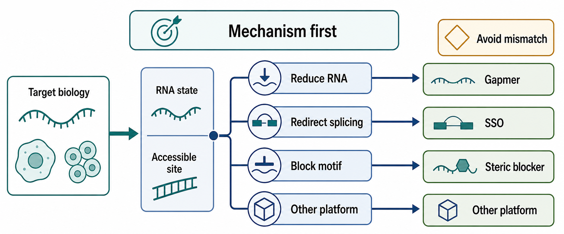
Figure 150.1. Choosing an ASO Mechanism. “Mechanism-first ASO design. Target biology determines whether a sequence-programmed oligonucleotide should recruit RNase H1, mask a splice element, block an RNA-protein or RNA-RNA interaction, or modulate translation.”
Length and melting temperature shape both potency and specificity. Short ASOs may avoid some off-targets but may bind too weakly, especially when the target RNA is structured or protein-bound. Longer ASOs may bind more strongly but create more opportunities for partial hybridization to unintended transcripts. High-affinity modifications such as LNA, cEt, or 2′-MOE can make short ASOs potent, but excessive affinity can reduce mismatch discrimination, increase protein binding, or increase toxicity in sequence-dependent ways. Phosphorothioate backbones improve nuclease resistance and plasma protein binding, which extends circulation and tissue exposure, but phosphorothioate also changes protein interactions. Chapter 149 covers these chemical features in detail; the practical point here is that every chemical improvement creates a pharmacological tradeoff.
ASO design also depends on compartment. Nuclear targets such as pre-mRNA splice elements, many lncRNAs, and nascent transcripts are accessible to ASOs that reach the nucleus. Cytoplasmic mRNA knockdown can be efficient for gapmers, but ribosome occupancy, mRNP architecture, and subcellular localization can affect potency. Viral RNAs pose additional constraints: their replication compartments, RNA structures, protein occupancy, and replication kinetics can influence whether a gapmer has time and access to bind. Okamoto et al. (2023) provide local chapter evidence that RNase H-dependent gapmers can be tested against Japanese encephalitis virus RNA, but broad antiviral ASO generalization needs virus-specific evidence.
Table 150.1. ASO Mechanism Classes and Readouts. “ASO class is defined by mechanism and evidence, not by the word antisense alone.”
| Planned row | Intended comparison | Evidence or caveat |
|---|---|---|
| RNase H gapmer | DNA-like central gap plus modified wings binds nuclear or cytoplasmic RNA and recruits RNase H1 for transcript reduction. | Proximal readout is target RNA loss, but direct cleavage, dose response, off-target profiling, and protein or pathway effect strengthen the claim. |
| Splice-switching oligo | Non-gap PMO, 2′-OMe, or 2′-MOE designs mask splice sites or regulatory elements to promote exon skipping or inclusion. | Readouts should quantify exact junctions and protein output |
| Poison-exon or pseudoexon blocker | Steric SSO suppresses an unproductive poison exon or a variant-created pseudoexon to restore a productive isoform. | Requires RNA-level mapping of the aberrant junction; minigene screens need endogenous RNA confirmation when feasible. |
| UTR or RBP-motif steric blocker | Nondegrading ASO masks a miRNA site, RBP motif, start-region element, uORF, RNA-RNA interface, or structural switch. | RNA abundance alone is insufficient; compare motif occupancy, ribosome engagement, protein output, and transcriptome-wide perturbation. |
| Translation blocker | ASO near a start site, scanning region, uORF, or internal ribosome entry element changes ribosome access without planned cleavage. | Use reporter, polysome, ribosome-profiling, and protein assays to separate translational control from RNA decay or stress. |
A responsible ASO design workflow therefore uses multiple filters. First, define the disease mechanism and decide whether reducing RNA, changing splicing, blocking a motif, or altering translation is expected to help. Second, map the target transcript isoforms and confirm that the chosen site is present in the disease-relevant RNA. Third, choose chemistry compatible with the desired mechanism: DNA-like gap for RNase H, non-gap chemistry for steric blockade, or a specialized backbone such as PMO for splice switching. Fourth, screen multiple candidates in a relevant cellular system and measure direct RNA outcomes. Fifth, test mismatch and transcriptome-wide off-target risk. Sixth, connect RNA change to protein, pathway, and phenotype. A sequence that knocks down RNA in a transfected cell line is not yet a therapeutic candidate; it is an early design hypothesis.
The strongest design evidence includes a rescue or orthogonal confirmation step. For a gapmer, one can show target RNA reduction by quantitative PCR or RNA-seq, map cleavage products when feasible, use multiple nonoverlapping ASOs against the same transcript, and demonstrate that phenotype tracks with target reduction rather than with a single sequence. For a splice-switching oligo, one can show corrected isoform ratios by RT-PCR and sequencing, protein restoration by immunoassay or functional assay, and loss of effect when the target splice element is mutated or absent. For a steric blocker, one can show that the blocked protein or RNA no longer binds the target motif, ideally without widespread RNA degradation.
Box 150.1. Mechanism Is a Claim, Not a Name
Do not classify an oligonucleotide solely from its target sequence, short length, or therapeutic label. Ask what substrate the RNA-oligo duplex presents and which molecule executes the effect. A gapmer claim needs a DNA-like gap, target RNA loss, dose response, and preferably cleavage mapping or RNase H-dependence controls. A splice-switching claim needs junction-level RNA evidence plus protein or functional follow-up. A steric-blocker claim needs evidence that the intended protein, RNA, ribosome, or structural event is blocked, with RNA abundance measured rather than assumed stable. Mixed outcomes are common: a gapmer may occlude a motif before cleavage, and a non-gap oligo may indirectly lower RNA through altered splicing, translation, RBP binding, or cellular stress. Treat the mechanism label as a tested conclusion, not a starting assumption.
An ASO is often described as “turning off a gene.” That shorthand is too broad. Gapmers reduce RNA after transcription; they do not edit genomic DNA or stop transcription. Splice-switching oligos redirect processing; they may increase one isoform while decreasing another. Steric blockers can inhibit, restore, or reroute RNA function without changing total RNA abundance. This distinction matters for interpreting biomarkers and adverse effects.
Gapmer Design, RNase H Recruitment, and Transcript Knockdown
Gapmers are the major ASO class for transcript knockdown. A canonical gapmer has modified nucleotides at both ends and a central stretch of DNA-like nucleotides. When the gapmer binds the target RNA, the central region forms an RNA-DNA hybrid that can be recognized by RNase H1. RNase H1 cleaves the RNA strand, leaving the ASO intact enough to dissociate and bind another target. This gives gapmers a catalytic character: one ASO molecule can contribute to cleavage of multiple RNA molecules if intracellular conditions permit repeated binding and release.
The physical substrate for RNase H1 is the RNA-DNA heteroduplex, not the disease transcript alone. This point explains why non-gap steric blockers generally avoid RNase H recruitment and why gap length matters. If the central DNA-like segment is too short or distorted by modifications that RNase H1 cannot tolerate, cleavage may be weak. If the gap is too long or the wings are too weak, nuclease stability and affinity may suffer. Many designs use a “wing-gap-wing” architecture such as 3-10-3 or 5-10-5, but the exact pattern depends on chemistry, target, desired potency, toxicity profile, and manufacturing constraints. The gapmer is therefore a composite molecule: the wings solve affinity and stability problems, while the gap creates an enzymatic substrate.
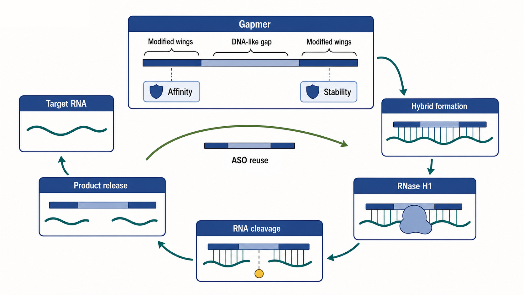
Figure 150.2. RNase H Gapmer Cleavage Cycle. “A gapmer uses modified wings for affinity and stability while the central DNA-like gap creates an RNase H1-compatible RNA-DNA hybrid.”
Gapmer target selection differs between mRNAs and noncoding RNAs. For a protein-coding mRNA, reducing transcript abundance is expected to reduce protein, but the relationship can be buffered by protein half-life, feedback regulation, translational compensation, or tissue turnover. For a nuclear lncRNA, gapmers can be useful because RNA interference machinery is often less effective in the nucleus. Maruyama and Yokota (2020) provide a local review anchor for lncRNA gapmer knockdown, including the practical idea that nuclear localization can make RNase H-dependent ASOs more suitable than siRNAs for some lncRNA targets. However, lncRNA biology also raises causality risks: a phenotype after one gapmer may result from off-target cleavage or protein-binding toxicity rather than from loss of the annotated lncRNA. Multiple ASOs, rescue strategies, and careful transcriptome analysis are especially important.
Gapmers can be designed for allele-selective silencing when a pathogenic allele differs from the healthy allele by a single-nucleotide variant, repeat-associated haplotype, or linked polymorphism. Huntington disease is a recurring example because lowering the mutant HTT transcript is a plausible disease-modifying strategy, while complete or chronic lowering of wild-type HTT may carry risk. Aslesh and Yokota (2020) anchor local coverage of Huntington gapmer development. The evidence challenge is that total HTT knockdown is easier to achieve than allele-selective knockdown. A mismatch centered in the ASO may reduce binding to the wild-type allele, but high-affinity wings can erase mismatch penalties. Therefore an allele-selective claim should report mutant reduction, wild-type reduction, selectivity ratio, dose range, target cell type, and whether the sequence variant exists in the intended patient population.
Table 150.2. Gapmer Design Variables. “Gapmer potency and safety emerge from combined sequence, chemistry, and target-context choices.”
| Planned row | Intended comparison | Evidence or caveat |
|---|---|---|
| Gap architecture | Central DNA-like gap creates the RNase H1-compatible substrate; modified wings support affinity and nuclease resistance. | Too little DNA-like character weakens cleavage, while excessive gap length or weak wings can reduce stability and tolerability. |
| Wing chemistry | 2′-MOE, LNA, cEt, and related high-affinity wings tune melting temperature, potency, and tissue behavior. | Higher affinity can reduce mismatch discrimination and may increase sequence- or protein-binding toxicity. |
| Target-site and isoform coverage | Candidate sites should be present in the disease-relevant transcript isoforms and reachable in the relevant compartment. | Folding predictions do not guarantee cellular access; tiling screens and isoform-aware assays remain important. |
| Allele-selective mismatch placement | Pathogenic or linked variants can be positioned to favor mutant-allele cleavage over wild-type cleavage. | Claims should report mutant and wild-type knockdown separately, because high-affinity chemistries can erase mismatch penalties. |
| Hybridization off-target filters | Partial complementarity, contiguous central matches, GC-rich segments, and motif-like sequences are screened before nomination. | Transcriptome profiling, mismatch controls, and multiple ASOs help distinguish target biology from off-target RNA loss. |
| Pharmacodynamic readout plan | RNA knockdown should be paired with protein, pathway, or phenotype measurements appropriate to target turnover. | Blood or bulk tissue biomarkers may not represent brain, muscle, retina, kidney-cell-type, or nuclear target engagement. |
Gapmer specificity must be evaluated at several levels. Hybridization-dependent off-targets arise when the ASO binds partially complementary RNAs, especially if a long contiguous seed-like region or central gap can support RNase H cleavage. Hybridization-independent effects arise when the oligonucleotide chemistry and sequence bind proteins, alter protein localization, trigger innate immune sensors, or perturb cellular stress pathways. Phosphorothioate ASOs are particularly protein-interactive. Some interactions are useful for pharmacokinetics because plasma protein binding extends circulation and improves tissue uptake. Other interactions can be harmful if they alter coagulation, complement, platelet behavior, hepatocyte stress, or kidney handling.
Hori et al. (2019) are important because they caution that RNA reduction and hepatotoxic potential can occur even with non-gapmer ASOs. That observation prevents a simplistic rule in which all RNA loss after ASO dosing is attributed to canonical RNase H gapmer cleavage. Toxic RNA loss, stress responses, and indirect pathway changes can masquerade as target knockdown. Mechanistic validation should therefore include chemistry controls, mismatch controls, transcriptome-wide profiling, dose-response separation of pharmacology from toxicity, and, when feasible, direct evidence of RNase H-dependent cleavage.
The experimental readout for gapmer activity is usually target RNA reduction, but the best readout depends on the target. Quantitative PCR is sensitive and efficient, but primer placement matters. If primers amplify a region downstream of the cleavage site, RNA fragments or alternative isoforms may complicate interpretation. RNA-seq can identify transcriptome-wide changes, but standard poly(A)-selected RNA-seq may miss non-polyadenylated RNAs, nuclear retained RNAs, or degradation intermediates. Rapid amplification of cDNA ends or specialized cleavage mapping can strengthen evidence for direct cleavage, although such assays are not always routine in therapeutic programs. Protein measurement is essential when the goal is protein reduction, especially when the protein is stable or when RNA knockdown triggers compensatory transcription.
Gapmer pharmacodynamics can be long-lived. ASOs can accumulate in tissues and remain active after plasma levels fall. This durability is therapeutically useful because infrequent dosing may maintain target reduction, but it complicates dose adjustment after adverse events. Tissue half-life, intracellular depot behavior, endosomal escape, nuclease resistance, and target turnover all contribute. A small RNA measurement in blood may not reflect target knockdown in brain, muscle, retina, or kidney. A clinical gapmer program therefore needs a biomarker strategy that links accessible samples to disease-relevant tissue exposure.
The boundary between gapmers and siRNAs is instructive. Both can reduce RNA abundance, but siRNAs are double-stranded triggers of Argonaute-mediated RNA interference, whereas gapmers are single-stranded ASOs that recruit RNase H1. siRNAs often benefit from GalNAc conjugation for hepatocyte delivery and have seed-mediated off-target risks; gapmers rely more heavily on phosphorothioate-driven distribution and RNase H-compatible chemistry. Chapter 151 compares siRNA therapeutics in detail. The practical decision is not which modality is generally superior, but which mechanism, tissue, duration, safety window, and manufacturing path fit the target.
150.2. Splice-switching oligos for exon skipping and inclusion
Splice-switching oligonucleotides change the interpretation of a pre-mRNA by the spliceosome. Pre-mRNA splicing depends on core signals such as the 5′ splice site, branch point, polypyrimidine tract, and 3′ splice site, but those signals are often weak and context-dependent in metazoans. Exonic and intronic splicing enhancers recruit positive regulators; exonic and intronic splicing silencers recruit negative regulators. RNA secondary structure, transcription kinetics, chromatin context, and tissue-specific RNA-binding proteins also influence exon choice. An SSO works by masking one of these cis-elements so that the splicing machinery assembles differently.
The most familiar exon-skipping strategy is frame restoration. In Duchenne muscular dystrophy, many pathogenic DMD variants disrupt the reading frame of dystrophin mRNA. Skipping a nearby exon can sometimes restore the downstream reading frame, producing a shorter internally deleted dystrophin protein that resembles milder Becker muscular dystrophy variants. PMO drugs for DMD exon skipping are approved examples in some jurisdictions, but the local Chapter 150 reference list lacks verified regulatory and pivotal-trial citations for these drugs. The mechanistic principle is clear: the SSO binds the pre-mRNA near a splice site or enhancer, the spliceosome excludes the targeted exon, the mature mRNA has a restored open reading frame, and translation produces a partially functional protein. The clinical evidence question is separate: how much dystrophin is restored, in which muscles, for how long, and whether that restoration changes patient-relevant outcomes.
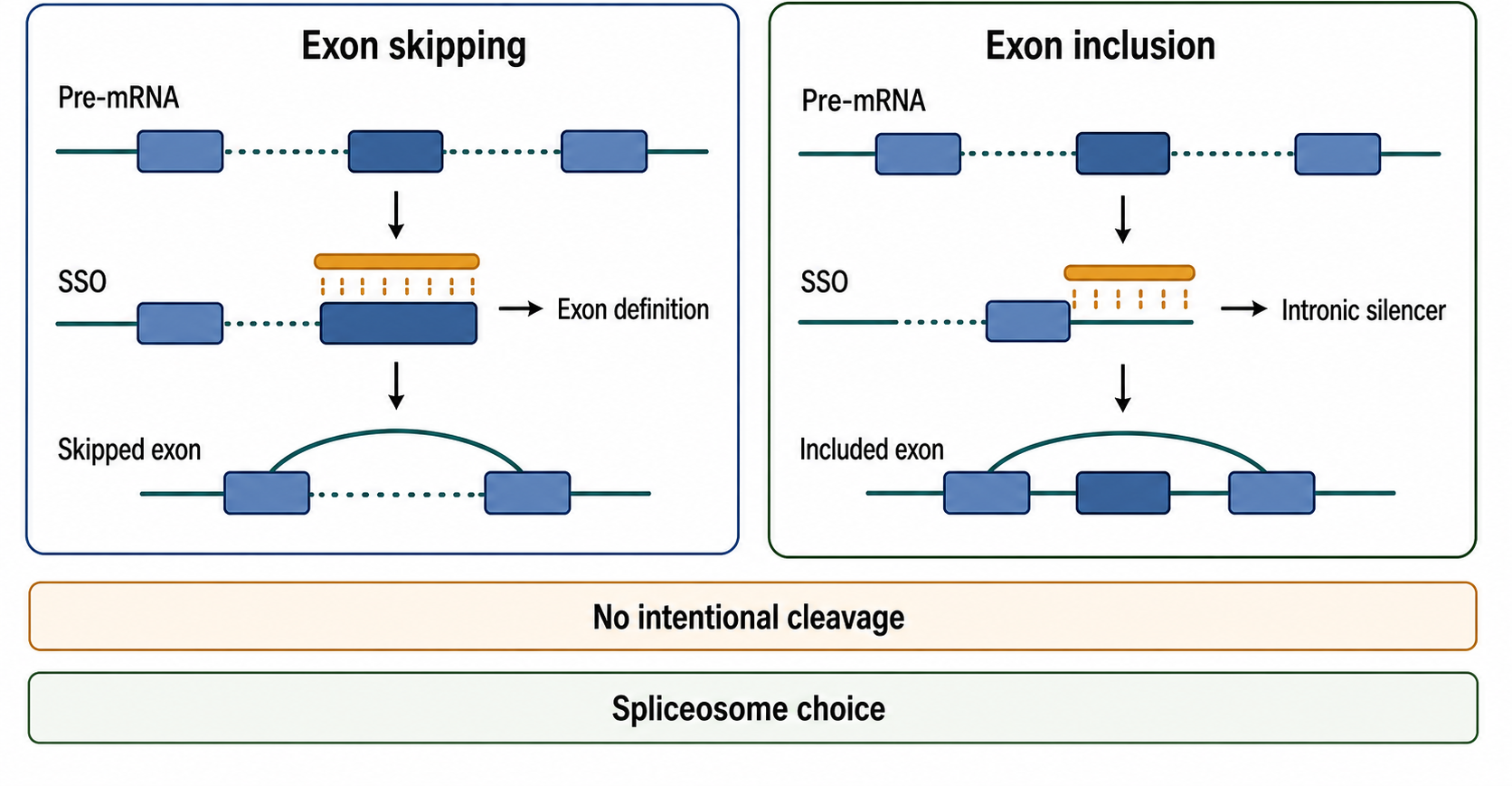
Figure 150.3. Exon Skipping Versus Exon Inclusion. “Splice-switching oligos change spliceosome interpretation of pre-mRNA without intentionally degrading the transcript.”
Exon inclusion uses the same steric logic in the opposite direction. Spinal muscular atrophy is the standard teaching case. Humans have SMN1 and SMN2 genes. Loss of SMN1 causes disease, while SMN2 mostly produces transcripts that skip exon 7 because a sequence difference weakens exon recognition and favors binding of inhibitory splicing factors. An intrathecally delivered SSO can bind an intronic silencer near SMN2 exon 7, promote exon inclusion, increase full-length SMN protein, and improve motor neuron survival. The mechanistic lesson remains central: a steric blocker can increase productive protein by preventing a negative splicing regulator from using an RNA element.
SSO design must consider whether the therapeutic goal is isoform replacement, partial correction, or deliberate production of a shorter protein. Exon skipping can be beneficial if the skipped exon is dispensable or if the resulting protein retains enough function. Exon skipping can be harmful if the exon contains essential catalytic residues, structural domains, localization signals, or regulatory motifs. Exon inclusion can restore function if the included exon is required, but it can also introduce premature termination codons, destabilize mRNA, or create a protein isoform with different properties. The therapeutic claim must therefore be made at the isoform and protein level, not only at the splice-band level.
SSO chemistries are chosen to avoid RNase H recruitment. PMOs have a morpholine ring and neutral phosphorodiamidate backbone. 2′-O-methyl and 2′-MOE phosphorothioate SSOs use ribose modifications that prevent the duplex from functioning as an RNase H substrate. These chemistries can be highly stable, but delivery differs. PMOs are neutral and often require high systemic doses for muscle exposure; peptide-conjugated PMOs can improve tissue uptake but introduce additional safety variables. Phosphorothioate SSOs bind proteins and distribute differently. Chemistry, target tissue, and dose cannot be separated from the splicing mechanism.
Evidence for splice switching begins with isoform assays. Reverse transcription PCR can show exon inclusion or skipping, and sequencing confirms the exact junctions. Quantitative assays are needed because visual band shifts can overstate effect size. RNA-seq can identify transcriptome-wide splicing changes, but depth and analysis pipeline matter, especially for rare junctions. Protein assays then test whether the corrected transcript yields meaningful protein. In diseases with low-abundance tissue-specific proteins, protein measurement may be difficult; in such cases, functional biomarkers and animal models become more important. Clinical benefit requires endpoints that fit the disease time course, developmental window, and tissue accessibility.
Splice-switching also has off-target risks. An SSO can bind partially complementary pre-mRNAs and alter splicing elsewhere. Even when sequence off-targets are limited, changing a splicing factor binding event at the intended target can indirectly alter networks if the target RNA encodes a regulator. In addition, high-dose oligonucleotides can cause chemistry-related toxicities independent of splice switching. A robust SSO program should use transcriptome-wide splicing analysis, chemistry controls, mismatch controls, and dose levels that separate intended splicing correction from cellular stress.
Exon skipping is sometimes equated with gene silencing. Exon skipping can reduce expression if it introduces nonsense-mediated decay, but therapeutic exon skipping often aims to preserve or restore expression by producing a different mature mRNA. Conversely, exon inclusion can either increase productive protein or trigger decay, depending on the exon. The words “skip” and “include” describe RNA processing, not the final biological direction.
150.3. Poison exon modulation and pseudoexon correction
Poison exons and pseudoexons illustrate why splicing therapeutics require precise transcript annotation. A poison exon is an exon whose inclusion reduces productive gene expression, commonly by introducing a premature termination codon that targets the transcript for nonsense-mediated decay. Poison exons are not always mistakes. Some are conserved regulatory switches that tune protein abundance in a tissue, developmental stage, or feedback circuit. For example, a splicing factor may promote inclusion of a poison exon in its own pre-mRNA, creating autoregulatory negative feedback. Therapeutic suppression of a poison exon is attractive when aberrant inclusion contributes to insufficient protein, but it is risky if the poison exon has a physiological regulatory role in tissues not captured by the disease assay.
A pseudoexon is different. It is usually an intronic segment that is not normally spliced into mature mRNA but becomes exonized when a variant creates or strengthens splice signals, disrupts silencing, or changes local context. Deep-intronic disease variants often act this way. The aberrant pseudoexon may contain a premature stop codon, frameshift the transcript, or insert amino acids that disrupt protein function. A splice-blocking oligonucleotide can mask the new splice site, enhancer, branch point region, or nearby sequence required for pseudoexon recognition, restoring the normal exon-exon junction.
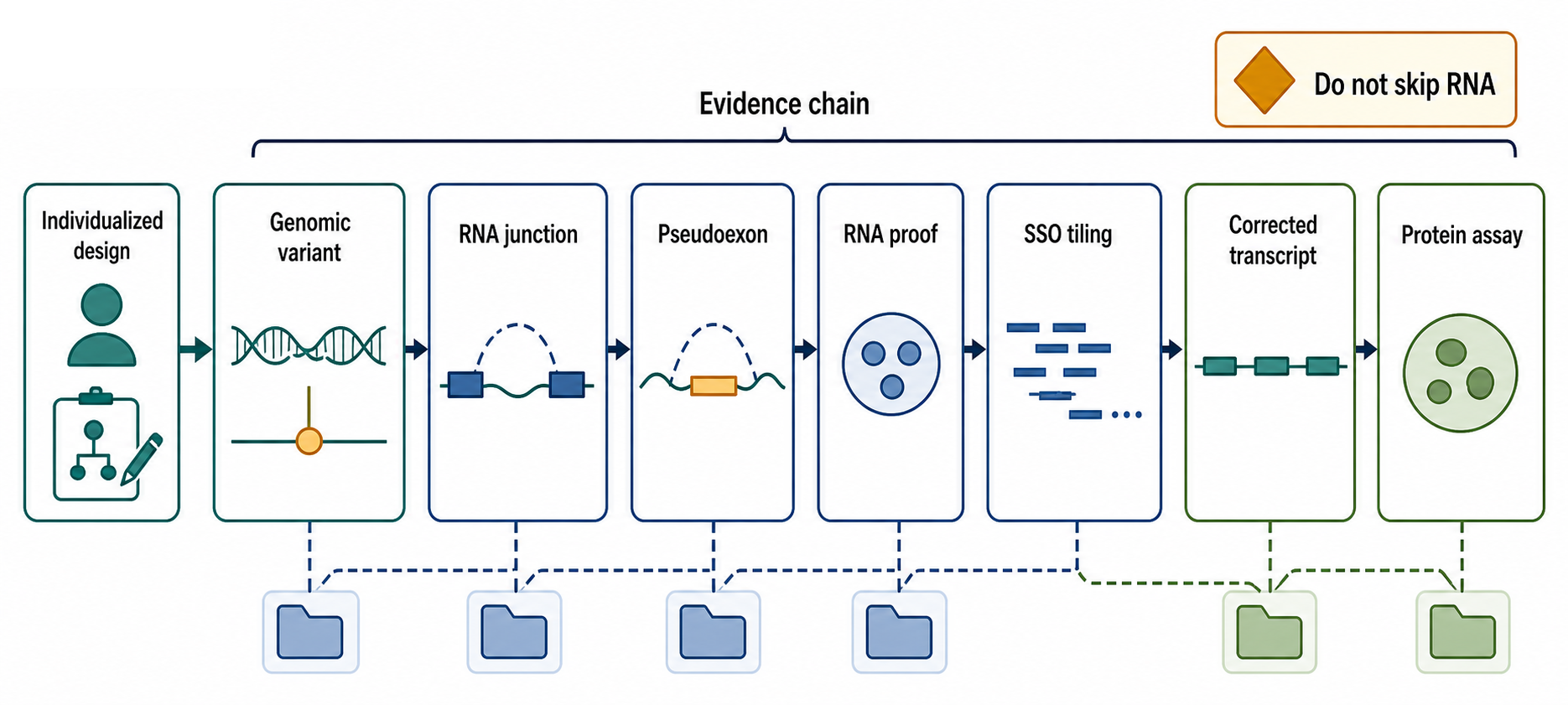
Figure 150.4. Pseudoexon Correction Workflow. “Pseudoexon correction requires RNA-level proof of the aberrant exon before an individualized or platform SSO can be rationally designed.”
The diagnostic path for pseudoexon correction begins before ASO design. Genomic sequencing may find a deep-intronic variant, but the causal claim requires RNA evidence from patient cells, disease tissue, organoids, or a minigene system. The aberrant transcript must be sequenced to define the pseudoexon boundaries. The reading-frame and nonsense-mediated decay consequences must be understood. If nonsense-mediated decay degrades the aberrant transcript, inhibitor experiments or targeted assays may be needed to detect it, but such experiments can create artifacts if interpreted without controls. Only after the aberrant splicing event is mapped can an SSO be designed rationally.
Box 150.2. Pseudoexon Correction Evidence Checklist
A credible pseudoexon-correction program should satisfy several checkpoints before a candidate SSO is treated as therapeutic. First, define the genomic variant, inheritance pattern, and disease fit. Second, show the RNA event directly: sequence the abnormal junctions, map pseudoexon boundaries, and account for nonsense-mediated decay if the aberrant transcript is unstable. Third, explain why the RNA event is damaging, such as frameshift, premature termination codon, domain insertion, or loss of productive transcript. Fourth, test tiled oligos against splice sites, enhancers, branch-region signals, or local structures rather than assuming the nearest splice site is best. Fifth, confirm correction with quantitative endogenous RNA assays when possible; minigenes are useful screens, not final proof. Finally, connect restored junctions to protein amount, localization, function, and safety monitoring for excessive or off-target splicing correction.
Poison-exon suppression and pseudoexon correction have a different evidence burden than broad knockdown. A gapmer can often be evaluated by target RNA reduction. A pseudoexon SSO must show that the disease-causing aberrant junction decreases and the normal productive junction increases. If the normal transcript is restored, the next question is whether protein expression and localization recover. In recessive loss-of-function diseases, partial restoration may be clinically meaningful if the dose-response curve is favorable. In dominant-negative contexts, merely reducing the aberrant isoform may not be enough if the mutant protein is already stable or if the corrected transcript still encodes a harmful variant.
The steric target for a pseudoexon SSO is not always obvious. Masking the new 5′ or 3′ splice site may work, but nearby enhancers, branch point sequences, or RNA structures can be better targets. A single pseudoexon may require several tiling oligos before a potent candidate is found. Minigene assays are useful for screening, but minigenes can fail to reproduce endogenous chromatin context, transcription kinetics, long introns, RNA structure, and tissue-specific splicing factor concentrations. Patient-derived cells or differentiated disease-relevant models provide stronger evidence, but they may be hard to obtain.
Individualized therapeutics often arise in this space. A private variant may affect one patient or a small family. If the molecular defect is a pseudoexon or poison-exon event, a custom SSO can be designed faster than a small-molecule program. The scientific logic can be compelling, but the clinical and regulatory logic is demanding. The patient may have advanced disease, no natural-history cohort, limited tissue access, and no conventional randomized trial path. Safety information may come from chemistry class, related sequences, animal toxicology, and in vitro assays rather than large exposed populations. This chapter returns to n-of-1 issues in Section 150.6.
The boundary between poison-exon therapy and normal regulation deserves attention. Some poison exons are part of natural homeostatic regulation. Blocking them chronically could overexpress a protein, disrupt autoregulation, or alter cell-type-specific isoform balance. Therefore the therapeutic window depends on disease mechanism. If a pathogenic variant creates a new pseudoexon absent from healthy people, correction is conceptually clean. If a therapy suppresses a conserved poison exon used in normal regulation, the program needs tissue-specific expression data, dose titration, and biomarkers that detect excessive protein restoration as well as insufficient correction.
150.4. Steric blockers, UTR targeting, and translation modulation
Steric blockade is the broadest antisense mechanism. A steric blocker binds RNA and prevents another molecular event from occurring at that site. The blocked event can be splice-factor binding, RNA-binding protein binding, miRNA recognition, ribosome scanning, start-codon selection, internal ribosome entry, RNA-RNA base pairing, or a structural switch. Unlike a gapmer, a steric blocker is not designed to destroy its target. The target RNA may remain stable and abundant while its interactions change.
UTRs are natural targets for steric blockers because untranslated regions encode regulatory information. A 5′ UTR can contain upstream open reading frames, internal ribosome entry elements, structured barriers to scanning, protein-binding motifs, and start-codon context. A 3′ UTR can contain miRNA target sites, AU-rich elements, localization motifs, polyadenylation-linked regulatory elements, and RBP binding sites. Blocking a UTR motif can increase or decrease translation depending on which factor is displaced. Masking a miRNA site may increase translation or mRNA stability; masking a stabilizing RBP site may decrease expression; blocking a ribosome entry element may inhibit translation.
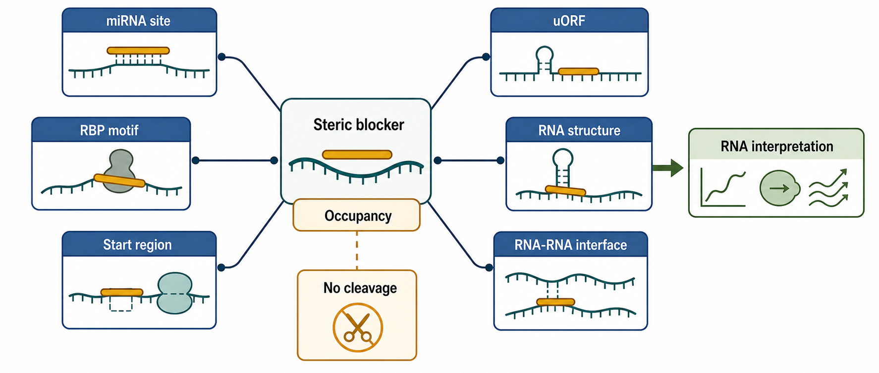
Figure 150.5. Steric Blockade of RNA Regulatory Motifs. “Steric blockers alter RNA interpretation by occupancy rather than by intentional cleavage.”
Translation modulation by steric blockers is conceptually simple but experimentally subtle. If an ASO binds near a start codon, it may inhibit ribosome scanning or initiation. If an ASO blocks an upstream open reading frame, it may increase downstream coding sequence translation. If an ASO masks an RNA structure required for internal initiation, it may reduce translation without changing mRNA abundance. Each case requires assays that separate RNA level from ribosome engagement and protein output. Total RNA measurement alone is insufficient. Ribosome profiling, polysome analysis, reporter assays, and protein quantification can distinguish translational effects from RNA decay, but each method has artifacts and should be interpreted with controls.
Steric blockers can also disrupt RNA-RNA interactions. Many regulatory RNAs work through base pairing: miRNAs bind 3′ UTRs, bacterial small RNAs bind mRNAs, snRNAs base-pair with splice sites, and some long RNAs pair with other transcripts or genomic RNA intermediates. An oligonucleotide can mask one partner and prevent the endogenous interaction. This logic underlies anti-miR oligonucleotides, which are treated in Chapter 152, but it also applies to blocking a disease-relevant RNA-RNA contact or viral RNA structure. The challenge is proving that the endogenous interaction is direct, functional, and blocked at tolerable ASO concentrations.
RNA-protein steric blockers are similarly powerful but evidence-intensive. Many RNA-binding proteins recognize short motifs that occur thousands of times in the transcriptome. Blocking one motif may have a clean effect if the motif is uniquely positioned, for example near a disease-causing splice event. Blocking a repeated motif family is harder because the ASO may have many partial targets, and the biological effect may reflect broad RNP perturbation. CLIP-seq, mutational reporters, protein occupancy assays, and transcriptome-wide RNA analysis can help distinguish direct motif blockade from indirect stress responses.
Steric blockers are sometimes described as “nondegrading” ASOs. That term is useful but not absolute. A steric blocker can indirectly change RNA stability by altering RBP binding, miRNA access, translation state, or splicing outcome. Hori et al. (2019) also warn that non-gapmer ASOs can be associated with RNA reduction and hepatotoxic potential. Therefore a non-gap chemistry lowers the likelihood of canonical RNase H cleavage but does not prove that RNA abundance will remain unchanged. A clean steric-blocker claim should report target RNA abundance, isoform composition, protein output, and transcriptome-wide perturbation.
The main therapeutic advantage of steric blockade is reversibility at the information-processing level. The genome is unchanged, and the RNA molecule can be interpreted differently while the ASO is present. This is attractive for splicing defects, dominant toxic motifs, and regulatory elements where partial modulation is enough. The main disadvantage is stoichiometry. Because steric blockers do not catalytically destroy target molecules, they often require enough intracellular ASO to occupy a meaningful fraction of target sites. High target abundance, rapid transcription, poor tissue exposure, or limited nuclear uptake can reduce efficacy.
150.5. Tissue delivery, toxicity, pharmacology, and biomarkers
ASO pharmacology begins with the route of administration. Systemically administered phosphorothioate ASOs bind plasma proteins and distribute strongly to liver, kidney, spleen, and some other tissues. They do not efficiently cross the intact blood-brain barrier, so central nervous system programs often use intrathecal or intracerebroventricular delivery. Local delivery can expose eye, ear, or other compartments. Muscle delivery remains a persistent challenge for many ASO classes, especially when the target is widespread skeletal and cardiac muscle and the chemistry is neutral or large. Chapters 156-158 provide broader delivery and pharmacology treatment; this section focuses on features that shape antisense mechanism.
Tissue exposure is not the same as productive intracellular exposure. An ASO can bind serum proteins, enter endosomes, accumulate in tissue, and still have limited access to nuclear or cytosolic RNA. Endosomal escape is often inefficient. A small fraction of internalized oligonucleotide may drive most pharmacology. This creates a measurement problem: total tissue ASO concentration can overestimate active concentration, while pharmacodynamic RNA change can underestimate exposure in cell types not represented by the sampled tissue. Cell-type specificity matters. A liver biopsy average may miss cholangiocyte, Kupffer cell, endothelial, or zonated hepatocyte differences; a cerebrospinal fluid biomarker may not report all brain regions equally.
Table 150.3. Toxicity and Biomarker Matrix. “ASO safety monitoring must cover sequence-dependent, chemistry-dependent, route-dependent, and tissue-accumulation risks.”
| Planned row | Intended comparison | Evidence or caveat |
|---|---|---|
| Productive exposure versus tissue burden | Total ASO in tissue is compared with active nuclear or cytosolic target engagement. | Endosomal trapping and cell-type heterogeneity can make bulk concentration misleading; use mechanism-matched PD markers. |
| Hybridization-dependent off-target toxicity | Unintended RNA binding can change RNA abundance, splicing, translation, or pathway state. | RNA-seq, splice analysis, mismatch controls, and dose-response separation are needed to distinguish off-target biology from intended pharmacology. |
| Liver stress | Hepatic accumulation, protein binding, and sequence or chemistry effects can produce hepatocellular injury. | Monitor transaminases, bilirubin, histopathology in preclinical studies, and toxicogenomic stress signatures when available. |
| Kidney handling | Tubular uptake and tissue persistence can create renal exposure and renal safety liabilities. | Monitor renal function, proteinuria, tubular injury markers, and kidney histology in nonclinical studies. |
| Platelet, coagulation, complement, and immune effects | Phosphorothioate and other chemistries can interact with proteins or immune sensors. | Track platelet counts, coagulation parameters, complement activation, cytokine or inflammatory signals, and clinical bleeding or infusion reactions. |
| CNS or local-route monitoring | Intrathecal, intracerebroventricular, ocular, or other local delivery changes compartment-specific risk. | CSF cells and protein, neurofilament light chain, neurological examination, imaging, and disease-specific biomarkers may be needed. |
| Biomarker lag and accumulation | RNA, protein, clinical function, and toxicity can change on different time scales after repeated dosing. | PK/PD models should include tissue half-life, target turnover, protein half-life, dosing interval, and stopping rules. |
Toxicity has multiple sources. Hybridization-dependent toxicity occurs when the ASO binds unintended RNAs and changes their abundance, splicing, or translation. Chemistry-dependent toxicity occurs when the backbone or sugar pattern interacts with proteins, membranes, complement, coagulation factors, or innate immune receptors. Sequence-dependent but hybridization-independent toxicity can occur when certain motifs bind proteins or form structures that perturb cellular pathways. Dose-dependent class toxicity can emerge from tissue accumulation even when the target mechanism is correct. Hori et al. (2019) anchor local evidence that non-gapmer ASOs can have hepatotoxic potential, reinforcing the need to test toxicity across ASO classes rather than assuming that only RNase H gapmers are risky.
The main organ systems watched in ASO toxicology include liver, kidney, immune system, platelets and coagulation, central nervous system for intrathecal agents, and injection-site or infusion reactions. Liver safety is monitored with transaminases, bilirubin, histopathology in preclinical studies, and sometimes transcriptomic stress signatures. Kidney safety includes tubular uptake, proteinuria, histology, and renal function markers. Platelet reductions and complement activation require hematology and immune monitoring. CNS programs may monitor cerebrospinal fluid cell counts, protein, neurofilament light chain, imaging, neurological examination, and disease-specific biomarkers. The exact panel depends on chemistry, route, target, species, and expected tissue distribution.
Pharmacokinetic and pharmacodynamic modeling helps choose dose and interval. Plasma half-life may be short relative to tissue half-life because ASOs leave plasma and persist intracellularly. Pharmacodynamic effects can lag behind tissue exposure because target RNA and protein turnover take time. Protein restoration after splice correction may lag behind RNA correction, and clinical function may lag further behind protein change. Conversely, toxicity can appear after repeated dosing as tissue burden accumulates. Good models include loading dose, maintenance interval, tissue half-life, target turnover, protein half-life, and biomarker sampling schedule.
Biomarkers differ by mechanism. For a gapmer, the proximal biomarker is target RNA reduction in the relevant tissue or a credible surrogate. For a secreted protein target, plasma protein may be a useful downstream biomarker. For a CNS target, cerebrospinal fluid protein or neurofilament light chain may help, but each biomarker has disease-specific interpretation limits. For a splice-switching oligo, the proximal biomarker is corrected isoform ratio; the next biomarker is protein restoration or reduction; the distal biomarker is clinical function. For a steric blocker of translation, ribosome engagement and protein output are more relevant than RNA abundance.
Box 150.3. Reading Biomarkers as a Ladder
An ASO program should not collapse every measurement into one success-or-failure number. First ask whether drug reached the relevant compartment: plasma, cerebrospinal fluid, tissue, or a credible surrogate. Second ask whether the target RNA changed in the mechanism-specific direction: knockdown for a gapmer, junction shift for an SSO, or altered ribosome engagement and protein output for a translation blocker. Third ask whether downstream protein or pathway state changed on the expected time scale. Fourth ask whether clinical function can plausibly respond given disease stage and tissue damage. Discordance is informative. Strong RNA correction without clinical benefit may reflect irreversible pathology, incomplete cell-type coverage, wrong target biology, or an insensitive endpoint. Apparent clinical benefit without proximal target engagement raises concern for confounding or off-target pharmacology. Safety biomarkers must be read in parallel because toxicity can emerge while RNA correction looks strong.
Delivery technologies can shift the ASO field but also change risk. Ligands such as GalNAc are best established for hepatocyte delivery of siRNAs and some oligonucleotides; antibody, peptide, lipid, polymer, and exosome-like systems are under investigation for other tissues. Conjugating a ligand to an ASO changes receptor uptake, endosomal trafficking, tissue distribution, immunogenicity, and manufacturing. Peptide-PMO conjugates can improve muscle uptake but may introduce renal or immune liabilities. A delivery improvement is therefore not only a potency improvement; it is a new drug substance or product design with its own evidence burden.
Because ASOs are sequence-specific, they are sometimes expected to be intrinsically safer than small molecules. Sequence specificity helps, but ASOs are still drugs with chemistry, distribution, metabolism, protein binding, immune recognition, and dose-limiting toxicity. A 20-mer can have many partial transcript matches and many protein contacts. Safety emerges from integrated design and testing, not from complementarity alone.
150.6. Approved drugs, n-of-1 therapy, trials, and regulation
Approved antisense drugs demonstrate several mechanisms. Nusinersen promotes SMN2 exon 7 inclusion for spinal muscular atrophy. Eteplirsen and related PMO exon-skipping drugs target DMD exons in Duchenne muscular dystrophy. Inotersen is an RNase H gapmer that reduces transthyretin RNA for hereditary transthyretin amyloidosis. Volanesorsen targets APOC3 RNA in familial chylomicronemia syndrome in some regulatory settings. Tofersen is an intrathecal ASO that lowers SOD1 for SOD1 amyotrophic lateral sclerosis.
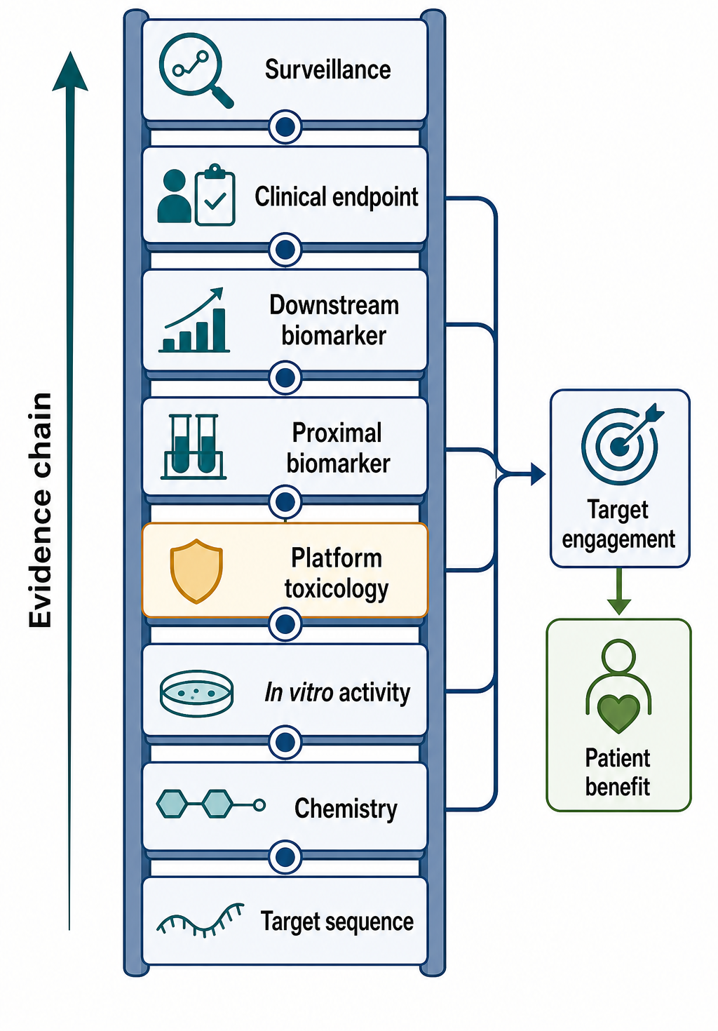
Figure 150.6. Clinical Translation Evidence Ladder. “Clinical antisense development links molecular target engagement to patient benefit through a chain of increasingly disease-specific evidence.”
The trial design depends on mechanism and disease. A gapmer for a secreted liver protein can often use blood protein reduction as an early pharmacodynamic marker. A splice-switching oligo for a neuromuscular disorder may require tissue biopsy, RT-PCR, protein quantification, motor scales, respiratory outcomes, or time-to-event endpoints. A CNS ASO may use cerebrospinal fluid biomarkers, neurofilament light chain, imaging, and clinical scales. Rare diseases often have small cohorts, heterogeneous progression, prior supportive care differences, and developmental timing effects. A trial that starts after irreversible tissue loss may show target engagement but limited functional recovery.
Table 150.4. Approved and Individualized ASO Evidence Patterns. “The smaller the clinical population, the more explicit the molecular diagnosis, biomarker rationale, and stopping rules must be.”
| Planned row | Intended comparison | Evidence or caveat |
|---|---|---|
| Approved gapmer for target reduction | A population-level program reduces a disease-relevant RNA such as a liver or CNS target. | Evidence pattern links target RNA or protein reduction, organ safety monitoring, clinical endpoint, and post-approval surveillance; verified labels and pivotal citations still need curation. |
| Approved splice-switching program | A genotype-defined SSO promotes exon inclusion or exon skipping to restore a functional isoform. | Requires junction quantification, protein restoration or reduction, tissue-relevant delivery, and clinical-function endpoints; local DMD and nusinersen citations remain gaps. |
| Rare-disease small trial | Small cohorts use molecular entry criteria, natural-history data, proximal biomarkers, and disease-specific scales. | Target engagement may be convincing even when functional recovery is limited by disease stage or irreversible tissue loss. |
| N-of-1 pseudoexon or poison-exon ASO | A private sequence is designed after genetic and RNA evidence define a causal splicing lesion. | Needs clear mechanism, target-tissue access, independent review, GMP-quality manufacturing, informed consent, monitoring, and stopping rules. |
| Platform extrapolation across ASO chemistries | Prior class experience can inform dose, route, toxicology, and analytics for related molecules. | Sequence changes can alter hybridization off-targets and protein interactions, so platform evidence cannot replace sequence-specific risk assessment. |
| Surrogate and post-approval evidence | Accelerated or small-trial approvals may rely heavily on RNA, protein, or fluid biomarkers. | Real-world follow-up should test durability, genotype-specific response, long-term safety, age-at-treatment effects, and rare adverse events. |
N-of-1 antisense therapy occupies a special category. The scientific case is strongest when a patient has a genetically defined defect, the aberrant RNA event is directly demonstrated, the proposed ASO has a clear sequence-specific correction mechanism, and the target tissue is reachable. The ethical case depends on disease severity, lack of alternatives, plausible benefit, preclinical safety, manufacturing quality, informed consent, monitoring, and transparency about uncertainty. The regulatory case is difficult because a single patient cannot provide conventional efficacy evidence, yet the drug is still administered to a human and can cause harm. Individualized ASO programs therefore need rigorous molecular documentation, independent review, dose rationale, stopping rules, adverse-event monitoring, and data sharing that respects privacy.
Clinical translation also requires chemistry-class knowledge. Regulators and clinicians may accept some extrapolation from related ASO chemistries, but sequence changes can alter hybridization off-targets and protein interactions. A custom SSO for a private pseudoexon cannot be assumed safe merely because a different PMO or 2′-MOE ASO was tolerated. Conversely, requiring a full conventional drug-development program for every private sequence may make individualized therapy impossible. The field is still negotiating how to balance platform evidence, sequence-specific risk, urgency, and patient protection. This is a major open regulatory question.
Endpoint selection should align with the disease mechanism. For a disease caused by toxic RNA or toxic protein, target reduction may be a useful proximal endpoint but not a complete clinical endpoint. For a loss-of-function splicing disorder, restored protein may be more meaningful than corrected RNA if the protein is measurable. For progressive neurodegeneration, slowing decline may be realistic even if improvement is not. For pediatric developmental disease, timing may determine whether molecular correction can rescue function. Natural-history data, patient-reported outcomes, caregiver burden, and objective functional measures all matter, but each can be noisy in ultra-rare populations.
Manufacturing and quality control are part of the clinical evidence chain. ASOs require sequence identity confirmation, purity assessment, impurity profiling, stereochemical or diastereomeric characterization when relevant, endotoxin and bioburden control, formulation stability, and container compatibility. PMOs, phosphorothioate ASOs, LNA-containing ASOs, and conjugated ASOs have different analytical challenges. Batch-to-batch consistency is especially important when pharmacodynamic windows are narrow or when repeated dosing creates accumulation.
Post-approval evidence remains important. Some ASO approvals have relied heavily on surrogate biomarkers, accelerated pathways, or small trials. Real-world data can clarify durability, long-term safety, genotype-specific response, age-at-treatment effects, and rare adverse events. Post-marketing commitments should not be treated as clerical formalities; they are part of the scientific process that connects RNA mechanism to patient benefit.
Recent Consensus
The current consensus is that ASOs are mature enough to be a recurring therapeutic platform but not mature enough to be treated as plug-and-play sequence drugs. The platform logic is strong: base pairing can direct a chemically optimized oligonucleotide to a defined RNA, and the chemistry can be chosen to recruit RNase H, redirect splicing, or block an RNA interaction. The clinical record shows that this logic can produce approved medicines. The unresolved part is prediction. Potency, tissue exposure, off-target behavior, and toxicity still require empirical screening and disease-specific validation.
Consensus point 1: mechanism must be designed and demonstrated. A gapmer should be shown to reduce RNA through an RNase H-compatible design, not merely to correlate with lower target abundance. A splice-switching oligo should be shown to alter the intended junctions and produce the intended protein or functional output. A steric blocker should be shown to block the intended interaction while preserving or predictably changing RNA abundance.
Consensus point 2: chemistry creates both efficacy and risk. Phosphorothioate, 2′ modifications, LNA, cEt, PMO, and conjugates are not interchangeable carriers for the same sequence. Chemistry controls nuclease resistance, affinity, RNase H recruitment, protein binding, tissue distribution, immune stimulation, and manufacturability. Chapter 149 provides the chemical basis, but clinical development must treat chemistry as pharmacology.
Consensus point 3: delivery remains a major bottleneck. Liver and CNS programs have clearer paths than many muscle, heart, lung, kidney-cell-type-specific, and immune-cell targets. Local or intrathecal delivery can solve some distribution barriers but introduces procedure-related burden and compartment-specific monitoring. New conjugates and delivery systems may broaden the field, but each must prove that increased uptake is productive and safe.
Consensus point 4: clinical interpretation must connect molecular correction to a disease-relevant endpoint. RNase H gapmers, splice-switching oligos, and steric blockers can all produce convincing proximal pharmacodynamic signals, but target RNA reduction, exon inclusion, exon skipping, pseudoexon suppression, or motif blockade is not automatically clinical benefit. Biomarkers are strongest when they are mechanistically close to the disease driver, measured in or credibly linked to the affected tissue, and interpreted with the expected lag between RNA change, protein change, tissue repair, and patient function.
Open Questions, Controversies, Deprecated Models, and Common Misconceptions
Open questions:
- How predictable is sequence-specific ASO toxicity? Some toxic effects can be reduced by motif filters, transcriptome off-target prediction, protein-binding screens, and chemical optimization, but many liabilities still emerge empirically. The field needs better models connecting ASO sequence, chemistry, protein interactome, subcellular localization, and stress pathways.
- How should individualized ASOs be regulated? A platform-based framework could make private-sequence therapies feasible for severe rare diseases, but individualized dosing and limited preclinical packages can miss risks. The unresolved policy problem is how to protect patients while allowing rational treatment when no commercial trial is possible.
- How should surrogate biomarkers be weighted? RNA correction is necessary for many ASO mechanisms, but it may not be sufficient for clinical benefit. Protein restoration, target reduction, neurofilament changes, dystrophin levels, or fluid biomarkers can be compelling when linked to disease biology, yet each can fail if the biomarker is not causally close enough to function.
- How far can delivery improvements be generalized across tissues and chemistries? Better uptake in liver, CNS, muscle, or another compartment does not prove productive exposure in the relevant cell type or nuclear/cytosolic compartment. Conjugates, peptides, and formulations can change receptor engagement, endosomal escape, protein binding, immune activation, renal handling, and manufacturing control, so delivery advances must be evaluated as part of the drug mechanism rather than as neutral add-ons.
Common misconceptions:
- “ASOs are just short pieces of DNA.” Therapeutic ASOs are chemically engineered molecules. Their backbones, sugars, stereochemistry, terminal patterns, conjugates, and formulations determine their behavior.
- “A perfect sequence match guarantees specificity.” Off-target binding can occur through partial complementarity, and protein-binding effects can be independent of Watson-Crick targeting.
- “If an ASO fixes RNA in a cell line, it should work in patients.” Patient benefit requires tissue delivery, durable pharmacodynamics, sufficient correction in disease-relevant cells, acceptable safety, and endpoints sensitive to the expected biological change.
- “Non-gap ASOs cannot reduce RNA.” Non-gap designs are intended to avoid RNase H recruitment, but RNA abundance can change indirectly through splicing, translation, RBP displacement, stress, or toxicity-associated pathways.
Deprecated or weakened claims:
- Deprecated or overgeneralized model: ASO classes are sometimes treated as if each chemistry maps to one clean mechanism. That model is too rigid. Gapmers can have steric effects before cleavage or in compartments with limited RNase H1 access; non-gap splice-switching or steric ASOs can alter RNA abundance indirectly through splicing, RBP displacement, translation state, stress responses, or toxicity; and a target-site blocker can behave differently across isoforms, cell types, and disease states.