Chapter 84. miRNA Biogenesis, Targeting, Repression Mechanisms, and Quantitative Regulation
Scope Note
MicroRNAs (miRNAs) are short regulatory RNAs, usually about 21-23 nucleotides long in animals, that guide Argonaute proteins to partially complementary transcripts and thereby tune gene expression after transcription. This chapter explains how animal miRNAs are transcribed, processed, exported, loaded into Argonaute, and used to recognize target messenger RNAs (mRNAs). It then follows the target from initial binding to translational repression, deadenylation, decapping, and decay, with special attention to quantitative regulation: why most single miRNA-target interactions are modest, why the combined effect of many sites can be biologically large, and why competing endogenous RNA (ceRNA) claims require strict stoichiometric evidence. Plant miRNA pathways are mentioned only as comparative boundary cases because plant small-RNA pathways are treated separately elsewhere.
Executive Summary
Animal miRNAs are genomic regulatory RNAs that usually begin as RNA polymerase II transcripts. A primary miRNA transcript (pri-miRNA) contains an imperfect hairpin embedded in flanking single-stranded RNA. The nuclear Microprocessor complex, composed of the RNase III enzyme Drosha and the double-stranded RNA-binding protein DGCR8, recognizes this hairpin architecture and cleaves near the base of the stem to release a precursor miRNA (pre-miRNA). Exportin-5 carries the pre-miRNA through the nuclear pore in a Ran-GTP-dependent step. In the cytoplasm, Dicer removes the terminal loop to create a short duplex with characteristic ends. One strand is selected as the guide strand and loaded into an Argonaute protein, whereas the other strand is usually discarded. This canonical pathway has important exceptions: some miRNAs are made from spliced introns called mirtrons, some derive from small nucleolar RNA-like or transfer RNA-like precursors, and Drosha or Dicer can have noncanonical roles beyond ordinary miRNA maturation. Wei et al. 2023 is a relevant listed review for Drosha roles, although broader canonical pathway review coverage remains a Final bibliography item.
The mature miRNA does not act as a free RNA. It functions as the guide component of an Argonaute-containing ribonucleoprotein complex. Nucleotides 2-8 from the miRNA 5′ end form the seed region, which is the main determinant of target recognition in animals. A typical animal target site lies in a 3′ untranslated region (3′ UTR) and pairs to the seed with Watson-Crick complementarity. Target repression is not determined by the seed alone. Site context matters: local accessibility, 3′ UTR position, adenosine-rich flanking sequence, site number, spacing, transcript abundance, and possible 3′ supplementary pairing can shift efficacy. McGeary et al. 2019 provides a strong quantitative biochemical anchor for how seed pairing, supplementary pairing, and site context affect miRNA targeting efficacy. Vega-Badillo et al. 2026, listed for flies, appears relevant for comparative biochemical targeting principles but should be rechecked during citation curation.
After target engagement, most animal miRNAs reduce gene expression through a combination of translational repression and accelerated mRNA decay. Early repression can reduce protein output before large mRNA abundance changes are visible, but stable repression in many mammalian systems is dominated by deadenylation, decapping, and exonucleolytic decay of the target mRNA. GW182/TNRC6 proteins are central adaptors: Argonaute binds the miRNA and target RNA, while GW182/TNRC6 recruits effector complexes such as CCR4-NOT and PAN2-PAN3. Deadenylation shortens the poly(A) tail, weakens poly(A)-binding protein support for translation and stability, and can promote decapping and 5′ to 3′ decay. The mechanistic balance differs by cell type, target site, time point, and assay. Czarnocka-Cieciura et al. 2024 is useful for the broader principle that deadenylation and decay are quantitatively coupled but not identical. Li et al. 2025 is most directly relevant to the boundary between target engagement and miRNA turnover, a topic developed further in Chapter 85.
The quantitative logic of miRNA regulation is often misunderstood. A single canonical site may cause only a small reduction in protein output, but a miRNA can affect hundreds of transcripts, and a transcript can integrate multiple miRNAs and RNA-binding proteins. Biological importance therefore may come from network-level tuning, threshold setting, noise buffering, developmental timing, or reinforcing transcriptional programs rather than from one target being silenced like an on/off switch. At the same time, not every predicted seed match is functional, and not every correlated expression pattern implies direct regulation. ceRNA models, in which one RNA affects another by competing for a shared miRNA, require unusually favorable stoichiometry: the competitor must be abundant enough, accessible enough, and sufficiently occupied by the relevant miRNA to measurably change miRNA availability. Most transcriptome-scale ceRNA networks inferred only from correlations are therefore hypothesis-generating rather than demonstrated mechanisms.
Disease and biomarker studies use miRNAs in three different ways that should not be conflated. A miRNA can be a causal regulatory molecule in a disease pathway, a passive marker of tissue injury or cell-type composition, or a therapeutic handle that can be mimicked or inhibited. Circulating miRNA biomarker studies face pre-analytical problems including hemolysis, platelet contamination, extraction bias, normalization choice, and batch effects. Target prediction tools are useful for prioritization but have high false-positive rates unless paired with perturbation, site mutation, Argonaute occupancy, and endogenous target readouts. Schlegel et al. 2022 illustrates the therapeutic importance of seed-mediated off-target biology in the adjacent siRNA field. Nappi 2024 and Ranasinghe et al. 2023 provide broader noncoding RNA and siRNA therapeutic context, but they do not substitute for miRNA-specific clinical curation.
Concept Inventory
- miRNA: A short endogenous regulatory RNA that guides Argonaute to partially complementary target RNAs. In animals, most miRNAs repress target mRNAs through seed-mediated recognition and recruitment of translational and decay machinery.
- pri-miRNA: The primary transcript containing a miRNA hairpin before Drosha cleavage. A pri-miRNA can be an independent transcript or a hairpin embedded in an intron or exon of a host gene.
- pre-miRNA: The hairpin precursor generated by nuclear processing, usually about 60-80 nucleotides in animals, exported to the cytoplasm for Dicer processing.
- Microprocessor: The nuclear complex centered on Drosha and DGCR8 that recognizes pri-miRNA hairpins and cuts them near the stem base.
- Seed region: miRNA nucleotides 2-8, counted from the 5′ end. This region provides the main target-recognition code for animal miRNAs.
- Argonaute loading: The process by which a small-RNA duplex enters an Argonaute protein and one strand is retained as the guide.
- GW182/TNRC6: A family of effector proteins that bind Argonaute and recruit repression machinery, including deadenylase complexes.
- Deadenylation: Shortening of the mRNA poly(A) tail. In miRNA repression, deadenylation often precedes decapping and decay.
- ceRNA: A proposed competing endogenous RNA whose binding sites titrate miRNA activity away from another target. The concept is plausible in restricted quantitative regimes but often overclaimed.
- Target validation: The process of showing that a miRNA directly regulates an endogenous transcript through a specific site, not merely that miRNA and mRNA abundances are correlated.
What to Know Before Reading This Chapter
Readers should know that eukaryotic mRNAs contain coding sequences flanked by untranslated regions and that the 3′ UTR often carries regulatory sites for RNA-binding proteins and small RNAs. The chapter uses mRNA, 3′ UTR, poly(A) tail, translation initiation, decapping, and exonuclease decay in the same sense developed in Chapters 26, 29, 35, 72, and 73. Readers should also know that RNase III enzymes cut double-stranded or hairpin RNA and that ribonucleoprotein complexes can recognize RNA structure, sequence, or both. No prior specialized knowledge of miRNA nomenclature is assumed.
The running example is a mammalian miRNA that is transcribed from a genomic locus, processed through Drosha and Dicer, loaded into AGO2, and used to repress several mRNAs through seed-matched 3′ UTR sites. The chapter repeatedly contrasts that ordinary animal case with three boundary cases: noncanonical miRNA biogenesis, plant miRNAs with extensive target pairing, and synthetic siRNA-like therapeutics whose seed-mediated off-target effects reveal how small-RNA target recognition works.
84.1. miRNA genes, pri-miRNA processing, Drosha, Exportin-5, and Dicer
miRNA Genes and Primary Transcripts
A miRNA gene is a genomic region whose transcript can fold into a hairpin that ultimately yields a mature miRNA guide strand. Some miRNA genes lie in intergenic regions with their own promoters. Others lie inside introns of protein-coding or long noncoding host genes and are transcribed as part of a larger RNA. Many animal miRNA transcripts are made by RNA polymerase II, capped at the 5′ end and polyadenylated at the 3′ end, although the hairpin itself is later excised from the larger transcript. This origin matters because miRNA expression can be linked to promoter regulation, host-gene transcription, chromatin state, splicing, and developmental signaling.
The primary transcript, or pri-miRNA, is not simply a perfect double-stranded stem. A typical animal pri-miRNA hairpin contains a lower stem, an upper stem, a terminal loop, and flanking single-stranded regions. The mature miRNA is encoded on one arm of the hairpin, and the opposing arm gives rise to the passenger strand, often annotated with a star or 3p/5p arm designation. The 5p and 3p nomenclature describes which side of the hairpin produces the mature strand, not whether one strand is automatically functional and the other is waste. In many loci, one arm is strongly dominant; in others, both arms can be loaded into Argonaute in different tissues or conditions.
The chapter’s first figure should show the pathway as a physical sequence of RNA forms rather than as a list of protein names. Figure 84.1 starts with a genomic locus and ends with an Argonaute-loaded guide strand, making clear where each processing enzyme acts and which RNA ends are created.
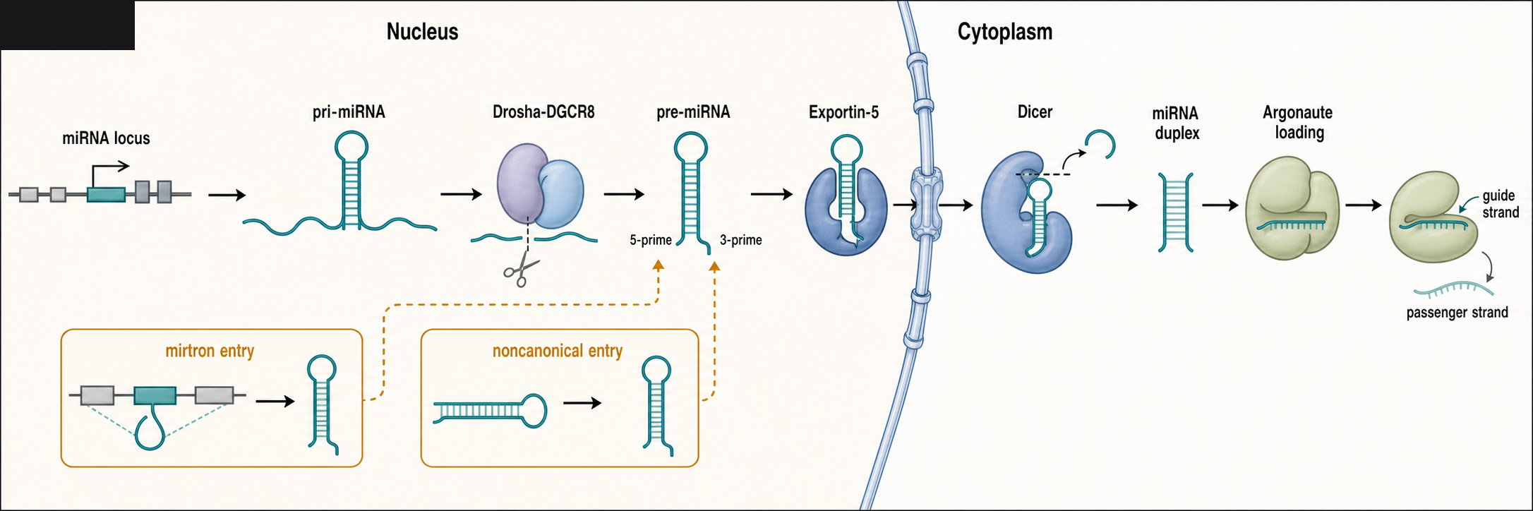
Figure 84.1. Canonical and Selected Noncanonical Animal miRNA Biogenesis. Canonical animal miRNA maturation begins when a genomic miRNA locus is transcribed into a pri-miRNA hairpin. Drosha-DGCR8 cleaves the pri-miRNA in the nucleus, Exportin-5 transports the pre-miRNA to the cytoplasm, Dicer removes the terminal loop to produce a short duplex, and Argonaute loading selects one strand as the guide. Insets show mirtrons and other noncanonical precursors entering downstream steps.
miRNA genes are frequently clustered. A polycistronic pri-miRNA can contain multiple hairpins, each processed into a different miRNA. Clustered miRNAs can be co-expressed and may regulate related biological programs, but clustering alone does not prove that all products share the same targets or functions. A cluster can contain miRNAs with different seed sequences, different processing efficiencies, and different tissue-specific arm usage. Conversely, unrelated genomic loci can encode miRNAs with identical seed sequences, forming seed families that regulate overlapping target sets.
Drosha, DGCR8, and Nuclear Processing
The Microprocessor is the entry point into canonical animal miRNA maturation. Drosha is an RNase III enzyme that cuts the two strands of a pri-miRNA stem near the junction between the double-stranded stem and the basal single-stranded flanking RNA. DGCR8, called Pasha in some organisms, binds the pri-miRNA and helps Drosha position the cut. The result is a pre-miRNA hairpin with a short 3′ overhang, a structural feature recognized by downstream export and processing machinery.
This step is best understood as molecular measurement. The Microprocessor evaluates the hairpin’s shape, stem length, basal junction, apical region, and sequence or structural motifs that recruit auxiliary proteins. A hairpin that is too short, too irregular, too tightly embedded in other RNA structure, or bound by inhibitory proteins may be inefficiently processed. The Microprocessor therefore converts transcriptional information into mature miRNA output with its own layer of regulation. A cell can express a pri-miRNA without producing much mature miRNA if processing is blocked or inefficient.
Several proteins modulate pri-miRNA processing. Some RNA-binding proteins bind terminal loops or flanking regions and increase processing of specific pri-miRNAs; others inhibit processing or redirect the transcript to alternative fates. Signaling pathways can change Drosha, DGCR8, or accessory factor abundance, localization, or post-translational modification. The provided reference list includes Wei et al. 2023, a review on noncanonical Drosha roles, which is relevant as a reminder that Drosha biology is broader than the simple textbook Microprocessor step. The listed bibliography does not yet contain a strong general review focused on canonical Drosha-DGCR8 recognition; that gap should be corrected before final publication.
Not all miRNAs require Drosha. Mirtrons are short introns that, after splicing and debranching, fold into pre-miRNA-like hairpins and enter the pathway downstream of Microprocessor cleavage. Some other small RNAs derive from snoRNA-like, tRNA-like, endogenous short hairpin, or repeat-associated precursors. These noncanonical routes are not rare curiosities in evolutionary terms, but the canonical Drosha-DGCR8 pathway remains the central mechanism for most annotated animal miRNAs.
Exportin-5, Dicer, and Duplex Formation
After Drosha cleavage, the pre-miRNA must move from the nucleus to the cytoplasm. Exportin-5 recognizes the pre-miRNA hairpin and its 3′ overhang in cooperation with Ran-GTP. The export step protects the precursor from inappropriate nuclear degradation and places it in the compartment where Dicer and Argonaute loading occur. Exportin-5 is sometimes described as a passive carrier, but the carrier also contributes to pathway specificity by favoring hairpins with appropriate length and end structure.
Dicer is a cytoplasmic RNase III enzyme that cuts near the terminal loop of the pre-miRNA to release an approximately 22-nucleotide duplex. Dicer uses domains that contact the end of the hairpin and measure a defined distance before cleavage. In mammalian cells, Dicer functions with double-stranded RNA-binding partners such as TRBP and PACT, which can influence processing accuracy, strand selection, and pathway regulation. The duplex generated by Dicer has two strands with different end stability and sequence features. These properties help decide which strand becomes the guide strand.
Processing precision is biologically important because a one-nucleotide shift at the 5′ end changes the seed region. Such 5′ isomiRs can redirect the target spectrum, whereas 3′ end variation often affects stability, tailing, trimming, or target-directed miRNA turnover more than seed identity. Chapter 85 treats miRNA tailing, trimming, and decay in detail. For this chapter, the key point is that biogenesis is not merely a production line; it also establishes the identity of the regulatory sequence that Argonaute will read.
Plants use related but distinct logic. Plant MIRNA genes are also transcribed into hairpin precursors, but Dicer-like enzymes usually perform nuclear processing steps, and mature plant miRNAs are commonly 2′-O-methylated at their 3′ ends. Plant miRNAs often pair extensively with targets and can direct slicing or strong repression. Yu et al. 2026, listed as a plant miRNA review, is useful for this comparative boundary. Animal miRNA rules should not be projected directly onto plant systems.
84.2. Argonaute loading, seed pairing, target-site context, and repression
Argonaute Loading and Strand Selection
Argonaute proteins are the core effectors of miRNA function. A mature miRNA guide strand binds inside an Argonaute protein so that the guide’s 5′ phosphate is anchored in a pocket and the seed region is preorganized for target recognition. Humans have four major AGO proteins, AGO1 through AGO4. AGO2 is unique among them in retaining robust endonucleolytic slicing activity, but most animal miRNA targets are not sliced because their pairing is usually incomplete. All AGO proteins can support translational repression and mRNA destabilization through recruitment of effector proteins.
Loading begins with a small-RNA duplex rather than a single mature strand. The duplex enters an Argonaute-loading pathway, and the strand with the less stably paired 5′ end is often favored as the guide. The passenger strand is removed by unwinding or, for highly paired duplexes loaded into slicer-competent AGO2, by cleavage. Strand choice is probabilistic, not absolute. Duplex structure, nucleotide identity at the 5′ end, Argonaute isoform, cell type, and precursor abundance can all influence guide selection. This is why mature miRNA annotation should specify arm usage and, when necessary, isoform or isomiR identity.
The loaded Argonaute-miRNA complex can be considered a programmable RNA-binding protein. The miRNA supplies sequence specificity, but Argonaute supplies binding geometry, stability, protein interactions, subcellular localization, and effector recruitment. This distinction is important for interpreting experiments. Overexpressing a miRNA mimic does not simply add a free RNA to the cell; it competes for Argonaute loading, may saturate pathway components, and may create nonphysiological target occupancy.
Seed Pairing, Site Types, and Target-Site Context
In animals, most productive target recognition starts with seed pairing. The seed region, especially nucleotides 2-7 or 2-8 of the miRNA, pairs with a complementary sequence in the target RNA. Common site classes include 6-mer sites, 7-mer sites with either an adenosine opposite miRNA position 1 or pairing to position 8, and 8-mer sites that combine both features. These labels are useful shorthand, but they should not be mistaken for a complete mechanism. A weak site in an accessible region can outperform a stronger seed match buried in structure or occluded by another protein.
Target-site context integrates several features. Sites in 3′ UTRs are often more effective than sites in coding sequences, partly because translating ribosomes can disrupt coding-region interactions and because 3′ UTRs are hubs for regulatory proteins. Sites away from the stop codon and poly(A) tail can be more effective than sites crowded by competing complexes, although exact positional effects depend on the transcript. Local AU-rich sequence tends to increase accessibility. Multiple sites can act additively or cooperatively when positioned favorably, but site crowding can also create diminishing returns. Some targets use 3′ supplementary pairing between miRNA nucleotides beyond the seed and complementary target bases to increase affinity or specificity.
Figure 84.2 and Table 84.1 should be read together. Figure 84.2 shows how the guide strand is presented by Argonaute and how different target-site geometries align with the seed and supplementary regions. Table 84.1 separates evidence types, because a reporter assay, an Argonaute CLIP peak, and an endogenous mRNA response do not prove the same claim.
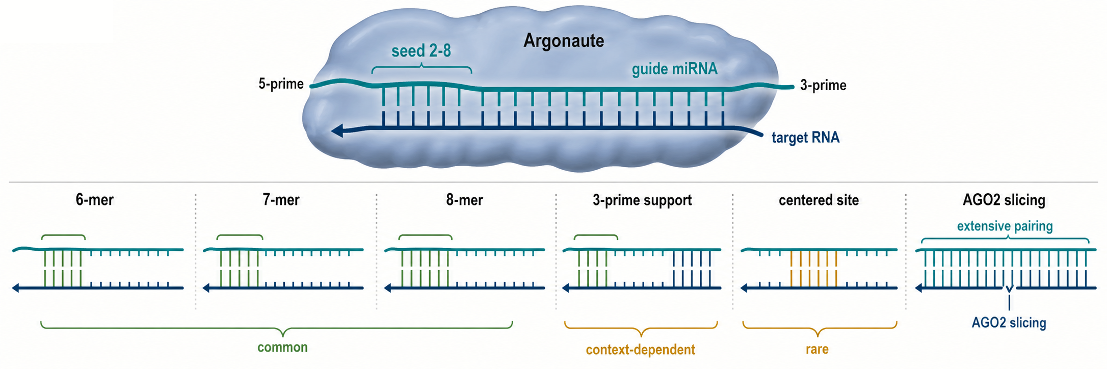
Figure 84.2. miRNA Target-Site Recognition Geometry. Animal miRNA targeting is dominated by seed pairing, especially guide nucleotides 2-8, but site efficacy depends on the entire target context. The diagram compares 6-mer, 7-mer, and 8-mer seed sites, a site with 3′ supplementary pairing, a noncanonical centered site, and an extensively paired target capable of AGO2 slicing.
Table 84.1. Evidence Ladder for miRNA Target Assignment. Different assays support different meanings of “miRNA target.” Seed prediction supports a hypothesis; Argonaute CLIP supports occupancy; reporter assays support site capability; endogenous perturbation supports regulation; site editing and rescue support direct causal assignment.
| Evidence type | What it supports | Key caveat or next validation step |
|---|---|---|
| Seed match | Plausible guide-target pairing, especially at miRNA positions 2-8 in animal 3′ UTRs. | Many seed matches are unused because expression, accessibility, affinity, or context is wrong. |
| Conserved site | Evolutionary constraint consistent with functional regulation across related species. | Conservation does not identify the active cell type or prove present-day repression. |
| Biochemical affinity or efficacy model | Quantitative ranking of site type, supplementary pairing, local context, and likely occupancy. | Models generalize best within assayed contexts and still need endogenous validation. |
| Argonaute CLIP peak | Physical occupancy or proximity of AGO-miRNA complexes near a candidate site. | CLIP does not prove repression and can be affected by crosslinking, mapping, and peak-calling bias. |
| Reporter repression | A cloned site or UTR fragment can confer miRNA-dependent repression. | Reporter context may miss endogenous RNA structure, isoform usage, RBP occupancy, localization, and dose. |
| miRNA mimic or inhibitor response | Changing miRNA activity shifts candidate target RNA or protein output. | Mimics can overfill AGO and inhibitors can be incomplete or indirect; use physiological perturbations when possible. |
| Endogenous target mRNA or protein response | The native transcript or protein changes in the relevant cell state. | Response may be secondary unless timing, AGO occupancy, and site dependence are tested. |
| Target-site mutation or deletion | A specific endogenous site is required for regulation of the transcript. | Editing can disturb overlapping UTR elements; use minimal edits and matched rescue where feasible. |
| Phenotype rescue | The miRNA-target relationship contributes to a biological phenotype. | Rescue must separate one direct target from broad miRNA network effects. |
McGeary et al. 2019 is a major listed anchor for this section because it used quantitative biochemical measurements to explain why site type and sequence context change miRNA targeting efficacy. The paper is valuable pedagogically because it shows that target prediction can be treated as a biochemical problem, not only a motif search problem. Vega-Badillo et al. 2026 appears to extend biochemical targeting analysis in flies; because that entry is recent and should be checked during curation, this chapter treats it as relevant but pending expert metadata review.
Do not overgeneralize seed rules. A seed match is neither necessary in every exceptional case nor sufficient in most transcriptomes. Some noncanonical sites, centered pairing, 3′ compensatory pairing, or extensive pairing arrangements can function under specific conditions, but they are harder to predict and often less common than canonical seed sites in mammalian 3′ UTRs. Conversely, the genome contains many seed matches that are never detectably used because the transcript is not co-expressed with the miRNA, the site is inaccessible, or the predicted interaction is too weak to matter at physiological concentrations.
84.3. Translational repression, deadenylation, decay, and target engagement
Target engagement begins when the Argonaute-miRNA complex binds a target RNA long enough to recruit effector proteins or alter translation. In animal cells, the central effector bridge is the GW182/TNRC6 family. GW182/TNRC6 proteins bind Argonaute through glycine-tryptophan motifs and interact with poly(A)-binding protein and the CCR4-NOT deadenylase complex. The consequence is that a target-bound miRNA complex can connect sequence recognition at a short target site to broader mRNP remodeling.
The repression sequence is usually described in four linked steps. First, target binding can interfere with translation initiation or early translation events. Second, GW182/TNRC6 and associated factors recruit deadenylases, especially CCR4-NOT and PAN2-PAN3, causing poly(A)-tail shortening. Third, a shortened poly(A) tail weakens translation and makes the mRNA more susceptible to decapping. Fourth, decapped transcripts are degraded by 5′ to 3′ exonucleases such as XRN1, while other decay routes can also contribute. The order and relative contribution of these steps vary. Some systems show rapid translational repression before detectable mRNA loss; many steady-state mammalian measurements show that mRNA destabilization explains much of the long-term reduction in protein output.
The chapter’s repression figure should show time as well as mechanism. Figure 84.3 places translational repression, deadenylation, decapping, and decay on a timeline so readers can distinguish early mechanistic events from later steady-state outcomes.
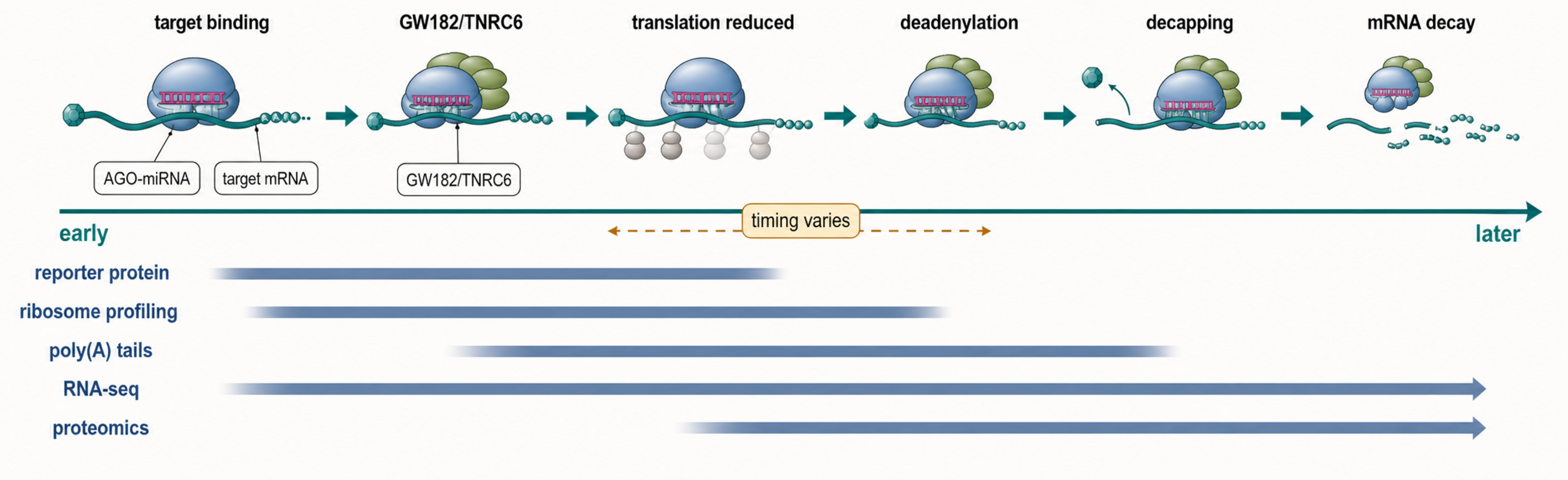
Figure 84.3. Timeline of miRNA-Mediated Repression. After Argonaute-miRNA target engagement, GW182/TNRC6 proteins recruit repression machinery. Early effects can reduce translation before large mRNA abundance changes are detected. Over time, deadenylation, decapping, and exonucleolytic decay often dominate steady-state repression. Different assays observe different stages of this timeline.
This distinction resolves an older false dichotomy. The question is not whether miRNAs repress translation or degrade mRNAs. The answer can be both, with different emphasis depending on time scale, cell state, target architecture, and measurement method. Ribosome profiling, polysome gradients, proteomics, RNA-seq, metabolic RNA labeling, poly(A)-tail assays, and reporter time courses each observe different parts of the process. A target can show reduced protein output before a large RNA-seq change, and the same target can later show mRNA decay. Czarnocka-Cieciura et al. 2024, although not miRNA-specific, is relevant to the general caution that deadenylation rates and mRNA decay rates have a complex relationship rather than a simple one-to-one mapping.
Extensive target pairing can change the outcome. When a miRNA pairs nearly perfectly with a target in slicer-competent AGO2, the target RNA can be cleaved directly between bases paired to miRNA positions 10 and 11. This slicing mechanism is central to many siRNA experiments and plant miRNA systems but is not the typical fate of animal miRNA targets. A target with unusual pairing can also trigger target-directed miRNA degradation, in which the target promotes miRNA tailing, trimming, and Argonaute unloading or decay. Li et al. 2025 is relevant to the interaction between translation and target RNA-mediated miRNA decay, but Chapter 85 provides the main treatment of that topic.
84.4. Quantitative models, stoichiometry, and competing endogenous RNA claims
miRNA regulation is quantitative rather than binary. A strong miRNA perturbation may shift hundreds of mRNAs and proteins, but an individual endogenous site often produces a modest change. This modesty is not a failure of the pathway. Many regulatory systems use small effects to tune thresholds, reduce noise, stabilize cell identity, and reinforce transcriptional decisions. For example, a differentiation-associated miRNA may slightly repress many mRNAs from an earlier cell state, making the new program more robust without requiring complete silencing of each target.
Several quantities determine the effect of a miRNA-target pair. The abundance of the miRNA-loaded Argonaute complex sets the supply of regulators. The abundance of the target site sets the demand. Binding affinity and off-rate determine occupancy. Site accessibility and site context determine whether occupancy recruits repression efficiently. The target’s synthesis rate, translation rate, poly(A)-tail dynamics, and decay rate determine how a given repression event appears in RNA and protein measurements. These quantities explain why the same seed match can matter in one cell type and be invisible in another.
Table 84.2 summarizes the difference between common evidence statements and what each one can justify. This table is especially important for quantitative claims, because the strongest causal claims require perturbing the site or miRNA at endogenous levels and measuring the right output.
Table 84.2. Quantitative Variables Controlling miRNA Output. miRNA repression depends on regulator abundance, target-site abundance, binding and dissociation, site accessibility, repression efficiency, target synthesis and decay rates, translation rate, and cellular compartment. Each variable has different experimental proxies and failure modes.
| Variable | What it changes | Measurement caveat |
|---|---|---|
| miRNA-loaded AGO abundance | Sets the effective supply of active guide complexes available for targeting. | Total mature miRNA abundance is an imperfect proxy for loaded, functional AGO-miRNA. |
| Target transcript abundance | Sets the number of candidate binding sites competing for the same guide. | RNA-seq abundance may not reflect isoform-specific 3′ UTR site exposure. |
| Effective site number | Multiple accessible sites can increase repression or create cooperative effects. | Site crowding and weak sites can produce diminishing returns rather than additivity. |
| Site affinity and off-rate | Controls occupancy lifetime and probability of effector recruitment. | Sequence models need context-aware validation; a strong seed can fail if inaccessible. |
| Site accessibility | Determines whether AGO can productively engage the RNA site. | RNA structure and RBP binding are cell-state dependent and hard to infer from sequence alone. |
| 3′ UTR isoform usage | Alternative polyadenylation can include or remove miRNA sites. | Gene-level expression hides transcript isoforms with different regulatory architectures. |
| Poly(A)-tail dynamics | Deadenylation links miRNA engagement to translation loss and mRNA decay. | Tail shortening and decay rates are coupled but not identical quantities. |
| Translation rate | Determines how quickly repression appears as protein-output change. | Ribosome profiling and proteomics observe different time windows and have distinct biases. |
| Target decay rate | Shapes whether miRNA effects are visible in RNA abundance at steady state. | RNA-seq can miss early translational repression or confound direct and secondary decay effects. |
| Competitor abundance | Governs whether a ceRNA or sponge can measurably titrate miRNA-loaded AGO. | Correlation is insufficient; competition requires favorable stoichiometry and site dependence. |
| Subcellular co-localization | Allows the miRNA, AGO, target, and competitor RNAs to encounter each other. | Bulk measurements can falsely imply interaction between molecules in different compartments or cell types. |
Stoichiometry is the central issue in ceRNA models. A ceRNA is proposed to regulate another RNA by sharing miRNA binding sites and thereby titrating the miRNA away. This mechanism can work in principle, especially in engineered systems or special endogenous cases with high competitor abundance, many effective sites, and a miRNA concentration near the sensitive part of the occupancy curve. However, a low-abundance transcript with one weak site cannot usually sequester enough miRNA-loaded Argonaute to change repression of other targets. Correlated expression between a long noncoding RNA, circular RNA, pseudogene transcript, and mRNA is therefore not sufficient evidence for ceRNA regulation.
Figure 84.4 should present ceRNA behavior as a dose-response problem. The useful visual is not a hairball network of arrows. It is a graph showing how competitor concentration, miRNA abundance, site affinity, and target abundance determine whether competition is negligible, detectable, or saturating.
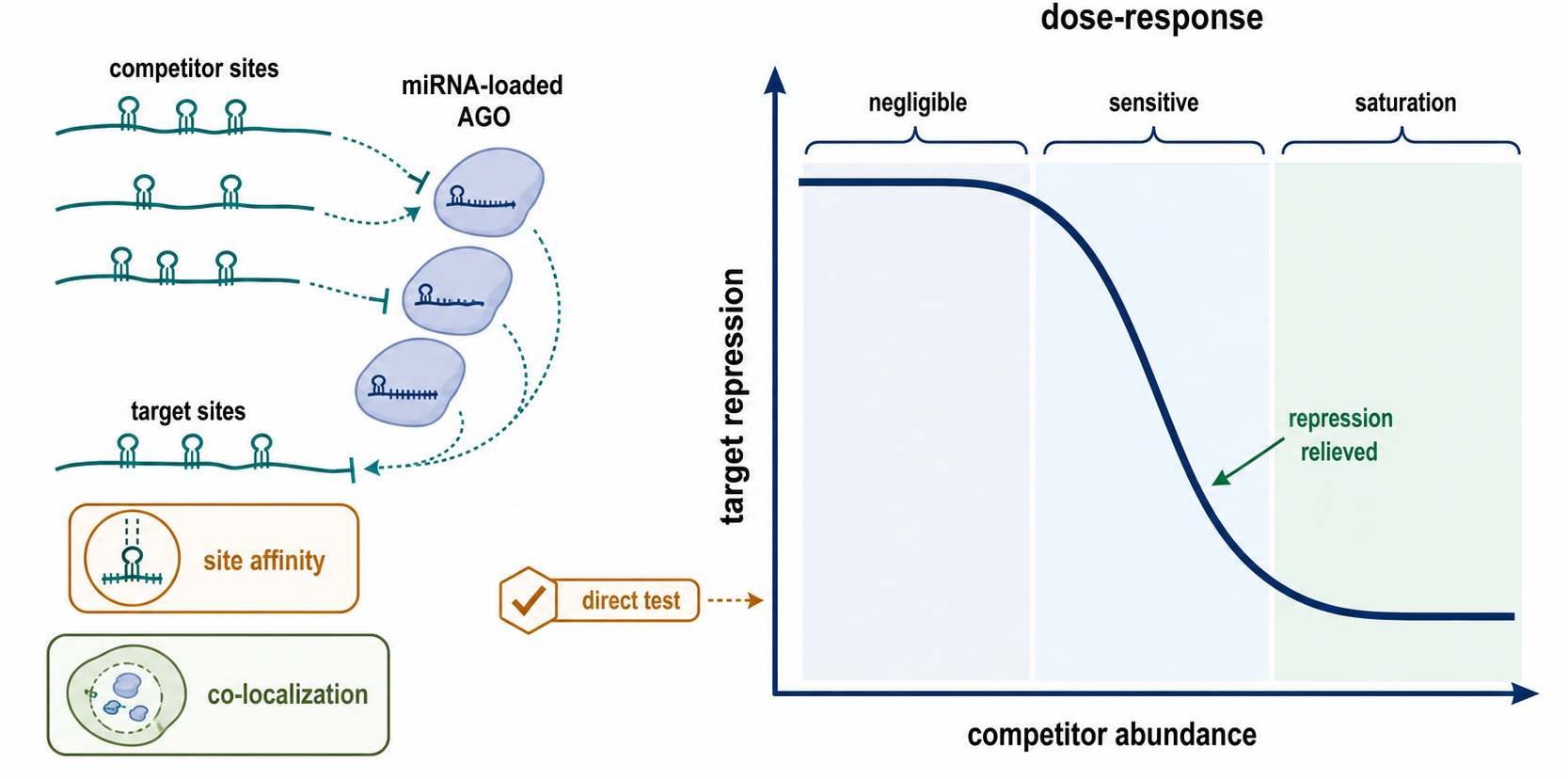
Figure 84.4. Quantitative ceRNA Dose-Response. A competing endogenous RNA can affect a second target only within a sensitive concentration range. When competitor sites are too scarce or weak, miRNA occupancy on other targets barely changes. When the competitor is abundant and effective enough, it can titrate miRNA-loaded Argonaute and reduce repression of other targets. At saturating regimes, further competitor increases may have little additional effect.
The reference list for this chapter contains Xiao et al. 2022, a disease-associated ceRNA network paper, but such studies often rely heavily on computational correlations and database predictions. The entry can be used as an example of the type of literature requiring careful validation, not as a general proof that ceRNA networks are pervasive. Robust ceRNA evidence should include absolute or carefully normalized abundance measurements, demonstration that the candidate competitor is in the same cells and compartments as the miRNA and target, perturbation of the competitor at physiological levels, mutation or deletion of the relevant sites, and measurement of the predicted target response.
84.5. Disease, biomarkers, target prediction, and validation caveats
Disease, Biomarkers, and Clinical Interpretation
miRNAs are attractive disease molecules because they sit at the intersection of regulatory networks, tissue identity, and extracellular RNA measurement. Some miRNAs contribute causally to disease by repressing tumor suppressors, oncogenes, differentiation factors, immune regulators, metabolic enzymes, or fibrosis pathways. Others change because the cell types in a tissue sample have changed. Still others appear in blood or other fluids because cells release extracellular vesicles, protein-bound RNA complexes, or injury-associated RNA fragments. These categories require different evidence.
In cancer, a miRNA can act like an oncogenic miRNA if increased expression represses genes that restrain proliferation, apoptosis resistance, invasion, or immune escape. A miRNA can act like a tumor-suppressive miRNA if loss of the miRNA releases oncogenic targets. The same miRNA can have different apparent roles in different tissues because the available targets and transcriptional context differ. Therefore, a label such as “oncogenic miRNA” is shorthand for a context, not an intrinsic property of a 22-nucleotide sequence.
Circulating miRNAs and other extracellular RNAs are studied as biomarkers because they can be measured in blood, plasma, serum, urine, cerebrospinal fluid, and other fluids. A biomarker claim must specify the sample type, collection conditions, extraction method, normalization strategy, disease comparator, and intended clinical decision. Hemolysis can release abundant red blood cell miRNAs. Platelet activation can change extracellular RNA profiles. Differences between plasma and serum can be substantial. A marker that distinguishes severe inflammatory injury from healthy controls may fail when tested against clinically similar diseases. Chang et al. 2024, although focused on circulating cell-free RNA in tuberculosis rather than miRNA alone, is relevant to the broader lesson that host-response RNA biomarkers require careful clinical comparator design.
miRNA therapeutics are treated in later chapters, but this chapter provides the regulatory logic. A mimic attempts to restore a miRNA-like activity. An inhibitor, antagomir, locked nucleic acid anti-miR, sponge, or target-site blocker attempts to reduce a miRNA’s effect. Because one miRNA can regulate many targets, both desired pathway-level effects and unwanted off-target effects are expected. Nappi 2024 is a broad noncoding RNA-targeted therapy review, and Ranasinghe et al. 2023 provides small interfering RNA therapeutic context. Schlegel et al. 2022 is especially useful for illustrating how seed-pairing destabilization can reduce siRNA off-target effects, a principle that reinforces the importance of seed-mediated interactions for miRNA-like repression.
Target Prediction and Validation Caveats
Target prediction begins with sequence, but valid target assignment cannot end there. A simple seed-match search usually produces many candidate sites. Algorithms improve ranking by adding conservation, site type, local sequence context, 3′ supplementary pairing, UTR length, site number, target abundance, and sometimes machine-learning features. Yet prediction remains a prioritization tool. It identifies hypotheses that require biological testing.
A strong validation chain asks increasingly specific questions. Is the miRNA expressed in the relevant cell type? Is the target transcript expressed in the same cells? Is there a candidate site with plausible context? Does miRNA perturbation change endogenous target mRNA or protein abundance? Does Argonaute occupancy map near the site? Does mutation or deletion of the site abolish regulation without disrupting unrelated UTR features? Does rescue of the target escape the miRNA effect? Does the phenotype depend on the specific target rather than on broad miRNA overexpression?
Box 84.1. What Does “miRNA Target” Mean?
The phrase “miRNA target” can mean a predicted seed match, an Argonaute-occupied RNA segment, a transcript whose abundance changes after miRNA perturbation, a protein whose output changes, or a gene that mediates a phenotype. These meanings require different evidence.
Reporter assays are useful but limited. A reporter containing a 3′ UTR fragment can show that a site is capable of conferring repression, but the reporter may not reproduce endogenous transcript abundance, RNA structure, alternative polyadenylation, RNA-binding protein occupancy, localization, translation rate, or decay kinetics. Conversely, failure in a reporter does not always prove that a site is irrelevant in its native mRNP context. CLIP-family methods help identify Argonaute-bound regions, but crosslinking biases, mapping ambiguity, peak-calling thresholds, and indirect occupancy must be considered. RNA-seq after miRNA overexpression or inhibition can reveal broad responses, but secondary transcriptional and stress effects appear quickly when perturbations are strong.
The most convincing target studies triangulate across methods. Endogenous site editing, physiological miRNA perturbation, protein-level measurement, and matched time courses are stronger than any single assay. For disease claims, validation also requires the right cell state and clinically relevant specimens. A miRNA-target pair that is real in a transfected cell line can still be irrelevant in the disease tissue if the miRNA, target isoform, or 3′ UTR isoform is not present at the required level.
Experimental Foundations and Evidence
The earliest evidence for miRNA function came from genetics and developmental timing, where loss or gain of small regulatory RNAs produced clear organismal phenotypes. Modern evidence is broader and more quantitative. Small-RNA sequencing identifies mature miRNA species, arm usage, isomiRs, and tissue specificity, but library preparation can bias length, end chemistry, and sequence recovery. Pri-miRNA and pre-miRNA measurements distinguish transcription from processing, but precursor detection is technically harder than mature miRNA detection because precursors are less abundant and more transient.
Biochemical assays define enzyme mechanisms. Microprocessor assays can measure pri-miRNA cleavage in extracts or purified systems. Dicer assays can map processing products. Argonaute loading assays can test duplex asymmetry and guide selection. Quantitative target-binding assays, including the logic exemplified by McGeary et al. 2019, connect site sequence to binding and repression efficacy. These biochemical approaches provide mechanistic clarity, but they simplify the crowded cellular environment.
Cellular perturbation provides function. miRNA mimics, inhibitors, genetic knockouts, Dicer or Drosha perturbation, and CRISPR editing of target sites can reveal regulatory effects. Each perturbation has a failure mode. Global Dicer or Drosha loss changes many miRNAs and non-miRNA substrates, so it is not evidence for one miRNA-target pair. Mimic overexpression can create nonphysiological guide abundance and off-target repression. Anti-miRs can vary in potency across tissues and may leave residual miRNA activity. Site editing can perturb overlapping regulatory elements in the same 3′ UTR. Good experimental design chooses the least disruptive perturbation that can answer the causal question.
High-throughput approaches define networks but require careful interpretation. Argonaute CLIP-family methods map RNA fragments crosslinked to Argonaute, identifying candidate binding regions. Transcriptomics and proteomics after miRNA perturbation identify responsive genes. Ribosome profiling can distinguish translation efficiency changes from mRNA abundance changes, although ribosome profiling itself has biases related to nuclease digestion, footprint assignment, and translation-state interpretation. Poly(A)-tail assays connect miRNA repression to deadenylation. The strongest studies integrate these data types rather than treating any one genome-wide assay as definitive.
Box 84.2. Animal miRNA Repression versus Plant miRNA Slicing and siRNA Cleavage
Animal miRNAs usually use partial pairing and recruit repression machinery, whereas many plant miRNAs and experimental siRNAs use extensive pairing and AGO-catalyzed cleavage. The same Argonaute fold supports different outcomes depending on guide-target pairing and organismal pathway context.
Biological Contexts Across Organisms and Cell States
In animals, miRNAs are deeply involved in developmental timing, cell differentiation, tissue identity, metabolism, immunity, and stress responses. Their effects often appear as canalization rather than as simple switches. A miRNA can make a transcriptome less permissive for an alternative cell fate, sharpen transitions between developmental states, or dampen noisy expression of targets that should remain low. This logic explains why some miRNA knockouts have subtle phenotypes under laboratory conditions but stronger phenotypes under stress, sensitized genetic backgrounds, or developmental transitions.
Cell type matters because the relevant quantities change. A neuronal cell, immune cell, epithelial cell, and cancer cell may express different Argonaute proteins, different RNA-binding proteins, different 3′ UTR isoforms, and different sets of abundant miRNAs. Alternative polyadenylation can shorten 3′ UTRs and remove miRNA sites, a common mechanism by which proliferating or transformed cells alter post-transcriptional regulation. RNA-binding proteins can mask miRNA sites, recruit miRNAs indirectly, or cooperate with miRNA-mediated decay. Thus, a target site is not a universal property of a gene; it is a property of a transcript isoform in a cellular context.
Viral infection and host defense add another layer. Some viruses encode miRNAs, especially DNA viruses with nuclear phases, and viral miRNAs can regulate viral or host transcripts. Host miRNAs can also bind viral RNAs, sometimes restricting viral replication and sometimes being co-opted by the virus. RNA viruses are less commonly stable sources of canonical miRNAs because producing a miRNA through Drosha or Dicer can threaten the viral genome, although exceptions and engineered systems exist. Viral and antiviral RNA biology is developed in later chapters, but miRNA target logic remains relevant whenever short Argonaute-bound RNAs engage viral or host transcripts.
Plants provide an instructive contrast. Plant miRNAs often have near-perfect complementarity to targets and can direct AGO-mediated slicing. Plant miRNA maturation is also more nuclear and Dicer-like enzyme centered than the simplified animal pathway. Yu et al. 2026 is the listed review anchor for plant miRNA maturation and function. The comparison helps prevent a common misconception: “miRNA” is a broad class name, but target recognition, processing compartments, and effector outcomes differ substantially across eukaryotes.
Technology, Computational, Clinical, and Engineering Links
miRNA biology created a toolkit for gene regulation. Synthetic miRNA target sites can be placed in expression constructs to restrict transgene expression away from cells that express a particular miRNA. For example, adding sites for a tissue-specific miRNA can reduce expression in that tissue while allowing expression elsewhere. The same principle is used in viral vectors, cell therapies, and synthetic circuits, although success depends on target-site number, site strength, transcript dose, and avoiding saturation of endogenous miRNA machinery.
Target prediction and network modeling connect this chapter to computational RNA biology. A computational model may use seed sequence, conservation, UTR context, and expression data to prioritize targets. More mechanistic models use binding energies, occupancy, transcript abundance, and repression kinetics. The best models are explicit about what they predict: physical binding, Argonaute occupancy, mRNA abundance change, protein output, or phenotype. These are related but distinct outputs. Chapter 142 treats algorithm classes and benchmarking in more detail.
Clinical measurement connects miRNAs to diagnostics. A clinically useful miRNA assay must be reproducible across collection sites, extraction batches, platforms, and patient populations. It also must outperform or complement existing markers for a defined decision. “Differentially expressed in cases versus controls” is an early discovery claim, not a validated diagnostic claim. The biomarker literature is especially vulnerable to small cohorts, batch effects, poor normalization, and inadequate comparators.
Therapeutic engineering connects miRNA and siRNA fields. Both depend on Argonaute loading and seed-mediated off-target biology, but their design goals differ. A therapeutic siRNA is usually designed for one intended target with extensive guide-target pairing and AGO2 slicing. A miRNA mimic intentionally recreates a multi-target regulatory activity, while an anti-miR blocks an endogenous multi-target regulator. Schlegel et al. 2022, although centered on GalNAc-siRNA safety, is relevant because seed-pairing destabilization shows that seed effects are not only a target-prediction abstraction; they can influence therapeutic safety.
Recent Consensus
The current consensus is that canonical animal miRNA biogenesis proceeds through a Drosha-DGCR8 nuclear step, Exportin-5-mediated export, Dicer cytoplasmic processing, and Argonaute loading, but each step is regulated and has noncanonical alternatives. Mature miRNA abundance cannot be inferred from host-gene transcription alone because processing, export, loading, and stability all matter.
The consensus on targeting is that seed pairing is the dominant recognition rule for most animal miRNA effects, especially in 3′ UTRs, but target efficacy is a quantitative function of site type, context, accessibility, supplementary pairing, and expression. McGeary et al. 2019 provides strong support for this biochemical framing. Perfect or near-perfect pairing leads to slicing in appropriate Argonaute contexts, but incomplete pairing and recruitment of repression machinery are the common animal miRNA mode.
The consensus on repression is that translational repression and mRNA decay are coupled rather than mutually exclusive. GW182/TNRC6-mediated recruitment of deadenylation and decay machinery explains many durable effects, whereas early translational repression can be visible before substantial mRNA loss. Assay timing is therefore part of the biological claim.
The consensus on quantitative regulation is that most single endogenous miRNA sites have modest effects, while networks of sites can have substantial biological consequences. ceRNA regulation is possible but requires favorable stoichiometry and direct evidence. Large ceRNA networks inferred only from expression correlations should be treated as hypotheses.
Open Questions, Controversies, Deprecated Models, and Common Misconceptions
Open questions:
- How much cell-to-cell variation in miRNA biogenesis and Argonaute loading contributes to phenotypic heterogeneity? Bulk small-RNA sequencing can miss variation in rare cell states, and single-cell small-RNA methods remain technically challenging.
- How does subcellular localization shape miRNA targeting? A miRNA and target must encounter each other in the same molecular environment, but many models still treat the cell as a well-mixed compartment.
- Which step dominates under a given time point, cell type, target, and assay: translational repression or decay? The old model that miRNAs primarily block translation without affecting mRNA abundance is too narrow for many mammalian systems. The opposite model, that mRNA decay explains everything and translation is irrelevant, is also too narrow.
Controversies:
- CeRNA biology is controversial because the mechanism is real in principle but frequently overextended. A correlation-based ceRNA network with thousands of edges does not demonstrate widespread miRNA titration. Strong ceRNA claims need measurements of abundance, occupancy, site dependence, and physiological perturbation. Circular RNAs, pseudogene transcripts, long noncoding RNAs, and mRNAs can all be candidate competitors, but the burden of proof is quantitative.
Common misconceptions:
- “A seed match proves miRNA regulation.” A seed match is a candidate site; regulation requires accessibility, Argonaute occupancy, expression context, and functional evidence.
- “A miRNA inhibitor phenotype proves every predicted target mediates the phenotype.” Inhibitor phenotypes can reflect a subset of targets, off-target effects, delivery effects, or network responses.
- “A reporter assay is endogenous target validation.” Reporter assays test a simplified sequence context; endogenous validation requires the native transcript, cell state, and regulatory machinery.
- “A circulating miRNA signature is automatically a mechanistic disease pathway.” Circulating miRNAs can be biomarkers of tissue damage, inflammation, blood-cell composition, or sample processing without causing the disease state.
- “MiRNAs are merely weak repressors.” Modest individual effects can be decisive when miRNAs act across a network or near a biological threshold.