Chapter 59. Structural and Biophysical Methods for RNA and RNPs
Scope Note
This chapter explains how structural and biophysical methods turn RNA and ribonucleoprotein (RNP) complexes into interpretable molecular models. It covers X-ray crystallography, cryogenic electron microscopy, nuclear magnetic resonance spectroscopy, small-angle scattering, native electrospray ionization mass spectrometry of intact RNPs, ion mobility, single-molecule fluorescence and force methods, chemical probing, crosslinking-derived restraints, integrative modeling, sample preparation, heterogeneity analysis, and validation. The emphasis is not on memorizing method names, but on matching each method to the physical question it can answer, recognizing the proxy each method actually measures, and avoiding overinterpretation of static structures, ensemble averages, gas-phase ions, low-resolution restraints, or artifact-prone samples. This chapter owns native mass spectrometry as a structural and biophysical method for intact RNA-protein and RNA-ligand assemblies: stoichiometry, oligomeric state, occupancy distributions, activation behavior, and mobility-derived collision cross sections. Sequence- and composition-resolved RNA mass spectrometry, including bottom-up and top-down analysis, belongs to Chapter 132; peptide-centric and protein-capture proteomics belongs to Chapter 138. Transcriptome-scale RNA structure probing, computational secondary-structure prediction, comparative/covariance modeling, and tertiary simulation are treated in more detail in Chapters 60 to 64 and Chapter 131.
Executive Summary
Structural and biophysical methods ask related but distinct questions about RNA and ribonucleoprotein complexes. X-ray crystallography and cryogenic electron microscopy can reveal three-dimensional architecture at high resolution, but they usually require purified, compositionally defined material and careful interpretation of static or class-averaged states. Nuclear magnetic resonance spectroscopy, small-angle X-ray scattering, and other solution methods observe molecules in solution and can measure dynamics, ensembles, and interactions that are partly hidden in a crystal or frozen grid. Single-molecule fluorescence and force methods watch individual molecules fluctuate, fold, bind, translocate, or remodel. Chemical probing and crosslinking convert structural features into biochemical constraints. Integrative modeling combines incomplete observations into structural hypotheses that must remain accountable to the underlying data.
RNA makes structural biology difficult for reasons that are also biologically important. RNA is a charged polymer with many near-degenerate conformations, extensive metal-ion and protein dependence, and strong sensitivity to sequence context, termini, modifications, folding path, and buffer conditions. Many functional RNAs are not a single rigid structure. A riboswitch aptamer, RNase P RNA, viral genome element, spliceosomal RNA, or messenger RNA regulatory element may populate several interconverting conformations. RNPs add additional heterogeneity because protein occupancy, RNA processing state, nucleotide state, assembly intermediate, ligand binding, and post-translational modification can change the observed structure. Modern studies therefore increasingly describe conformational ensembles rather than only one best model.
No method measures “RNA structure” in the abstract. Every method measures a physical proxy. Crystallography measures diffraction from an ordered crystal. Cryo-EM measures electron scattering from vitrified particles and reconstructs class-averaged density. NMR measures magnetic environments and distance- or angle-sensitive restraints. SAXS measures global scattering from all species in solution. Native mass spectrometry measures mass-to-charge distributions of desolvated ions that survived transfer from a deliberately prepared solution; ion mobility additionally measures gas-phase transport and can support a condition-specific collision cross section. smFRET measures distance-sensitive dye behavior between labeled positions. Optical tweezers measure extension under force. Chemical probing measures reactivity, accessibility, or covalent modification. Crosslinking measures spatial proximity under a defined chemistry. A sound interpretation states the proxy, the model assumptions, the sample state, the validation criteria, and what alternative explanations remain. The practical goal for RNA and RNP structural biology is not to choose a universally superior method. The goal is to match the method to the biological question. Atomic detail may require crystallography, cryo-EM, or NMR. Conformational exchange may require NMR, smFRET, stopped-flow, hydrogen-deuterium exchange where applicable to proteins, or ensemble-aware cryo-EM classification. Large RNP architecture may require cryo-EM, crosslinking, native mass spectrometry, and integrative modeling. Native mass spectrometry can distinguish intact stoichiometries and ligand occupancies that a class-averaged structure may conceal, but those spectra require solution-phase and gas-phase controls. Transcriptome-scale structure-function hypotheses may begin with chemical probing and sequencing, then return to targeted biochemical or structural assays. Confidence grows when independent methods agree on the same mechanistic model and when the modeled state explains biochemical function.
Concept Inventory
- Structural model: a representation of molecular geometry. For RNA, a model may include base identities, base-pairing, helical axes, tertiary contacts, metal ions, ligands, proteins, and waters. A model is not the molecule itself. It is an inference from data, restraints, stereochemistry, prior knowledge, and refinement choices.
- Resolution: the scale at which structural features can be distinguished. In crystallography and cryo-EM, high resolution can support base identity, backbone conformation, side-chain placement, ligand orientation, and ion assignment, but resolution is not uniform across a model. Flexible RNA loops, peripheral helices, protein tails, and transient contacts can be worse resolved than the stable core.
- Conformational ensemble: a population of structures sampled by the same molecule or complex. The ensemble may reflect thermal fluctuation, alternative folded states, ligand-bound and ligand-free states, assembly intermediates, or mixtures produced during purification. For RNA, ensemble thinking is often essential because secondary and tertiary contacts can rearrange without covalent change.
- Restraint: an experimental observation translated into a geometric or energetic condition for modeling. A NOE in NMR, a crosslink, a FRET distance range, a SAXS envelope, a SHAPE reactivity pattern, or a cryo-EM density feature can all act as restraints, but each has different uncertainty and different artifact modes.
- Validation: testing whether a structural model is compatible with the data and with independent evidence. Validation includes map-model agreement, stereochemical quality, cross-validation, reproducibility, sample homogeneity, biochemical activity, mutational tests, ligand or protein dependence, and comparison with orthogonal methods.
- Native mass spectrometry: electrospray-ionization mass spectrometry configured to transfer intact noncovalent assemblies from solution and resolve their mass-to-charge distributions. In this chapter, “native” describes sample preparation and transfer intended to preserve informative associations; it does not certify that every detected gas-phase conformation is identical to the solution structure.
- Charge-state deconvolution: inference of neutral molecular masses and component distributions from families of peaks corresponding to different ion charge states. Deconvolution is model-dependent when charge envelopes overlap, adducts broaden peaks, or heterogeneous complexes have closely spaced masses.
- Collision cross section (CCS): an ion-mobility-derived measure related to how an ion collides with a specified bath gas under defined instrumental conditions. CCS can test relative compaction or expansion across controlled states, but RNA ions can compact after desolvation, so agreement with a calculated solution model is supportive rather than definitive.
What to Know Before Reading This Chapter
Readers should know that RNA has primary, secondary, tertiary, and quaternary levels of organization. A primary sequence folds into stems, loops, bulges, junctions, pseudoknots, and long-range contacts. Proteins, metabolites, ions, nucleotide cofactors, and other RNAs can stabilize or remodel these structures. Chapters 3, 43, 56, and 57 introduce the needed folding, RBP-recognition, and RNP-machine background.
This chapter uses four running examples: a riboswitch aptamer, RNase P RNA, a viral polymerase-RNA complex, and a small engineered or therapeutic RNA. These examples connect ligand recognition, ensemble behavior, large RNP architecture, and design-relevant structure-function tradeoffs. Native-mass-spectrometry examples include a neomycin-sensing riboswitch aptamer with discrete ligand occupancies, coronavirus nucleocapsid-RNA assemblies, SARS-CoV-2 nsp10/nsp16 complexes with RNA substrate or product, and heterogeneous ribosomal particles.
Do not treat a high-resolution structure as automatically more biological than a lower-resolution measurement. A crystal structure of a truncated RNA may be precise but represent one stabilized state. A lower-resolution in-cell probing experiment may be closer to the native environment but less specific about atomic contacts. The useful question is whether the method observes the state relevant to the mechanism being claimed.
59.1. X-ray crystallography and cryo-EM for RNA/RNPs
X-ray crystallography determines structure from diffraction by an ordered crystal. The sample must form a periodic lattice, and each repeating unit must be similar enough to produce interpretable diffraction. For RNA, this requirement is often the central obstacle. RNA surfaces are highly charged, hydrated, and conformationally flexible. Many RNAs have few large hydrophobic patches, limited natural crystal contacts, and mobile termini. Crystallization may require construct engineering, removal of flexible regions, stabilizing mutations, ligand addition, protein chaperones, antibody fragments, crystallization modules, or careful ion conditions. Recent reviews emphasize that RNA crystallography remains powerful, but that success depends heavily on sample design and stabilization strategy.
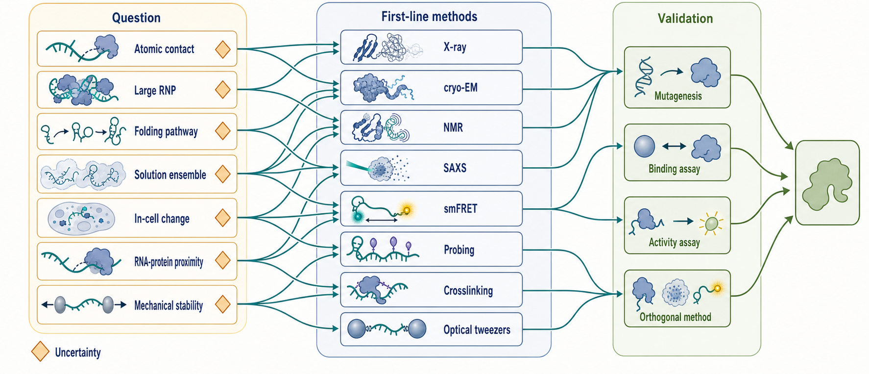
Figure 59.1. Method-Choice Map for RNA and RNP Structural Questions. No method measures RNA structure in the abstract. Each method measures a physical proxy under defined sample conditions, and the strongest mechanistic claims align the method, the structural state being observed, and the functional readout being tested. This figure maps biological question categories—including atomic contact, time-resolved crystalline change, large RNP architecture, folding pathway, solution ensemble, in-cell structural change, RNA-protein proximity, computational docking hypothesis, and mechanical stability—to recommended first-line methods and to the validation layer that must accompany each measurement.
Table 59.1. Comparison of Major Structural and Biophysical Methods for RNA and RNPs. Each method reports a different physical proxy for molecular geometry and is best paired with complementary validation approaches.
| Method | Primary readout | Typical sample state | Strengths | Common artifacts | Best validation partners |
|---|---|---|---|---|---|
| X-ray crystallography, including SFX | Atomic coordinates from electron-density map; serial diffraction patterns from many microcrystals | Ordered crystal or suspension of RNA/RNP nano- or microcrystals | High local detail; base-triple, ion, and ligand geometry; SFX can reduce radiation-damage bias and support room-temperature or triggered time series | Lattice-selected state; stabilized non-native contacts; flexible regions absent; microcrystal size/density heterogeneity, precipitate, delivery, or trigger artifacts | Mutagenesis; ligand-binding assay; NMR or SAXS in solution; independent reaction kinetics for time-resolved SFX |
| Cryo-EM | 3D density map reconstructed from particle images | Vitrified particles in aqueous buffer | Multiple conformational states by classification; large RNP architecture; no crystal required | Preferred orientation; blurred flexible regions; RNA base ambiguity at moderate resolution | Mutagenesis; biochemical activity; X-ray for domain detail |
| NMR spectroscopy | Chemical shifts, NOEs, RDCs, relaxation parameters | Isotope-labeled RNA or RNP in solution | Dynamics, exchange rates, local interactions, solution conditions | Spectral overlap with increasing size; exchange broadening; concentration dependence | SAXS; biochemical binding assay; mutagenesis |
| SAXS | Scattering profile; global size and shape | Purified RNA or RNP in solution | Solution-based; ensemble modeling; ligand- or folding-induced compaction | Aggregation; interparticle interference; mixture averaging | NMR; SEC-MALS; analytical ultracentrifugation |
| Native ESI-MS and IM-MS | Intact-ion mass-to-charge distributions, deconvolved masses, activation products, arrival times, and derived CCS | Purified RNA or RNP transferred from a volatile electrolyte into gas phase | Resolves stoichiometry, oligomeric states, ligand occupancy, and compositional heterogeneity; compares controlled gas-phase conformers | Adduction; overlapping charge states; droplet-induced association; in-source dissociation; response bias; gas-phase compaction | SEC-MALS or analytical ultracentrifugation; solution binding/activity; cryo-EM, NMR, or SAXS |
| smFRET | FRET efficiency trajectories per molecule | Dye-labeled RNA or RNP, surface-immobilized or freely diffusing | Single-molecule state separation; kinetics; cotranscriptional folding | Dye stacking; photobleaching; surface perturbation; linker distance uncertainty | Biochemical activity; cryo-EM or X-ray for state identity |
| Optical tweezers | Force-extension curves and unfolding intermediates | RNA or RNP tethered between beads via handles | Energy landscape; mechanical stability; RNA motor kinetics | Handle effects; off-pathway force intermediates; low throughput | smFRET; biochemical reconstitution; cryo-EM |
| Chemical probing (SHAPE, DMS) | Nucleotide reactivity or modification profile | RNA in vitro, in lysate, or in cell | Transcriptome-scale; in-cell context; detects condition-dependent structural changes | Reagent-access bias; protein-occupancy confounds; normalization assumptions | Mutagenesis; biochemical activity; NMR or cryo-EM |
| Crosslinking (RNA-RNA, RNA-protein, XL-MS) | Proximity restraints from covalent crosslinks | Purified complex or in-cell | Constrains large flexible RNPs; sparse but informative distance upper bounds | Chemistry- and geometry-dependent yield; transient-contact bias; non-specific crosslinks | Cryo-EM; mutagenesis; biochemical binding assay |
| Nanopore single-molecule sequencing | Ionic current trace; sequence and modification per molecule | Individual RNA molecules threaded through nanopore | Long reads; molecule-to-molecule heterogeneity; modification and isoform detection | High error rate; indirect structure readout; current signal interpretation | Mass spectrometry for modifications; targeted chemical probing |
| Computational tertiary modeling and protein-RNA docking | Ranked structures, poses, scores, or construct hypotheses | Input sequences, coordinate models, templates, and optional restraints | Rapid hypothesis generation; prioritizes contacts, truncations, and experiments | Template dependence; incomplete conformational sampling; scoring and rank failures; false precision from one best model | Withheld experimental restraints; mutagenesis; binding and activity assays; targeted structure determination |
| Integrative modeling | Structural ensemble satisfying multiple data types | Combined inputs from multiple experimental methods | Accommodates incomplete data; quantifies uncertainty; represents large RNPs | Overfitting; undetected data inconsistency; underdetermination from sparse restraints | Withheld-data cross-validation; mutagenesis; biochemical activity |
The strength of crystallography is atomic interpretability when diffraction and phasing are adequate. A good RNA crystal structure can reveal base triples, ribose puckers, metal-binding sites, local hydration, ligand contacts, and protein-RNA recognition geometry. Such detail is essential for mechanistic enzymology, riboswitch ligand recognition, RNA-targeted drug discovery, and validation of computational predictions. The limitation is that the crystal lattice can select one state from a broader solution ensemble. Crystal contacts can distort peripheral helices, stabilize non-native contacts, or hide dynamic transitions. A structure determined from a minimal construct can be exactly right for that construct and still incomplete for the full biological RNA.
Serial femtosecond crystallography (SFX) extends diffraction experiments to streams of nano- or microcrystals interrogated with ultrashort pulses from an X-ray free-electron laser (XFEL). The pulse can record diffraction before radiation damage develops fully, permitting room-temperature and, with a synchronized trigger, time-resolved studies. For RNA, however, SFX shifts rather than eliminates the sample problem. An adenine-riboswitch protocol shows that crystal identity, size distribution, number density, precipitate load, mother-liquor compatibility, and delivery stability must all be controlled because nonuniform crystals or granular precipitate lower diffraction hit rates and can obstruct sample injection. SFX still observes a lattice-selected crystalline population, and a sequence of time points becomes a mechanistic trajectory only when reaction initiation and kinetics are independently established.
Cryogenic electron microscopy avoids crystallization by imaging vitrified particles. In single-particle cryo-EM, many particle images are computationally aligned and averaged to reconstruct density. Cryo-EM is especially effective for large RNPs such as ribosomes, spliceosomal complexes, viral polymerases, packaging assemblies, and RNA-processing machines. For RNA and RNPs, cryo-EM can capture multiple compositional or conformational states through classification, which is one reason it has transformed large-complex structural biology. Reviews of negative-sense RNA virus polymerases illustrate how cryo-EM structures can organize mechanistic understanding of RNA synthesis, template engagement, and protein-RNA architecture.
RNA-only cryo-EM remains harder than protein-rich cryo-EM. RNA has lower chemical diversity, strong preferred orientations in some grids, radiation sensitivity, and flexible regions that blur during averaging. Small RNAs often lack enough mass or distinctive shape for robust alignment. Sample optimization can include stabilizing proteins, Fab fragments, aptamer-binding modules, ligands, crosslinking with caution, grid chemistry changes, buffer screening, and construct redesign. A recent protocol-focused review specifically addresses RNA sample optimization for cryo-EM and underscores that grid behavior, particle distribution, and conformational stability are method-defining variables rather than afterthoughts.
The output of cryo-EM is a density map and a model fitted to that map. The model is strongest where density is continuous and chemically interpretable. RNA base identity can be difficult at moderate resolution because bases have similar shapes and density can be averaged across flexibility. Magnesium ions, waters, modified nucleotides, and transient ligands require especially careful assignment. Local resolution, map sharpening, particle-class occupancy, model-map cross-validation, and biochemical activity of the imaged sample should be reported. A plausible model placed into weak density is a hypothesis, not a demonstrated contact.
Crystallography and cryo-EM are complementary. Crystallography can provide high local detail for stable domains, small RNAs, and ligand pockets. Cryo-EM can place these domains into larger assemblies and identify multiple states. Hybrid strategies are common: crystal or NMR structures of domains may be fitted into cryo-EM maps; cryo-EM maps may suggest constructs for crystallography; biochemical mutants may test contacts inferred from either method. For RNP machines, the most persuasive story often combines structure with assembly assays, kinetics, mutagenesis, and substrate or product trapping.
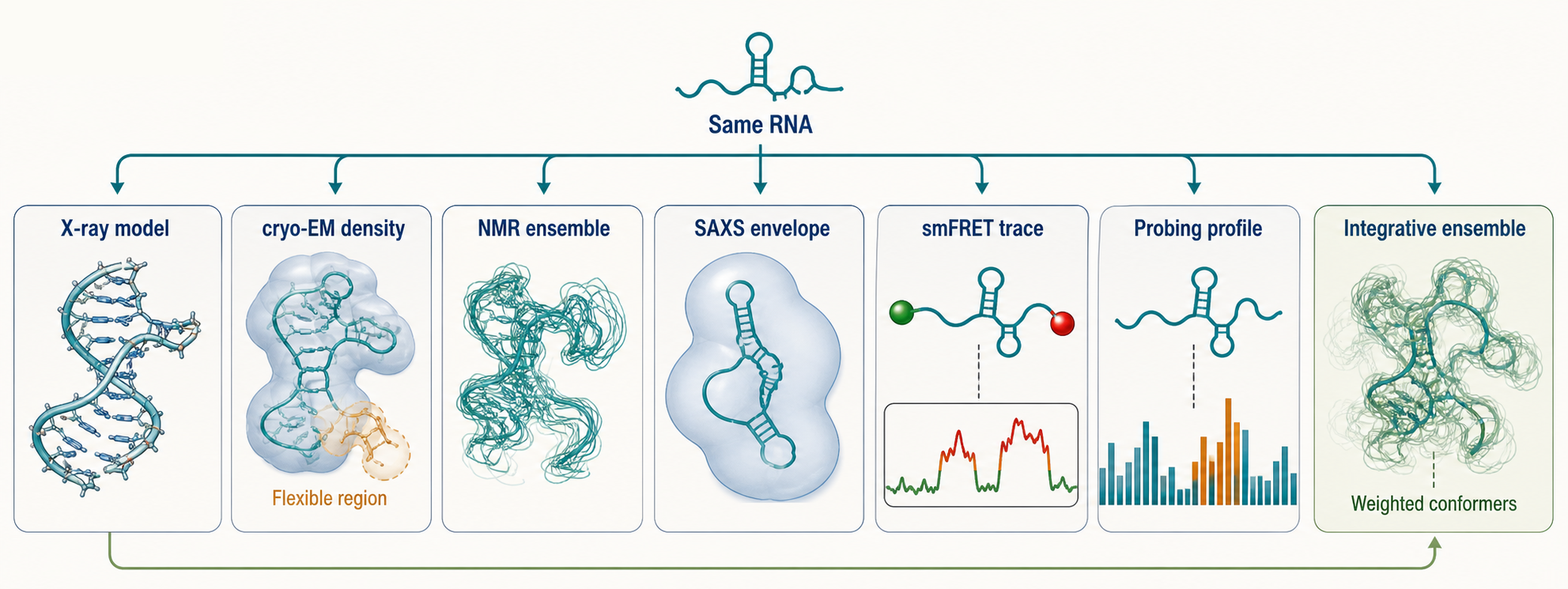
Figure 59.2. One RNA Viewed Through Multiple Measurement Proxies. The same RNA molecule can appear as a single stabilized atomic model in a crystal structure, a class-averaged density with blurred flexible regions in cryo-EM, a set of solution conformers in an NMR ensemble, a low-resolution global envelope in SAXS, a state-switching trajectory in smFRET, a nucleotide-reactivity bar chart in a chemical probing profile, and a weighted multi-conformer model in integrative analysis. Structural disagreement across these representations is sometimes methodological complementarity rather than inconsistency, because each assay measures a different physical proxy of molecular geometry.
59.2. NMR, SAXS, and solution methods
Nuclear magnetic resonance spectroscopy observes nuclei in a magnetic field and reports on their local chemical environments and interactions. In RNA NMR, commonly observed nuclei include protons, carbons, nitrogens, and phosphorus atoms, often with isotope labeling. NMR can determine local structure through nuclear Overhauser effects, scalar couplings, residual dipolar couplings, relaxation measurements, chemical-shift perturbations, and exchange experiments. The central pedagogical point is that NMR is not merely a smaller version of crystallography. NMR can measure dynamics, weak interactions, exchange between states, and local changes during ligand or protein binding.
NMR is particularly useful for RNA hairpins, aptamer domains, internal loops, riboswitch elements, protein-binding motifs, and dynamic regions that resist crystallization. Chemical-shift perturbation can identify nucleotides affected by protein or ligand binding. Relaxation dispersion can reveal microsecond-to-millisecond exchange. Paramagnetic probes can supply long-range distance information. Selective labeling can simplify spectra and make larger RNAs more tractable. The method’s limitations are equally important: spectral overlap increases with RNA size, conformational exchange can broaden peaks beyond detection, and sample concentration or buffer conditions may differ from cellular conditions.
Small-angle X-ray scattering measures how X-rays scatter at low angles from molecules in solution. SAXS does not usually give atomic coordinates by itself. Instead it reports global size, shape, compactness, flexibility, and population-averaged distance distributions. For RNA, SAXS is valuable because it can compare folded and unfolded states, ligand-bound and ligand-free conformations, compact and extended ensembles, and protein-bound RNP architectures. The method is solution-based, relatively tolerant of size, and compatible with ensemble modeling, but it is also sensitive to aggregation, interparticle effects, concentration dependence, and mixtures.
NMR and SAXS together can be more powerful than either alone. NMR provides local restraints and dynamics; SAXS constrains global shape and compaction. A study of a UCAAUC RNA oligonucleotide combined molecular dynamics simulations, SAXS, and NMR to analyze conformational heterogeneity, illustrating how a small RNA can require ensemble interpretation rather than a single conformation. Recent solution studies of 7SK RNP and 7SL signal recognition particle RNAs also show how solution characterization can address biologically important RNP RNAs whose full complexes are challenging. RNase P RNA work similarly highlights broad conformational space in solution.
Other solution and native-ion methods contribute to the same evidence ladder. Analytical ultracentrifugation, size-exclusion chromatography coupled to multiangle light scattering (SEC-MALS), native mass spectrometry, electrophoretic mobility shift assays, isothermal titration calorimetry, stopped-flow fluorescence, and absorbance melting do not all produce atomic structures, but they test stoichiometry, binding, oligomeric state, folding stability, and kinetics. These measurements are often the difference between a beautiful structural model and a biologically credible mechanism. If a modeled RNP has a 1:1 stoichiometry, the sample should show that stoichiometry under the conditions used for structure determination. If a ligand is proposed to lock a conformation, binding and folding assays should support that claim. Chapter 124 owns the detailed concentration regimes, active-fraction tests, equilibrium and kinetic models, immobilization artifacts, global fitting, and parameter identifiability behind quantitative binding measurements; this chapter uses their results to validate structural states.
Native electrospray ionization mass spectrometry, usually abbreviated native ESI-MS or native MS, has a more specific structural role than the phrase “mass spectrometry” alone suggests. A purified RNA, protein, RNA-protein complex, or RNA-ligand assembly is introduced from a volatile aqueous electrolyte through an electrospray emitter. Droplet evaporation, fission, and desolvation produce multiply charged intact ions whose measured mass-to-charge ratios can be converted into neutral masses when charge states are assigned. Comparing those experimental masses with the masses expected from the RNA, proteins, cofactors, ions, and ligands identifies which intact assemblies reached the detector. The direct readout is therefore a distribution of gas-phase ions. The useful structural inference is often discrete composition: one RNA with one protein dimer, an RNA-bound versus RNA-free enzyme complex, several ligand occupancies on one aptamer, or a mixture of assembly intermediates.
This intact-assembly view can separate species that an ensemble average obscures. A native spectrum may contain free RNA, free protein, a 1:1 RNP, higher oligomers, and complexes carrying zero, one, or several ligands. Peak areas can show how these populations change across a controlled titration, mutation, ionic condition, or reaction time. The result is particularly informative when component masses differ enough to resolve stoichiometries and when each assignment is reproduced across charge states. For a 40-nucleotide neomycin-sensing riboswitch aptamer, native spectra resolved discrete 1:1, 1:2, and 1:3 RNA:neomycin populations; tandem activation localized binding behavior, while NMR supplied solution-state information and helped distinguish specific from additional nonspecific binding. The intact occupancy distribution belongs to this chapter’s structural evidence; sequence-localizing top-down fragmentation belongs to Chapter 132.
RNP examples demonstrate the same logic at larger scales. Native and hybrid mass spectrometry of ribosomal particles resolved heterogeneous subunit compositions and associations of viral internal ribosome entry site RNAs with ribosomal particles, complementing structural models of the ribosome rather than replacing them. Native MS of SARS-CoV-2 nsp10/nsp16 distinguished the allosteric heterodimer and its interactions with RNA substrate or product, illustrating how mass-resolved states can connect enzyme assembly to a reaction cycle. Native MS of coronavirus nucleocapsid proteins with RNA resolved monomeric and dimeric protein-RNA stoichiometries and used controlled perturbations to compare the persistence of protein-protein and protein-RNA contacts. These cases are concrete evidence that native MS can reveal which RNP populations coexist. They do not, by themselves, supply atomic interfaces or prove that detector abundances equal intracellular abundances.
Charge-state distributions contain information but require discipline. One neutral complex normally produces a family of peaks because otherwise similar ions carry different numbers of charges. Charge-state deconvolution groups those peaks and estimates neutral mass. Closely spaced proteoforms, variable RNA lengths, salt adduction, incomplete desolvation, ligand exchange, and overlapping oligomers can create interleaved envelopes. Algorithms can deconvolve such spectra, but a smooth reconstructed mass distribution is not independent evidence that the component assignments are correct. Analysts should inspect the underlying charge-state series, mass error, peak width, adduct pattern, reproducibility, and expected component masses. Heterogeneous complexes are scientifically valuable, yet they are also where deconvolution is most underdetermined.
Collision or activation series add a limited form of architectural evidence. After selecting an intact ion population, the instrument can increase collisional energy or use another activation method and record which ligands, RNAs, protein subunits, or subcomplexes are lost. A reproducible dissociation pathway may identify a labile peripheral component, distinguish nested assembly intermediates, or support a subunit-connectivity model. Comparing activation thresholds across mutants or liganded states can show that one gas-phase ion is more resistant to dissociation than another. The correct inference is about behavior along the chosen gas-phase activation coordinate. Collision energy is not temperature, a gas-phase dissociation threshold is not a solution dissociation constant, and asymmetric charge partitioning or unfolding can make the ejected species reflect ion physics as much as native interface strength. Activation series should therefore be interpreted with intact stoichiometry, solution binding, activity, and structural data, not translated directly into solution free energies.
Ion mobility mass spectrometry (IM-MS) separates ions according to their transport through a bath gas under an electric field. The primary measurement is an arrival time or mobility; a collision cross section (CCS) is derived through a physical model or calibration that depends on ion charge, bath gas, temperature, pressure, electric-field regime, and instrument method. Under matched conditions, IM-MS can test whether a ligand, RNA, protein, or activation step produces a more compact, expanded, or heterogeneous gas-phase population. A measured CCS can also be compared with CCS values calculated from structural candidates, allowing grossly incompatible architectures to be rejected. Reporting should preserve the distinction between measured mobility, derived CCS, and the structural model used for comparison.
The solution-to-gas transition is the major boundary condition. Noncovalent contacts can survive electrospray, but solvent removal changes electrostatic screening, hydrogen-bond competition, counterion organization, and the energetic cost of exposed charge. RNA is especially sensitive because its phosphate backbone is highly charged. Ion-mobility experiments on RNA hairpins and kissing complexes showed substantial gas-phase compaction, and calculated structures did not fully explain the measured CCS values. A compact CCS is therefore not proof that the RNA had the same compact tertiary conformation in solution. Conversely, dissociation in the source does not prove that an assembly was absent from solution. Source conditions, ion polarity, charge reduction, collision voltages, residence time, and instrument pressure can determine which species survive. Reduced-pressure ionization can improve transmission of some assemblies and tolerate conditions that are otherwise difficult, but it changes the measurement pathway and still requires condition-specific controls.
Binding titrations require another caution. A series of native spectra can support stoichiometry, competitive binding, or cooperative population changes when free and bound species are resolved and controls demonstrate similar detection behavior. However, observed ion abundance can be altered by solution equilibration, nonspecific association during droplet concentration, different ionization efficiencies, charge-state-dependent transmission, in-source dissociation, detector response, and depletion of one binding partner. An apparent bound fraction is not automatically an equilibrium fraction. Estimating a dissociation constant from native MS requires an explicit binding model, known active concentrations, equilibrium controls, response-factor assessment, and evidence that transfer does not selectively lose or create complexes. Orthogonal calorimetry, fluorescence, analytical ultracentrifugation, or other solution assays should establish the thermodynamic claim.
These boundaries define ownership across the book. Chapter 59 treats intact native-MS readouts as structural and biophysical evidence: RNP stoichiometry, oligomeric states, ligand and RNA occupancy, collision/activation behavior, and IM-MS/CCS comparisons. Chapter 132 treats RNA mass analysis that asks which nucleotide, modification, sequence, terminus, fragment, or covalent composition is present, including bottom-up and top-down workflows. Chapter 138 treats peptide-centric identification and quantification of proteins recovered by RNA-centered capture or interaction-proteomics experiments. A single study can cross these boundaries—for example, intact spectra can define a riboswitch’s ligand occupancy while top-down fragments localize binding—but each inference should be attributed to the method layer that actually supports it.
Table 59.2. What Each Data Type Can and Cannot Prove. Each observation type supports a conservative inference and is commonly overinterpreted in a specific way; an orthogonal test helps distinguish the supportable from the unsupported claim.
| Observation | Conservative inference | Overinterpretation to avoid | Orthogonal test |
|---|---|---|---|
| High SHAPE reactivity | Ribose is locally flexible or nucleotide is unpaired | Specific tertiary contact is absent | NMR or mutagenesis to test local pairing |
| Low DMS reactivity | Watson-Crick face is not solvent-accessible | Nucleotide is base-paired (could be protein-protected or modified) | RBP footprinting; chemical modification mapping |
| RNA-protein crosslink | RNA and protein are in proximity under crosslinking conditions | Direct stable contact or defined binding site | Binding affinity assay; contact-residue mutagenesis |
| FRET state change | Distance between labeled positions changes | Full tertiary structure of both states is known | Cryo-EM or X-ray to characterize each state |
| Cryo-EM class | Distinct particle population exists in the sample | Class represents a productive biochemical intermediate | Activity assay of trapped or enriched class |
| SAXS compaction | Global shape becomes more compact under tested condition | Specific tertiary contacts are formed | NMR or chemical probing to identify contacts |
| NMR chemical-shift perturbation | Local magnetic environment changes at shifted nucleotide | Shifted nucleotide is a direct binding-contact site | FRET; affinity measurement; contact mutagenesis |
| Native-MS mass matching an RNA plus one protein dimer | An intact ion with that nominal stoichiometry survived transfer and detection | The same stoichiometry is dominant in cells or has a unique atomic architecture | SEC-MALS or analytical ultracentrifugation; cryo-EM; activity assay |
| Native-MS ligand-occupancy series | Resolved ions carry different numbers of ligands under the tested preparation and transfer conditions | Peak areas are unbiased equilibrium populations or directly yield a dissociation constant | Solution titration by calorimetry or fluorescence; response-factor and concentration controls |
| Higher collision energy required for subunit loss | Selected gas-phase ions differ in resistance along the tested activation pathway | The solution interface has a proportionally stronger binding free energy | Solution affinity/kinetics; mutagenesis; structural interface analysis |
| Different ion-mobility CCS values | Selected gas-phase ions differ in transport cross section under reported conditions | Each CCS uniquely identifies a solution conformation | NMR, SAXS, smFRET, or cryo-EM; replicate CCS under controlled charge states |
| Force-extension intermediate | A mechanically stable state exists under applied force | Same intermediate is populated at zero force | smFRET at zero force; biochemical characterization |
| Predicted tertiary model or protein-RNA docking pose | A plausible fold or interface hypothesis exists under the input model, sampling, and score | The top-ranked structure is correct, unique, or independent of its template | Chemical probing; interface mutagenesis; binding/activity assay; targeted structure determination |
Solution methods also reveal why RNA structure is conditional. Magnesium concentration, monovalent salt, pH, temperature, ligand concentration, crowding agents, RNA termini, transcription method, purification history, and modifications can shift the ensemble. A ribozyme folded in magnesium-rich buffer may be mostly unfolded under another condition. A viral RNA motif may require protein binding to adopt the state seen in an RNP. A synthetic RNA may behave differently after chemical synthesis than after transcription if termini, modifications, or impurities differ. Therefore method sections and figure captions should state conditions precisely.
59.3. Single-molecule FRET, optical tweezers, and force methods
Single-molecule methods ask what individual molecules do rather than what an ensemble average implies. Single-molecule fluorescence resonance energy transfer, usually called smFRET, labels two positions with donor and acceptor dyes and measures distance-sensitive energy transfer. For RNA, smFRET can monitor folding, docking, strand exchange, riboswitch switching, RNA-RNA interaction, protein-induced remodeling, and co-transcriptional folding. Method papers and reviews describe smFRET applications to RNA structural rearrangements, RNA-RNA interactions, and co-transcriptional folding.
The key value of smFRET is that it can separate states averaged together in bulk. A bulk signal might suggest a partially folded RNA, while single-molecule traces reveal two-state switching between folded and unfolded conformations. Dwell times can estimate kinetic rates, and hidden Markov models or related analyses can infer state transitions. Co-transcriptional experiments can ask whether a nascent RNA folds before downstream sequence emerges. For riboswitches, this matters because gene regulation often depends on folding path and timing, not only final thermodynamic stability.
smFRET has its own artifacts. Dyes are not passive mathematical points. They can stack with bases, change local folding, respond to environment, blink, bleach, or rotate anisotropically. Linkers introduce distance uncertainty. Surface immobilization can perturb RNA or protein behavior. Oxygen scavengers and triplet quenchers can alter chemistry. A single FRET efficiency is not a direct distance without calibration and assumptions. The strongest smFRET studies therefore test labeling positions, preserve activity, compare surface and freely diffusing formats where possible, use controls for dye behavior, and connect FRET states to independent structural or biochemical evidence.
Optical tweezers and related force methods hold molecules under controlled mechanical load. In a typical RNA experiment, an RNA or RNP is tethered between beads or surfaces, and force-extension behavior reports folding, unfolding, refolding, or motor activity. Force can destabilize helices, reveal intermediates, measure energy landscapes, and test how proteins stabilize or remodel RNA. Optical tweezers have also been applied to viral systems, where genome packaging, uncoating, polymerase movement, or protein-nucleic-acid interactions can be studied under mechanical constraints. The same logic applies broadly to RNP machines: force can expose mechanochemical steps that are invisible in static structures.
Force methods are powerful because they perturb the molecule in a defined way. That perturbation is also their limitation. A force applied through engineered handles may not match forces experienced in a cell. The tether geometry selects a mechanical coordinate, usually end-to-end extension, while biologically relevant motions may occur elsewhere. Handles, linkers, surfaces, and pulling rates can alter the observed pathway. A force-unfolding intermediate may be real under force but not populated at zero force. Interpretation requires connecting mechanical states to biochemical states through constructs, controls, and orthogonal assays.
Nanopore-based single-molecule RNA methods occupy a related but distinct space. Direct RNA nanopore sequencing can report sequence, modifications, and structure-sensitive signals at the single-molecule level, and long-read RNA structure sequencing can preserve information across extended molecules. These methods are not substitutes for atomic structure determination, but they can reveal molecule-to-molecule heterogeneity and long-range structural patterns. They are especially relevant when the biological question concerns transcript isoforms, modification heterogeneity, or structural variation across long RNAs.
59.4. Chemical probing and crosslinking constraints
Chemical probing measures how nucleotides react with reagents that prefer certain structural environments. Selective 2′ hydroxyl acylation analyzed by primer extension, abbreviated SHAPE, detects flexible ribose conformations. Dimethyl sulfate, abbreviated DMS, modifies accessible Watson-Crick faces of adenine and cytosine under common conditions. Other reagents target different bases, backbone features, solvent exposure, or in-cell chemistry. In modern workflows, modification sites can be read by primer extension, mutational profiling, or sequencing. Reviews of in vivo probing and the dynamic RNA structurome emphasize that probing has become a bridge between molecular structural biology and transcriptome-scale RNA biology.
The first rule is that probing reports reactivity, not structure directly. High SHAPE reactivity often means local flexibility, but it is affected by nucleotide identity, local chemistry, tertiary contacts, protein binding, ligand binding, and reagent accessibility. Low DMS reactivity may indicate base pairing, protein protection, tertiary contact, chemical modification, or poor reagent access. A probing profile becomes structural information only after controls, normalization, replicate analysis, and model assumptions are applied. Probing is excellent for detecting changes between conditions, but the molecular explanation for a change requires additional evidence.
Crosslinking methods capture proximity by forming covalent bonds between nearby molecules or residues. RNA-RNA crosslinking can identify duplexes or contacts; RNA-protein crosslinking can identify binding regions; protein-protein crosslinking mass spectrometry can constrain RNP architecture. Crosslinks are sparse and chemistry-dependent, but they can be invaluable for large flexible assemblies where high-resolution structure is incomplete. A crosslink is usually an upper-bound proximity restraint, not proof of a stable contact in every molecule. Crosslink yield depends on geometry, chemistry, residence time, irradiation or reagent exposure, and detection bias.
Chemical probing and crosslinking can be used in vitro, in lysates, in cells, or in organisms. In-cell probing better preserves cellular context, but it also introduces interpretation problems: reagent uptake, compartment access, RNA abundance, protein occupancy, RNA modification, reverse-transcription bias, and cell-state mixtures all affect signal. In vitro probing is cleaner but may miss co-transcriptional folding, crowding, RNP occupancy, and cellular ion conditions. The best studies use the contrast productively, asking which features persist across contexts and which are cell-state dependent.
Probing-derived constraints can improve computational folding and integrative modeling. SHAPE or DMS data can guide secondary-structure prediction by rewarding or penalizing paired states, while crosslinks can restrain long-range contacts. The limitation is that incorrect constraints can force an incorrect model. For example, a protected nucleotide in a protein-bound RNA should not automatically be encoded as a Watson-Crick base pair. A crosslink caused by transient sampling should not be treated as a fixed contact. Bonilla and colleagues frame this broader problem as structural and biophysical dissection of RNA conformational ensembles, where data types must be interpreted according to the population they sample.
59.5. Integrative modeling and computational refinement
Integrative modeling builds structural hypotheses from multiple incomplete data types. A large RNP may have cryo-EM density for the stable core, crosslinks for flexible regions, NMR structures for domains, SAXS profiles for solution compactness, chemical probing for RNA accessibility, and biochemical data for stoichiometry. Integrative structural biology combines these observations into models that satisfy the data within uncertainty. A review on integrative structural biology of protein-RNA complexes provides a useful framework: the aim is not to decorate a favored model with many assays, but to encode each data type with appropriate uncertainty and then test which architectures remain possible.
Computational refinement can mean several things. In crystallography and cryo-EM it may mean optimizing coordinates against diffraction or density while maintaining stereochemistry. In NMR it may mean calculating structures that satisfy restraints. In SAXS it may mean selecting or weighting conformers to match scattering data. In molecular dynamics it may mean simulating conformational fluctuations under a force field, sometimes with experimental restraints. In RNA-targeted drug discovery or viral RNA modeling, computational methods can prioritize pockets, conformations, or ligandable states, but predictions need experimental validation.
Protein-RNA docking makes this evidence boundary especially clear. A docking workflow begins with coordinate models or sequences, constructs any missing RNA or protein model, samples relative placements, and ranks candidate complexes with a scoring function. HDOCK, for example, can accept single- or double-stranded RNA sequence input and uses template-based or template-free modeling before docking; its illustrative cases also show that the top-ranked pose can fail while a more compatible pose appears deeper in the ranked list. Template-supported success is useful, but it is not independent evidence when the template already encodes a related interface. A complementary aptamer case combined Vfold secondary- and tertiary-structure models with protein docking to guide truncation of the prostate-specific membrane antigen A9 RNA aptamer, then used an enzymatic assay to test whether the shortened RNA retained function. These workflows generate testable contact and construct hypotheses. They do not establish an interface, binding mode, or active conformation without independent restraints, mutagenesis, binding measurements, or functional rescue.
RNA refinement is difficult because RNA force fields, ion models, and sampling remain imperfect. Magnesium ions are especially challenging: some ions are specifically bound with defined coordination, while others contribute diffuse electrostatic screening. Protonation states, tautomeric forms, modified nucleotides, and noncanonical base pairs can be misrepresented. A simulation can look physically plausible while sampling the wrong ensemble. Conversely, a simulation can reveal transient conformations that are real but low-population and hard to capture experimentally. The standard should be agreement with independent observables, not visual plausibility.
Ensemble modeling is often more honest than single-model fitting. If SAXS, NMR relaxation, smFRET, or probing data indicate multiple states, forcing all data into one structure may produce an averaged model that no molecule actually adopts. Ensemble approaches represent the molecule as a population with weights. The challenge is underdetermination: many different ensembles can fit the same data. Modelers should report uncertainty, test alternative ensembles, withhold data for cross-validation when possible, and avoid overinterpreting low-resolution restraints as atomic details.
Machine learning has entered RNA structural modeling, but this chapter treats it as a source of hypotheses rather than authority. Learned models may predict secondary structure, tertiary contacts, binding pockets, or RNP interfaces. Their reliability depends on training data, representation of RNA chemistry, treatment of modified nucleotides and ions, and benchmarking against held-out experimental cases. A computational model that predicts a plausible RNA fold should be checked against chemical probing, mutational covariation, biochemical activity, and targeted structure determination before it becomes a mechanistic claim.
59.6. Sample preparation, heterogeneity, and validation
For RNA structural biology, sample preparation is often the experiment. RNA must have the intended sequence, termini, modification state, folding history, and purity. Transcribed RNA can contain abortive products, 3′ heterogeneity, double-stranded contaminants, misincorporations, or template-derived additions. Chemically synthesized RNA can contain protecting-group remnants, truncations, depurination products, or incomplete deprotection. Refolding protocols can trap alternative states. RNP assembly can leave substoichiometric protein occupancy or mixed assembly intermediates. These problems are not technical trivia; they determine which structure is measured. Structural-sample optimization remains here, whereas Chapter 124 provides the reusable framework for concentration accuracy, active fractions, label perturbation, ligand depletion, and activity qualification in quantitative biochemical assays.
Chemical identity can also change during preparation. In a targeted tRNA-modification study, cyclic N6-threonylcarbamoyladenosine reacted during RNA hydrolysis with glycerol from enzyme-storage buffers under mildly alkaline conditions, producing an ester that initially resembled a biological modification. This example concerns destructive nucleoside LC-MS preparation rather than intact structural specimens, so it does not show that every structural buffer creates the same artifact. It establishes the narrower control principle that storage components, pH, hydrolysis, and work-up chemistry must be tested when an RNA contains labile modifications; sequence- and modification-resolved analysis of such products belongs to Chapter 132.
Heterogeneity can be compositional or conformational. Compositional heterogeneity means different particles contain different components, such as variable protein occupancy, ligand occupancy, RNA length, or processing state. Conformational heterogeneity means the same composition samples different shapes. Cryo-EM classification may separate some conformations, but small classes can be missed or overclassified. NMR may show line broadening or multiple peaks. SAXS may average everything into one profile. smFRET may reveal subpopulations. Chemical probing may average across many molecules unless single-molecule or long-read strategies are used. Method choice therefore determines which heterogeneity is visible.
Native MS makes compositional heterogeneity visible only after a demanding sample transition. Conventional structural buffers often contain phosphate, sulfate, high concentrations of sodium or potassium salts, glycerol, detergents, nonvolatile reducing agents, or other additives that suppress electrospray and leave broad unresolved adduct distributions. Native-MS samples are therefore commonly exchanged into volatile electrolytes, often ammonium acetate or related formulations, by spin desalting, dialysis, size-exclusion, online exchange, or repeated concentration and dilution. The goal is to remove nonvolatile material without dissociating the RNP, stripping essential metal ions, changing oligomerization, or selecting only the most stable subpopulation. Recovery, activity, and stoichiometry should be checked before and after exchange.
Nucleoprotein complexes add method-specific failure modes. Magnesium or another nonvolatile cation may be required for assembly while simultaneously broadening peaks; free RNA or DNA and metal ions can suppress analyte signal; and strongly electrostatic interfaces may resist conventional collision-induced dissociation. A weak, missing, or unusually persistent ion population can therefore reflect ionization and activation physics rather than absence, presence, or exceptional strength of the solution assembly. Component-omission controls, ion-retention checks, and an orthogonal solution assay are especially important for RNPs.
The common phrase “ammonium acetate buffer at neutral pH” is chemically misleading. Ammonium and acetate have widely separated acid-base equilibria, so an equimolar solution near pH 7 has weak buffering capacity there. Moreover, evaporating electrospray droplets do not preserve bulk-solution composition or pH in a simple way; measurements and simulations support acidification of nominally neutral ammonium acetate droplets. An RNP whose binding or folding is proton-sensitive can therefore experience a different chemical trajectory from the one implied by the starting vial. Investigators should report concentration, adjusted pH, temperature, additives, exchange method, time after exchange, emitter geometry, polarity, and source settings rather than treating “native conditions” as a complete recipe.
Droplet acidification is not equivalent to instantaneous unfolding. The acidic droplet environment can persist for less than a millisecond, whereas many bulk unfolding processes require milliseconds to seconds; the practical risk therefore depends on analyte-specific protonation and rearrangement kinetics. This time-scale qualification does not rescue a pH-sensitive assignment, but it explains why ammonium acetate can remain operationally useful despite being a poor neutral-pH buffer.
Desalting must be balanced against biological cofactors. Many RNAs require magnesium or monovalent cations for folding, yet metal and salt adducts can broaden native spectra and make mass assignments ambiguous. A narrow peak after aggressive desalting is not necessarily the most native sample; it may be a depleted or collapsed sample. Conversely, a broad high-mass shoulder may reflect unresolved sodium, potassium, magnesium, water, or buffer adducts rather than a genuine extra protein or ligand. Useful controls include a no-RNA protein sample, RNA alone, component-mass standards, titration of essential ions, source-energy series, replicate buffer exchanges, and orthogonal confirmation of folding or activity. If a heterogeneous envelope cannot distinguish adduction from compositional occupancy, the assignment should remain unresolved.
Validation should begin before structure determination. Denaturing gels, capillary electrophoresis, covalent mass confirmation, native gels, size-exclusion profiles, multiangle light scattering, analytical ultracentrifugation, activity assays, ligand-binding assays, and negative-stain EM can all prevent wasted high-resolution work on the wrong sample. Intact native MS contributes a mass-resolved check of stoichiometry and occupancy, but it should be compared with SEC-MALS, analytical ultracentrifugation, native electrophoresis, or another solution measurement whenever the central claim depends on oligomerization. For RNPs, stoichiometry and activity matter. A viral polymerase-RNA complex should synthesize or bind RNA as expected under relevant conditions. An RNA-processing enzyme complex should process substrate. A riboswitch aptamer should bind ligand with plausible affinity and specificity. Structure without function is still useful, but the claim should be limited.
Validation after modeling asks whether the model explains independent observations. Mutating a proposed base triple should alter folding or activity. Removing a protein contact should change binding or catalysis. A ligand pocket should show binding dependence. A cryo-EM class assigned as an intermediate should be enriched under trapping conditions. Probing changes should map to the modeled structural transition. The point is not to make every structure require every assay. The point is to avoid treating a coordinate file as self-validating.
For native MS, validation should bracket the solution-to-gas transition. Before electrospray, verify the solution ensemble by activity, binding, size, or separation. During measurement, acquire gentle-to-strong source and collision-energy series to determine whether a putative subcomplex was already present or was created by activation. After interpretation, compare the inferred stoichiometry with component concentrations, known molecular masses, solution-phase oligomerization, and a structural model. Ion mobility adds value when mobility distributions are reproducible and the CCS comparison tests explicit alternatives, but CCS should not be used as a decorative confirmation of a favored structure. The strongest study explains both agreement and disagreement among the orthogonal readouts.
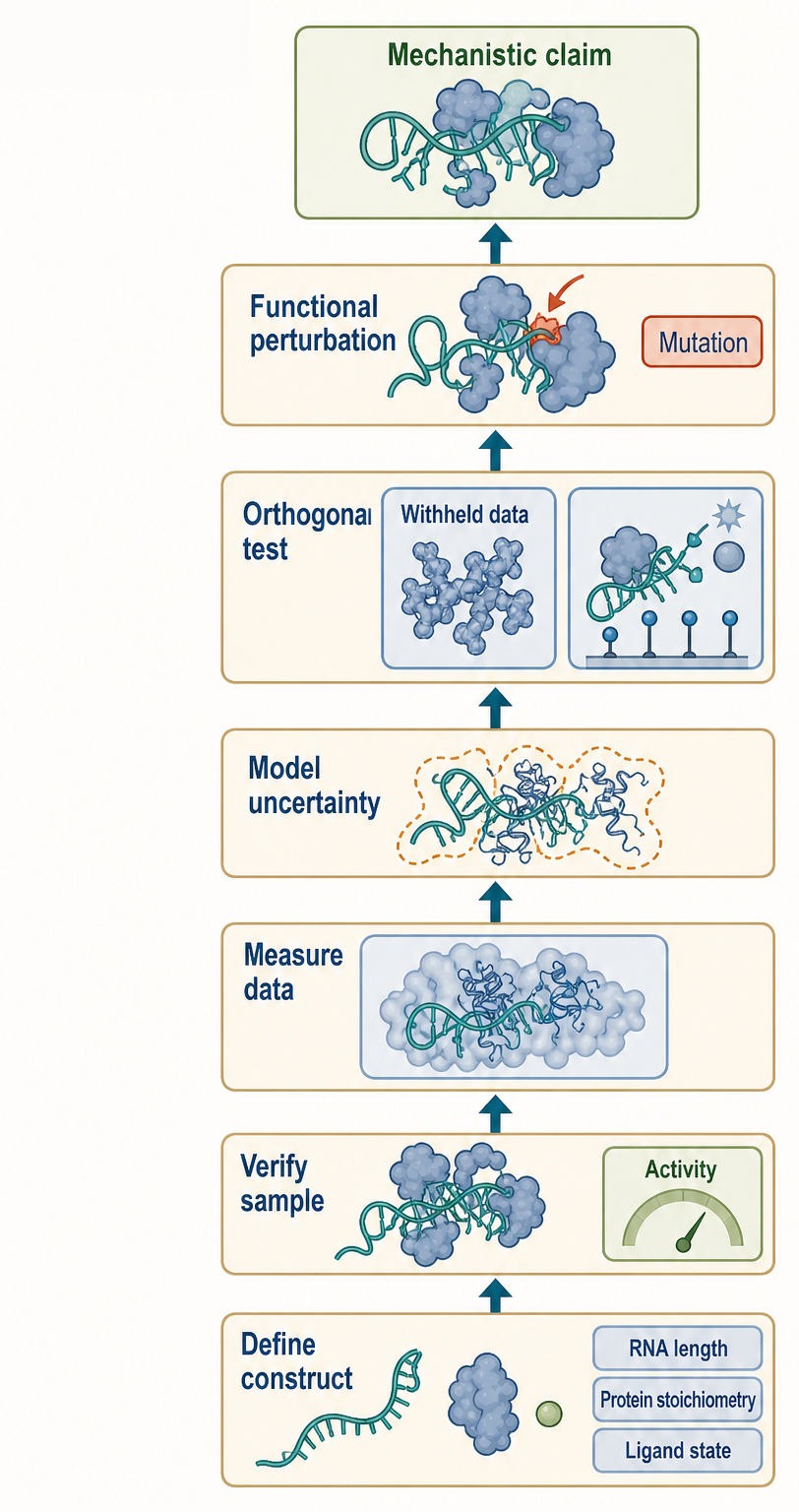
Figure 59.3. Validation Ladder for an RNP Structural Model. Building a trustworthy RNP structural model requires sequential checkpoints beginning before data collection: defining the RNA construct, protein composition, ligand state, and buffer condition; verifying purity, length, stoichiometry, and biochemical activity; acquiring and interpreting structural or biophysical data with explicit uncertainty; cross-validating the model against withheld or orthogonal observations; and finally perturbing predicted contacts to test biochemical or cellular consequences. An RNP coordinate model becomes a mechanistic model only when it explains independent observations and withstands functional perturbation.
59.7. Method choice and interpretation caveats
Method choice should start with the biological claim. If the claim is that a protein recognizes a specific RNA motif through atomic contacts, high-resolution crystallography, NMR, or cryo-EM plus mutagenesis may be needed. If the claim is that an RNA switches between two conformations during transcription, smFRET, time-resolved probing, or co-transcriptional assays may be more relevant than a static endpoint model. If the claim is that a large RNP has several assembly intermediates, cryo-EM classification, native mass spectrometry, crosslinking, and biochemical reconstitution may be appropriate. Native MS is a first-line choice when the discriminating question is whether intact particles contain zero, one, or several copies of an RNA, protein, or ligand. It is not the first-line method for locating an atomic contact or determining a complete solution fold. If the claim is transcriptome-scale structural remodeling, chemical probing and sequencing can nominate candidates, but targeted validation remains necessary.
The native-MS question should be written before the spectrum is acquired. For stoichiometry, list all plausible component masses and specify which mass difference would distinguish competing models. For oligomerization, measure a concentration series and compare with a solution method so that droplet-induced association and in-source dissociation can be recognized. For ligand or RNA binding, include inactive or specificity-defective mutants, unrelated ligands or RNAs where appropriate, and titrations that reveal nonspecific high-occupancy binding. For activation experiments, state whether the intended inference is subunit connectivity, ligand lability, or comparative gas-phase stability. For IM-MS, predefine whether the test concerns relative compaction, conformational heterogeneity, or compatibility with candidate models, and report the CCS method and uncertainty.
Heterogeneous spectra should be interpreted as distributions, not forced into a single nominal complex. A broad charge-state envelope can contain true assembly intermediates, salt adducts, unresolved RNA length variants, or conformers with different charging behavior. Replicate preparation, sharper desalting, component omission, isotope or mass shifts, and independent separation can distinguish some possibilities. If multiple assignments remain compatible, the structural conclusion should be correspondingly coarse. This is the native-MS analogue of resisting overfitting in SAXS or weak-density model building: resolution gained by computation is not necessarily information gained from the specimen.
Orthogonal validation should target the weakest link in the inference. SEC-MALS or analytical ultracentrifugation can test solution oligomeric mass; native gels or size-exclusion chromatography can test whether multiple assemblies pre-exist electrospray; NMR or solution scattering can test ligand-induced structural change; cryo-EM or crystallography can localize an architecture; activity assays can establish functional competence; and quantitative binding assays can test affinity or cooperativity. Agreement need not mean identical apparent populations because each method samples a different condition and weights species differently. The important questions are whether discrepancies are mechanistically explainable and whether the central conclusion survives them.
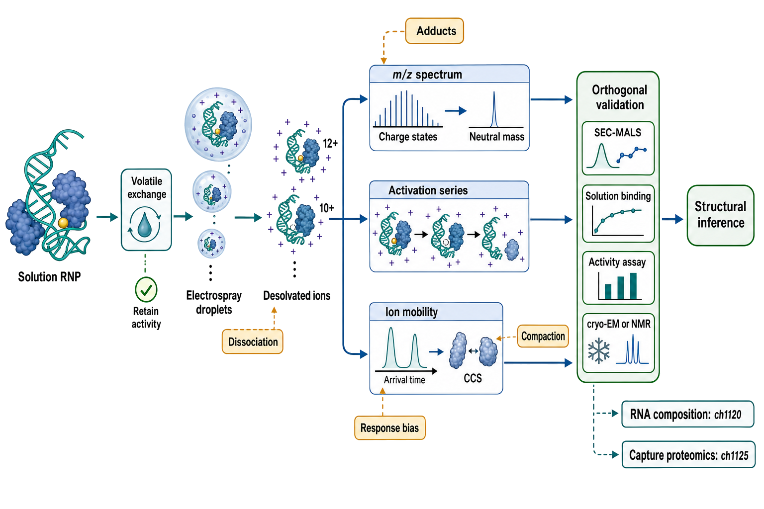
Figure 59.4. Native-MS Evidence Ladder for an Intact RNP. An intact RNP begins in a characterized solution ensemble, is exchanged into a volatile electrolyte with explicit retention checks, and enters electrospray droplets before becoming desolvated multiply charged ions. The spectrum directly reports mass-to-charge peaks and charge-state envelopes; deconvolution supports neutral masses, stoichiometries, oligomeric states, and ligand occupancies. Controlled activation can test gas-phase dissociation pathways, while ion mobility reports arrival times and condition-specific collision cross sections. A central visual boundary separates direct observations from structural inferences and flags possible adduction, in-droplet association, in-source dissociation, gas-phase compaction, and transmission bias. SEC-MALS or analytical ultracentrifugation, solution binding, biochemical activity, and cryo-EM/NMR provide orthogonal validation. The figure ends with explicit handoffs: nucleotide-, modification-, sequence-, and fragment-level analysis to Chapter 132, and peptide-centric RNA-capture proteomics to Chapter 138.
A common misconception is that agreement between two low-resolution methods automatically proves an atomic model. It does not. SAXS and chemical probing can both support compactness without specifying the same tertiary contacts. A crosslink and a FRET state can both imply proximity without proving a direct interaction. A cryo-EM map and a predicted RNA model can appear compatible because many RNA helices fit tube-like density. Agreement is powerful when the data constrain different aspects of the same model and when alternative models have been tested.
Another misconception is that in-cell data always outrank purified data. In-cell probing, imaging, and crosslinking preserve biological context, but they may mix cell states, splice isoforms, protein-bound and unbound RNAs, and indirect effects. Purified assays remove many confounders and can test mechanism, but they may omit native cofactors and crowding. The strongest interpretation often requires both: purified reconstitution to show sufficiency and cellular perturbation to show relevance.
Researchers should also distinguish occupancy from consequence. A protein can bind an RNA without changing its function. An RNA can change conformation without changing gene expression. A crosslink can identify proximity without stable regulation. A cryo-EM class can exist without being a productive intermediate. Functional interpretation requires readouts such as catalysis, splicing, translation, decay, replication, localization, immune activation, or phenotype. This caution applies to RBP biology broadly, where reviews emphasize that RNA-binding proteins can edit, process, transport, stabilize, repress, and scaffold RNAs through context-dependent mechanisms.
Finally, structural interpretation should avoid false precision. A model may justify saying that two helices are coaxially stacked, but not that a particular magnesium ion is essential. A probing experiment may justify saying that a region becomes protected after protein binding, but not that a specific base pair forms. A single-molecule trace may justify saying that two states interconvert, but not that the complete tertiary structure is known. Good structural biology is explicit about the level of inference.
The same rule fixes the boundaries among mass-spectrometric chapters. An intact mass difference consistent with one additional RNA or ligand supports an occupancy assignment here when alternative adducts and components are excluded. Fragment ions that identify a nucleotide, modification site, sequence segment, or covalent composition belong to Chapter 132. Peptides used to identify or quantify proteins enriched by an RNA bait belong to Chapter 138. If a hybrid experiment uses all three layers, the paper should separate intact-assembly evidence, RNA compositional evidence, and protein-identification evidence rather than presenting “mass spectrometry” as one undifferentiated proof.
Box 59.1. Misconception Check: Resolution Is Not Relevance
- A high-resolution crystal or cryo-EM structure is not automatically more biologically meaningful than a lower-resolution measurement if it captures the wrong state or a truncated construct.
- A lower-resolution in-cell or kinetic measurement may be closer to the native mechanism even if it cannot specify atomic contacts.
- The relevant question is whether the method observes the structural state responsible for the biological function being claimed.
- Both high- and low-resolution data require targeted validation; resolution does not substitute for functional evidence.
Box 59.2. Practical Sample Questions Before Starting RNA Structural Work
- Is the RNA sequence and length verified by denaturing gel, capillary electrophoresis, or mass spectrometry?
- Are the termini and modification state fully defined?
- Could storage, buffer exchange, hydrolysis, or work-up chemistry alter a labile nucleotide or cofactor?
- Is the RNP assembled with the intended stoichiometry, confirmed by SEC-MALS or analytical ultracentrifugation?
- If native MS is planned, does exchange into a volatile electrolyte preserve activity, folding, and solution oligomerization?
- Are broad peaks caused by compositional heterogeneity or by salt, metal, water, and buffer adducts?
- Which charge states, source-energy controls, and deconvolution assumptions support each intact-mass assignment?
- Is the sample biochemically active under the conditions used for structure determination?
- Are multiple folded conformations expected, and if so, is the preparation enriched for a defined state?
- Which buffer condition, ion concentration, and temperature reflect the biologically relevant environment?
- Which orthogonal method will be used to validate the main structural model?
Recent Consensus
Recent consensus is that RNA and RNP structural biology is increasingly ensemble-aware, hybrid, and validation-driven. Crystallography remains essential for high local detail when stable crystals can be obtained, and synthetic antibodies or other stabilizers can accelerate RNA crystallography. Cryo-EM is now the dominant high-resolution approach for many large RNPs, but RNA sample optimization and heterogeneity control remain central bottlenecks. NMR and SAXS retain unique value for dynamics, solution ensembles, and local interaction mapping. Native MS is now a mature complement for resolving intact stoichiometry, oligomeric state, occupancy, and compositional heterogeneity, especially when combined with ion mobility, controlled activation, and solution-phase validation. Single-molecule and force methods expose kinetic pathways that ensemble structures average away. Chemical probing and crosslinking connect structural hypotheses to cellular and transcriptome contexts.
The shared methodological consensus is that no single measurement is enough for most mechanistic claims. The field has moved from “solve the structure” toward “explain the state, ensemble, transition, and function.” For a large RNP, the best answer may be a set of states with occupancies, uncertainties, and biochemical roles. For a dynamic RNA, the best answer may be a restrained ensemble rather than one coordinate model.
Open Questions, Controversies, Deprecated Models, and Common Misconceptions
Open questions:
- How can structural biologists best represent RNA ensembles without overfitting sparse data?
- How should cryo-EM classes be connected to kinetic pathways rather than treated as snapshots along a presumed reaction coordinate?
- Which in-cell probing signals reflect RNA structure directly, and which reflect protein occupancy, modification, compartment access, or RNA abundance?
- How reliable are current RNA force fields and machine-learning models for modified nucleotides, ions, ligand binding, and large RNP interfaces?
- Which native-MS transfer and charge-reduction conditions best preserve informative RNA-protein contacts without selecting only the most gas-phase-stable states?
- How can native-MS response factors and in-droplet chemistry be calibrated well enough to recover quantitative solution populations for heterogeneous RNPs?
- When does an RNA or RNP collision cross section retain useful memory of solution architecture, and when is gas-phase compaction dominant?
Common misconceptions:
- “A high-resolution structure is automatically the native functional state.” Resolution describes distinguishable detail in the measured specimen; construct design, sample condition, lattice contacts, vitrification, and state selection still determine biological relevance.
- “Low chemical reactivity proves base pairing, and a crosslink proves a stable contact.” Reactivity and crosslinking are chemistry-dependent proxies that can also reflect protein protection, modification, accessibility, or transient proximity.
- “A FRET efficiency is a precise geometric distance without additional assumptions.” Dye orientation, linker behavior, photophysics, labeling geometry, and state averaging limit the structural precision of a FRET-derived distance.
- “A cryo-EM class is automatically a biochemical intermediate, and a predicted model is independent evidence.” A class requires kinetic or functional connection to a pathway, while a prediction becomes evidence only through comparison with independent observations.
- “The word native means that an electrosprayed RNP has the same conformation in solution and in the gas phase.” Native MS is designed to preserve informative noncovalent assemblies, but desolvation changes electrostatics and RNA ions can compact, unfold, or dissociate.
- “Native-MS peak intensities directly report equilibrium populations and affinities.” Ionization, droplet concentration, response, transmission, and dissociation can weight species unequally; equilibrium constants require explicit calibration and orthogonal solution controls.
- “A higher collision threshold proves a stronger solution interface.” Activation measures gas-phase behavior along an instrument-specific pathway and cannot be converted directly into a solution binding free energy.
- “A collision cross section uniquely identifies an RNA fold.” CCS can reject incompatible gross architectures or compare controlled states, but different structures can have similar CCS values and gas-phase rearrangement can erase solution distinctions.