Chapter 27. Splicing Chemistry, Spliceosome Assembly, Catalysis, and Intron Evolution
Scope Note
Splicing is the RNA-processing reaction that removes introns and joins exons to make a continuous RNA product. In the best-studied nuclear example, a precursor messenger RNA, or pre-mRNA, is cut at a 5′ splice site, folded through a branch-point adenosine to form a lariat intermediate, cut again at a 3′ splice site, and ligated across the two exons. The reaction is chemically simple when drawn as two phosphotransesterification steps, but it is biologically demanding because splice sites are short, introns vary enormously in length, and wrong splice-site choices can change reading frames, produce premature termination codons, or alter regulatory RNA elements.
This chapter explains constitutive spliceosome chemistry and assembly, including the biological mechanisms of recursive splicing, inter-RNA trans-splicing, and spliced-leader trans-splicing. Alternative splicing is treated in Chapter 28; exon-junction complexes and export in Chapters 30 and 31; RNA decay in Chapters 32-35; and product-specific design of engineered RNA trans-splicing in Chapter 154. Group I and group II introns remain comparative pathway examples here because they clarify spliceosomal chemistry, while Chapter 9 owns their full comparison with other catalytic RNAs, including metal-ion strategies, kinetics, evolution, and engineering.
Executive Summary
Pre-mRNA splicing removes introns through two transesterification reactions. In spliceosomal introns, the 2′ hydroxyl of a branch-point adenosine attacks the phosphate at the 5′ splice site, releasing the 5′ exon and forming a lariat intron-3′-exon intermediate. The free 3′ hydroxyl of the 5′ exon then attacks the phosphate at the 3′ splice site, ligating the exons and releasing the lariat intron. This chemistry preserves the number of phosphodiester bonds, but it changes RNA connectivity and depends on correct positioning of reactive groups, catalytic metal ions, and spliceosomal RNA elements.
The major spliceosome recognizes most nuclear pre-mRNA introns through a set of short and degenerate sequence signals: the 5′ splice site, the branch point, a downstream polypyrimidine tract in many metazoan introns, and the 3′ splice site. The minor spliceosome recognizes a much smaller class of U12-type introns with distinct consensus features and snRNA components, although both systems converge on a related two-step lariat-splicing chemistry. Splice-site recognition is therefore not simple motif matching. It is a kinetic and structural decision made by RNA-RNA pairing, RNA-binding proteins, exon definition, intron definition, chromatin and transcription context, and competition with nearby cryptic sites.
Spliceosome assembly is a staged remodeling pathway rather than the action of a preformed enzyme. U1 small nuclear ribonucleoprotein, or snRNP, initially pairs with the 5′ splice site. SF1/BBP and U2AF help define the branch-point region and 3′ splice-site region in many metazoan introns. U2 snRNP then pairs with the branch site in a way that bulges the branch-point adenosine. Recruitment of the U4/U6.U5 tri-snRNP creates a precatalytic spliceosome. Activation then ejects U1 and U4, allows U6 to pair with the 5′ splice site and U2, and organizes the catalytic RNA core.
The spliceosome is an RNA-metalloenzyme embedded in a protein machine. U2 and U6 snRNAs form the conserved catalytic RNA network, while proteins such as PRPF8, the SF3B complex, Cwc25, Slu7, Prp18, and multiple ATP-dependent DExD/H-box proteins stabilize, inspect, remodel, or discard spliceosomal states. Proteins do not merely hold RNA in place; they tune substrate choice, coordinate remodeling, prevent premature catalysis, and couple splicing to transcription and downstream mRNP assembly.
Splicing fidelity arises from kinetic competition rather than from one static proofreading checkpoint. Correct splice-site pairs must assemble fast enough and stably enough to proceed through ATPase-driven transitions, while poor substrates are delayed, rearranged, or rejected. ATP hydrolysis by spliceosomal helicases can proofread branch-site pairing, first-step chemistry, second-step chemistry, exon ligation, and post-catalytic disassembly. This logic explains why mutations in splice sites, branch points, auxiliary motifs, or core factors can activate cryptic exons rather than simply eliminating splicing.
Group I and group II introns show that RNA itself can catalyze intron removal. Group I introns use an exogenous guanosine nucleophile and do not form a lariat. Group II introns usually use an internal branch-point adenosine and form a lariat, making them mechanistically and evolutionarily closer to spliceosomal introns. Current consensus favors an evolutionary relationship between group II introns and spliceosomal introns, but the exact path from mobile group II ribozymes to the modern nuclear spliceosome remains reconstructed from comparative genomics, structural homology, and plausible cellular transitions rather than from direct historical observation.
Splicing topology must be named explicitly. Ordinary cis-splicing joins splice sites carried on one precursor RNA. Recursive splicing is also cis-splicing: a long intron is removed in consecutive segments through temporary recursive splice sites or ratchet points on that same precursor. Inter-RNA trans-splicing instead joins exons carried on separate RNA molecules. Spliced-leader trans-splicing is a specialized trans-splicing pathway in which a short capped exon from a dedicated spliced-leader RNA is transferred to an acceptor pre-mRNA. These pathways can use related spliceosomal branch-and-lariat chemistry without being interchangeable, and none is automatically a form of alternative splicing.
Splicing is measured and manipulated by assays with different levels of mechanistic resolution. RNA-seq can reveal exon inclusion, intron retention, and cryptic splice products, but it often cannot prove the immediate biochemical cause. In vitro splicing, minigene reporters, branch-point mapping, lariat analysis, crosslinking, and cryo-electron microscopy provide complementary evidence. Therapeutic correction can use antisense oligonucleotides, small molecules, trans-splicing or RNA-editing concepts, and genome editing, but each strategy must be evaluated by target mechanism, tissue delivery, off-target splicing effects, durability, and disease context.
Concept Inventory
- Intron: an RNA segment removed from a precursor RNA during processing. In protein-coding pre-mRNA, introns interrupt exons before mature mRNA formation. Introns are not inherently nonfunctional, and intron removal is not always spliceosomal.
- Exon: a sequence retained in a mature RNA product after splicing. Exons can encode protein sequence, untranslated regions, or noncoding RNA sequence; exon does not mean protein-coding segment.
- Splice site: an intron-exon boundary recognized during splicing. The 5′ splice site is at the exon-intron junction upstream of an intron, and the 3′ splice site is at the intron-exon junction downstream of an intron.
- Branch point: the intronic nucleotide, usually adenosine in spliceosomal and group II lariat splicing, whose 2′ hydroxyl attacks the 5′ splice-site phosphate to form a branched lariat intermediate.
- Lariat: a branched RNA produced when the branch-point 2′ oxygen links to the 5′ end of the intron through an unusual 2′-to-5′ phosphodiester bond. Lariat detection is strong evidence for lariat splicing but not by itself a complete map of splice-site regulation.
- Major spliceosome: the U2-dependent spliceosome that removes most eukaryotic nuclear pre-mRNA introns using U1, U2, U4, U5, and U6 snRNPs.
- Minor spliceosome: the U12-dependent spliceosome that removes a rare class of nuclear introns using U11, U12, U4atac, U5, and U6atac snRNPs. Minor does not mean biologically unimportant.
- Small nuclear RNA: a nuclear RNA component of snRNPs. In the spliceosome, snRNAs recognize splice signals, rearrange by base pairing, and form parts of the catalytic center.
- Small nuclear ribonucleoprotein: an RNP particle containing an snRNA and associated proteins. Spliceosomal snRNPs are dynamic modules rather than passive RNA carriers.
- Transesterification: a reaction in which one phosphodiester linkage is exchanged for another. Splicing uses transesterification rather than hydrolytic cutting followed by energy-consuming ligation.
- Group I intron: a self-splicing ribozyme intron that uses an external guanosine nucleophile and generally releases a linear intron rather than a lariat.
- Group II intron: a self-splicing ribozyme intron that usually forms a lariat through an internal branch-point adenosine and is widely discussed as an evolutionary relative of spliceosomal introns.
- Splicing fidelity: the ability of the splicing system to select correct splice sites and reject incorrect or suboptimal splice-site combinations. Fidelity reflects kinetic competition, ATP-dependent proofreading, RNA-protein recognition, and transcript context.
- Cryptic splice site: a sequence that is normally ignored but can be used when canonical signals are weakened, regulatory context changes, or nearby recognition is altered.
- Splice-switching oligonucleotide: an antisense molecule designed to alter splice-site or regulatory-element use by sterically blocking RNA-protein or RNA-RNA interactions without necessarily degrading the RNA.
- Cis-splicing: splicing in which the reacting splice sites and exons are carried on the same precursor RNA. Recursive splicing is an intramolecular form of cis-splicing even though it proceeds through more than one reaction cycle.
- Recursive splicing: stepwise removal of a long intron through one or more internal recursive splice sites. A first splice creates or exposes a splice donor at a ratchet point, and later splicing removes the next intron segment.
- Trans-splicing: spliceosome-mediated joining of exon sequences carried on separate RNA molecules. Trans-splicing describes substrate topology, not whether the final product is constitutive or alternatively selected.
- Spliced-leader trans-splicing: a specialized trans-splicing reaction that transfers the short 5′ exon of a dedicated spliced-leader RNA to an acceptor pre-mRNA.
- Spliced-leader RNA: a small noncoding donor RNA with a capped leader exon, a 5′ splice site, an intron-like region, and RNP-binding features that support trans-splicing.
- Ratchet point: an internal recursive-splicing site that acts first as a splice acceptor and, after the upstream intron segment is removed, as a reconstituted or exposed splice donor for the next step.
What to Know Before Reading This Chapter
RNA chains have polarity and reactive hydroxyl groups. The conventional backbone contains 3′-to-5′ phosphodiester linkages, and a ribose 2′ hydroxyl can act as a nucleophile when correctly positioned. Chapter 2 explains phosphodiester chemistry in detail. For splicing, the important point is that RNA can be cut and rejoined by exchanging phosphodiester bonds without consuming ATP directly in the chemical step. ATP is instead used by spliceosomal remodeling enzymes to build, inspect, and dismantle the active machine.
A pre-mRNA is a newly transcribed RNA that is still undergoing processing. In a typical eukaryotic protein-coding gene, exons are segments retained in the mature mRNA and introns are segments removed. This definition is operational. An exon can be part of a coding sequence or an untranslated region, and an intron can contain regulatory elements, noncoding RNAs, repeat fragments, or evolutionary relics. Do not equate exon with “protein-coding” or intron with “junk.” Chapters 18 and 28 return to transcript models and regulated isoform choice.
The running example in this chapter is a human RNA polymerase II pre-mRNA with a U2-type intron. The upstream exon ends at a 5′ splice site, the intron contains a branch-point region and a downstream polypyrimidine tract, and the intron ends at a 3′ splice site. The major spliceosome must recognize these features in a long RNA molecule crowded with similar short motifs. This recognition problem is why splicing is not reducible to the sequence “GU.AG.” That shorthand captures common intron-end dinucleotides, not the whole recognition code.
Small nuclear ribonucleoproteins, or snRNPs, are central spliceosomal modules. Each snRNP contains a small nuclear RNA, or snRNA, plus proteins. U1, U2, U4, U5, and U6 snRNPs participate in major-spliceosome splicing. The minor spliceosome uses U11, U12, U4atac, U5, and U6atac. The snRNAs base-pair with the pre-mRNA and with one another, and those pairings change as the spliceosome matures. Spliceosome assembly is therefore a controlled RNA-folding and RNP-remodeling pathway.
Finally, evidence matters. A sequencing read spanning two exons proves that a spliced product exists in the sampled RNA pool, but it does not by itself prove which spliceosomal step changed, which factor bound the RNA, or whether the event is direct. A cryo-electron microscopy structure can show a catalytic state, but it may represent a trapped or enriched state rather than the full kinetic ensemble. A minigene reporter can isolate local sequence logic, but it may miss chromatin, transcription, and full-length intron effects. The sections below pair mechanisms with the assay types that can and cannot support them.
The same evidence caution becomes more important for non-collinear products. A chimeric complementary-DNA read can be produced by genuine inter-RNA splicing, but also by a genomic rearrangement, read misalignment, library ligation, reverse-transcriptase template switching, or PCR mispriming. Conversely, a recursive-splicing intermediate can be short-lived and absent from poly(A)-selected mature-RNA data even when the pathway is active. Mechanistic assignment therefore requires topology-aware controls rather than a single junction sequence.
27.1. Splice-site signals, branch points, and intron classes
Splice-site signals are the local RNA features that tell the splicing machinery where an intron begins, where an intron ends, and which internal nucleotide should initiate the first chemical step. In a common metazoan U2-type intron, the 5′ splice site includes the last nucleotides of the upstream exon and the first nucleotides of the intron. The intron usually begins with GU in the RNA sequence. Near the downstream end, a branch-point sequence contains a branch-point adenosine, a polypyrimidine tract is enriched in cytidine and uridine, and the 3′ splice site often ends with AG. These signals are short and degenerate, meaning that many functional introns deviate from a single ideal consensus.
The recognized signals place the reactive groups for the two-step chemistry summarized in Figure 27.1.
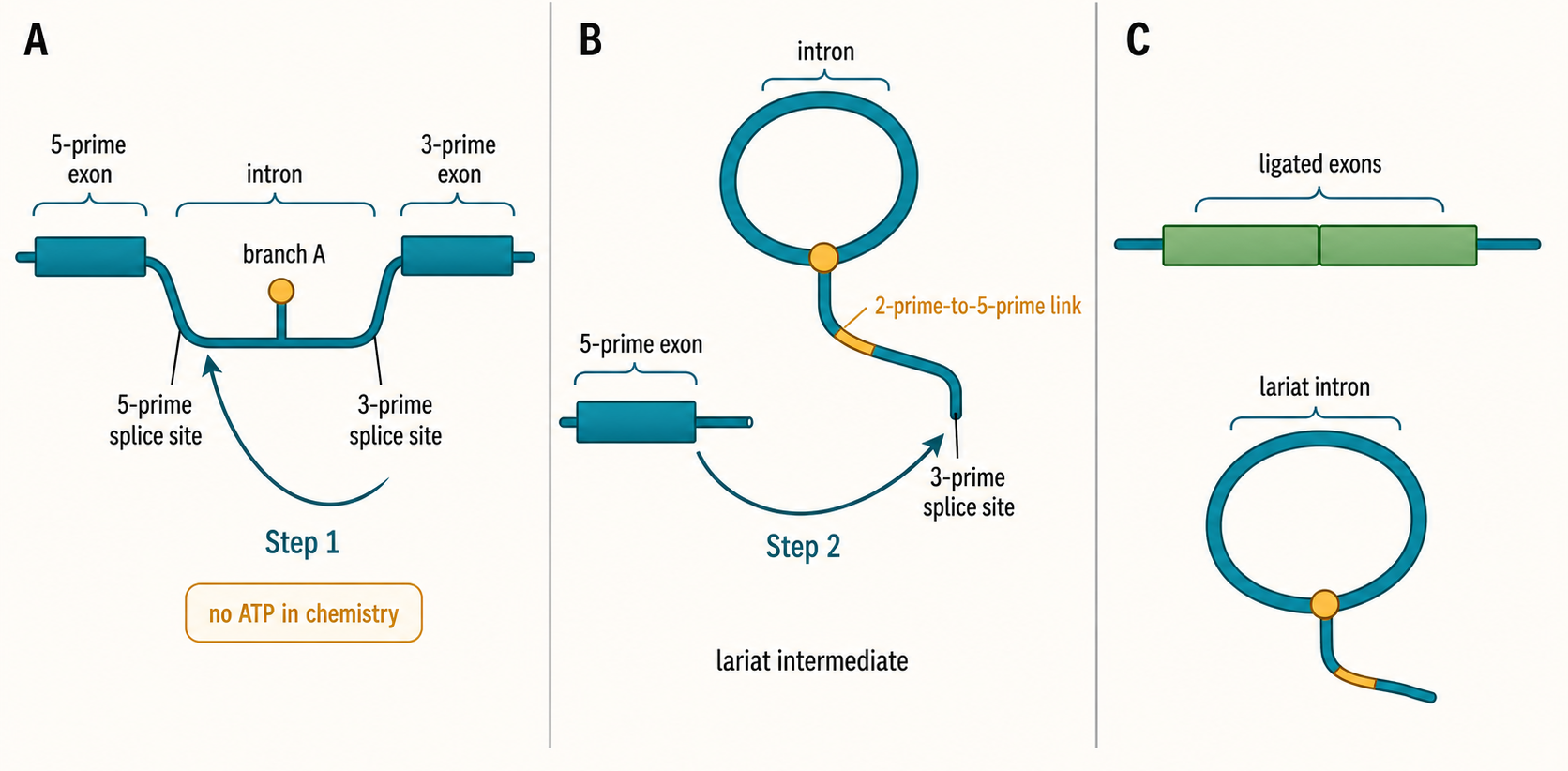
Figure 27.1. Two-Step Lariat Splicing Chemistry. The figure illustrates the two transesterification reactions of spliceosomal pre-mRNA splicing. First, the 2′-hydroxyl of the branch-point adenosine attacks the phosphate at the 5′ splice site, releasing the 5′ exon with a free 3′-hydroxyl and forming a lariat intermediate joined by an unusual 2′-to-5′ phosphodiester bond. Second, the free 3′-hydroxyl of the 5′ exon attacks the 3′ splice site, ligating the exons and releasing the lariat intron. Because each step exchanges one phosphodiester bond for another, no bond-formation energy from ATP is consumed at the chemical steps themselves.
The branch point deserves special attention because it is both a recognition signal and a chemical participant. During the first transesterification reaction, the 2′ hydroxyl of the branch-point adenosine attacks the phosphate at the 5′ splice site. The resulting 2′-to-5′ linkage creates the lariat branch. U2 snRNA pairs with the branch-site region but leaves the branch-point adenosine unpaired or bulged, which helps position that adenosine for catalysis. The same nucleotide is therefore recognized by base-pairing context and then used as a nucleophile.
The major spliceosome removes U2-type introns. In humans and other metazoans, this class accounts for the overwhelming majority of spliceosomal introns. A smaller U12-type class is removed by the minor spliceosome. U12-type introns have distinct consensus features and are recognized by U11 and U12 instead of U1 and U2 at early stages; U4atac and U6atac substitute for U4 and U6, while U5 is shared. The name “minor spliceosome” refers to relative abundance, not to marginal importance. Some genes are highly sensitive to minor-spliceosome activity, and defects in minor-spliceosome components can have developmental consequences.
Intron classes also include self-splicing group I and group II introns. These introns are not removed by the nuclear spliceosome. Group I introns use an external guanosine as the initiating nucleophile and typically release a linear intron. Group II introns use an internal branch-point adenosine in most cases and release a lariat intron. Group II introns are common in bacterial and organellar contexts and often behave as mobile genetic elements with encoded maturase or reverse transcriptase functions. These distinctions matter because “intron” names an interrupted RNA segment, not one universal removal mechanism.
Splice-site recognition can occur by intron definition or exon definition. In intron definition, factors assemble across a relatively short intron, connecting the 5′ and 3′ splice-site regions directly. In exon definition, factors initially define an exon across its flanking splice sites, a strategy useful in organisms with long introns and relatively short exons. Metazoan genes often use exon-definition logic because introns can span thousands to hundreds of thousands of nucleotides. This architecture helps explain why a variant in one splice site can affect recognition of a neighboring exon rather than only the immediately adjacent intron.
Concrete disease examples show why motif degeneracy matters. A mutation that disrupts the invariant GU at a 5′ splice site or AG at a 3′ splice site often has severe consequences, but many pathogenic variants occur outside those dinucleotides. A branch-point change can weaken U2 recognition. A deep intronic variant can create a new splice site and activate a cryptic exon. A synonymous coding variant can disrupt an exonic splicing enhancer. In each case, the sequence remains transcribed, but the spliceosome reads the RNA differently.
The evidence basis for splice-site signals comes from several layers. Comparative genomics reveals conserved motifs and intron-class distributions. Mutational analysis and massively parallel splicing assays test how sequence variants alter splice products. Crosslinking and immunoprecipitation methods map factor binding. Structural studies reveal how snRNAs and proteins contact splice sites. RNA-seq discovers the products that accumulate in cells. No single assay covers all levels. A motif score predicts risk; it is not proof of use in a particular cell type.
Box 27.1. Do Not Overgeneralize Splicing Signals
- GU-AG at intron ends is a common dinucleotide pattern, not the complete recognition code; many functional introns use rare boundary dinucleotides and depend on auxiliary context.
- A sequence that scores well on a splice-site matrix is not proof that the site is recognized or used in a given cell type or developmental stage.
- A weak splice-site motif can support efficient splicing when SR proteins, exon definition, cooperative factor binding, or RNA structural context compensates.
- Minor-spliceosome introns are numerically rare, but the genes they interrupt can be highly dosage-sensitive, and defects in minor-spliceosome components have documented developmental consequences.
- Exon does not mean protein-coding; exons can encode untranslated regions, noncoding RNA sequences, or regulatory elements and remain subject to spliceosomal removal of flanking introns.
The main boundary case is that splice-site consensus is a probability model, not a rule book. Some authentic introns use rare dinucleotides, such as GC-AG or AT-AC in DNA notation, and some introns depend heavily on auxiliary factors. Conversely, a sequence that resembles a splice site may be ignored because it is occluded by RNA structure, positioned poorly, embedded in a repressive RNP context, or outcompeted by a stronger site. Chapter 28 develops the regulated use of competing sites; this section establishes the core recognition vocabulary.
Table 27.1. Splice-Site Signals and Recognition Factors. Short and degenerate sequence features at intron boundaries and within exons and introns direct spliceosome assembly; the table summarizes each signal, its recognition factors in the major and minor spliceosomes, the main evidence type, and key caveats.
| Signal or feature | Molecular location | Major-spliceosome recognition factors | Minor-spliceosome counterpart | Main evidence type | Common perturbation | Caveats |
|---|---|---|---|---|---|---|
| 5′ splice site | Exon-intron junction at intron start | U1 snRNP via U1 snRNA base-pairing | U11 snRNP | Crosslinking, mutational analysis, cryo-EM | GU→GC or GU→AU disrupts splicing | GU-AG is a pattern, not the full recognition code |
| Branch point | ~15–50 nt upstream of 3′ splice site | U2 snRNP via U2 snRNA base-pairing; SF3B complex | U12 snRNP | Branch-point mapping, lariat sequencing, CLIP | Branch-point A mutation stalls assembly or shifts branch-point use | Bulged adenosine geometry required for catalysis |
| Polypyrimidine tract | Between branch point and 3′ splice site | U2AF2 (U2AF65) | Less conserved in U12 introns | SELEX, CLIP, mutational analysis | Purine substitution or deletion weakens 3′ splice-site recognition | Prominent in metazoans; less critical in yeast |
| 3′ splice site | Intron-exon junction at intron end | U2AF1 (U2AF35); later spliceosome remodeling proteins | U12 snRNP region | Mutational analysis, crosslinking | AG→AC mutation blocks exon ligation | Some introns use non-AG dinucleotides |
| Exonic splicing enhancer | Exon body near splice sites | SR proteins (SRSF1, SRSF2, and others) | Not well characterized | SELEX, CLIP, reporter assays | Synonymous variant can disrupt enhancer and cause exon skipping | Context-dependent; sequence score alone insufficient |
| Intronic splicing enhancer | Intron body near regulated exon | SR proteins or hnRNP proteins (context-dependent) | Not well characterized | CLIP, mutational analysis | Deletion or mutation can cause exon skipping | May overlap regulatory secondary structure |
| Exonic splicing silencer | Exon body | hnRNP proteins (hnRNP A1, hnRNP I/PTB) | Not well characterized | Reporter assays, CLIP | Mutation can cause exon inclusion | Some silencers are cell-type specific |
| Intronic splicing silencer | Intron body | hnRNP proteins | Not well characterized | CLIP, reporter assays | Mutation can activate a nearby cryptic exon | Can act at long range from the regulated exon |
| Cryptic splice site | Anywhere in pre-mRNA | Same factors as canonical sites but lower affinity | Minor-spliceosome counterparts where applicable | RNA-seq from patient samples, reporter assays | Canonical site weakening activates cryptic site | Often tissue-specific; competition controls use |
| Pseudoexon | Deep intronic region | SR proteins and splicing complex if a variant activates it | Rare | Long-read RNA-seq, minigene reporters | Deep intronic variant creates or strengthens a cryptic donor or acceptor | Clinical interpretation requires endogenous RNA evidence |
27.2. Spliceosome assembly and snRNA rearrangements
The spliceosome is assembled anew on each intron. This feature distinguishes it from enzymes that bind a substrate in a fully formed active site. Assembly begins with recognition of splice signals and proceeds through a series of RNP states often named E, A, B, Bact, B, C, C, P, and intron-lariat spliceosome states. The labels vary somewhat across organisms and experimental systems, but the logic is consistent: the pre-mRNA is first recognized, then combined with snRNP modules, then remodeled into an active RNA-centered catalytic machine, then dismantled after exon ligation.
Table 27.2. Assembly States and Remodeling Events. Each spliceosomal assembly state is defined by its snRNP composition, characteristic RNA-RNA pairings, and the ATP-dependent transition that drives progression to the next state; the intron-lariat spliceosome state marks the end of catalysis before disassembly.
| Assembly state | Core snRNP or factor composition | Key RNA-RNA pairing | Main ATP-dependent transition | Product or intermediate | Representative assay |
|---|---|---|---|---|---|
| E (early) complex | U1 snRNP, SF1/BBP, U2AF1, U2AF2 | U1 snRNA:5′ splice site | Prp5 promotes U2 snRNP engagement | Pre-splicing recognition complex on pre-mRNA | Native gel, affinity capture |
| A complex | U1 snRNP, U2 snRNP | U1 snRNA:5′ splice site; U2 snRNA:branch site (branch-point A bulged) | Prp5 completion | Branched snRNA:pre-mRNA duplex with bulged branch-point adenosine | Native gel, crosslinking |
| B complex | U1, U2, U4/U6·U5 tri-snRNP | U4:U6 extensive pairing; U5:exon sequences | Brr2/SNRNP200 begins U4/U6 unwinding | Fully assembled but catalytically inactive spliceosome | Cryo-EM, native gel |
| Bact | U2, U5, U6 (U1 and U4 released) | U6:5′ splice site; U2:U6 helix I and II | Prp2/DHX16 opens branch-site region | Activated spliceosome awaiting first catalytic step | Cryo-EM |
| B* | U2, U5, U6, step-1 factors including Cwc25 | U2:U6 catalytic network established | None required; catalysis follows remodeling | First-step active conformation | In vitro splicing time course |
| C complex | U2, U5, U6 with lariat-intermediate-bound RNA | U2:U6 maintained; U5:downstream exon | Prp16/DHX38 promotes second-step rearrangement | Lariat intron-3′ exon intermediate; free 5′ exon | Native gel, lariat detection |
| C* | U2, U5, U6, step-2 factors (Slu7, Prp18, Prp22) | Second-step RNA geometry for exon ligation | Prp16/DHX38 completion | Second-step active conformation | Cryo-EM, in vitro splicing |
| P (product) complex | U2, U5, U6 with ligated exons and retained lariat | Exons ligated; lariat still associated with snRNPs | Prp22/DHX8 releases mature mRNA | Mature mRNA released; lariat remains in spliceosome | In vitro splicing, lariat sequencing |
| Intron-lariat spliceosome | U2, U5, U6 with disassociating intron lariat | snRNA:intron lariat | Prp43/DHX15 drives disassembly | Released intron lariat; snRNPs returning to free pool | Cryo-EM, native gel |
| Disassembled/recycled snRNPs | Individual U1, U2, U4, U5, U6 snRNPs | U4:U6 pairing reformed during recycling | Various snRNP recycling and reassembly factors | Recycled snRNP modules available for new assembly round | Biochemical reconstitution |
Early assembly solves the recognition problem. U1 snRNP pairs with the 5′ splice site through U1 snRNA. At the 3′ end of many metazoan introns, SF1/BBP helps recognize the branch-point region, U2AF2 binds the polypyrimidine tract, and U2AF1 contacts the 3′ splice-site region. These proteins do not simply mark positions; they stabilize weak RNA signals and recruit later factors. The early complex is dynamic, and recent structural and biochemical work emphasizes transient interactions rather than a single locked recognition state.
The A complex forms when U2 snRNP engages the branch site. U2 snRNA base-pairs with intronic sequence around the branch point, leaving the branch-point adenosine bulged. The SF3B complex, a major protein component of U2 snRNP, helps stabilize the branch-site region and prevents premature use of the branch adenosine. This stage is a checkpoint because a poor branch site can slow or redirect assembly. Many small-molecule splicing inhibitors and cancer-associated spliceosome mutations affect branch-region recognition through SF3B-centered mechanisms, although the disease and pharmacology details require target-specific evidence.
The B complex forms after recruitment of the U4/U6.U5 tri-snRNP. U4 and U6 are extensively paired in the incoming tri-snRNP, which keeps U6 from prematurely forming the catalytic RNA network. U5 is positioned to interact with exon sequences near splice junctions. The B complex is therefore a loaded but inactive assembly. It contains the parts needed for catalysis, but key snRNA pairings still have to be rearranged.
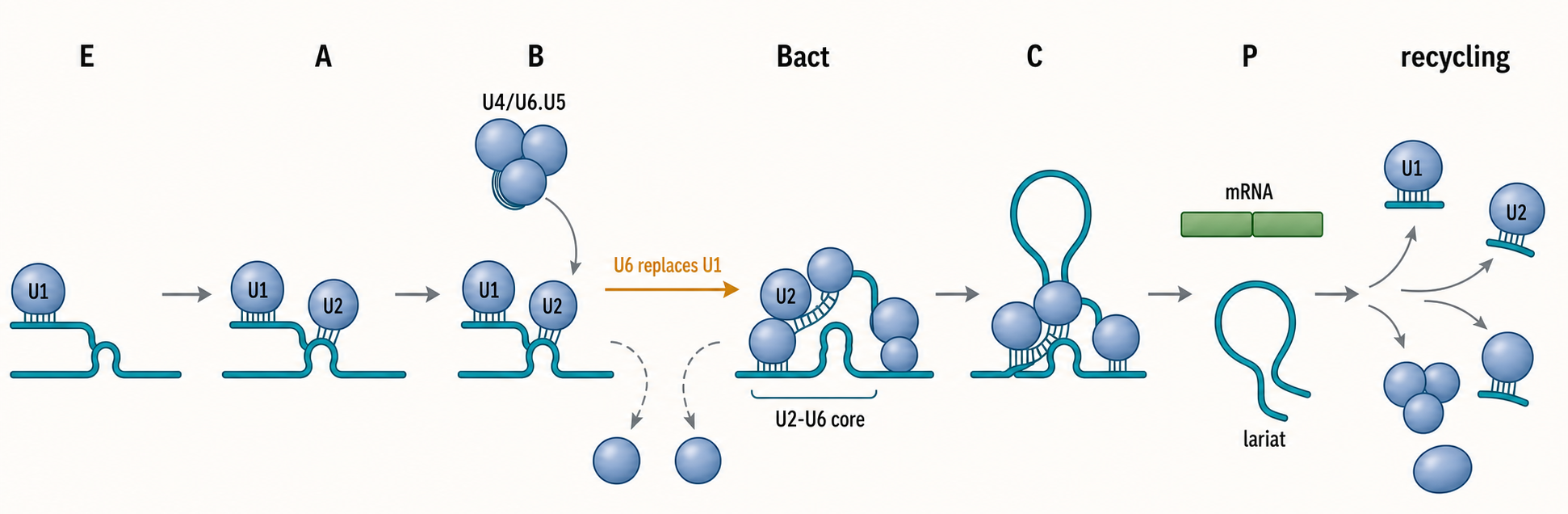
Figure 27.2. Spliceosome Assembly and snRNA Rearrangements. The major spliceosome is assembled anew on each intron through staged snRNP recruitment and RNA remodeling events rather than as a preformed enzyme. U1 snRNP initially pairs with the 5′ splice site while auxiliary factors define the branch-point and 3′ splice-site regions, after which U2 snRNP engages the branch site and bulges the branch-point adenosine for catalysis. Subsequent recruitment of the U4/U6.U5 tri-snRNP creates a fully assembled but inactive B complex, and activation then ejects U1 and U4, allowing U6 to pair with the 5′ splice site and U2 to form the catalytic RNA core. Post-catalytic disassembly releases the mature mRNA and the intron lariat, returning snRNPs to free pools for recycling.
Activation is one of the most important remodeling transitions in RNA biology. U1 must leave the 5′ splice site, U6 must replace U1 at that site, U4 must be unwound from U6, and U6 must pair with U2 to form catalytic structures. The RNA helicase Brr2, called SNRNP200 in humans, helps unwind U4/U6, while other ATP-dependent proteins coordinate additional rearrangements. Once U4 leaves, U6 can form the intramolecular stem-loop and U2/U6 helices that organize the catalytic metal-binding environment.
The first catalytic conformation, often called B*, is prepared for branching. Proteins and RNAs position the branch-point adenosine near the 5′ splice site. Cwc25 and related factors stabilize first-step chemistry in well-studied systems. After the first step, the spliceosome must remodel again so that the 3′ hydroxyl of the 5′ exon can attack the 3′ splice site. This transition from first-step to second-step geometry is not automatic. It requires repositioning of substrates and factors such as Prp16, Slu7, Prp18, and Prp22 in yeast-centered nomenclature, with related human factors in metazoan spliceosomes.
After exon ligation, the spliceosome still has work to do. The mature mRNA product must be released, the intron lariat remains associated with spliceosomal components, and snRNPs must be recycled. Prp22-like and Prp43-like ATPases participate in product release and disassembly, and recent structural work has captured states that illuminate how disassembly begins. Recycling matters biologically because spliceosomal components are limiting, and failure to disassemble can trap factors in dead-end complexes.
The evidence for this pathway comes from genetic suppressor studies, in vitro assembly systems, native gel complexes, affinity purification, crosslinking, single-particle cryo-electron microscopy, and substrate-specific kinetic assays. Cryo-electron microscopy has transformed the field by showing many spliceosomal states at near-atomic or high molecular resolution. The limitation is that structures are snapshots. They must be interpreted with kinetics, perturbation, and biochemical state assignment because spliceosomes are heterogeneous and many factors bind transiently.
Box 27.2. What Counts as Evidence for a Splicing Mechanism?
- RNA-seq product evidence identifies which splice products accumulate in a cell population but does not by itself distinguish altered spliceosome catalysis from altered nuclear retention, RNA stability, or transcription.
- Lariat and intermediate assays (lariat sequencing, in vitro time courses, native gels) can assign a defect to a specific step before or after branching or exon ligation.
- Factor-binding evidence from CLIP or crosslinking identifies candidate regulatory sites but requires perturbation and rescue experiments to demonstrate that binding affects splice-site choice.
- Perturbation and rescue evidence (factor depletion, complementation, antisense oligonucleotide, inhibitor washout) is needed to move from correlation to causality.
- Structural-state evidence from cryo-EM reveals conformational geometry, RNA pairings, and metal-ion positions but represents enriched or trapped states rather than the full kinetic ensemble.
- Kinetic evidence from time-course assays and rate measurements connects structural states to the actual path through assembly, catalysis, and disassembly.
- Clinical variant evidence requires endogenous RNA from disease-relevant tissue or isogenic engineered cell models, not reporter constructs alone, to establish physiological effect.
- Recursive-splicing evidence should combine intermediate junctions, step-specific lariats, coverage patterns, and site perturbation; a sawtooth profile by itself is not conclusive.
- Candidate trans-splicing evidence must exclude genomic fusion and contiguous transcription and control library ligation, reverse-transcriptase template switching, PCR mispriming, and alignment artifacts.
Do not overgeneralize a diagram of assembly into a rigid universal clock. Yeast and human spliceosomes share core logic but differ in factor number, intron architecture, alternative-splicing regulation, and co-transcriptional context. Minor-spliceosome assembly follows related principles but uses different early snRNPs and distinct intron signals. Some protists and highly reduced eukaryotes have unusual spliceosome compositions. Chapter 25 covers co-transcriptional processing; this section focuses on the intron-level RNP choreography.
27.3. Catalytic RNA-metal mechanisms and protein cofactors
Spliceosomal chemistry is a two-step phosphoryl-transfer reaction. In the first step, the branch-point 2′ hydroxyl attacks the phosphate at the 5′ splice site. The 5′ exon is released with a free 3′ hydroxyl, and the intron becomes joined to the branch adenosine through a 2′-to-5′ linkage. In the second step, the 3′ hydroxyl of the 5′ exon attacks the phosphate at the 3′ splice site. The exons are ligated, and the intron lariat is released. Because each step exchanges one phosphodiester linkage for another, no ATP is consumed directly by bond formation.
The catalytic center is RNA-rich. U6 snRNA pairs with the 5′ splice site and with U2 snRNA. Conserved U6 nucleotides and U2/U6 structures help coordinate divalent metal ions and align the reacting groups. This is why the spliceosome is often compared with group II intron ribozymes. Both systems use RNA architecture and metal ions to catalyze lariat formation and exon ligation. Proteins surround and stabilize the active site, but the catalytic core is not simply a protein enzyme with RNA decorations.
Metal ions are essential because phosphoryl transfer requires stabilization of charged transition states, activation or positioning of nucleophiles, and organization of leaving groups. A common framework for ribozyme and spliceosome catalysis is a two-metal-ion model, in which divalent metals help align the nucleophile and stabilize the scissile phosphate environment. Structural studies of human and yeast spliceosomes show catalytic metal positions consistent with RNA-mediated metal-ion chemistry, but exact assignments can depend on trapped state, substrate analog, metal identity, and resolution. Therefore, “metal-dependent catalysis” should be stated with the observed structural and biochemical context.
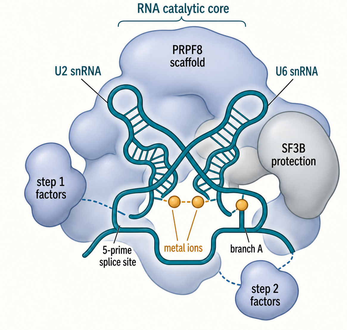
Figure 27.3. RNA-Metal Catalytic Core with Protein Scaffolding. The spliceosomal catalytic center is organized around U2 and U6 snRNA elements that base-pair with each other and with the 5′ splice site, creating a framework that coordinates divalent metal ions essential for phosphoryl transfer. PRPF8 and other proteins scaffold this RNA architecture and stabilize step-specific conformations, but the chemical heart of catalysis is RNA-centered rather than protein-centered. The SF3B complex protects the branch-site region before activation, and distinct step-1 and step-2 protein factors then stabilize the successive RNA geometries required for each transesterification reaction.
Proteins make the RNA enzyme usable in cells. PRPF8 is a large, conserved spliceosomal protein that sits near the catalytic center and helps scaffold the active site. The SF3B complex stabilizes the branch-region architecture before catalysis. U5 snRNP proteins help align exons. Step-specific factors stabilize first-step or second-step conformations. ATP-dependent DExD/H-box proteins remodel RNA-RNA and RNA-protein interactions before and after catalysis. The result is a protein-rich RNP enzyme whose chemical heart is RNA-centered but whose specificity, timing, and robustness depend on proteins.
The transition between catalytic steps is a useful example of why proteins matter. After the first step, the branch-site region and 5′ splice-site region have already reacted. The spliceosome must now position the 3′ splice site and the 5′ exon for exon ligation. Factors that stabilize the first-step conformation can become obstacles to the second-step conformation. Remodeling ATPases help move the spliceosome from one productive geometry to another. A mutation or inhibitor that changes this transition can produce lariat intermediates, exon-skipping products, or stalled complexes rather than a simple all-or-none loss of splicing.
In vitro self-splicing introns help clarify what the spliceosome added to ribozyme chemistry. A group II intron RNA can fold into a catalytic structure that recognizes its exon boundaries and performs lariat splicing with far fewer protein components. However, in cellular contexts group II introns often require maturases or host factors for efficient folding and mobility. The spliceosome can be viewed as a trans-acting descendant or analog of such RNA machines: intron-encoded catalytic RNA information was externalized into snRNAs and many proteins, allowing numerous introns to be removed by a shared apparatus.
The main misconception is to ask whether splicing is “RNA-catalyzed or protein-catalyzed” as if those were mutually exclusive categories. The spliceosome is an RNP catalyst. The RNA components provide conserved catalytic architecture and substrate base-pairing, while proteins enforce assembly order, stabilize conformations, reject bad substrates, and connect splicing to the rest of gene expression. Strong mechanistic claims should specify which catalytic feature is RNA-mediated, which factor contacts a substrate, and which experiment distinguishes direct chemistry from stabilization.
Box 27.3. Spliceosome as an RNP Catalyst
- U1 and U2 snRNAs recognize substrate splice signals by direct base-pairing with pre-mRNA sequences, making the spliceosome an RNA-guided recognition machine.
- The U2/U6 snRNA network forms the conserved catalytic RNA core that coordinates divalent metal ions and aligns the reactive groups for each transesterification step.
- Divalent metal ions, commonly magnesium, are essential for phosphoryl-transfer chemistry in both catalytic steps and are coordinated primarily by RNA.
- PRPF8 and the broader spliceosomal protein complement scaffold, inspect, and stabilize RNA conformations throughout assembly and catalysis but do not substitute for the RNA catalytic center.
- ATP-dependent DExD/H-box helicases remodel RNA-RNA and RNA-protein interactions, driving the spliceosome through productive states, enforcing fidelity checkpoints, and dismantling spent complexes.
- Asking whether splicing is “RNA-catalyzed or protein-catalyzed” presents a false dichotomy; the spliceosome is an RNP enzyme in which RNA provides the chemical scaffold and proteins provide specificity, timing, and coupling to gene expression.
27.4. Fidelity, proofreading, and kinetic competition
Splicing fidelity is the ability to remove the intended intron and join the intended exons while avoiding nearby incorrect sites. The challenge is severe because splice-site motifs are short. A long human intron can contain many sequences resembling partial 5′ splice sites, branch points, or 3′ splice sites. If the spliceosome used only consensus matching, cryptic splicing would be common. Instead, fidelity emerges from sequential recognition, cooperative assembly, kinetic competition, and ATP-dependent rejection.
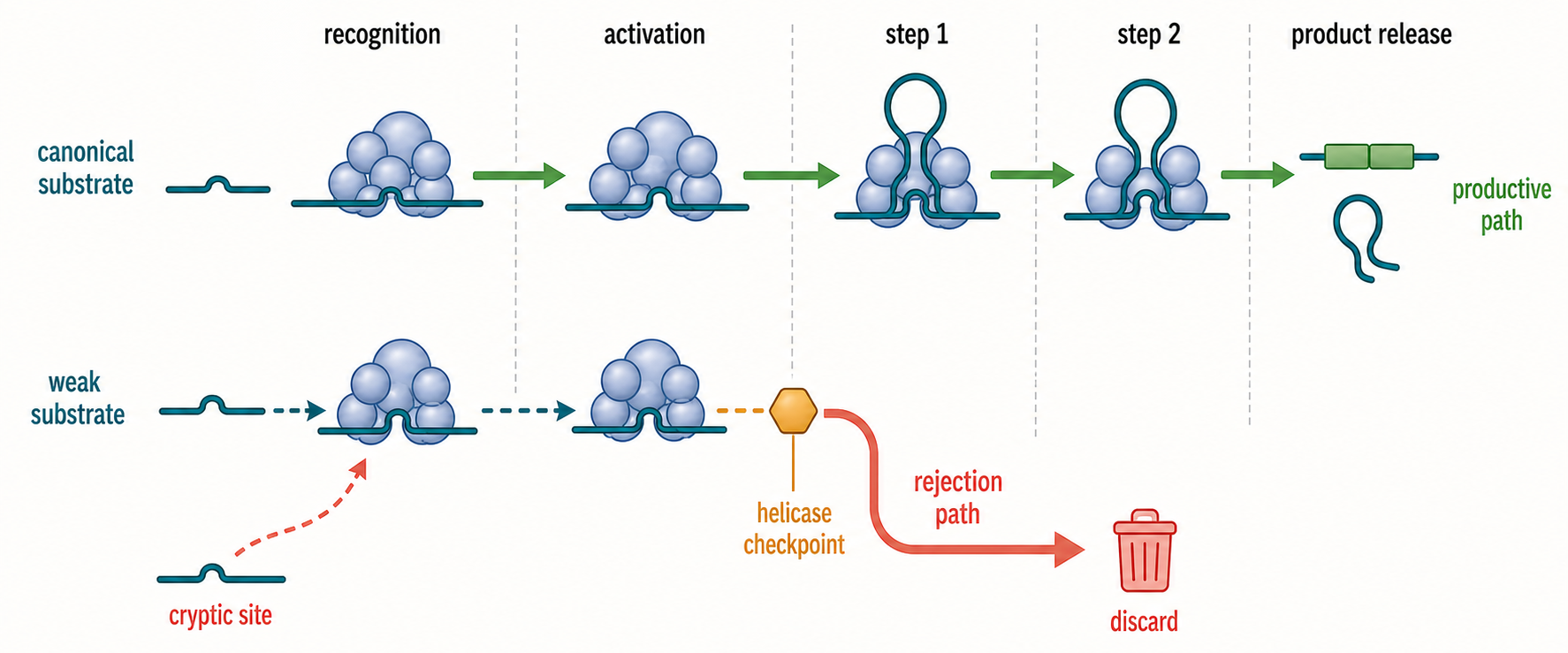
Figure 27.4. Fidelity as Kinetic Competition. Splicing fidelity arises from kinetic competition across the entire assembly and catalytic pathway rather than from a single static proofreading checkpoint. A substrate with strong canonical splice signals progresses rapidly through recognition, activation, first-step chemistry, second-step chemistry, and product release, whereas a substrate with a weak branch site or poor splice-site geometry may stall or be discarded at an ATP-dependent helicase transition. When a canonical splice site is weakened, nearby cryptic sites can compete for spliceosome engagement, and structural studies of rejection complexes such as the DHX35-GPATCH1 system illustrate how the spliceosome can be actively driven away from productive catalysis when substrate features are suboptimal.
Kinetic competition means that multiple possible RNP states compete over time. A strong 5′ splice site may pair efficiently with U1 and transition productively to later states. A weak site may still be recognized if auxiliary proteins stabilize it or if competing sites are absent. A decoy site may bind a factor but fail a later rearrangement. The observed spliced RNA pool is therefore the outcome of rates: binding, release, remodeling, catalysis, discard, RNA synthesis, and RNA decay. This framework is essential for interpreting variants that partly reduce, rather than abolish, splicing.
Proofreading in splicing is not identical to polymerase nucleotide proofreading. There is no copied base that can simply be excised and replaced. Instead, ATP-dependent spliceosomal factors create opportunities to discard or redirect complexes that have poor substrate geometry. In yeast-centered models, Prp5 influences early branch-site engagement, Prp16 promotes rejection or remodeling after first-step decisions, Prp22 can proofread exon ligation and promote mRNA release, and Prp43 participates in discard and disassembly. Human homologs and associated factors implement related logic in larger spliceosomes.
Recent structural work extends this idea to explicit rejection states. The DHX35-GPATCH1 system has been implicated in rejection of aberrant splicing substrates in human spliceosomes, providing a structural view of how a spliceosome can be driven away from productive catalysis when substrate features are wrong. This kind of evidence is important because it moves fidelity models from genetic inference toward molecular state descriptions. It does not mean one factor is responsible for all splice-site fidelity; rather, fidelity is distributed across the pathway.
Fidelity is also shaped before catalysis. Exon definition, SR proteins, hnRNP proteins, RNA structure, transcription speed, chromatin state, and nearby competing splice sites all influence which substrate enters the catalytic pathway. FUBP1, for example, has been reported as a factor that facilitates 3′ splice-site recognition and splicing of long introns, illustrating that splice-site choice can depend on factors beyond the canonical U2AF-SF1 branch-region apparatus. Such examples belong at the boundary between constitutive splicing and regulated splicing, which Chapter 28 develops more fully.
Errors can arise from several mechanistic classes. A weakened canonical splice site can cause exon skipping or intron retention. A newly created cryptic splice site can insert a pseudoexon. Branch-point disruption can stall assembly or shift to an alternative branch point. Spliceosome-factor mutations can change recognition preferences across many transcripts. Small molecules can stabilize or destabilize specific branch-region states. The same RNA-seq phenotype, such as intron retention, can therefore reflect different molecular defects.
Evidence for fidelity requires more than counting splice products. RNA-seq identifies altered products, but proofreading claims require perturbations of candidate ATPases or fidelity factors, substrates with defined mutations, kinetic measurements, stalled intermediates, or structures of rejection states. Genetic suppressors can reveal fidelity checkpoints, but suppressors may act indirectly by changing factor abundance or competing pathways. Minigene reporters can isolate local sequence logic but may miss long intron architecture. Strong fidelity analysis combines product profiling with mechanistic assays.
The practical caution is that “weak splice site” is not a complete explanation. A weak site in one transcript context can be used efficiently in another. A variant predicted to weaken splicing may have little effect in a tissue where an auxiliary factor is abundant. Conversely, a modest motif change can be pathogenic if it activates a strong cryptic site, introduces a premature termination codon, or shifts an isoform balance in a dosage-sensitive gene. Chapter 5 provides general evidence standards for causality; in splicing, causality must connect sequence, RNP mechanism, RNA product, protein or RNA function, and phenotype.
27.5. Group I, group II, and spliceosomal intron evolution
Intron evolution asks how interrupted genes, self-splicing ribozymes, mobile elements, and modern spliceosomes became connected. The field is historical and comparative, so the evidence is indirect. Researchers compare intron distributions across genomes, intron phases within coding sequences, ribozyme structures, snRNA structures, organellar intron mobility, and phylogenetic patterns. Strong claims distinguish observations from reconstructed scenarios.
Group I introns are ribozymes that initiate splicing with an external guanosine. The guanosine 3′ hydroxyl attacks the 5′ splice site, and the 3′ hydroxyl of the upstream exon later attacks the 3′ splice site. This pathway removes the intron without a lariat branch. Group I introns are found in bacteria, bacteriophages, organelles, and some eukaryotic nuclear contexts. Many encode homing endonucleases or depend on proteins that help folding, mobility, or maturation. They show that RNA can solve intron removal by chemistry different from spliceosomal lariat formation.
Group II introns are closer to the spliceosomal story. A group II intron folds into conserved domains, uses an internal branch-point adenosine in many reactions, forms a lariat, and often encodes a reverse transcriptase-like protein with maturase activity. Group II introns can behave as retroelements: the intron RNA and encoded protein can insert into DNA through RNA-templated mechanisms. These features make group II introns plausible ancestors or close relatives of spliceosomal introns and retroelements.
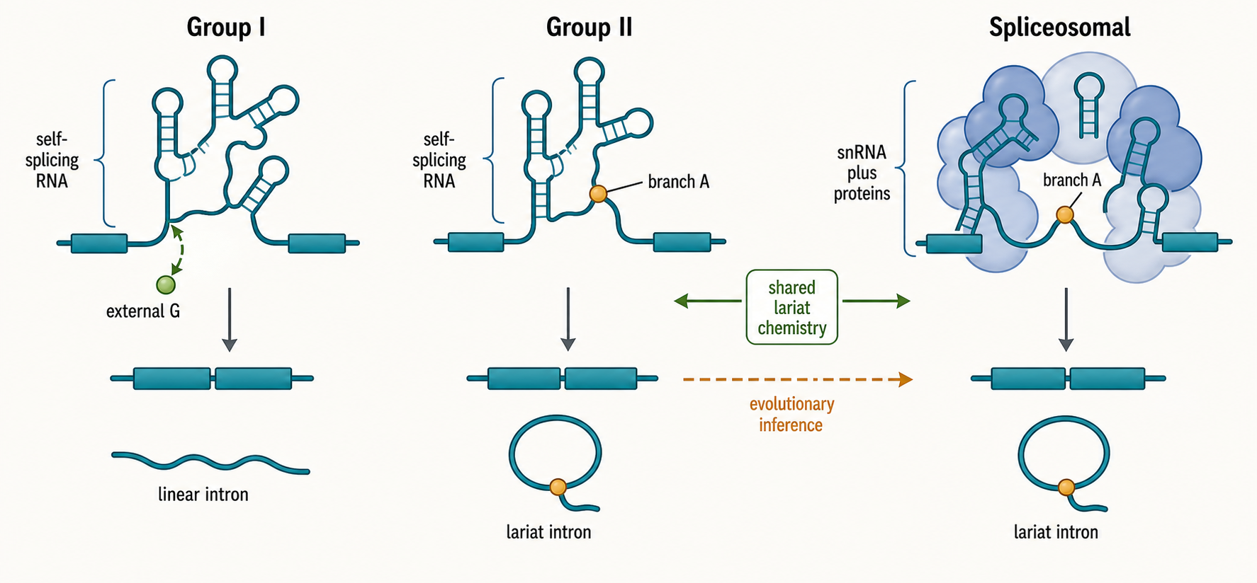
Figure 27.5. Intron Classes and Evolutionary Relationships. Group I, group II, and spliceosomal introns differ fundamentally in initiating nucleophile, cofactor requirement, and product topology, illustrating that “intron” does not name one universal removal mechanism. Group I introns use an exogenous guanosine 3′-hydroxyl as the nucleophile and release a linear excised intron, whereas group II introns use an internal branch-point adenosine to form a lariat, a chemistry mechanistically shared with spliceosomal introns. The modern spliceosome can be interpreted as a system in which catalytic RNA functions analogous to group II intron domains have been externalized into reusable snRNAs and elaborated by dozens of protein factors, though the precise evolutionary steps connecting mobile group II ribozymes to the nuclear spliceosome remain reconstructed from comparative genomic and structural evidence rather than directly observed.
The modern spliceosome can be interpreted as a trans-acting fragmentation and elaboration of group II intron-like functions. Instead of each intron carrying a full ribozyme, snRNAs provide reusable RNA elements that recognize and catalyze splicing across many introns. U2 and U6 snRNAs resemble, at a functional level, the catalytic RNA parts needed to position the branch site and metal ions; U5 helps align exons; U1 and U2-like recognition modules identify substrate features. Proteins supply folding assistance, regulation, fidelity, and coupling to nuclear gene expression. This scenario is widely discussed but remains a reconstructed model, not a directly observed evolutionary sequence.
The timing of spliceosomal intron expansion is debated. Eukaryotic genomes vary widely in intron density, from intron-rich vertebrates and plants to reduced intron-poor lineages. The last eukaryotic common ancestor probably had a complex spliceosome and spliceosomal introns, but exact intron gains, losses, and ancestral intron numbers are inferred with uncertainty. Intron gain can occur through mobile-element activity, transposon insertion, genomic duplication, intron transposition, or repair-associated processes; intron loss can occur through reverse-transcribed mRNA recombination, deletion, or genome streamlining. Different lineages can have different dominant mechanisms.
Organellar introns provide living examples of intron mobility and dependence on protein cofactors. Fungal, algal, and plant mitochondria and chloroplasts contain group I and group II introns with lineage-specific distributions, maturases, homing endonucleases, and host factors. These systems are valuable because they show introns as mobile and regulated elements rather than static interruptions. They also warn against treating nuclear spliceosomal introns as the only intron biology that matters.
Splicing evolution also intersects with regulation. Once introns are common, exon-intron structure can support alternative splicing, exon shuffling, nonsense-mediated surveillance, mRNA export marks, and coupling between transcription and RNA processing. This does not mean introns evolved “for” alternative splicing in a simple adaptive story. Evolutionary explanations must separate origin, persistence, later co-option, and lineage-specific expansion. A feature that is now regulatory may have originated as a mobile element or nearly neutral insertion.
Table 27.3. Intron Classes. Six major intron systems differ in initiating nucleophile, product topology, catalytic machinery, and genomic distribution; the table highlights key distinctions and common misconceptions.
| Intron class | Initiating nucleophile | Product topology | Main catalysts or cofactors | Typical genomic contexts | Mobility features | Evolutionary relevance | Common misconceptions |
|---|---|---|---|---|---|---|---|
| Spliceosomal U2-type | 2′-OH of internal branch-point adenosine | Lariat intron; ligated exons | Major spliceosome (U1, U2, U4, U5, U6 snRNPs) plus protein factors | Nuclear pre-mRNAs of most eukaryotes | Not intrinsically mobile | Proposed descendant of group II intron-like elements; substrate for alternative splicing | GU-AG dinucleotides alone define an intron |
| Spliceosomal U12-type | 2′-OH of internal branch-point adenosine | Lariat intron; ligated exons | Minor spliceosome (U11, U12, U4atac, U5, U6atac snRNPs) | Nuclear pre-mRNAs; rare class present across most eukaryotes | Not intrinsically mobile | Parallel spliceosomal recognition system; evolutionarily distinct from U2-type | Dispensable because these introns are numerically rare |
| Group I | 3′-OH of exogenous guanosine nucleotide | Linear excised intron; ligated exons | Self-splicing RNA; maturase or host proteins may assist folding | Bacteria, bacteriophage, organelles, some eukaryotic nuclear rRNA | Homing endonuclease-mediated mobility | Shows RNA can catalyze intron removal without lariat or spliceosome | Forms a lariat like spliceosomal introns |
| Group II | 2′-OH of internal branch-point adenosine | Lariat intron; ligated exons | Self-splicing RNA; intron-encoded reverse transcriptase or maturase often required | Bacteria, mitochondria, chloroplasts | Retroelement mechanism via reverse transcriptase | Closest mechanistic and evolutionary comparator for spliceosomal introns | Abundant in nuclear eukaryotic genes |
| tRNA introns | 2′-OH of internal ribose | Ligated tRNA halves; linear removed segment | Protein-based tRNA splicing endonuclease and RNA ligase | tRNA genes in eukaryotes and archaea | Not mobile | Distinct protein-enzyme system unrelated to spliceosomal chemistry | Same removal mechanism as pre-mRNA splicing |
| Archaeal introns | 2′-OH of internal ribose | Ligated RNA halves; excised segment | Archaeal splicing endonuclease (protein-based) | rRNA, tRNA, and some mRNA genes in archaea | Not mobile | Probable evolutionary origin of the eukaryotic tRNA splicing endonuclease pathway | Related to spliceosomal introns mechanistically |
The main misconception is to collapse all introns into one origin model. Group I introns, group II introns, spliceosomal introns, tRNA introns, archaeal introns, and protein-spliced inteins are different systems. They share the broad theme of interrupted genetic information but differ in chemistry, enzymes, distribution, and evolution. Chapter 13 covers mobile elements and ancient RNA-linked genome innovation; this section supplies the spliceosome-centered evolutionary framework.
Box 27.4. Open Questions in Intron Evolution
- How group II intron-like catalytic RNA domains were fragmented and externalized into the reusable snRNAs of the modern spliceosome remains mechanistically unresolved and is reconstructed from structural homology and comparative genomics.
- The timing of spliceosomal intron expansion in early eukaryotic evolution is debated and is inferred from genome comparisons across extant lineages, not from direct observation of ancestral states.
- The relative contributions of intron gain versus intron loss to the highly variable intron densities seen across eukaryotic lineages are still being estimated, with different mechanisms likely dominant in different lineages.
- Whether organellar group I and group II introns in fungi, algae, and plants represent true evolutionary intermediates toward spliceosomal introns or divergent mobile-element lineages is not settled.
- Features of alternative splicing regulation that are secondary co-options of an established intron landscape require separate evolutionary explanations from the mechanisms that originally gave rise to spliceosomal introns.
27.6. Trans-splicing chemistry, spliced-leader RNAs, and recursive-splicing boundaries
Several mechanisms are easily confused because all produce exon junctions, but the location of the reacting splice sites distinguishes them. In ordinary cis-splicing, the upstream exon, intron, and downstream exon are covalently connected within one precursor RNA before the reaction. In recursive splicing, that same precursor contains a long intron that is removed in consecutive segments; the RNA remains an intramolecular substrate at every step. In inter-RNA trans-splicing, the 5′ splice site and 3′ splice site are carried on different RNA molecules. Spliced-leader trans-splicing is a specialized inter-RNA reaction in which a dedicated small RNA donates a short 5′ exon to many acceptor transcripts. Alternative splicing is a separate dimension: it describes regulated choice among product isoforms and does not specify whether the reacting sites are in cis or in trans.
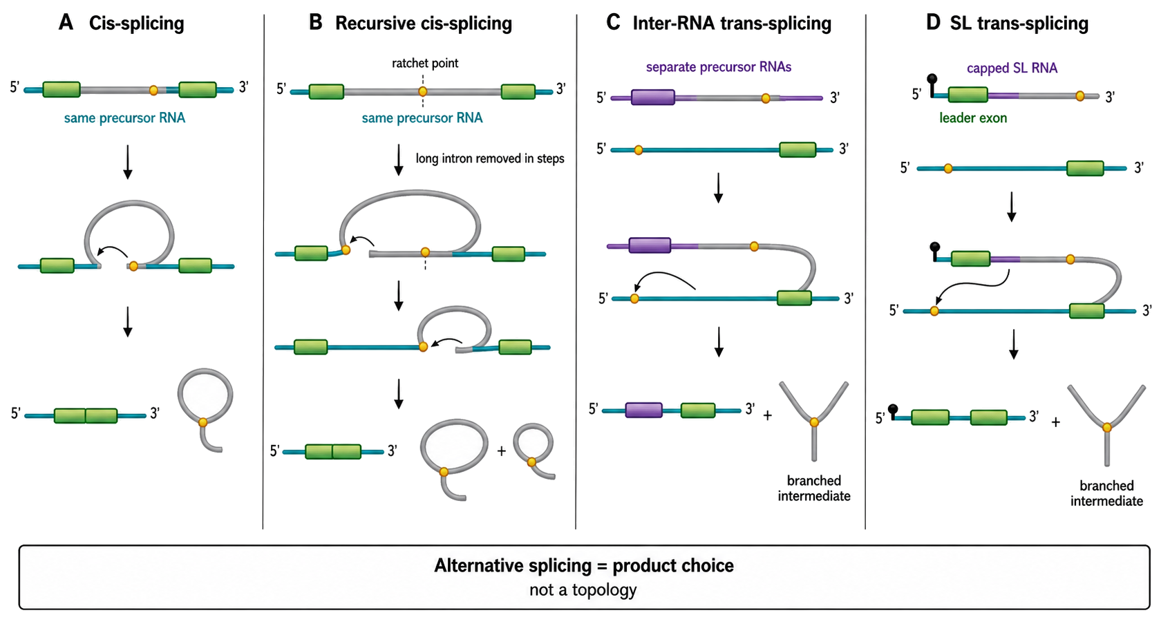
Figure 27.6. Four Splicing Topologies That Must Not Be Confused. A four-panel mechanism comparison. Panel A shows ordinary cis-splicing with both splice sites on one precursor. Panel B shows recursive cis-splicing of one long intron through an internal recursive site and a transient intermediate; a split inset contrasts a predominant Drosophila zero-nucleotide ratchet-point drawing with vertebrate initial recursive-exon definition followed by competition between the reconstituted donor and the recursive-exon donor. Label the Drosophila survey evidence as total-RNA sawtooth plus successive lariats and U2AF sensitivity, and label the vertebrate perturbation boundary: blocking selected sites did not necessarily disrupt accurate final exon joining. Panel C shows general inter-RNA trans-splicing with donor and acceptor exons on separate precursors. Panel D shows SL trans-splicing with a capped SL RNA donating a short leader exon to an acceptor pre-mRNA through a branched Y intermediate; only the leader is retained, so the SL donor is consumed rather than recycled like a U snRNA. Beneath panel D, add two compact lineage callouts: T. brucei uses a 39-nucleotide cap-4 leader, an extended acceptor-side polypyrimidine tract, and divergent U2/U4 Sm cores, with U1 participation unresolved; surveyed dinoflagellates use an approximately 22-nucleotide leader from a major 50–60-nucleotide donor class, with a candidate rather than proven Sm-binding motif in the retained exon. A separate product-choice axis states that alternative splicing is orthogonal to all four topologies. Use molecule-continuity colors and explicit “same RNA” versus “separate RNAs” labels; do not imply that every SL system processes polycistrons, every long intron requires recursion, or a predicted RNP-binding motif proves protein occupancy.
The chemical relationship between cis- and trans-splicing is close. In a spliceosome-mediated inter-RNA reaction, a branch-point adenosine on the acceptor-side intron sequence can attack the 5′ splice-site phosphate carried by the donor RNA. This first phosphotransfer releases the donor exon 3′ hydroxyl and creates a branched intron intermediate whose arms originated on different precursors. The donor-exon 3′ hydroxyl then attacks the acceptor 3′ splice site, joining the two exons. Classic in vitro experiments showed that separated RNA substrates bearing the two sides of an intron can be joined, particularly when complementary sequences help bring the molecules together. The spliceosome still has to recognize a 5′ splice site, a branch region, and a 3′ splice site, but it must solve an additional encounter problem because those elements are not tethered within one molecule.
Inter-RNA trans-splicing is a topology class, not one universal biological pathway. Some systems join two coding precursor RNAs whose exon fragments together produce one complete open reading frame. Other systems use trans-splicing to repair or diversify transcripts. Still others use a dedicated leader donor whose exon is short and largely invariant. Complementary intronic sequences can increase the local concentration and alignment of two general trans-splicing substrates, as in experimental systems, but natural lineages differ in the extent to which substrate pairing, RNP recruitment, or transcriptional proximity specifies partner choice. A chimeric mature RNA therefore does not reveal its assembly pathway by sequence alone.
A spliced-leader RNA, abbreviated SL RNA, is a small noncoding donor RNA with two functional regions. Its 5′ segment is the leader exon that will remain in mature messenger RNA. Immediately downstream lies a 5′ splice site followed by an intron-like segment that is discarded after the reaction. SL RNAs fold into lineage-specific stem-loops and assemble with proteins into SL ribonucleoproteins. Many contain an Sm-protein-binding site or a related RNP-assembly element. The leader is capped before transfer, so trans-splicing can deliver both a defined 5′ sequence and a cap to an acceptor RNA. Unlike a spliceosomal U snRNA, which remains intact and can be recycled, an SL RNA is consumed as a donor substrate: its leader exon enters mature mRNA and its remaining intron-like segment enters the branched discard product. This architecture makes each SL RNA molecule a single-use donor-exon module that the cell must continually replenish, rather than a conventional messenger-RNA precursor or a reusable spliceosomal snRNA.
The phrase “Sm-protein-binding site” also requires lineage-specific evidence. In well-characterized SL RNPs, Sm proteins assemble on a single-stranded element in the intron-like portion of the donor RNA. A dinoflagellate study instead identified an Sm-consensus-like motif within the 22-nucleotide leader exon of unusually short, approximately 50–60-nucleotide SL RNAs. The motif position and folding model made Sm binding plausible, but the study did not directly demonstrate protein occupancy or show whether Sm proteins remain on the transferred leader in mature mRNA. Sequence resemblance should therefore be described as a candidate RNP-assembly site, not as proof of an assembled Sm ring.
The cap cannot be generalized across all SL systems. In Trypanosoma brucei and related kinetoplastids, the 39-nucleotide leader carries cap 4, a hypermethylated structure built from an m7G cap plus extensive ribose and base methylation of the first four transcribed nucleotides. Cap 4 maturation proceeds on the SL RNA before the leader is transferred; capping and successive modifications are coupled to SL-RNA transcription and RNP maturation. By contrast, nematode and other metazoan SL RNAs have their own leader lengths, caps, secondary structures, and RNP requirements. “The SL cap” is therefore not a single conserved chemical object. The safe general statement is that the donor leader is capped and RNP-packaged, with lineage-specific cap chemistry and architecture.
During SL trans-splicing, the acceptor precursor supplies the branch site, polypyrimidine-rich region where applicable, 3′ splice site, and downstream exon. The SL RNA supplies the leader exon and 5′ splice site. Branching joins the acceptor-side branch adenosine to the 5′ end of the SL intron, producing a Y-shaped branched intermediate rather than the closed lariat drawn for a one-molecule intron. Exon ligation then attaches the capped leader to the acceptor exon. In a classic Trypanosoma brucei experiment, metabolic phosphate labeling, nuclease digestion, and two-dimensional nucleotide chromatography identified an adenosine-centered 2′-5′ branch containing the 5′ guanosine of the SL intron; trypanosome extracts also contained debranching activity with properties similar to the activity assayed from HeLa cells. This direct chemical analysis established that the discard RNA is conventionally branched even though its two arms came from separate precursors. Shared chemistry does not imply identical assembly: trypanosomatid spliceosomal proteins and snRNP interactions are divergent, and the SL RNP supplies a donor module that an ordinary cis-intron does not require.
Trypanosomatids provide the clearest example of SL trans-splicing as an obligatory gene-expression system. Long RNA polymerase II transcription units contain tens to hundreds of protein-coding regions, often with unrelated functions, and are transcribed as polycistronic precursors. Processing at an intercistronic region couples addition of an SL to the downstream mRNA with cleavage and polyadenylation of the upstream mRNA. The result is a set of capped, polyadenylated monocistronic RNAs that can be translated or degraded independently. The downstream trans-splice acceptor helps position upstream 3′-end formation, although processing sites can be skipped and maturation need not be completed in one strictly cotranscriptional pass. In Trypanosoma cruzi, stable leader-bearing dicistronic RNAs and RNAs with unusually long unprocessed 3′ regions were found in the cytoplasmic fraction and could yield properly trans-spliced, polyadenylated monocistrons in reporter and transcription-block time-course analyses. These results establish delayed processing capacity, but the proposed use of such intermediates as a regulated translationally latent store, the factors that time their maturation, and whether the later splice occurs in the nucleus or cytoplasm remained hypotheses rather than demonstrated pathway-wide rules.
The trypanosomatid machinery is not simply a smaller mammalian spliceosome. T. brucei contains the five spliceosomal U snRNAs, yet its proteins are sufficiently sequence-divergent that many orthologs were initially missed by genome annotation. Biochemical work identified U2- and U4-specific variants of the Sm core, and acceptor recognition places strong weight on an extended polypyrimidine tract because branch-point and 3′-splice-site sequences show little obvious conservation. The role of U1 snRNP in SL trans-splicing remains a useful example of unresolved lineage-specific assembly: nematode experiments indicated that U1 was dispensable for SL transfer, whereas trypanosome complexes contained both SL and U1 RNAs and abundant U1 proteins, leaving open a structural or trans-splicing-specific role. Shared two-step chemistry therefore does not license importing the mammalian assembly pathway unchanged.
This coupled processing architecture has major regulatory consequences. Because initiation at individual protein-coding genes is not the dominant control point within a polycistronic unit, trypanosomatids rely heavily on trans-splice-site selection, alternative 3′-end formation, RNA-binding proteins, mRNA stability, and translation to tune gene expression. SL addition supplies a common capped 5′ end, while variable acceptor-site use changes 5′ untranslated regions and can alter translation or RNA stability. Nevertheless, the leader should not be described as a generic expression enhancer independent of context. Cap 4 and leader sequence contribute to productive translation in kinetoplastids, but the quantitative consequence of SL addition depends on the species, transcript, processing site, and downstream RNP.
Nematodes use an independently elaborated SL system with different transcript architectures. In Caenorhabditis elegans, SL1 is added mainly at outrons, which are intron-like 5′ leader regions upstream of the first retained exon. SL2-family leaders are used preferentially for downstream genes in operons, where 3′-end formation at an upstream gene is coordinated with trans-splicing of the downstream pre-mRNA. Sequence analysis distinguished a U-rich, or Ur, element typically centered about 40–60 nucleotides downstream of the upstream cleavage site from the more proximal U-rich region associated with cleavage and polyadenylation. Together with perturbation work cited by that analysis, the positioning supports a model in which the Ur element and cleavage stimulation factor help recruit or retain the SL2 machinery on the downstream RNA. Standard operons often place the cleavage and trans-splice sites about 100–120 nucleotides apart; as the distance increases, SL1 use rises, whereas a distinct SL1-type operon can place the two processing sites nearly adjacent. An upstream UC-rich candidate “Ou” element may help define SL1 outrons, but its computational enrichment is not equivalent to direct factor-binding evidence. Thus operon arrangements and leader preferences are mechanistically structured but not absolute categories. Basal and derived nematodes can encode distinct repertoires of SL RNAs, so the C. elegans SL1/SL2 scheme is a useful example rather than a universal nematode blueprint.
Tunicates demonstrate that SL trans-splicing is not confined to parasitic protists or nematodes. In the ascidian Ciona intestinalis, a major leader is trans-spliced to a large but incomplete subset of expressed genes, and both monocistronic and polycistronic transcription contexts occur. Some genes are frequently trans-spliced, some infrequently, and some not detectably trans-spliced. The original 5′ segment of an acceptor precursor can be removed during the reaction, which makes mature-mRNA 5′ ends unreliable markers of transcription start sites. Promoter mapping in such genes requires methods that recover precursor 5′ ends, perturb the acceptor site, or otherwise separate transcription initiation from leader addition.
Dinoflagellates have another distinct SL system. A primary survey recovered the conserved approximately 22-nucleotide leader from hundreds of full-length complementary DNAs across 15 species representing all major dinoflagellate orders. The leader occurred on transcripts encoding diverse proteins, including nuclear-encoded proteins destined for organelles, but targeted assays did not detect it on tested organelle-encoded messenger RNAs or ribosomal RNAs. Genomic-to-cDNA comparisons and the conserved SL–acceptor junction support trans-splicing, while the survey design does not establish that every nuclear mRNA in every dinoflagellate is modified. Across a later six-species analysis, the major SL RNA transcripts were approximately 50–60 nucleotides long, with an additional lower-abundance 70–92-nucleotide class in one Karenia brevis strain. SL genes occurred in SL-only tandem arrays, mixed SL–5S ribosomal-RNA arrays, and other lineage-specific configurations; these arrangements showed no simple progression along the sampled phylogeny even though major transcript length and broad fold were conserved. The conserved leader has been exploited as a lineage-selective primer in transcriptomics, but incomplete 5′ capture can falsely suggest absence of the leader, whereas primer-driven recovery can overstate universality. Dinoflagellate mitochondrial trans-splicing, in which separately transcribed mitochondrial RNA pieces can be joined, is a different pathway and should not be inferred from the nuclear DinoSL system. Other protists, including kinetoplastids, dinoflagellates, and scattered amoeboid or parasitic lineages, use trans-splicing in lineage-specific forms; “protist trans-splicing” is not a single conserved mechanism.
The patchy phylogenetic distribution of SL trans-splicing argues against projecting one lineage’s mechanism onto another. SL systems occur in trypanosomatids, nematodes, flatworms, tunicates, dinoflagellates, and additional scattered animal and protist groups, while many well-studied fungi, plants, arthropods, and vertebrates lack a comparable pervasive SL pathway. A survey of more than 70 metazoan expressed-sequence-tag datasets found additional systems in amphipod and copepod crustaceans, ctenophores, and hexactinellid sponges, yet also found many close relatives without detectable leaders. Under equal gain and loss costs, the authors’ parsimony reconstruction required six to ten independent metazoan gains; even when losses were assumed to be twice as likely, it required two to five gains. This analysis supports repeated origins and the experimental plausibility that an existing Sm-bound small RNA can acquire donor-exon function, but incomplete 5′ ends, shallow taxon sampling, and undetected low-frequency trans-splicing make absence less secure than presence. An ancient origin followed by extensive loss therefore remains a competing historical model. Similar outputs—a common leader at mature 5′ ends—can be homologous within a lineage but convergent across deep phylogenetic distances.
Recursive splicing solves a different physical problem. A very long intron can be difficult to recognize and remove as one unit, especially while the downstream portion is still being transcribed. In recursive splicing, the spliceosome first joins the upstream exon to an internal recursive splice site. That junction creates or exposes a new 5′ splice site at the same neighborhood, called a ratchet point, allowing a second splice to remove the next intron segment. Repetition shortens the effective distance handled in each reaction. Both reacting sites remain on the same nascent precursor, so recursive splicing is intramolecular cis-splicing, not trans-splicing.
In Drosophila, many ratchet points appear in mature RNA as zero-nucleotide exons because an AG acceptor is juxtaposed to a reconstituted GT donor. Ribo-depleted total-RNA sequencing across developmental stages, tissues, and cultured cells identified 197 ratchet points in 130 introns, while recursive junctions, sawtooth coverage, and lariat reads from successive internal segments established stepwise removal. In that dataset, failure to detect lariats that skipped ratchet points and correlation of recursive-junction abundance with host-gene expression supported predominantly constitutive use, although low-frequency regulated skipping could have escaped detection. Depleting either U2AF subunit reduced recursive junctions, and combined depletion eliminated detectable recursive junctions without a comparable loss of ordinary junction reads; this result implicates U2AF in ratchet-point recognition but is not a rate-normalized comparison of unstable nascent intermediates with stable mature products. Genetic and reporter experiments further show that an apparently zero-nucleotide ratchet point can be embedded in a cryptic recursive exon. Competition between the donor at the ratchet point and a downstream donor normally suppresses stable inclusion of that recursive exon; weakening the ratchet-point donor can expose exon inclusion. The zero-nucleotide drawing is thus a useful product-level abstraction, not always a complete account of local exon-definition events.
Vertebrate recursive sites share the principle of segmental intron removal but need not copy the predominant Drosophila architecture. Conserved recursive sites have been identified in exceptionally long vertebrate genes, including genes expressed in neural tissues. In nine high-confidence human cases, recognition of the recursive acceptor required initial definition of a downstream recursive exon: masking that exon’s donor reduced recursive-site use in human cells and at a conserved zebrafish site. After the first splice, the donor reconstituted at the recursive site competes with the recursive-exon donor; blocking the reconstituted donor or changing their relative strengths switches the outcome toward recursive-exon inclusion, often creating a premature termination codon and nonsense-mediated-decay substrate. Importantly, antisense inhibition of selected human and zebrafish recursive sites did not cause inaccurate final exon joining in the assayed transcripts, and RNA-abundance effects differed among genes. Recursive-splicing architecture, necessity, and regulatory consequence are therefore gene- and lineage-dependent. The existence of some human sites does not mean that all long human introns are recursively spliced.
Recursive splicing subdivides the recognition problem posed by some long introns, but evidence that it is required for timely or accurate mature-mRNA production must be established transcript by transcript. A ratchet-point mutation can reduce completion of intron removal, activate a cryptic recursive exon, change reading frame, or alter RNA surveillance. Recursive intermediates can receive exon-junction-complex-related marks and compete among donor sites, linking a nominally constitutive pathway to local isoform decisions. Those product choices are alternative-splicing consequences layered onto recursive chemistry; they do not make recursive splicing itself synonymous with alternative splicing. Long-gene expression, transcription kinetics, and cell-type-specific factor availability can determine when the pathway is most consequential.
Evidence for recursive splicing should demonstrate sequence order and intermediates rather than rely on a single mature junction. Useful signals include total- or nascent-RNA junctions from a constitutive upstream exon to an internal recursive site, the expected sawtooth coverage gradient, lariat branch reads for successive intron segments, short-lived intermediate enrichment, conservation of the composite acceptor-donor motif, and loss or rerouting of products after site mutation. Poly(A)-selected RNA alone is often insensitive because recursive intermediates are rapidly processed. A sawtooth profile alone is also not decisive because transcriptional gradients, mapping biases, and degradation can shape intronic coverage.
Evidence for inter-RNA trans-splicing requires a different control ladder. A candidate junction should be reproduced with independent RNA preparations, strand-aware methods, unique mapping, and multiple junction offsets or molecules rather than PCR duplicates. Genomic DNA and long genomic reads should exclude a structural fusion or unannotated contiguous locus. Direct-RNA or ligation-independent evidence is valuable because reverse transcriptases can switch templates and PCR can join molecules at short homologous sequences. Detection of the predicted branched intermediate, dependence on splice sites or spliceosomal factors, compensatory substrate-pairing tests, and perturbation-rescue experiments move the inference from “chimeric RNA exists” toward “the spliceosome made it in trans.”
SL systems offer a powerful extra signature because the same defined leader maps to a dedicated SL-RNA gene and appears precisely at acceptor sites across many transcripts. Even here, amplification with an SL-specific primer enriches only leader-bearing products and cannot by itself measure the fraction of transcripts that are trans-spliced. 5′ truncation can create false negatives, while internal priming, misassignment among similar genes, and PCR recombination can create false positives. Quantitative conclusions require matched unenriched libraries, molecule-level counting, precursor evidence, and lineage-appropriate controls.
Natural trans-splicing provides a mechanistic foundation for engineered transcript repair. A designed donor can in principle replace a defective 5′, internal, or 3′ region of a target RNA while leaving the genome unchanged. The biological constraints described here—splice-site compatibility, branch and polypyrimidine-region recognition, donor-target encounter, cis-versus-trans competition, RNP assembly, subcellular colocalization, and aberrant-product surveillance—set the feasibility boundary. Product-specific donor architectures, pharmacology, delivery, manufacturing, safety, and comparative therapeutic design belong to Chapter 154, not this mechanism section.
The final terminology rule is simple. Use recursive splicing for stepwise intron removal within one precursor; use trans-splicing when the joined exons came from different RNA molecules; use SL trans-splicing when a dedicated SL RNA donates the leader exon; and use alternative splicing only when discussing regulated choice among different RNA products. A pathway can occupy more than one category—for example, alternative acceptor choice within an SL system—but the categories answer different questions and should never be substituted for one another.
27.7. Splicing assays, inhibitors, disease variants, and correction strategies
Splicing assays should be matched to the claim. Reverse-transcription PCR can quickly test whether a predicted exon is included or skipped, but it is semi-quantitative unless carefully controlled. RNA-seq provides transcriptome-wide product information, but short reads can struggle with full isoform structures and repetitive regions. Long-read RNA sequencing can connect distant exons in individual molecules, but error profiles, coverage, and library preparation biases must be controlled. Lariat sequencing and branch-point mapping can identify branch usage, but lariats are rapidly debranched and degraded, so absence of a lariat read is not proof that a branch was never used.
Table 27.4. Splicing Assays and What They Can Claim. Each splicing assay provides distinct direct outputs and supports specific inferences while leaving other questions unanswered; matching assay to claim is essential for mechanistic and therapeutic interpretation.
| Method | Direct output | Strongest inference | Common limitation | Best orthogonal validation | Useful for therapeutic development? |
|---|---|---|---|---|---|
| RT-PCR | Amplified splice product bands | Exon inclusion or skipping for a targeted event | Semi-quantitative; primer design bias | Quantitative RT-qPCR or RNA-seq | Yes; fast, low-cost screening of splice changes |
| Short-read RNA-seq | Junction reads and per-exon coverage | Transcriptome-wide exon usage and intron retention | Cannot resolve full isoform structures; repetitive regions problematic | Long-read RNA-seq for complete isoform connectivity | Yes; broad off-target splicing profiling |
| Long-read RNA-seq | Full-length transcript sequences per molecule | Complete isoform connectivity in individual molecules | Higher error rate; lower depth; library preparation bias | Short-read RNA-seq for quantification accuracy | Yes; resolves complex multi-exon isoform landscapes |
| Lariat sequencing | Reads spanning 2′-to-5′ branch junctions | Branch-point identity and usage for detected introns | Lariats are rapidly debranched and low-abundance | Branch-point mapping by primer extension or CLIP | Limited; primarily a mechanistic research tool |
| Branch-point mapping | Position of the branch adenosine for a given intron | Branch-point identity and sequence context | Labor-intensive; not easily transcriptome-scale | Lariat sequencing, crosslinking with U2 snRNP | Limited; primarily mechanistic and variant studies |
| CLIP | Protein-RNA contact sites transcriptome-wide | Candidate binding locations for a given factor | Binding is not regulation without perturbation and rescue | Factor depletion and RNA-seq to test functional effect | Moderate; identifies therapeutic target-binding sites |
| Minigene reporter | Splice products from a defined exon-intron construct | Local sequence logic for splice-site or exon choice | May miss long intron, chromatin, elongation, or promoter context | Endogenous locus editing with RNA-seq readout | Yes; standard tool for variant characterization |
| Massively parallel splicing assay | Splice products from thousands of sequence variants | Sequence-activity map across a splice-site or regulatory region | Reporter context differs from the endogenous gene | Endogenous validation of high-priority hits | Yes; variant prioritization for disease and correction design |
| In vitro splicing | Splicing intermediates and products in nuclear extract | Step-specific biochemistry and factor requirements | Extract may not reproduce chromatin, elongation, or in vivo context | Cellular splicing assay with matched substrate | Yes; mechanistic validation of inhibitor mode of action |
| Native gel assembly assay | Spliceosomal complex migration pattern over time | RNP state assignment at defined assembly time points | State heterogeneity; calibration against known markers needed | Cryo-EM, factor depletion, crosslinking | Limited; mechanistic research |
| Cryo-EM | Atomic or near-atomic spliceosome structure | Conformational state, RNA pairing, protein contacts, metal positions | Captures stable or enriched states; not a kinetic ensemble | Kinetic assays, mutational analysis, biochemical factor depletion | Limited; informs structural basis of drug-target interactions |
| Inhibitor profiling | Dose-response curves and splice product shifts | Compound potency and apparent step specificity | Indirect effects on splicing network at high concentrations | In vitro splicing assay, target crosslinking after inhibitor treatment | Yes; lead identification and mechanistic characterization |
| Patient-derived RNA analysis | Endogenous splice products from disease-relevant tissue | Physiological variant effect in a clinically relevant cell type | Tissue access, cell heterogeneity, confounding genetic background | Isogenic cell model, minigene reporter with matched sequence | Yes; clinical biomarker development and validation |
| Recursive-splicing analysis | Upstream-exon-to-recursive-site junctions, intronic coverage gradients, and step-specific lariat or intermediate reads | Candidate stepwise removal of a long intron | Sawtooth coverage alone is not specific; poly(A) selection loses transient intermediates | Recursive-site mutation plus total or nascent RNA and lariat analysis | Limited; mechanism and variant interpretation |
| Candidate trans-splicing validation | Chimeric junction molecules, precursor topology, and splice-site dependence | Inter-RNA spliceosome-mediated joining after alternatives are excluded | Genomic fusion, read-through transcription, mapping, ligation, RT switching, and PCR artifacts can mimic the product | Direct-RNA or ligation-independent assay, genomic exclusion, spliceosome perturbation, branch-intermediate detection | Yes; required before engineered-product claims |
Table 27.5. Correction Strategies for Splicing Defects. Seven correction strategies differ in mechanistic target, delivery requirements, off-target risks, and the evidence standards needed before clinical interpretation.
| Strategy | Mechanistic target | Example use case | Delivery challenge | Off-target concern | Evidence needed before clinical interpretation |
|---|---|---|---|---|---|
| Splice-switching oligonucleotide | Specific RNA sequence blocked sterically to alter splice-site or regulatory-element use | SMN2 exon 7 inclusion in spinal muscular atrophy | CNS penetration; sustained cellular uptake; tissue targeting | Altered splicing of off-target transcripts sharing partial sequence complementarity | Transcriptome-wide RNA-seq off-target profiling; functional rescue in disease model; clinical biomarker |
| Small-molecule splicing modifier | Protein-RNA complex stabilization or inhibition (e.g., SF3B branch-site interaction) | SMN2 exon 7 inclusion; cancer spliceosome mutation sensitization | Oral bioavailability; tissue distribution; therapeutic dose window | Global spliceosome perturbation at supratherapeutic doses | Transcriptome-wide RNA-seq; mechanistic selectivity assays; animal efficacy and toxicity data |
| Genome editing of splice variants | DNA sequence at canonical splice site or deep intronic variant | Removal of a pseudoexon-activating variant; repair of a splice-site mutation | In vivo delivery of editor (AAV or LNP); mosaicism risk | Unintended edits at off-target genomic sites | Deep sequencing for on-target efficiency and off-target DNA changes; RNA correction confirmation; long-term safety data |
| Base or prime editing | Single nucleotide correction at a pathogenic DNA variant | Conversion of a splice-disrupting base to wild-type sequence | Editor size limits some delivery vectors; same delivery challenges as genome editing | Bystander editing at adjacent bases; RNA editing activity at off-target sites | Edit efficiency at target; RNA splicing correction measurement; proteomics or functional rescue assay |
| RNA editing | Enzymatic A-to-I or C-to-U change in the RNA itself | Editing of a splice-site or regulatory element base to restore recognition | Guide RNA delivery; reliance on endogenous ADAR expression levels | Transcriptome-wide A-to-I editing at off-target sites | Editing efficiency at the target site; demonstrated splicing change; functional protein or RNA outcome |
| Engineered trans-splicing | Replacement of a defective 5′, internal, or 3′ transcript region by spliceosome-mediated joining across two separate RNA molecules | Transcript repair without permanent genomic editing | Donor delivery, target-site accessibility, subcellular colocalization, and competition from cis-splicing | Aberrant fusion products, unintended partners, and low-level junction artifacts | Quantification of endogenous corrected product by artifact-resistant methods; partner specificity; absence of aberrant fusions; functional rescue; product design in Chapter 154 |
| NMD modulation (indirect) | Nonsense-mediated decay machinery, to stabilize a partially functional isoform | Stabilizing a retained-intron transcript that encodes functional protein if translated | Risk of global NMD inhibition; short duration of achievable effect | Upregulation of NMD substrate transcripts that may encode toxic proteins | Demonstration that stabilized RNA is translated productively; protein functional assay; safety profiling |
In vitro splicing assays give mechanistic control. A defined pre-mRNA substrate can be incubated with nuclear extract or purified components, and intermediates can be monitored over time. Mutating splice sites, changing branch points, depleting factors, adding recombinant proteins, or applying inhibitors can reveal which step is affected. The limitation is that extract systems may not reproduce chromatin, transcription speed, nuclear organization, long intron context, or cell-type-specific factor concentrations. Biochemical assays are strongest when paired with cellular validation.
Reporter assays occupy a middle ground. A minigene reporter places an exon-intron segment into an expression construct and measures splice products in cells. Massively parallel reporter assays can test thousands of variants or sequence contexts. These approaches are powerful for mapping local splicing grammar and variant effects, but they can miss long-range intronic elements, endogenous promoter effects, RNA polymerase II elongation context, and chromatin-dependent regulation. A reporter result should be treated as evidence about a constructed substrate unless the endogenous transcript is also tested.
Structural and binding methods answer different questions. Cryo-electron microscopy reveals spliceosomal states and active-site geometry. CLIP and related crosslinking methods map where proteins bind RNA in cells. Chemical probing can reveal RNA structure and accessibility. These methods do not directly measure final mRNA output unless combined with product assays. A factor binding near a splice site is not proof that the factor regulates that splice event; perturbation and rescue are needed for causality.
Splicing inhibitors are both tools and drug leads. Natural products and synthetic molecules can perturb SF3B, U2 snRNP function, spliceosome activation, or other steps. Some compounds globally inhibit splicing and are mainly mechanistic probes or oncology leads with narrow therapeutic windows. Other small molecules act more selectively by stabilizing a particular RNA-protein interaction, as in splice-modifying approaches that alter SMN2 exon 7 inclusion in spinal muscular atrophy. The broad category “splicing inhibitor” therefore includes global blockers, selective modulators, and target-specific stabilizers.
Disease variants can affect splicing through many routes. Canonical splice-site variants often disrupt intron removal. Branch-point and polypyrimidine-tract variants can weaken assembly. Exonic and intronic regulatory variants can alter enhancer or silencer binding. Deep intronic variants can create pseudoexons. Variants in core spliceosome genes, such as SF3B1, U2AF1, SRSF2, or minor-spliceosome components, can alter many transcripts and contribute to cancer or developmental disease. Clinical interpretation must connect the DNA variant to RNA evidence and then to protein, RNA, cellular, and patient phenotype.
Correction strategies follow the mechanism. A splice-switching oligonucleotide can block a cryptic splice site, mask a silencer, promote exon inclusion, or force exon skipping. This approach is useful when changing splice-site choice can restore a reading frame, remove a toxic exon, or increase a functional isoform. Nusinersen, an antisense oligonucleotide that modifies SMN2 splicing, and risdiplam, a small-molecule SMN2 splicing modifier, illustrate how the same transcript can be modulated by different therapeutic chemistries when the RNA mechanism is well defined. Beta-globin IVS2-654 studies provide classic experimental examples in which antisense or RNA-repair approaches corrected aberrant splicing in model systems. Genome editing can remove a deep intronic pseudoexon trigger or repair a splice-site mutation, but editing raises delivery, specificity, and permanence questions. The natural trans-splicing mechanisms in the preceding section define the biochemical constraints on engineered transcript repair; product-specific RNA editing and trans-splicing strategies are treated in Chapter 154.
The review by Malard and colleagues on 5′ splice-site selection emphasizes a practical principle: correction requires knowing why a 5′ splice site is selected incorrectly, not merely knowing which product is abnormal. If the disease mechanism is a strengthened cryptic donor, blocking that donor may work. If the mechanism is loss of a canonical donor, adding an antisense oligonucleotide elsewhere may not restore correct assembly unless an alternative productive path exists. If the mechanism is a core-factor mutation, transcript-specific correction may be harder than pathway-level or disease-context intervention.
The assay caveat for therapeutic splicing is off-target measurement. Any intervention that changes splice-site recognition can potentially alter other transcripts. RNA-seq is useful for global profiling, but low-abundance transcripts, rare cell types, and transient intermediates may be missed. Proteomics, functional rescue, animal models, and clinical biomarkers can be needed to determine whether RNA correction is sufficient. The relevant endpoint is not always perfect restoration of the canonical isoform; in some diseases, producing enough functional protein or reducing enough toxic product is the therapeutic goal.
Box 27.5. Splicing Correction Decision Tree
- First characterize the RNA product defect: determine whether the disease-relevant product arises from exon skipping, pseudoexon inclusion, intron retention, or altered alternative splice-site choice.
- Identify the causal variant location: canonical splice site, branch point, exonic or intronic enhancer or silencer, deep intronic cryptic site, or a core spliceosome factor gene.
- Assess whether steric blocking of a competing or aberrantly activated sequence could restore a productive isoform; if so, a splice-switching oligonucleotide targeting that sequence is a candidate.
- Evaluate whether a small-molecule stabilizer or inhibitor of a specific RNA-protein interaction can achieve adequate transcript selectivity at doses that do not broadly perturb spliceosome function.
- Consider permanent DNA correction through genome editing, base editing, or prime editing when the pathogenic variant is well-defined, delivery to the relevant tissue is feasible, and durable correction is the therapeutic goal.
- In all cases, require endogenous RNA correction evidence from disease-relevant cells or tissue, functional rescue of the protein or RNA product, and transcriptome-wide off-target profiling before drawing clinical conclusions.
The chapter-level synthesis is that splicing is a chemical reaction embedded in a recognition and quality-control network. The reaction itself is two RNA-mediated transesterification steps. The biological system that makes the reaction specific is a dynamic spliceosome assembled on a transcript in cellular context. The evolutionary history explains why the system looks like a ribozyme buried inside a protein-rich machine. The medical relevance follows from the same fact: small changes in recognition, timing, or remodeling can reroute RNA products across the transcriptome.
Experimental Foundations and Evidence Standards
Splicing mechanisms should be assigned to evidence types explicitly. Product assays such as RT-PCR, RNA-seq, and long-read sequencing establish what RNA isoforms accumulate. They do not by themselves distinguish altered spliceosome assembly from altered RNA stability. Intermediate assays such as lariat detection, native gels, and in vitro time courses can place defects before or after branching or exon ligation. Binding assays and CLIP identify candidate regulators, but binding must be linked to perturbation and rescue.
Structural biology has exceptional power in this chapter because spliceosomal states are physical assemblies. Structures can show snRNA pairings, substrate positions, metal-ion coordination, and protein contacts. Yet structures can overrepresent stable or trapped states. A complete mechanistic claim usually requires structures plus biochemical kinetics, mutations, or factor depletion. The most defensible statements in this draft therefore use structures as direct support for state geometry and reviews for pathway-level consensus.
Noncanonical topology requires additional evidence. Recursive-splicing claims should combine intermediate junctions, step-specific lariats, coverage patterns, and site perturbation. Inter-RNA trans-splicing claims should exclude genomic fusion and contiguous transcription, control reverse-transcription and PCR artifacts, and demonstrate splice-site or spliceosome dependence. A chimeric read is product evidence, not a mechanism assignment.
Variant interpretation requires endogenous RNA evidence whenever possible. Computational splice predictors can prioritize variants, and reporter assays can test local sequence effects, but patient tissue, disease-relevant cells, or engineered isogenic models are needed to establish physiological effect. Splicing variants are especially prone to context dependence because factor expression differs across tissues and developmental stages.
Biological Contexts Across Organisms and RNA Classes
Metazoan pre-mRNA splicing is dominated by long introns, short exons, exon definition, widespread alternative splicing, and coupling to transcription and chromatin. Budding yeast has shorter introns, fewer intron-containing genes, and historically provided many core mechanistic insights because genetic and biochemical systems were tractable. Plants have extensive splicing regulation, intron-mediated expression effects, and stress-linked splice regulation, while still using the same broad spliceosomal logic of short signals interpreted in transcript and cellular context.
Organellar systems broaden the view. Mitochondria and chloroplasts in fungi, algae, and plants can contain group I and group II introns whose removal depends on ribozyme folding, intron-encoded proteins, and nuclear-encoded cofactors. These introns connect splicing to genome mobility and organelle evolution rather than to nuclear mRNP export.
Protists and parasites provide several independent boundary cases. Trypanosomatids use obligatory SL trans-splicing coupled to processing of long polycistronic precursors; dinoflagellates use a distinct conserved leader on nuclear messenger RNAs; and other protist lineages show their own split-intron or leader systems. Nematodes and tunicates independently demonstrate that SL trans-splicing also occurs in metazoans, with different relationships to outrons and operons. Recent work on Trichomonas vaginalis illustrates how intron plasticity and trans-splicing capability can expose spliceosome diversification in a deeply divergent eukaryote, but broad comparative claims still require lineage-by-lineage evidence.
Recent Consensus
The current consensus is that spliceosomal catalysis is RNA-centered, metal-dependent, and deeply dynamic. U2 and U6 snRNAs form the conserved catalytic RNA network, while proteins provide essential scaffolding, regulation, and fidelity. The major spliceosome and minor spliceosome are distinct recognition systems that converge on related lariat chemistry. Group II introns remain the strongest evolutionary comparator for spliceosomal introns, although details of the transition from mobile ribozymes to the modern spliceosome remain unsettled. Recursive splicing is a stepwise cis pathway for selected long introns, whereas inter-RNA and spliced-leader trans-splicing join separate precursor molecules. SL systems have patchy phylogenetic distributions and lineage-specific architectures, so trypanosomatid, nematode, tunicate, and dinoflagellate mechanisms cannot be collapsed into one universal model. Splicing regulation cannot be cleanly separated from core splicing because splice-site choice is determined by both basal recognition and regulatory context.
Open Questions, Controversies, Deprecated Models, and Common Misconceptions
Open questions:
- How exactly did group II intron-like elements give rise to spliceosomal introns and snRNAs? The broad relationship is persuasive, but the path, timing, and intermediate states remain reconstructed.
- How many fidelity checkpoints exist in human spliceosomes, and how transcript-specific are they? Structural snapshots are rapidly improving this area, but kinetic integration is still incomplete.
- Which disease-associated spliceosome mutations act mainly through altered branch recognition, altered kinetics, altered factor interactions, or downstream RNA surveillance?
- How many long introns use recursive splicing in specific vertebrate cell types, and which apparent recursive sites are constitutive intermediates versus regulated recursive-exon switches?
- Why have SL trans-splicing systems originated or persisted in some eukaryotic lineages but not closely related ones, and which molecular features are homologous rather than convergent?
- Which candidate chimeric RNAs in animals represent reproducible spliceosome-mediated trans-splicing rather than genomic fusion, read-through transcription, or library artifacts?
Common misconceptions:
- “All introns are removed by the spliceosome.” Group I, group II, tRNA, archaeal, and other introns use different systems.
- “GU-AG is enough to define an intron.” It is a common boundary pattern, not a complete recognition code.
- “RNA-seq evidence of intron retention is direct proof of failed catalysis.” Intron-containing RNAs can also accumulate because of transcription, nuclear retention, surveillance, or decay changes.
- “The minor spliceosome is dispensable because U12-type introns are rare.” U12-type introns are rare but often highly conserved, and some control essential or dosage-sensitive genes; rarity is not dispensability.
- “Recursive splicing joins exons from separate RNA molecules.” Recursive splicing removes one long intron in steps on the same precursor RNA; joining separate precursors is trans-splicing.
- “Trans-splicing is a type of alternative splicing.” Trans-splicing describes inter-RNA substrate topology, whereas alternative splicing describes regulated product choice; an event can be one, both, or neither.
- “All spliced-leader trans-splicing resolves polycistronic precursors.” Coupled polycistron processing is central in trypanosomatids and some operons, but SL addition also occurs at outrons and monocistronic transcripts in other lineages.
- “A chimeric RT-PCR product proves trans-splicing.” Reverse-transcriptase template switching, PCR mispriming, library ligation, genomic rearrangement, and mapping errors must be excluded before assigning an inter-RNA splicing mechanism.