Chapter 25. Co-Transcriptional RNA Folding, Processing, Modification, and RNP Assembly
Scope Note
This chapter follows RNA while the RNA is still being made. A nascent RNA is a newly synthesized RNA chain that remains physically near its polymerase, template, or site of synthesis. During this interval the RNA is not only a passive product. It can fold, receive a 5′ cap or another end structure, expose splice sites, recruit cleavage and polyadenylation factors, acquire chemical modifications, bind proteins, assemble into ribonucleoprotein particles, trigger surveillance, and feed back on chromatin or transcription. Polymerase elongation, pausing, and termination are treated in Chapter 24. Mature 5′ cap chemistry, spliceosome catalysis, 3′-end formation, RNA modification systems, nuclear mRNP export, RNA decay, and stable RNP biogenesis are developed in later specialist chapters.
The chapter’s central claim is that RNA maturation is a kinetic pathway. The mature RNA observed at steady state is the result of synthesis plus time-ordered folding, factor binding, processing, modification, and quality control. Many of these events are reversible, delayed, or remodelled after transcript release, so “co-transcriptional” should not be read as “universal,” “complete,” or “functionally decisive” without evidence for the specific RNA, organism, and condition.
Executive Summary
RNA begins to mature before transcription ends. The order in which nucleotides emerge from an RNA polymerase creates a vectorial folding pathway: 5′ sequences appear first, downstream sequences appear later, and the early structures or protein contacts can bias all later choices. A full-length RNA refolded in vitro is therefore not always a faithful model of the pathway used by the same RNA in a cell. Elongation speed, transcriptional pausing, ionic conditions, ribosomes in bacteria, chromatin in eukaryotes, RNA-binding proteins, RNA helicases, and processing factors all alter the local time window in which competing structures can form.
Co-transcriptional folding has a clear mechanistic basis. A newly emerged RNA segment can form a hairpin, bind a protein, pair with upstream RNA, pair with DNA in an R-loop, or remain unstructured. Each choice changes what is available to later factors. Ribosomal RNA illustrates the scale of the problem: rRNA transcription, folding, modification, processing, ribosomal protein binding, and assembly-factor action are interleaved, and the pathway is actively guided to avoid long-lived misfolded intermediates.
For many RNA polymerase II transcripts, 5′ capping is one of the earliest stable processing events. The nascent 5′ end emerges from Pol II, capping enzymes are recruited in coordination with early elongation and C-terminal domain phosphorylation, and the cap becomes an identity mark read by downstream processing, export, translation, and surveillance systems. The cap model must nevertheless be broadened. RNA 5′ ends include canonical m7G caps, cap variants, metabolite-linked caps, uncapped triphosphate or monophosphate ends, and products of recapping or end repair in selected contexts.
Splicing and 3′-end formation are also coupled to transcription. As splice sites emerge, spliceosomal components and regulatory RNA-binding proteins recognize the nascent pre-mRNA in a moving kinetic environment. Elongation rate, pausing, chromatin, RNA structure, CTD state, and factor concentration can influence whether a splice-site interaction becomes productive. Recent evidence that U2AF can cycle through phases of co-transcriptional recognition supports a dynamic model in which early binding, release, proofreading, and commitment are separated rather than collapsed into one static event. Cleavage and polyadenylation similarly connect nascent RNA sequence, processing factors, chromatin, and termination.
RNA modifications can be installed or regulated while RNA is still chromatin-associated or otherwise nascent, but modification timing and modification function are different claims. Pseudouridine synthases can modify human pre-mRNA co-transcriptionally in some settings, and m6A-linked events can connect RNA-binding proteins, transcription termination, and genome stability in particular mammalian contexts. A mark detected on nascent RNA may alter structure, recruit readers, change processing, or reflect the RNP environment in which the RNA was made. Functional interpretation requires perturbation of the relevant writer, reader, eraser, RNA site, or transcriptional context.
Ribonucleoprotein assembly is the broadest consequence of co-transcriptional maturation. rRNA, tRNA, snRNA, snoRNA, telomerase RNA, long noncoding RNA, and mRNA all become biologically meaningful through protein association. The first stable fold or binding event can recruit a processing enzyme, block a competing factor, protect an RNA from decay, or send the RNA into surveillance. RNP assembly is therefore a kinetic competition among folding, protein binding, processing, modification, export, localization, translation, and decay.
Nascent RNA also participates in quality control and genome regulation. Defective caps, unprocessed introns, abnormal RNPs, unresolved R-loops, or stalled processing reactions can trigger nuclear retention, termination, decay, or chromatin-associated responses. Nascent RNA, RNA-DNA hybrids, and RNA-bound proteins can influence chromatin state and transcription, but evidence must distinguish direct feedback from indirect association. Nascent RNA methods now capture folding intermediates and RNA contacts with increasing resolution, yet these methods depend on capture chemistry, crosslinking, extraction, library construction, and modeling assumptions.
Concept Inventory
- Co-transcriptional RNA folding: the formation of RNA secondary or tertiary structure while the RNA is being synthesized. The defining feature is timing: folding begins before the full-length RNA has been released. The main boundary case is that a structure observed after in vitro refolding of a completed transcript may not represent the structures that formed during cellular transcription.
- Vectorial folding: folding constrained by the order of RNA emergence, usually 5′ to 3′. A 5′ hairpin can form before a downstream competing helix exists. Later helicases, ribosomes, ribosomal proteins, spliceosomal factors, or RNA chaperones can remodel the early structure, so vectorial folding is not destiny.
- 5′ capping: the addition or formation of a protective and regulatory chemical structure at the 5′ end of selected RNAs, especially Pol II transcripts. The canonical m7G cap is the central eukaryotic example, but cap biology also includes cap1 and cap2 methylation, noncanonical metabolite caps, uncapped ends, recapping, and cap quality control.
- Co-transcriptional splicing: that splice-site recognition, spliceosome assembly, or intron removal occurs while the RNA remains associated with the transcribing gene or polymerase. It does not mean every intron is removed before transcription ends. Intron length, exon architecture, cell type, elongation rate, chromatin state, and factor availability all influence timing.
- Cleavage and polyadenylation: the coupled 3′-end processing reaction in which many Pol II transcripts are cleaved downstream of a polyadenylation signal and receive a poly(A) tail. CPA is linked to termination, but cleavage, poly(A) addition, downstream RNA degradation, and polymerase release are separable steps.
- Co-transcriptional RNA modification: installation or regulation of a chemical modification before transcript release. Examples include selected pseudouridine and m6A-linked events on nascent transcripts, as well as extensive modification during rRNA and tRNA maturation. Detection on chromatin-associated RNA is not by itself proof of direct installation during transcription.
- RNP assembly: the ordered association of RNA with proteins to form a ribonucleoprotein complex. An RNP can be catalytic, structural, regulatory, transport competent, or a quality-control intermediate. Some assembly steps occur co-transcriptionally; other steps occur after nuclear export, localization, or compartmental transfer.
- Co-transcriptional ribosome assembly: folding, processing, modification, and protein association of rRNA during or soon after rRNA transcription. Bacterial and eukaryotic ribosome assembly both involve co-transcriptional events, but they differ greatly in compartment, precursor organization, assembly factors, and surveillance pathways.
- Nascent RNA surveillance: quality control acting while an RNA is being made or before the RNA leaves the synthesis site. Surveillance can remove defective products, terminate unproductive transcription, retain abnormal RNPs, or regulate normal transcript output. It is not merely a trash-disposal pathway.
- Nascent RNA-chromatin feedback: the influence of nascent RNA, RNA-DNA hybrids, RNA-bound proteins, or processing complexes on chromatin state and transcription behavior. The boundary case is essential: a transcript found near chromatin may be a product, regulator, scaffold, passenger, or artifact of extraction.
What to Know Before Reading This Chapter
Transcription synthesizes RNA in the 5′ to 3′ direction. The 5′ end of the RNA is made first, and later nucleotides are added to the 3′ end. In DNA-dependent transcription, a short RNA-DNA hybrid forms inside the polymerase before the RNA separates from the template and exits through an RNA channel. Chapter 24 explains elongation complexes, pausing, backtracking, termination, and nascent RNA methods. This chapter uses that machinery as the physical context for RNA maturation.
RNA folding is driven by base pairing, base stacking, tertiary contacts, ions, solvent, temperature, and protein binding. A hairpin is a simple secondary structure in which complementary segments of one RNA strand pair to make a stem and leave a loop. More complex RNAs, such as rRNA and riboswitches, use many helices and tertiary contacts. Chapters 3 and 4 cover the thermodynamic and structural principles. The new idea here is timing. A full RNA sequence does not become available all at once during transcription. The RNA is built piece by piece, and early pieces can make choices that affect the later path.
Processing means chemical or enzymatic alteration of the RNA after initial phosphodiester-bond synthesis. Examples include capping, cleavage, poly(A) addition, intron removal, trimming, nucleotide modification, editing, and end repair. RNP assembly means protein association with the RNA. Processing and assembly are coupled because many enzymes recognize RNA-protein complexes rather than naked RNA, and many proteins bind only after an RNA segment has folded or been modified.
The reader should also distinguish timing from causation. If a modification, protein, or RNA structure is detected on chromatin-associated RNA, the event may be co-transcriptional. That observation alone does not prove that the event caused transcriptional output, splice-site choice, RNA stability, or chromatin change. Strong causal claims usually require time-resolved measurement, acute perturbation, site-specific mutation, rescue, and an assay that separates direct RNA effects from indirect changes in transcription or cell state.
Two running examples will recur. The first is a metazoan Pol II pre-mRNA that is capped early, spliced during elongation, cleaved and polyadenylated near the end of the transcription unit, and packaged into an export-competent mRNP. The second is rRNA, which folds and assembles with proteins and assembly factors while it is transcribed. These examples are deliberately different: mRNA processing produces an informational RNA for translation, whereas rRNA assembly produces a large structural and catalytic RNP. The shared principle is kinetic coupling.
25.1. Nascent RNA folding during elongation
Nascent RNA folding begins with a physical asymmetry. The 5′ end of a transcript emerges before downstream sequence exists. If the first 40 nucleotides can make a hairpin, that hairpin can form while nucleotide 200 has not been synthesized. If a later segment could pair with the same 5′ region, the later segment must compete with a structure that may already exist. This is vectorial folding. It is not a special property of a few regulatory RNAs; it follows from the direction of transcription itself.
Table 25.1. Co-Transcriptional Events by RNA Class. Co-transcriptional events and the key caveat for each major RNA class, spanning polymerase identity through RNP assembly.
| RNA class | Polymerase | Early folding event | Processing event | Modification event | Assembly event | Key caveat |
|---|---|---|---|---|---|---|
| mRNA | RNA Pol II | 5′-UTR secondary structure; aptamer folding (bacteria) | 5′ capping; splicing; cleavage and polyadenylation | m6A; pseudouridine (pre-mRNA) | Cap-binding complex; spliceosomal factors; mRNP export factors | Not all introns removed co-transcriptionally; timing varies by intron and gene |
| rRNA | RNA Pol I (eukaryotes); bacterial RNAP | 5′ rRNA domain folding; ribosomal protein binding | Pre-rRNA cleavage at early spacer sites | 2′-O-methylation; pseudouridine (snoRNP-guided) | Ribosomal proteins; DEAD-box helicase assembly factors | Assembly is guided; misfolded intermediates require factor-mediated correction |
| tRNA | RNA Pol III | Cloverleaf secondary structure | 5′ leader removal (RNase P); 3′ trailer trimming; intron splicing (some); CCA addition | Extensive base and ribose modifications | Aminoacyl-tRNA synthetase; surveillance factors | Modification order varies by organism and tRNA species |
| snRNA | RNA Pol II (most); Pol III (U6) | Stem-loop structures | 5′ capping; 3′-end trimming; nuclear export; cap hypermethylation; nuclear re-import | Trimethylguanosine cap | Sm protein ring; snRNP-specific proteins | Assembly crosses nuclear and cytoplasmic compartments |
| snoRNA | RNA Pol II (intronic); Pol III (some) | Guide element folding | Excision from intron or processing from independent transcript | None installed on snoRNA itself | Box C/D proteins (fibrillarin) or H/ACA proteins (dyskerin) | snoRNA guides rRNA and snRNA modification; protein assembly required for function |
| Telomerase RNA | RNA Pol II (vertebrates); Pol III (yeast) | Template pseudoknot folding | 3′-end processing; H/ACA domain maturation | H/ACA-guided pseudouridylation (vertebrates) | TERT; dyskerin; accessory proteins | Ordered transport and assembly steps required; biogenesis defects cause dyskeratosis congenita |
| Long noncoding RNA | RNA Pol II | Local secondary structures; chromatin-associated folding | 5′ capping; variable splicing; polyadenylation | m6A (some lncRNAs) | Chromatin-regulatory proteins; Polycomb; coactivators | Function often inferred from locus perturbation; RNA-specific evidence frequently lacking |
The difference between vectorial folding and equilibrium refolding is pedagogically important. In an equilibrium refolding experiment, the full-length RNA is denatured and then allowed to fold with all sequence elements present simultaneously. That experiment can reveal stable structures and useful thermodynamic preferences. During transcription, however, the RNA may spend seconds or minutes in intermediates that would be rare or absent when the full sequence is available. A transient stem, a protein-protected loop, or a local tertiary contact can guide processing even if it is not part of the final mature structure.
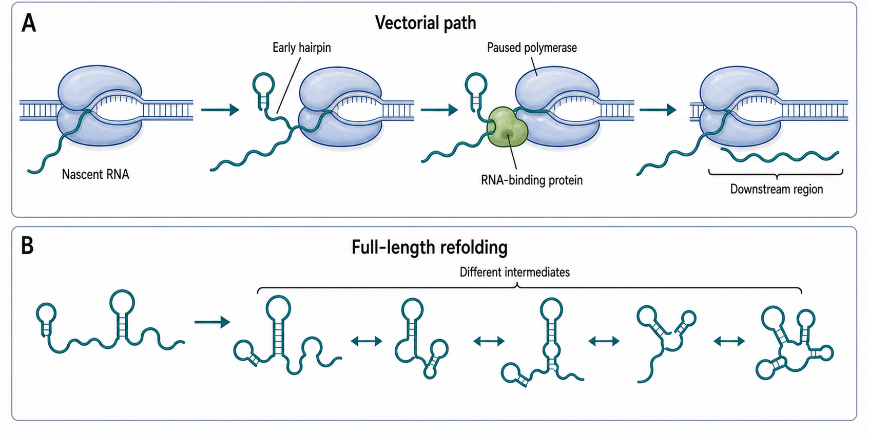
Figure 25.1. Vectorial Folding During Transcription. The 5′ region of a nascent RNA emerges first from the polymerase, allowing early hairpin structures to form before downstream complementary sequences are available. When the polymerase pauses, upstream RNA gains additional time to fold or bind protein, altering the structures accessible to later factors. Because folding begins before the full-length sequence exists, co-transcriptional folding can differ fundamentally from equilibrium refolding of a completed transcript in vitro.
Elongation rate changes the folding problem. A rapidly elongating polymerase gives downstream sequences less delay before they appear. A paused polymerase gives upstream RNA more time to fold, bind ligand, bind protein, or be modified before downstream competitors emerge. In bacteria, a pause can determine whether a terminator hairpin or an antiterminator structure forms. In riboswitches, ligand binding to a nascent aptamer can bias the later expression platform. In eukaryotic pre-mRNA, transcription speed can change the time interval between presentation of a splice site and arrival of a competing downstream element. These examples are not identical mechanisms, but they share one kinetic principle: pausing changes the time available for competing molecular events.
Protein binding can either stabilize or remodel nascent structures. An RNA-binding protein may bind an unstructured motif and prevent it from pairing elsewhere. Another protein may recognize a hairpin and protect it. A helicase may unwind an early structure, allowing a new fold. A ribosome translating a bacterial mRNA can cover RNA and prevent some structures or Rho loading. A ribosomal protein binding rRNA can stabilize a local domain and help recruit later factors. For this reason, cellular RNA folding should be described as RNA folding in an RNP environment, not as RNA folding in isolation.
rRNA is the most visually obvious example of co-transcriptional folding and assembly. Ribosomal RNA molecules are long, structured, and densely associated with proteins. If rRNA were left to find its mature fold without guidance, misfolded intermediates would be a severe problem. Instead, rRNA transcription is coordinated with folding domains, ribosomal protein binding, nucleotide modification, precursor cleavage, and assembly-factor action. In bacteria, rRNA can begin to fold and bind proteins while the transcript is still emerging from the rRNA operon. In eukaryotes, pre-rRNA transcription occurs in the nucleolus and feeds into a larger assembly network involving small nucleolar RNPs, processing enzymes, and many assembly factors. The bacterial and eukaryotic pathways differ, but both show why ribosome biogenesis is a pathway rather than one spontaneous folding reaction.
Sequential structure-probing methods make the kinetic nature of folding experimentally accessible. Chemical probes such as dimethyl sulfate or SHAPE reagents can report nucleotide accessibility or local flexibility. When probing is timed during transcription or applied to defined transcription intermediates, the experiment can reveal structures populated before the full-length RNA exists. The limitation is that chemical reactivity is an indirect structural signal. A reactive nucleotide may be unpaired, flexible, solvent-exposed, or affected by protein binding. Conversely, an unreactive nucleotide may be paired, buried, protein-protected, or inaccessible to the reagent. Structure-probing data become more persuasive when combined with mutational tests, orthogonal probes, biochemical folding assays, polymerase-speed perturbations, and functional readouts.
Table 25.2. Evidence for Co-Transcriptional Timing. Methods used to infer co-transcriptional timing, with the molecular signal each reports, the inference it supports, its main limitation, and the best orthogonal validation strategy.
| Method | Molecular signal | Temporal inference | Limitation | Best orthogonal validation |
|---|---|---|---|---|
| Chromatin-associated RNA-seq | RNA co-purified with chromatin fraction | Suggests transcription-site proximity of RNA | Cannot distinguish active synthesis from chromatin retention | Metabolic labeling; nascent RNA sequencing |
| Nascent RNA sequencing | RNA ends co-purified with elongating Pol II | Maps active transcription positions genome-wide | Sensitive to RNA cleavage and library preparation artifacts | Pol II ChIP-seq; live-cell imaging |
| Live imaging | Fluorescent signal at transcription locus in real time | Directly observes co-transcriptional events | Reporter design may alter RNA folding or processing | Endogenous RNA tagging; nascent RNA sequencing |
| Structure probing | Chemical reactivity of nucleotides (DMS, SHAPE) | Infers base-pairing state during or after elongation | Reactivity reflects multiple structural states including protein binding | Compensatory mutagenesis; orthogonal probe chemistry |
| ChIP/RIP | Protein co-immunoprecipitated with chromatin or RNA | RNA-protein co-occupancy at the gene | Antibody cross-reactivity and crosslinking artifacts | Sequential ChIP; endogenous epitope tagging |
| Mass spectrometry | Chemical identity of RNA modification | Establishes modification chemistry directly | Loses transcript-level and site-level resolution | Site-specific sequencing; reverse-transcription signature assays |
| Metabolic labeling | Newly synthesized RNA (e.g., 4-thiouridine incorporation) | Captures recently transcribed RNA in a defined time window | Incorporation bias; can perturb cellular physiology | Chromatin fractionation; Pol II co-immunoprecipitation |
The main boundary case is post-transcriptional remodeling. Many RNAs do not keep the first structure they form. Helicases, chaperone proteins, ribosomes, spliceosomes, ribosomal proteins, and RNA decay factors can remodel the RNA after the first fold. A mature RNA structure can therefore be consistent with multiple histories. The strongest claims about co-transcriptional folding specify both a structure and a timing mechanism.
Box 25.1. Mature RNA Does Not Reveal the Whole Folding Path
- A mature RNA fold reflects the endpoint of synthesis, processing, and remodeling—not the sequence of transient intermediates that shaped the pathway.
- Structures formed early during transcription may have directed splice-site choice, recruited modification enzymes, or triggered surveillance, then been replaced by the final fold.
- In vitro refolding of a full-length RNA can miss kinetically important intermediates that arise only when sequence is added one segment at a time.
25.2. Cotranscriptional capping and 5-prime processing
The 5′ end is the first chemical end of a transcript to appear. For many eukaryotic Pol II transcripts, this end is rapidly capped. The canonical mRNA cap is an N7-methylguanosine linked to the first transcribed nucleotide through an unusual 5′-to-5′ triphosphate bridge. Chapter 26 treats cap chemistry in detail. The point for this chapter is timing: capping begins while the transcript is short and still associated with Pol II, so the cap becomes an early marker of transcript identity.
The mechanistic sequence can be stated causally. A newly initiated Pol II transcript has a 5′ triphosphate. The capping apparatus acts on that end through triphosphatase, guanylyltransferase, and methyltransferase activities, producing a capped RNA. Recruitment is coordinated with the early elongation complex and the phosphorylation state of the Pol II C-terminal domain, or CTD. The CTD is a repetitive tail on the largest Pol II subunit that provides a changing interaction platform during transcription. Different phosphorylation patterns help recruit or release factors involved in capping, elongation, splicing, 3′ processing, and termination. This does not mean the CTD is a simple code with one mark for one factor. It is better understood as a dynamic, context-dependent binding surface.
Capping protects RNA from 5′ exonucleases, but protection is only one function. The cap is bound by the nuclear cap-binding complex, which helps connect the transcript to splicing, 3′-end processing, export, pioneer translation, and quality control. A transcript that fails to acquire or retain the proper cap can be routed into surveillance. Because capping occurs early, capping status can influence downstream steps before the transcript is complete.
The phrase “5′ processing” is broader than canonical m7G capping. Some RNAs are generated by cleavage from longer precursors and therefore obtain 5′ monophosphate ends. Pol III tRNA precursors undergo 5′ leader removal, usually by RNase P, as part of tRNA maturation. Some small RNAs have specialized caps or cap-like structures. Some RNAs can be recapped after end processing or decay-associated events. Noncanonical caps such as coenzyme-linked or metabolite-linked ends have expanded the range of known RNA 5′ chemistry, although the prevalence and function of each cap type are RNA-class and organism-specific.
Evidence for co-transcriptional capping comes from factor recruitment, biochemical enzyme specificity, nascent RNA analysis, genetic perturbation, and 5′-end mapping. Each evidence type has a limitation. CTD association shows recruitment potential, not necessarily catalytic completion. 5′ end sequencing can be biased by RNA abundance, end chemistry, decapping during extraction, and adapter-ligation efficiency. Antibody or cap-binding enrichment can confuse cap presence with protein affinity unless specificity controls are used. Mass spectrometry can identify chemical species but often requires larger input and may lose transcript-level information. A strong capping-timing claim therefore combines end chemistry with nascent transcript context.
There are useful boundary cases. Bacterial primary transcripts are not canonically m7G-capped like Pol II mRNAs, but bacterial RNAs can have triphosphate ends, monophosphate ends after processing, and metabolite-linked caps in selected contexts. Viral RNAs may be capped by viral enzymes, host enzymes, cap snatching, or noncanonical strategies. Organellar transcripts use still other combinations of processing and end maturation. The universal principle is not “all RNAs are capped”; it is that the 5′ end is an early chemical interface that can determine RNA fate.
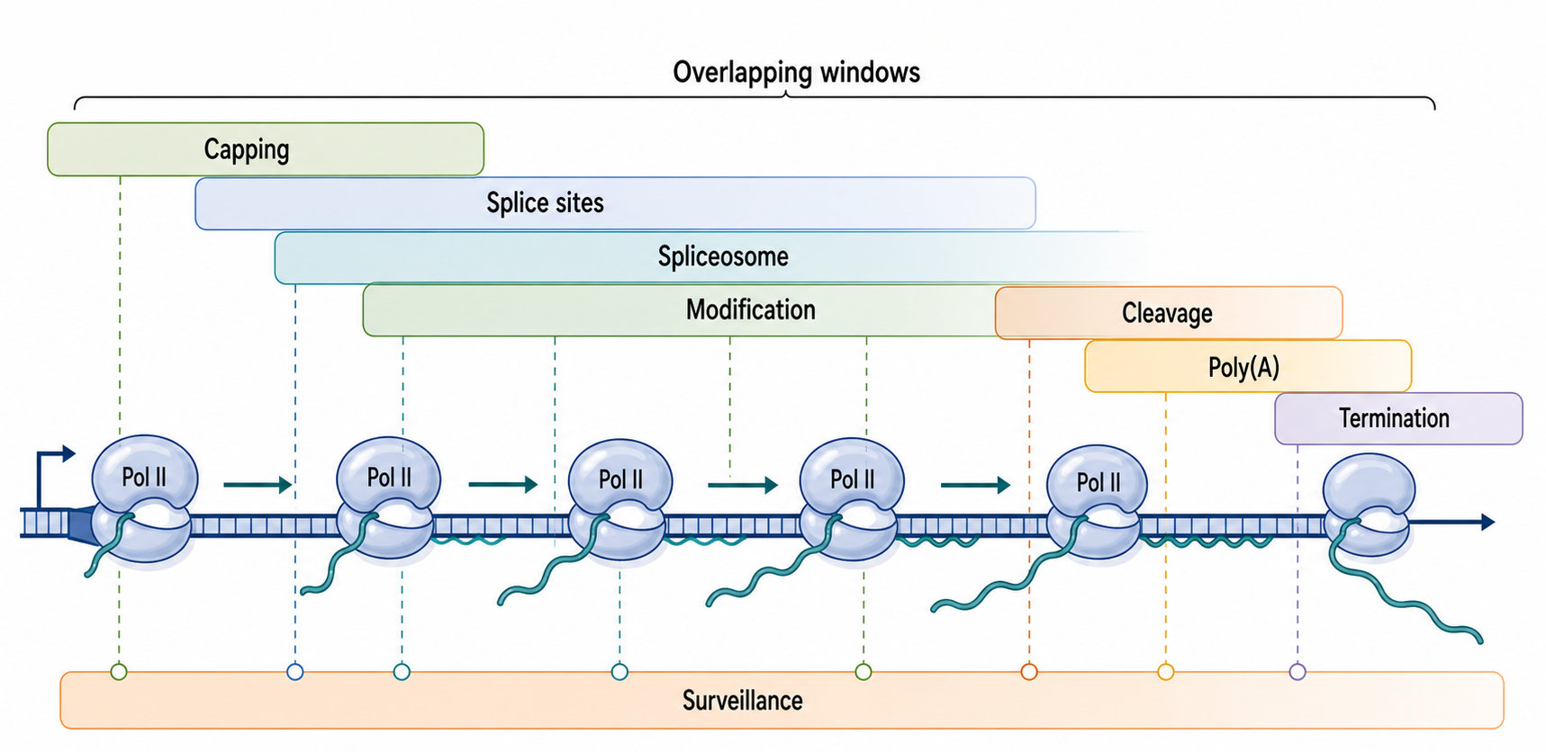
Figure 25.2. Pol II Nascent RNA Processing Timeline. During Pol II transcription, capping begins at the 5′ end while the transcript is still short, followed by progressive exposure of splice sites, assembly of spliceosomal components, chemical modification, and ultimately cleavage and polyadenylation near the 3′ end. These processing steps occur in overlapping time windows influenced by elongation rate, CTD phosphorylation state, and chromatin context. Transcription and RNA maturation are not sequential stages but interleaved events coordinated along the gene.
25.3. Cotranscriptional splicing, cleavage, and 3-prime processing
Splicing removes introns from pre-mRNA and joins exons. A spliceosome is a large RNP machine containing small nuclear RNAs and many proteins. The chemistry of spliceosome catalysis is treated in Chapter 27. This chapter focuses on how splice-site recognition and spliceosome assembly occur in the moving context of transcription. A splice site does not appear to the cell as part of a completed full-length pre-mRNA. It emerges from Pol II at a particular time, in a local chromatin state, near a CTD platform, with nearby RNA structure and RNA-binding proteins.
The simplest model of co-transcriptional splicing is that an upstream splice site is recognized while Pol II continues to transcribe downstream sequence. That model is useful but incomplete. Recognition can be transient. Factors can bind and release. A spliceosomal commitment step may occur before catalysis. Catalysis may occur before transcript release for one intron but after release for another intron in the same gene. An intron near the 5′ end of a long gene may have a long time window for co-transcriptional processing; a terminal intron close to a polyadenylation site may compete with 3′-end formation and transcript release.
U2AF provides a concrete example. U2 auxiliary factor helps recognize the polypyrimidine tract and 3′ splice-site region of many metazoan introns. Recent work showing dynamic U2AF cycling during co-transcriptional splicing supports a model in which recognition occurs in phases rather than as a single permanent loading event. This example should not be overgeneralized to every splice factor or intron, but it teaches a broad principle: a factor detected on nascent RNA may represent one step in a kinetic pathway rather than final commitment.
RNA structure can influence splicing in several ways. A local hairpin can hide a splice site, bring distant elements together, expose an enhancer, sequester a silencer, or alter the binding of regulatory proteins. Long-range RNA structures can influence alternative splicing or back-splicing, although proving a direct structural mechanism requires more than predicted base pairing. The strongest evidence combines compensatory mutations, structure probing, protein-binding assays, and splicing readouts. A predicted structure that correlates with exon inclusion is a hypothesis, not a mechanism by itself.
Elongation speed can influence alternative splicing through a kinetic window. If Pol II transcribes slowly across a region, an upstream weak splice site may have more time to recruit spliceosomal components before a downstream competing splice site emerges. If Pol II transcribes rapidly, a broader set of competing sites may be available at once. Chromatin can reinforce this logic by pausing Pol II, recruiting splicing factors, or changing nucleosome-associated elongation. The boundary case is important: changes in splicing after a polymerase or chromatin perturbation may be indirect. A perturbation can alter transcription level, nucleosome occupancy, RNA-binding-protein expression, cell stress, or RNA stability. Causal interpretation requires time-resolved and locus-aware experiments.
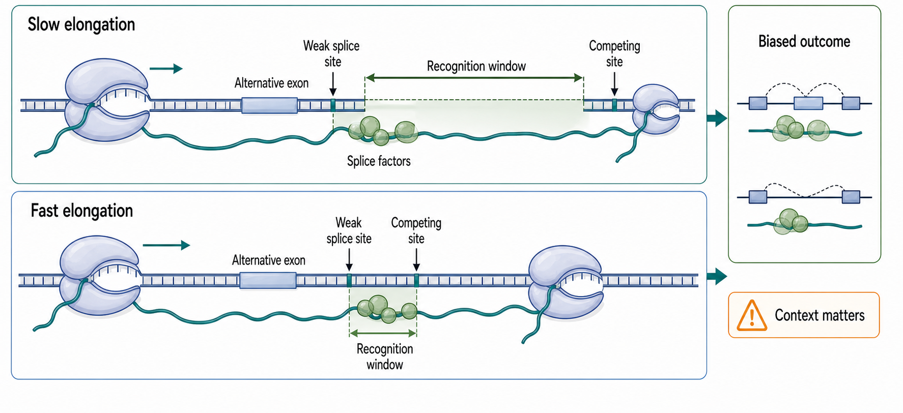
Figure 25.3. Co-Transcriptional Splicing Kinetic Competition. When Pol II elongates slowly across an alternative exon, an upstream weak splice site has more time to recruit spliceosomal factors before competing downstream elements emerge. A faster-elongating polymerase reduces this temporal window, potentially favoring a different splice outcome. The figure illustrates that elongation rate can bias alternative splicing by changing the kinetic opportunity for splice-site recognition, without being the sole determinant of splice choice.
Cleavage and polyadenylation create the mature 3′ end of many Pol II mRNAs. The nascent transcript contains sequence elements, including a polyadenylation signal and downstream elements, that recruit cleavage and polyadenylation factors. Cleavage separates the upstream pre-mRNA from downstream RNA still associated with Pol II, and poly(A) polymerase adds the poly(A) tail to the upstream product. Termination then follows through combined effects of cleavage, downstream RNA degradation, factor exchange, polymerase pausing, chromatin, and allosteric changes in the elongation complex.
Not all 3′ ends are made by the same pathway. Replication-dependent histone mRNAs in animals generally use a specialized 3′-end formation pathway rather than a long poly(A) tail. Many noncoding RNAs are cleaved by Integrator, RNase P, RNase Z, the exosome, or other factors depending on RNA class and organism. Animal mRNA cleavage and polyadenylation is supported by polyadenylation-signal and CstF recognition studies as well as recent alternative-polyadenylation reviews, while plant polyadenylation has distinctive regulatory features and extensive coupling to transcription, development, and stress responses. The useful generalization is that 3′-end formation is usually linked to transcript identity and termination, but the molecular route is transcript-class specific.
25.4. Cotranscriptional RNA modification
RNA modification means chemical alteration of a nucleotide after or during RNA synthesis. Common examples include methylation, pseudouridylation, ribose methylation, editing, thiolation, and many tRNA-specific or rRNA-specific changes. A modification can change base-pairing, stacking, local structure, protein recognition, enzymatic processing, translation, decay, immune sensing, or quality control. It can also be a mark whose function is subtle, context-specific, redundant, or unresolved.
Co-transcriptional modification has two meanings that should be kept separate. The first is strict timing: the chemical mark is installed while the transcript is still being synthesized or before release from the synthesis site. The second is regulatory coupling: transcription state, chromatin, polymerase behavior, or nascent RNP assembly influences whether a modification enzyme acts. A modification can be coupled to transcription without being installed at the exact nucleotide-addition moment.
Pseudouridine is an isomer of uridine in which the base is attached to ribose through a carbon-carbon bond rather than the usual nitrogen-carbon glycosidic bond. Pseudouridine is abundant in stable RNAs such as rRNA and tRNA, and it can also occur in pre-mRNA or mRNA. Human pre-mRNA pseudouridylation by pseudouridine synthases has been reported to occur co-transcriptionally and to affect pre-mRNA processing in some contexts. This does not imply that all mRNA pseudouridylation is co-transcriptional or that every detected site has the same functional consequence.
m6A, or N6-methyladenosine, is methylation of adenosine at the N6 position. In many eukaryotic mRNAs and noncoding RNAs, m6A is installed by writer complexes, interpreted by reader proteins, and sometimes removed by demethylases. The relationship among m6A, RNA structure, and RNP assembly is reciprocal: structure can influence writer access, m6A can alter local structure or protein binding, and reader proteins can reshape RNP fate. DDX21-mediated co-transcriptional m6A-linked regulation provides a mammalian example connecting nascent RNA modification, transcription termination, and genome stability. The appropriate conclusion is context-dependent coupling, not a universal rule that m6A always controls termination.
rRNA and tRNA modifications show that co-transcriptional timing is not limited to mRNA. rRNA modifications are guided by snoRNPs and assembly factors during ribosome biogenesis. tRNAs undergo extensive base and ribose modifications as part of maturation, and the ordering of leader removal, trailer processing, intron splicing, CCA addition, modification, aminoacylation, nuclear export, and surveillance varies across organisms and tRNA species. Some tRNA and rRNA modifications require a folded or partially folded substrate, whereas others occur early and help stabilize later structure.
Modification assays require special caution. Antibody-based enrichment can suffer from cross-reactivity and sequence-context bias. Reverse-transcription signatures can reflect modifications, damage, structure, or enzyme behavior. Chemical derivatization can be incomplete. Direct RNA sequencing can show modification-sensitive signal shifts but often requires careful calibration. Mass spectrometry can establish chemical identity but may sacrifice site and transcript information. A strong modification claim should specify the chemical identity, the RNA class, the site or region, the timing evidence, the perturbation, and the measured outcome.
Table 25.3. Modification Timing and Functional Interpretation. For each modification type, the timing of installation, examples of writer or guide machinery, possible functional outputs, and the most common overinterpretation risk.
| Modification | Possible timing | Writer or guide examples | Possible functional output | Overinterpretation risk |
|---|---|---|---|---|
| m7G cap | Co-transcriptional (early Pol II elongation) | RNA triphosphatase; guanylyltransferase; cap methyltransferase (RNMT) | Protects 5′ end; recruits nuclear cap-binding complex; promotes splicing, export, and surveillance | Assuming cap presence proves downstream function without decay or translation assay |
| m6A | Co-transcriptional to post-transcriptional (context-specific) | METTL3–METTL14 writer complex; YTHDF1–3 readers; FTO and ALKBH5 erasers | Alters local RNA structure; influences processing, decay, translation, and termination | Merging timing of detection with functional outcome without site-specific perturbation |
| Pseudouridine | Co-transcriptional for some pre-mRNAs; post-transcriptional for stable RNAs | PUS1, PUS7 (standalone synthases); H/ACA snoRNPs (for rRNA and snRNA) | Alters base stacking and local structure; can affect splicing or translation at specific sites | Generalizing from specific pre-mRNA examples to all mRNA pseudouridylation |
| 2′-O-methylation | Co-transcriptional during ribosome assembly; variable in other contexts | Box C/D snoRNPs (fibrillarin); FTSJ family tRNA methyltransferases | Stabilizes rRNA structure; confers nuclease resistance; influences translation fidelity | Applying stable RNA modification timing models to mRNA without separate evidence |
| A-to-I editing | Mostly post-transcriptional for ADAR editing; co-transcriptional for some substrates | ADAR1, ADAR2 (adenosine deaminases); ADAT (tRNA anticodon editing) | Recodes amino acids; alters splice sites or miRNA targets; expands RNA sequence diversity | Concluding regulation from editing frequency without functional perturbation |
| Organellar modifications | Variable; often coupled to organellar transcription and processing | Mitochondrial and chloroplast methyltransferases; organellar editing factors | Supports organellar rRNA, tRNA, and mRNA structure and function | Applying nuclear co-transcriptional timing models to organellar systems without separate evidence |
The most common overinterpretation is to merge three claims: “the mark exists,” “the mark is installed co-transcriptionally,” and “the mark causes a phenotype.” These claims are related but independent. A nascent m6A site may be functional, passive, redundant, or a consequence of local RNP assembly. A pseudouridine site may affect splicing at one transcript and be neutral at another. Modification biology becomes mechanistic only when the writer, site, reader, structure, processing step, and phenotype are connected by evidence.
25.5. RNP assembly and kinetic competition
An RNP is a ribonucleoprotein particle: an RNA molecule together with associated proteins. This definition includes mRNPs, ribosomes, spliceosomal snRNPs, snoRNPs, telomerase, RNase P, signal-recognition-particle RNA, small regulatory RNPs, and many long noncoding RNA complexes. The protein component is not decoration. Protein binding can stabilize RNA folds, remodel RNA structure, define localization, recruit enzymes, protect ends, expose or hide sequence elements, and determine whether the RNA becomes functional or degraded.
RNP assembly is kinetic competition because many events can act on the same nascent RNA. A newly emerged segment can fold before a protein binds. A protein can bind before a competing helix forms. A processing enzyme can cleave before an alternative RNP is assembled. A surveillance factor can recognize an unprotected end before a maturation factor arrives. The outcome depends on concentrations, affinities, transcription speed, pausing, compartment, post-translational modifications of proteins, and prior RNA modifications.
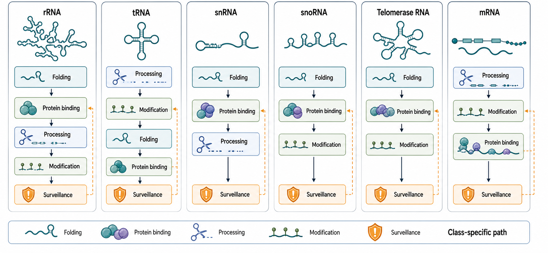
Figure 25.4. RNP Assembly Pathways by RNA Class. Different RNA classes—including rRNA, tRNA, snRNA, snoRNA, telomerase RNA, and mRNA—each follow a characteristic ordered pathway in which folding, protein binding, processing, modification, and surveillance steps are interdependent. Early folding or protein binding can stabilize intermediate states that recruit the next factor or protect the RNA from premature degradation. Despite these shared principles, the specific proteins, compartments, and timing differ substantially among RNA classes.
Ribosome assembly is the large-scale example. rRNA domains fold progressively while ribosomal proteins and assembly factors bind. Some factors prevent premature contacts; others stabilize intermediate states; still others act as checkpoints before subunit export or final maturation. In eukaryotes, nucleolar organization and snoRNP-guided modification add layers not present in the same form in bacteria. In bacteria, transcription-translation coupling does not apply to rRNA itself, but ribosome assembly still occurs in a cellular environment where transcription rate, rRNA processing, ribosomal protein availability, and growth state are tightly linked. Reviews of rRNA folding emphasize that assembly is a directed pathway with multiple checkpoints rather than a simple one-step transition to the mature ribosome.
tRNA biogenesis is compact but highly ordered. Pol III transcribes a precursor tRNA that may include a 5′ leader, a 3′ trailer, and sometimes an intron. RNase P removes the 5′ leader, 3′ processing removes the trailer, CCA may be added if not encoded, introns are removed for intron-containing tRNAs, and many nucleotides are modified. Aminoacyl-tRNA synthetases and surveillance pathways then test whether the tRNA can function in translation. These events are not necessarily a single rigid linear sequence, but coordination prevents defective precursors from accumulating.
Telomerase RNA provides a different type of RNP assembly problem. Telomerase RNA must form a template-bearing RNP with telomerase reverse transcriptase and accessory proteins. In yeast, ordered transport and assembly steps help build the mature telomerase RNP. This example is useful because the RNA is not simply processed into a mature standalone molecule. Its biological identity depends on assembling the correct RNP at the right time and place.
snRNAs and snoRNAs illustrate RNP assembly pathways that are both co-transcriptional and post-transcriptional. Some snRNAs are transcribed by Pol II, capped, processed, exported, assembled with Sm proteins, modified, and re-imported before functioning in the spliceosome. snoRNAs can be processed from introns or independent transcripts and assembled with guide proteins that direct rRNA modification. Chapter 27 treats spliceosomal snRNPs, whereas Chapter 42 and Chapter 49 treat sno/scaRNA-guided modification. Here the important point is that RNP assembly often crosses compartments and time scales. A nascent event may start the pathway, but the mature particle can require later remodeling.
Table 25.4. RNP Assembly Examples. Selected RNP classes illustrating the diversity of first assembly events, processing dependencies, surveillance routes, and connections to other chapters.
| RNP | RNA substrate | First assembly events | Processing dependencies | Surveillance route | Chapter cross-reference |
|---|---|---|---|---|---|
| Ribosome (small and large subunits) | Pre-rRNA (eukaryotic 35S/47S or bacterial 30S precursors) | Early rRNA domain folding; ribosomal protein binding during transcription | Pre-rRNA cleavage at multiple sites; snoRNP-guided 2′-O-methylation and pseudouridylation | Nuclear exosome; assembly-factor quality checkpoints before subunit export | Chapters 42 and 43 |
| Spliceosomal snRNP | snRNA (U1, U2, U4, U5, U6) | Sm protein ring assembly on exported Pol II-transcribed snRNAs | 5′ capping; 3′ trimming; cytoplasmic Sm assembly; cap hypermethylation; nuclear re-import | Cajal body remodeling; nuclear surveillance of unassembled snRNAs | Chapter 27 |
| snoRNP | Box C/D or H/ACA snoRNA | Core protein binding (fibrillarin for C/D; dyskerin for H/ACA) | Processing from intron host transcript or independent precursor | Exosome-mediated decay of unassembled snoRNA | Chapters 42 and 49 |
| Telomerase | Telomerase RNA (TERC in vertebrates; TLC1 in yeast) | Template pseudoknot folding; TERT catalytic subunit binding | 3′-end processing; H/ACA domain assembly with dyskerin (vertebrates) | PARN trimming pathway; exosome if biogenesis fails | Chapter 100 |
| tRNA-synthetase complex | Pre-tRNA | Cloverleaf fold of the tRNA body | 5′ leader removal (RNase P); 3′ trailer removal; intron splicing (some tRNAs); CCA addition; base and ribose modifications | TRAMP complex; nuclear exosome for defective precursors | Chapters 39 and 41 |
| mRNP (export-competent) | Pre-mRNA and mature mRNA | Cap-binding complex loading at m7G cap | Splicing; cleavage and polyadenylation; poly(A)-binding protein loading; exon-junction complex deposition | NMD; nuclear surveillance; NEXT and PAXT complexes | Chapters 30, 31, and 35 |
Long noncoding RNAs are a cautionary class. Many lncRNAs remain near chromatin, bind proteins, form local structures, or scaffold regulatory complexes. Some have well-supported mechanisms; many others have incomplete evidence. A chromatin-associated lncRNA signal does not automatically imply that the RNA product is a functional scaffold. It may reflect transcription through a locus, RNA processing, promoter activity, chromatin retention, or detection bias. Strong lncRNA RNP claims require perturbing the RNA molecule, not only the DNA locus, and measuring direct molecular partners.
Kinetic competition also explains why defects can have non-obvious phenotypes. A mutation that weakens an RNA hairpin may not simply unfold the mature RNA; it may expose a surveillance site during transcription. A slower polymerase may not only lower transcript output; it may change splice-site choice or RNP assembly order. A modification-enzyme depletion may not only remove a chemical mark; it may alter protein binding, folding, processing, and decay together. Mechanistic interpretation must therefore follow the pathway, not only the final abundance.
25.6. Nascent RNA surveillance, chromatin feedback, and single-molecule methods
Nascent RNA surveillance is quality control near the birth site of RNA. The cell must distinguish RNAs that are properly capped, folded, processed, modified, and assembled from RNAs that are aberrant, incomplete, antisense, cryptic, damaged, or stalled in maturation. Surveillance can cause nuclear retention, exonucleolytic decay, endonucleolytic cleavage, premature termination, RNP remodeling, or recruitment of chromatin-associated factors. Nuclear exosome cofactors and MTR4-associated adaptor pathways provide major routes for routing unstable or improperly processed nuclear RNAs toward remodeling or degradation. Later chapters treat decay enzymes in detail; this section focuses on why surveillance begins early.
A useful example is the fate of a Pol II transcript that fails early capping or processing. Without a proper cap-binding interaction, the transcript may become vulnerable to nuclear quality-control factors. If splicing fails, intron-containing RNA may be retained, degraded, or exported inefficiently depending on organism and transcript class. If 3′-end formation fails, Pol II termination can be delayed, downstream transcription can continue, and surveillance factors may degrade aberrant RNA. These events protect the cell from defective products but also regulate normal gene output.
Nascent RNA can feed back on chromatin and transcription. Chromatin is the DNA-protein organization of the genome, including nucleosomes, histone modifications, remodelers, and associated regulatory factors. Nascent RNA can remain near chromatin through direct RNA-DNA hybridization, RNA-binding proteins, processing complexes, or compartmental retention. An R-loop is a three-stranded structure containing an RNA-DNA hybrid and a displaced single-stranded DNA strand. R-loops can influence transcription, termination, recombination, DNA damage signaling, and chromatin marks, but their meaning depends on position, lifetime, sequence, protein context, and resolution.
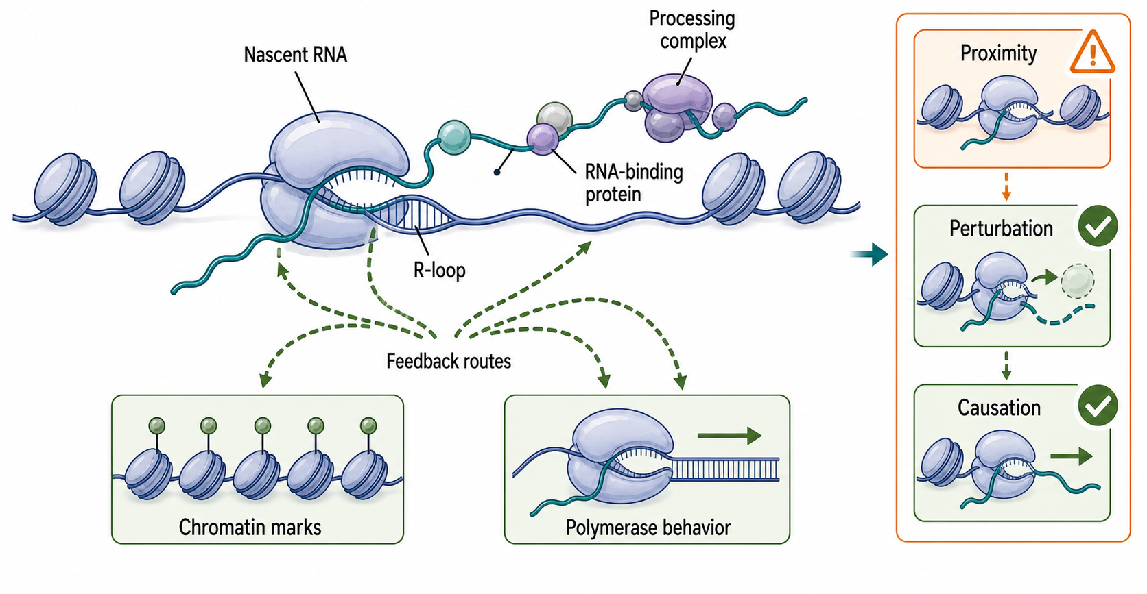
Figure 25.5. Nascent RNA-Chromatin Feedback. Nascent RNA, RNA-binding proteins, and processing complexes can remain associated with chromatin, and RNA-DNA hybrids known as R-loops can form when the nascent RNA reanneals to the template strand. These interactions can influence chromatin marks, polymerase behavior, and transcriptional output in specific contexts. However, the presence of nascent RNA near chromatin indicates proximity rather than direct regulatory causation, and strong feedback claims require perturbation of the RNA or hybrid combined with measurement of chromatin or transcription state.
The boundary for chromatin feedback is evidence. A nascent RNA found at a gene may regulate chromatin, but it may also be present because it was just made there. An RNA-binding protein detected at chromatin may bind the RNA, the polymerase, the DNA, histones, or another protein. R-loop mapping can be influenced by antibody specificity, nuclease treatment, extraction, strand bias, and genome sequence. Strong feedback claims usually combine orthogonal localization, perturbation of the RNA or hybrid, rescue, and measurement of transcription or chromatin state.
Single-molecule and time-resolved methods are powerful because co-transcriptional biology is inherently dynamic. Live-cell imaging can follow transcription sites, reporter RNA emergence, splicing timing, and release. In vitro single-molecule transcription can observe polymerase motion, pauses, RNA folding, or factor binding with controlled templates. Nascent RNA sequencing can locate polymerase-associated RNA ends or recently synthesized RNA genome-wide. Structure-probing and proximity-ligation methods can capture local folds or RNA contacts in chromatin-associated transcripts. CAR-SPLASH-like nascent RNA contact approaches, dynamic nascent-RNA imaging, CRISPR-Csm live-cell RNA imaging, and nano-COP-style nascent-processing measurements illustrate complementary operational definitions of timing.
These methods do not provide a complete movie by themselves. A fluorescent reporter may alter RNA folding or processing. Crosslinking can capture proximity rather than direct base pairing. Proximity ligation can create chimeras influenced by fragmentation and ligation bias. Nascent RNA sequencing can conflate polymerase pausing, RNA cleavage, processing delay, and RNA stability. Structure probing can perturb folding or miss protein-protected regions. The right interpretation depends on the question: timing of emergence, structure, protein binding, processing, or functional outcome.
The best studies triangulate. To test whether a nascent hairpin regulates splicing, one might combine structure probing, compensatory mutations, nascent RNA sequencing, acute perturbation of elongation rate, splice isoform measurement, and rescue of base pairing. To test whether m6A affects termination, one might combine writer depletion, catalytic rescue, site mutation, nascent RNA readthrough measurement, R-loop or genome-stability assays if relevant, and controls for global transcription changes. Co-transcriptional claims become reliable when the experiment separates correlation, timing, and causation.
Biological Contexts Across Organisms and RNA Classes
Bacteria couple RNA synthesis to translation for mRNAs. A ribosome can bind an mRNA while the mRNA is still being transcribed, and ribosome position can affect RNA folding, Rho-dependent termination, RNA decay, and regulatory leader function. Bacterial rRNA and tRNA are not translated while made, but they still undergo rapid folding, processing, modification, and RNP assembly in ways tied to growth state and transcriptional organization. Operons and attenuation systems are especially clear examples of RNA structure and transcription timing acting together.
Archaea provide important comparisons because archaeal transcription machinery shares features with eukaryotic systems, while archaeal RNA processing and RNP repertoires have lineage-specific properties. Archaeal RNAs undergo processing, modification, and RNP assembly, including tRNA and rRNA maturation. Archaeal examples help separate universal constraints, such as vectorial RNA emergence, from eukaryote-specific features such as the Pol II CTD.
Yeast and metazoan Pol II genes illustrate extensive coupling among capping, splicing, chromatin, elongation, cleavage, polyadenylation, export competence, and surveillance. Co-transcriptional splicing is common in many eukaryotic systems but varies by intron, gene, and condition. Long genes, alternative exons, terminal introns, weak splice sites, and developmentally regulated factors can all change timing. Cancer-associated splicing alterations often involve changes in splicing factors, chromatin, transcription, or RNA-binding proteins, but a disease association does not by itself prove a co-transcriptional mechanism.
Plants have strong evidence for co-transcriptional processing and modification as an integrated regulatory layer. Plant polyadenylation, splicing, RNA modification, small-RNA pathways, and development or stress responses are extensively connected to transcription and chromatin. Plant examples are useful because they prevent the chapter from treating mammalian Pol II as the only eukaryotic model.
Viruses broaden the logic. Some retroviral RNAs are host Pol II transcripts that must be capped, spliced or retained unspliced, exported, translated, packaged, and reverse-transcribed. Positive-strand RNA virus genomes assemble replication organelles and RNPs in the cytoplasm, a setting where “co-transcriptional” may refer to RNA synthesis by a viral RNA-dependent RNA polymerase rather than nuclear Pol II. The mechanistic question is always the same: what polymerase makes the RNA, where is the RNA made, what structures and proteins engage it during synthesis, and how is the product routed?
Organelles add further boundary cases. Mitochondrial and chloroplast RNAs are transcribed, processed, edited, modified, and assembled in organelle-specific RNP environments. Many organellar systems use extensive RNA editing or specialized processing factors. The chapter does not treat organelles in detail, but organelles reinforce that co-transcriptional maturation is not restricted to nuclear chromatin.
Box 25.2. Co-Transcriptional Does Not Mean Universal
- Whether a processing event occurs before or after transcript release depends on intron length, gene architecture, elongation rate, chromatin state, factor availability, cell type, and organism.
- Some introns in a single gene can be removed co-transcriptionally while others in the same pre-mRNA are processed post-transcriptionally.
- Describing a process as co-transcriptional requires specifying the RNA class, gene, condition, and assay used to determine timing; no single rule applies across all contexts.
Technology, Computational, Clinical, and Engineering Links
Co-transcriptional mechanisms shape experimental design. In vitro transcription can produce RNA for structure studies, nanotechnology, therapeutics, or biochemical assays, but the folding path can depend on polymerase speed, temperature, ion concentration, transcript length, sequence, and whether proteins or ligands are present. RNA origami and other engineered RNAs exploit co-transcriptional folding to build structures as the RNA emerges. Engineering success often depends on avoiding kinetic traps, not merely designing a favorable final minimum-energy structure.
Computational modeling of nascent folding must include time. Minimum-free-energy prediction of a full-length RNA is useful but incomplete for co-transcriptional biology. A nascent model may need to simulate transcript growth, polymerase pauses, local refolding, ligand binding, protein binding, and constraints from structure probing. The output should be interpreted as a kinetic hypothesis. Validation requires mutations or perturbations that change the predicted pathway and a measured RNA-processing or functional consequence.
Clinical links are strongest where co-transcriptional maturation affects gene expression programs. Splicing-factor mutations in cancer, repeat-expansion RNAs that form abnormal structures, R-loop-associated genome instability, defects in ribosome assembly, tRNA processing disorders, and altered m6A pathways can all intersect with nascent RNA biology. However, clinical association should not be oversold. A disease mutation may affect transcription, chromatin, RNA processing, translation, or stress pathways indirectly. Mechanistic claims should specify the RNA class, cell type, pathway, and evidence level.
Therapeutic RNA production also depends on synthesis-coupled folding and modification. Synthetic mRNA manufacturing must control cap status, poly(A) tail, modified nucleotides, double-stranded RNA impurities, sequence design, and purification. Those topics are treated in RNA therapeutic chapters, but the mechanistic bridge is clear: the path of RNA synthesis and processing affects final RNA identity, immune sensing, translation, and stability. For small RNAs, guide RNAs, and structured therapeutic RNAs, folding during synthesis can affect product quality and function.
Box 25.3. Modification Timing Is Not Modification Function
- A modification detected on chromatin-associated or nascent RNA establishes installation timing, not biological function.
- The same chemical mark may influence processing in one RNA class or cellular context and be neutral in another.
- Establishing function requires perturbing the writer enzyme, the modified site, or the reader protein and measuring a specific downstream molecular or cellular outcome.
Method development is moving toward higher temporal and molecular resolution. Long-read sequencing can connect distant splice choices, RNA modifications, and isoforms. Direct RNA sequencing may help read native modifications but requires calibration. Nascent proximity methods can link folding to processing but need controls for crosslinking and ligation bias. Live-cell imaging can reveal timing, but reporter design must preserve endogenous context. The best technology is not the newest assay by default; it is the assay whose operational definition matches the biological question.
Recent Consensus
Current consensus is that RNA maturation frequently begins during transcription. Nascent RNA folding is vectorial, elongation rate and pausing can influence folding and processing, and mature RNA state is often the product of a pathway rather than a single equilibrium endpoint. Co-transcriptional capping is established for many Pol II transcripts, co-transcriptional splicing is common but variable, and cleavage and polyadenylation are tightly linked to termination.
The field also agrees that RNA modification and RNP assembly can be co-transcriptionally coupled in specific contexts. rRNA assembly is a guided co-transcriptional and post-transcriptional pathway. tRNA maturation is ordered and quality controlled. m6A and pseudouridine provide strong examples of nascent modification biology, but broad generalizations remain risky. RNA-chromatin feedback and R-loop functions are real but context-dependent.
The strongest methodological consensus is cautionary. Nascent RNA assays are powerful because they enrich transient intermediates, but the word “nascent” has assay-specific meanings. A chromatin-associated RNA, a polymerase-associated RNA end, a newly labeled transcript, a live-cell reporter signal, and a crosslinked RNA contact are related but different measurements. Kinetic interpretation requires explicit models and perturbations.
Open Questions, Controversies, Deprecated Models, and Common Misconceptions
Open questions:
- How often does early RNA folding determine final RNA structure?
- Which genes use transcription speed as a major splicing regulator under physiological conditions?
- How many RNA modification sites are installed before release rather than shortly after release?
- How is nascent RNP assembly monitored at the level of individual molecules?
- When do R-loops act as regulated intermediates rather than harmful lesions?
- How does condensate-like organization influence co-transcriptional maturation? Condensate-like behavior has been implicated in some RNA-processing and modification systems, including plant SERRATE-associated regulation of m6A and miRNA biogenesis. The term “phase separation” should be used carefully. Concentration, multivalency, microscopy appearance, and perturbation sensitivity do not by themselves prove a thermodynamic phase transition or a necessary biological mechanism.
Common misconceptions:
- “Mature RNA structure is enough to infer the folding pathway.” A mature structure can hide transient intermediates that influenced processing, assembly, or surveillance.
- “Co-transcriptional splicing means all introns are removed before transcription ends.” Splicing timing varies widely by intron, gene, organism, cell type, and condition.
- “Detection on chromatin-associated RNA proves direct co-transcriptional modification.” Chromatin association supports timing but does not prove catalytic timing or function without controls.
- “R-loops are always genome-damaging.” Some R-loops participate in normal regulation, but persistent or mislocalized R-loops can threaten genome stability.
- “A polymerase-speed perturbation reveals only elongation effects.” Changing elongation can indirectly alter chromatin, RNA processing, stress responses, RNA stability, and factor recruitment.