Chapter 30. mRNP Assembly, Nuclear Export, Retention, and Large RNP Logistics
Scope Note
This chapter follows a eukaryotic messenger RNA after synthesis begins but before the RNA has settled into its cytoplasmic life. A messenger ribonucleoprotein particle, abbreviated mRNP, is an mRNA molecule together with the proteins and sometimes additional RNA-associated factors bound to it. The term is broader than “mature mRNA.” An mRNP can be a nascent pre-mRNA still attached to chromatin, a splicing intermediate, an export-competent particle at a nuclear pore complex, a retained nuclear RNA, a stored RNP, or a cytoplasmic mRNA ready for translation, localization, storage, or decay. The chapter emphasizes nuclear mRNP assembly, export, retention, and large RNP movement. Chapter 25 introduced co-transcriptional RNP assembly as a general principle. Chapters 26 through 29 cover cap formation, splicing, and 3′-end formation in detail. Chapter 31 develops nuclear RNA surveillance and exosome targeting, and later chapters treat cytoplasmic translation, decay, granules, localization, and therapeutic mRNA delivery.
The central claim is that mRNA export is not a simple diffusion step performed after a transcript is “finished.” Export competence is built by ordered and partly reversible RNP assembly. The cap, exon junction complex, splicing history, cleavage and polyadenylation status, poly(A)-binding proteins, TREX-associated factors, export receptors, RNA-binding proteins, helicases, and quality-control factors create a molecular identity that helps decide whether an RNA is exported, retained, remodeled, degraded, or released later. This identity is context-dependent: intronless, intron-rich, long, structured, repeat-rich, viral, retroelement-derived, stress-regulated, and developmentally controlled transcripts can use different combinations of adaptors and checkpoints.
Executive Summary
An mRNP is assembled in layers. The nascent Pol II transcript first receives a 5′ cap and cap-binding proteins, then spliceosomal and RNA-binding proteins recognize introns and exons, 3′-end factors cleave and polyadenylate many transcripts, and export adaptors are loaded or stabilized by the transcript’s processing history. This assembly path is not a conveyor belt with one mandatory order for every mRNA. It is a probabilistic and actively remodeled pathway in which RNA sequence, exon-intron architecture, polymerase behavior, RBP concentration, helicase activity, and compartmental organization influence the final export state.
The exon junction complex, abbreviated EJC, is a protein complex deposited upstream of many exon-exon junctions after splicing. It marks splicing history, helps organize downstream mRNP composition, and contributes to nonsense-mediated decay, localization, and translation control in later compartments. The transcription-export complex, abbreviated TREX, is an export-coupling assembly that includes THO-related components, the RNA helicase UAP56 or DDX39B, ALYREF and related adaptors, and additional proteins depending on organism and context. TREX and EJC are not the same structure, but both connect processing to export competence. Export adaptors help recruit the principal metazoan mRNA export receptor NXF1 with its partner NXT1, or the yeast Mex67-Mtr2 system.
Nuclear pore complexes are large protein channels that perforate the nuclear envelope. Small proteins can use karyopherin-mediated transport cycles, whereas most bulk mRNA export is mediated by mRNP-specific receptors rather than by direct Ran-dependent karyopherin transport. A mature export-competent mRNP must engage the nuclear basket, pass through the central channel, and undergo cytoplasmic remodeling. The cytoplasmic remodeling step matters because export receptors and nuclear adaptors must be removed or rearranged so the mRNA cannot simply slide back into the nucleus and can instead enter translation, localization, storage, or decay pathways.
Nuclear retention is not only a failure mode. Some RNAs are retained because they are incompletely processed, damaged, incorrectly packaged, or targeted for surveillance. Other RNAs are retained as part of normal regulation. Retention can occur through chromatin association, RNA structure, repeat-derived sequence, intron retention, RBP binding, paraspeckle-like bodies, nuclear condensates, or attachment to nuclear pore-associated quality-control systems. Long noncoding RNAs make this point especially clear: some lncRNAs are designed by their biogenesis and sequence features to remain nuclear, whereas many mRNAs are normally exported unless processing or stress changes their RNP composition.
Large transcript export introduces physical constraints. Most mRNPs move through nuclear pore complexes after compaction, remodeling, and receptor engagement. Very large or unusually packaged RNPs can challenge the pore’s geometry, timing, and quality-control gates. Recent work in muscle cells supports nuclear-envelope budding as a specialized export route for large transcripts, which should be treated as a context-specific mechanism rather than a universal replacement for pore-mediated export. Viral and retroelement transcripts add another boundary case: host export pathways can be co-opted, altered, or used to license immune-relevant cytoplasmic RNA species.
The identity of an mRNA is encoded by more than its nucleotide sequence. A transcript carries marks of where and how it was made: cap state, splicing history, exon architecture, m6A patterns, 3′-end processing, poly(A) tail features, bound RBPs, RNP compaction, nuclear export path, and surveillance history. These identity marks are useful but not deterministic. For example, splicing can promote export, but intronless transcripts can be exported efficiently with alternative adaptors. A poly(A) tail usually supports mRNA maturation, but aberrant or excessive nuclear polyadenylation can mark RNAs for surveillance. A retained RNA may be defective, regulatory, or simply slow to complete processing. Mechanistic claims therefore need explicit evidence for RNA class, cell type, processing state, bound factors, and fate.
Concept Inventory
- Messenger ribonucleoprotein particle: an mRNA or pre-mRNA together with associated proteins and other RNP factors. The term includes export intermediates, retained nuclear particles, and cytoplasmic particles. It should not be used as a synonym for naked mRNA.
- Export competence: the molecular state in which an RNA has acquired the processing marks, RNP proteins, and receptor interactions needed for efficient passage from nucleus to cytoplasm. Export competence is not equivalent to transcription completion.
- Exon junction complex: a protein complex deposited upstream of many exon-exon junctions after splicing. It marks splicing history and can influence export, localization, translation, and nonsense-mediated decay.
- TREX: a transcription-export coupling assembly that helps load export adaptors and connect mRNA biogenesis to export. The exact composition differs across organisms and experimental definitions.
- Export adaptor: an RNA-binding or RNP-binding protein that connects an RNA substrate to an export receptor. Adaptors often recognize processing-linked RNP states rather than a universal RNA sequence.
- NXF1-NXT1: the major metazoan mRNA export receptor pair. It binds export adaptors and nucleoporins and supports mRNP movement through nuclear pore complexes.
- Nuclear pore handoff: the series of interactions by which an mRNP docks at the nuclear basket, traverses the pore, and is remodeled on the cytoplasmic side.
- Nuclear retention: the persistence of an RNA in the nucleus because of incomplete processing, surveillance, regulated storage, chromatin association, nuclear body localization, or physical export constraints.
- Retention body: a nuclear compartment or condensate-like structure enriched for retained RNAs and associated proteins. The term should be used with care because different bodies have different molecular composition and evidence.
- Large RNP logistics: the packaging, deformation, remodeling, and possible alternative export routes required for unusually long or bulky RNPs.
What to Know Before Reading This Chapter
Eukaryotic cells separate transcription and translation with a nuclear envelope. The nuclear envelope is a double membrane pierced by nuclear pore complexes. RNA polymerase II synthesizes most mRNA precursors in the nucleus, whereas ribosomes translate mRNAs in the cytoplasm. Therefore a protein-coding Pol II transcript must be processed, packaged, moved across the nuclear envelope, and handed to cytoplasmic machinery before it can be translated. This chapter assumes familiarity with capping, splicing, cleavage and polyadenylation, but it defines the export-specific terms when they first appear.
The reader should also distinguish RNA sequence from RNP state. A newly made pre-mRNA contains nucleotide sequence information, but the cell does not usually handle that RNA as a naked polymer. Proteins bind the cap, introns, exons, exon-exon junctions, poly(A) tail, structured regions, and sequence motifs. Some proteins bind transiently and remodel the RNP; others remain as marks that influence later fate. The same RNA sequence can therefore have different fates if it is capped differently, spliced differently, bound by a different adaptor, retained in a nuclear body, or produced during stress.
Three running examples organize the chapter. The first is a typical metazoan intron-containing mRNA, which receives a cap, undergoes splicing and 3′-end formation, recruits export factors, passes through nuclear pores, and begins cytoplasmic remodeling. The second is an intronless or weakly spliced transcript, which illustrates why export cannot be reduced to EJC deposition alone. The third is a very large or unusually retained RNA, which reveals physical and regulatory limits of standard pore-mediated export.
Experimental evidence for mRNP export comes from several sources. Fluorescence in situ hybridization can show whether RNA accumulates in the nucleus or cytoplasm, but it does not identify the direct export factor by itself. Nuclear-cytoplasmic fractionation can quantify compartmental enrichment, but leakage, incomplete fractionation, and long-RNA breakage can mislead interpretation. RNA immunoprecipitation and crosslinking can identify RNP contacts, but protein-RNA proximity is not automatically functional binding. Acute depletion or mutation of export factors can test requirement, but secondary effects on transcription, splicing, stress responses, and RNA decay must be controlled.
30.1. mRNP assembly from transcription to export
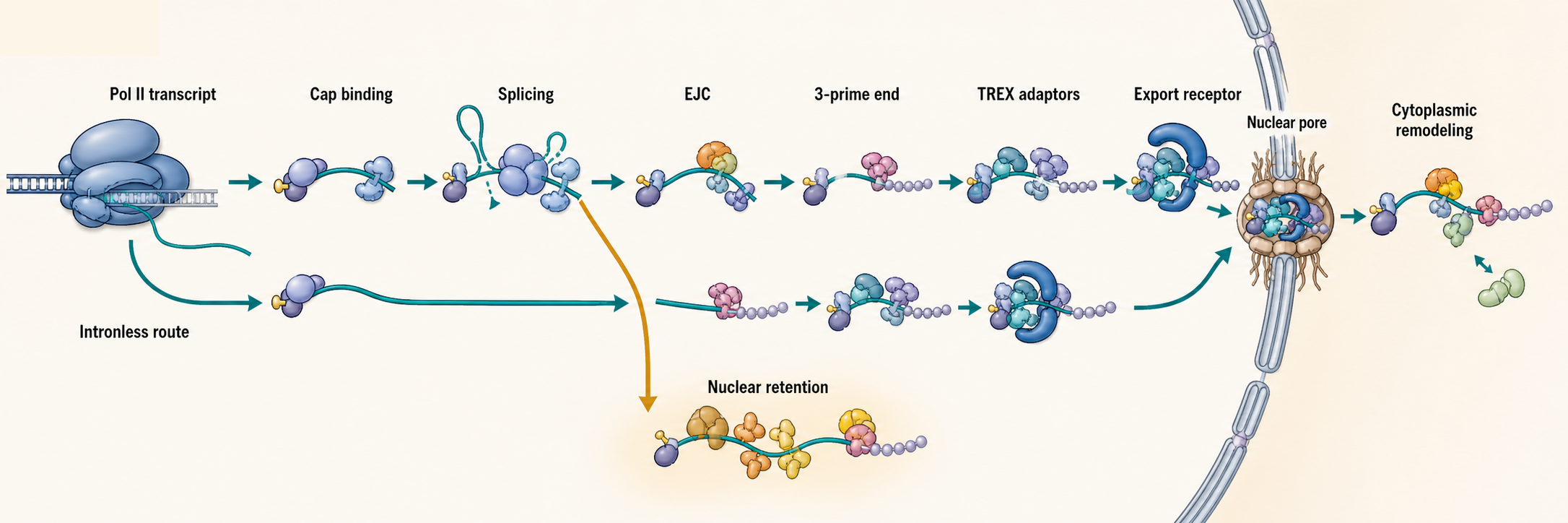
Figure 30.1. Assembly of an export-competent mRNP. A Pol II transcript acquires export competence through cap-binding, splice-linked protein deposition, 3′-end formation, TREX-associated adaptor loading, export receptor engagement, nuclear pore transit, and cytoplasmic remodeling. Alternative paths exist for intronless transcripts, which use cap-dependent or element-dependent adaptors without EJC deposition, and for retained transcripts, which accumulate in the nucleus because of incomplete processing, RBP binding, or regulated sequestration. The figure illustrates that mRNA export is the outcome of ordered, partly reversible RNP assembly rather than naked RNA diffusion.
Table 30.1. Molecular marks of mRNA identity. The table summarizes the combinatorial identity marks that together determine whether an mRNA is exported, retained, degraded, or regulated in the nucleus.
| Mark or feature | Molecular carrier | When acquired | Typical reader or consequence | Boundary case |
|---|---|---|---|---|
| 5′ cap | CBC (CBP80–CBP20) | Co-transcriptionally, shortly after initiation | Protects from 5′ decay; promotes export and translation | Capped RNAs can still be retained, degraded, or processed into noncoding products |
| Cap-binding complex | CBP80–CBP20 heterodimer | On the 5′ cap shortly after synthesis | Recruits processing and export factors; later replaced by eIF4E | eIF4E replacement marks cytoplasmic handoff; nuclear CBC and cytoplasmic eIF4E differ in function |
| Splice status | Spliceosomal proteins | During splicing | Remodels adaptor landscape; creates EJC deposition sites | Intronless transcripts are exported efficiently without splicing |
| EJC | eIF4A3, MAGOH, Y14, MLN51 core | After each exon-exon ligation | Marks splicing history; aids export, localization, and NMD surveillance | Absent from intronless mRNAs; coverage varies across junctions |
| TREX adaptor loading | ALYREF, SARNP, UAP56/DDX39B | Coupled to transcription and splicing | Recruits export receptor; links biogenesis to export | Adaptor composition varies by RNA class and organism |
| Poly(A) tail | PABPN1 (nuclear), PABPC1 (cytoplasmic) | After cleavage and polyadenylation | Signals 3′-end completion; stabilizes mRNA | Aberrant nuclear polyadenylation can flag RNAs for surveillance |
| m6A modification | METTL3–METTL14 methyltransferase; YTHDC1 reader | Post-transcriptionally in the nucleus | Influences splicing, nuclear export via YTHDC1, and translation efficiency | Can also promote mRNA decay; direction of effect is context-dependent |
| RBP occupancy | Sequence- or structure-specific RBPs | Throughout biogenesis | Can promote or inhibit export, stability, localization, or translation | Same RNA can carry competing RBPs depending on cell state |
| Export receptor engagement | NXF1–NXT1 (metazoa); Mex67–Mtr2 (yeast) | After adaptor recruitment near the nuclear basket | Drives pore transit and cytoplasmic release | Engagement may be transient; quality-control failure blocks progress |
| Nuclear pore modification state | O-GlcNAc-modified nucleoporins | Dynamically regulated by cellular metabolism | Tunes pore export capacity globally | This is a machinery state, not an mRNA identity mark per se |
An mRNP begins to assemble while an RNA polymerase II transcript is still being synthesized. The first practical point is physical: the RNA emerges from the polymerase in the 5′ to 3′ direction, so 5′ sequence, cap state, early RNA structure, and early protein binding are available before the transcript’s downstream sequence exists. Chapter 25 described this as kinetic and co-transcriptional RNP assembly. Here the focus narrows to mRNAs and export. Export competence is the state in which an mRNP can be recognized by export receptors and successfully pass to the cytoplasm. That state is built through a series of processing-linked handoffs rather than by one universal export signal.
The earliest mark on many Pol II mRNAs is the 5′ cap. The cap is a modified nucleotide structure added to the 5′ end soon after transcription initiation. Cap-binding proteins protect the RNA, help recruit processing factors, and contribute to downstream export and translation competence. The cap is not an export passport by itself, because capped RNAs can still be retained, degraded, or processed into noncoding products. It is better understood as one identity mark read together with splice status, 3′-end formation, RNP composition, and surveillance state.
Splicing can strengthen export competence because it deposits proteins and creates exon-exon junctions. A typical intron-containing metazoan pre-mRNA recruits spliceosomal components to introns, removes introns, and leaves an mRNP whose exon architecture has been changed by splicing. This produces binding platforms for the exon junction complex and associated proteins. However, splicing is not required for every exported mRNA. Histone mRNAs, many viral RNAs, naturally intronless genes, and engineered transcripts can use other sequence elements, cap-dependent factors, or specialized adaptors. The useful rule is therefore not “spliced RNAs are exported and unspliced RNAs are retained.” The better rule is that processing history changes the adaptor landscape, and different RNAs can reach export competence by different routes.
Box 30.1. Do not overgeneralize splicing and export
- Splicing deposits EJCs and remodels the adaptor landscape, typically strengthening export competence for intron-containing mRNAs.
- Intronless transcripts—including histone mRNAs, many viral RNAs, and engineered constructs—reach the cytoplasm using cap-dependent adaptors, sequence elements, or specialized RNA-binding proteins without EJC deposition.
- Some intron-containing cellular and viral RNAs are intentionally exported in partially spliced form under regulated conditions, as in the case of HIV-1 genomic RNA.
- Nuclear accumulation of an intron-containing RNA does not automatically indicate export failure; it may reflect regulated retention, intron-dependent nuclear function, or normal slow processing kinetics.
3′-end formation adds another checkpoint. For most Pol II mRNAs, cleavage and polyadenylation separate the transcript from downstream RNA and create a poly(A) tail that binds nuclear poly(A)-binding proteins. Proper 3′-end formation helps terminate transcription, release processing factors, and package the mRNP for export. A yeast study showed that assembly of an export-competent mRNP can be required for efficient release of the 3′-end processing complex after polyadenylation, which illustrates that export factor loading and processing-factor recycling are coupled rather than strictly sequential. A more recent study of the DEAD-box ATPase Dbp2 supports the related idea that helicase-dependent remodeling at gene ends contributes to an mRNP assembly checkpoint and cleavage-factor recycling.
RNA helicases are central because mRNP assembly creates both useful and obstructive contacts. A helicase is an ATP-dependent enzyme that can remodel RNA structure or RNA-protein interactions, although many DEAD-box proteins act locally rather than as long-range motors. In mRNP biogenesis, helicases can remove proteins, alter RNA folding, promote adaptor loading, or help test whether an mRNP is correctly packaged. DDX39B, also known as UAP56 in many contexts, functions in export adaptor recruitment and TREX-related assembly. Dbp2 in budding yeast acts at gene ends. DDX6/p54 has been implicated in nuclear assembly of stored mRNP particles in some systems. These examples should not be collapsed into a single helicase function; each helicase has specific substrates, partners, and timing.
A major conceptual shift in current mRNP biology is heterogeneity. Older diagrams often showed an assembly line from gene to pore: transcription, capping, splicing, polyadenylation, adaptor loading, receptor binding, export. That diagram remains useful as a teaching scaffold, but real mRNP populations are compositionally diverse. RNAs differ in length, intron number, exon architecture, RNA structure, modification status, synthesis site, stress history, and RBP occupancy. Export factors themselves can bind transiently or substoichiometrically. The 2025 review on dynamic and compositional heterogeneity of nuclear mRNPs emphasizes that a cell contains many related mRNP states rather than one uniform particle type.
Table 30.2. Interpreting nuclear RNA accumulation. The table provides an evidence checklist for distinguishing among the different explanations for why an RNA accumulates in the nucleus.
| Possible interpretation | Expected supporting evidence | Artifact risk | Follow-up experiment |
|---|---|---|---|
| Defective splicing | Unspliced introns detectable in nuclear RNA; splicing factor depletion phenocopies | Normal exported transcripts may co-accumulate | Test splicing in isolation; rescue with wild-type splicing factor |
| Defective 3′-end formation | Readthrough RNA beyond annotated poly(A) site; processing factor depletion phenocopies | Co-transcriptional termination defects alter RNA levels independently | Verify poly(A) tail integrity; map cleavage site; deplete individual 3′-end factors |
| Export receptor failure | Export receptor depletion phenocopies; capping and splicing confirmed intact | Upstream processing defects can mimic an export block | Acutely deplete export receptor; confirm intact processing before interpreting |
| Regulated intron retention | Intron at specific position in mature RNA; accumulation is condition-dependent | Contaminating pre-mRNA resembles retained intron | Splice-junction spanning RT-PCR; compare multiple conditions and cell states |
| lncRNA nuclear function | RNA lacks ribosome association; localizes near chromatin or nuclear body | Nuclear enrichment alone is insufficient evidence for function | Map chromatin association; define nuclear binding partners; test genetic knockout |
| Nuclear body storage | RNA co-localizes with known nuclear body marker; reversibly mobilized by signal | Condensate association may be incidental | Live-cell imaging; correlate RNA fate with body dynamics; perturb body components |
| Increased transcription | Nascent RNA signal elevated; RNA synthesis rate increased | Stable nuclear RNA accumulation mimics export block | Measure transcription rate; nuclear run-on; compare pre-mRNA and mature mRNA ratios |
| Decreased cytoplasmic decay | Cytoplasmic RNA also elevated; stabilized by decay factor depletion | Stability change can be mistaken for export enhancement | Measure mRNA half-life; deplete cytoplasmic decay factors selectively |
| Fractionation contamination | Cytoplasmic marker RNAs appear in nuclear fraction; compartment markers mislocalized | Common with suboptimal lysis or very abundant cytoplasmic RNA | Validate fractionation with compartment-specific marker RNAs and proteins |
The evidence basis for this section comes from genetics, factor depletion, imaging, biochemical purification, structural studies, and protein-RNA mapping. In yeast, mutations in export factors can cause nuclear accumulation of poly(A)+ RNA, which is a strong phenotype but not a complete mechanism. In mammalian cells, depletion of NXF1, TREX components, or adaptor proteins can alter nuclear export, but many of these proteins also influence splicing, transcription, or RNA stability. Microscopy can show where an RNA accumulates, but a nuclear FISH signal may reflect retention, slow export, increased transcription, decreased decay, or defective fractionation controls. The strongest mechanistic studies connect a defined RNA or RNP factor to a specific assembly step, rescue the perturbation, and distinguish export failure from upstream processing failure.
30.2. Exon junction complexes, TREX, and export adaptors
The exon junction complex, or EJC, is a multiprotein complex deposited upstream of many exon-exon junctions after pre-mRNA splicing. It is a mark of splicing history, not a generic label on all mRNAs. The EJC’s best-known downstream role is in nonsense-mediated decay, where a premature termination codon upstream of an EJC can help trigger mRNA surveillance. But the EJC also contributes to mRNA localization, translation, and export-linked mRNP organization. In export biology, the EJC teaches a broader principle: processing can leave physical protein marks on an RNA, and those marks can be read by later compartments.
TREX, the transcription-export complex, is a set of proteins that couples nuclear mRNA biogenesis to export. The name can be confusing because TREX does not mean a single invariant particle with the same subunits in all organisms, preparations, or papers. In many eukaryotic systems, TREX includes THO-complex components, the DEAD-box ATPase UAP56/DDX39B, ALYREF or related adaptor proteins, and additional proteins such as SARNP/CIP29 depending on context. The complex helps connect transcription, splicing, 3′-end formation, adaptor loading, and export receptor recruitment.
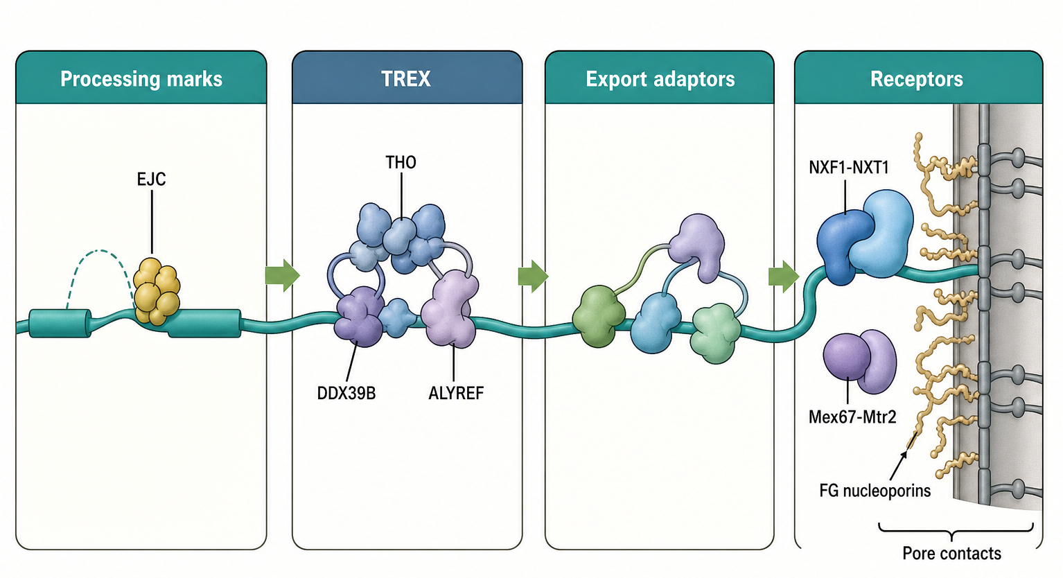
Figure 30.2. EJC, TREX, export adaptors, and export receptors. EJCs are deposited upstream of many exon-exon junctions after splicing and serve as marks of splicing history that influence later mRNP organization. TREX-related assemblies, including THO components, UAP56/DDX39B, and ALYREF, couple mRNA biogenesis to export adaptor loading, while export adaptors bridge mRNPs to the principal metazoan export receptor pair NXF1-NXT1, or the budding yeast analog Mex67-Mtr2, which engages FG nucleoporins during pore transit. The four categories—processing marks, adaptors, receptors, and pore components—are functionally distinct, even though their functions are linked in ordered steps.
An export adaptor is a protein that connects an RNA or mRNP to an export receptor. Adaptors are needed because the core export receptor cannot by itself read every feature of every mRNA with sufficient specificity. ALYREF is a classic adaptor. SR proteins, cap-associated factors, EJC-associated proteins, and other RNA-binding proteins can also contribute to export for particular RNA classes or conditions. The adaptor concept is more accurate than a search for one universal mRNA export sequence.
NXF1-NXT1 is the main metazoan mRNA export receptor pair. NXF1, historically called TAP, binds export adaptors and nucleoporins. NXT1 helps NXF1 function at the nuclear pore. In budding yeast, the analogous export receptor is Mex67 with Mtr2. These receptors interact with phenylalanine-glycine-rich nucleoporins, often called FG nucleoporins, in the central channel of nuclear pore complexes. This interaction allows mRNPs to move through the pore while maintaining selectivity.
Recent structural work illustrates how adaptor loading can be organized. A 2023 study reported structural information for a higher-order complex involving SARNP and DDX39B, supporting a model in which DDX39B-associated ATPase cycles and adaptor proteins help organize export-competent mRNP assembly. The mechanistic lesson is not merely that two proteins bind. The important point is that export adaptors can be stabilized by structured protein-protein and protein-RNA assemblies whose timing is linked to mRNA maturation.
Newer methods also broaden the evidence base. A 2024 method named TREX in the chapter bibliography identifies proteins that bind specific RNA regions in living cells; regardless of naming overlap, the method reminds the reader that region-specific RBP mapping can reveal how different segments of the same RNA carry different protein contacts. The limitation is that crosslinking and enrichment report contact or proximity, not necessarily a transport-causal interaction. A protein found on a 3′ untranslated region might regulate export, stability, localization, or translation; perturbation is needed to assign function.
Exon architecture can influence mRNP identity beyond simple EJC deposition. Exon number, exon length, intron position, splice timing, and RNA modification patterns can affect which proteins bind. A 2023 study connecting exon architecture with m6A suppression and gene expression supports the idea that exon-intron organization is read by nuclear processing and modification systems. For this chapter, the conservative inference is that exon architecture contributes to mRNP identity. It would be too strong to claim that every exon-architecture effect acts through export.
The boundary cases are important. First, intronless transcripts can be exported. They often rely on alternative adaptor recruitment through cap-binding proteins, sequence elements, RNA structure, or specialized viral or cellular mechanisms. Second, incompletely spliced RNAs are not always mistakes. Some viral RNAs and selected cellular RNAs intentionally export intron-containing forms under controlled conditions. Third, the same factor can have multiple roles. A protein classified as an export adaptor may also influence transcription, splicing, mRNA stability, or translation. These overlapping roles explain why factor depletion experiments require careful interpretation.
30.3. Nuclear pores and cytoplasmic handoff
The nuclear pore complex is a massive protein assembly embedded in the nuclear envelope. It contains scaffold nucleoporins that form a stable framework, FG nucleoporins that create a selective central channel, cytoplasmic filaments, and a nuclear basket facing the nucleoplasm. Protein import and export often use karyopherins and the Ran GTPase cycle. Bulk mRNA export is different: most mRNAs are exported as mRNPs by NXF1-NXT1 or Mex67-Mtr2-like receptors rather than by a standard Ran-dependent karyopherin cargo cycle.
An export-competent mRNP first has to find or be delivered to nuclear pores. The simplest model is diffusion: an mRNP moves through the nucleoplasm until receptor-nucleoporin interactions capture it. More organized models include gene gating, in which certain active genes or mRNPs are positioned near nuclear pores, and specialized pore states that favor particular RNA-processing outputs. A 2022 budding yeast study found that nuclear mRNA metabolism can drive selective assembly of nuclear baskets on a subset of nuclear pore complexes. This supports a view in which pores are not merely passive holes. Nuclear pore composition and mRNA metabolism can influence one another.
Traversal through the pore requires repeated weak interactions. NXF1-NXT1 or Mex67-Mtr2 contacts FG nucleoporins, allowing the mRNP to enter and move through the central channel. The mRNP must be compact enough and properly arranged to avoid clogging or retention. A long RNA does not pass as an extended naked strand. It passes as a dynamic RNP whose proteins, RNA structure, and export receptors shape its size and surface properties. This packaging is why mRNP assembly and pore transport cannot be separated cleanly.
The cytoplasmic handoff is a remodeling event. On the cytoplasmic side of the nuclear pore, export receptors and nuclear adaptors must be removed or rearranged. In budding yeast, the DEAD-box ATPase Dbp5, activated by Gle1 and inositol hexakisphosphate, remodels exported mRNPs at the cytoplasmic face. This remodeling helps make export directional because once receptor-adaptor interactions are disrupted, the mRNP is less able to re-enter the pore in the reverse direction. Mammalian cells contain related remodeling logic, although factor names, redundancy, and regulatory layers differ. Directionality in mRNA export therefore emerges from asymmetric remodeling, not simply from a concentration gradient.
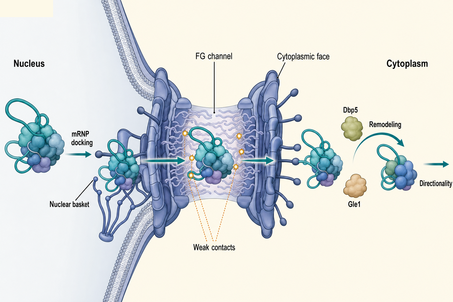
Figure 30.3. Nuclear pore handoff and directionality. Export-competent mRNPs dock at the nuclear basket, traverse the central FG-nucleoporin channel through repeated weak receptor-pore interactions, and emerge on the cytoplasmic face where helicase-dependent remodeling removes or rearranges export factors. This asymmetric cytoplasmic remodeling—driven by Dbp5 and Gle1 in yeast and related mechanisms in metazoa—makes export directional by disrupting the receptor interactions needed for nuclear re-entry. The remodeled mRNP is then available for translation, localization, storage, or cytoplasmic decay.
The handoff also connects nuclear history to cytoplasmic fate. After export, the mRNP can recruit translation initiation factors, localization machinery, decay factors, storage proteins, or granule-associated proteins. Nuclear events can influence these choices. For example, nuclear RNA-related processes can modulate the assembly of cytoplasmic RNA granules, indicating that cytoplasmic RNP organization may inherit information from earlier nuclear assembly states. The cautious interpretation is that nuclear RNP history can bias cytoplasmic organization; it does not mean every cytoplasmic granule phenotype is directly caused by export.
Table 30.3. Export factors and common names. The table lists the major mRNA export factors with their aliases, roles, and organism scope to reduce naming confusion across the literature.
| Factor or complex | Common aliases | Broad role | Organism notes | Caution |
|---|---|---|---|---|
| NXF1/TAP | TAP | Principal metazoan mRNA export receptor | Metazoa; requires NXT1 as co-factor | Not the same gene as yeast Mex67 despite analogous function |
| NXT1/p15 | p15 | Co-receptor for NXF1; supports NPC interaction | Metazoa | Functional analog of yeast Mtr2 but a distinct protein |
| Mex67 | — | Principal yeast mRNA export receptor | Budding and fission yeast | Functional analog of NXF1; sequence-divergent from metazoan NXF1 |
| Mtr2 | — | Co-receptor for Mex67 | Budding yeast | Functional analog of NXT1/p15; not the same gene |
| TREX | THO-TREX, TREX complex | Export-coupling assembly linking biogenesis to adaptor loading | Metazoa and yeast; composition varies | Name overlaps with an unrelated RNA-protein crosslinking method |
| THO | THOC1–THOC7 subunits | RNA packaging and TREX scaffolding; co-transcriptional | Conserved from yeast to human | Often studied as part of TREX; separable functions documented |
| UAP56/DDX39B | UAP56, DDX39B, Sub2 (yeast) | DEAD-box ATPase; export adaptor recruitment and mRNP remodeling | Conserved; yeast Sub2 is ortholog | Multiple cellular roles; depletion has pleiotropic effects on splicing and export |
| ALYREF | REF, Aly, Yra1 (yeast) | Export adaptor; bridges mRNP to NXF1 | Conserved; yeast Yra1 is functional ortholog | Not the only adaptor; many RNAs use alternative adaptors |
| SARNP/CIP29 | CIP29, SARNP | Export adaptor; part of TREX; structurally characterized with DDX39B | Metazoa primarily | Less studied than ALYREF; structural role in TREX complex established by 2023 work |
| Dbp5 | DDX19 (human ortholog) | DEAD-box ATPase; cytoplasmic mRNP remodeling at nuclear pore | Conserved; activated by Gle1 and inositol hexakisphosphate | Acts on the cytoplasmic face after export, not in the nucleus |
| Gle1 | hGle1 (human) | Co-activator of Dbp5/DDX19; binds inositol hexakisphosphate | Conserved | Gle1 mutations linked to motor neuron disease in humans |
| Dbp2 | DDX5 (human ortholog) | DEAD-box ATPase; mRNP assembly checkpoint at gene ends | Budding yeast; human DDX5 is a broad-function helicase | Yeast Dbp2 gene-end role may not translate directly to human DDX5 |
| EJC | eIF4A3–MAGOH–Y14–MLN51 core | Splicing mark; promotes export, NMD surveillance, and localization | Metazoa; less prominent in yeast | Not all exon-exon junctions receive equal EJC coverage |
| Nuclear basket | Nup153, TPR (metazoa); Mlp1, Mlp2 (yeast) | mRNP docking site; quality-control gate before pore entry | Conserved scaffold with organism-specific factors | Checkpoint function inferred from genetic interactions; direct mechanism not fully resolved |
Compartment assays are technically difficult. Nuclear-cytoplasmic fractionation can identify RNAs enriched in one compartment, but long RNAs break, nuclear membranes leak, and abundant cytoplasmic RNAs can contaminate nuclear fractions. A 2023 JoVE protocol for preparing cytoplasmic and nuclear long RNAs underscores how much the result depends on extraction and quality control. Imaging has complementary limitations: fixed-cell RNA FISH preserves spatial information but may under-detect compacted or protein-shielded RNA, whereas live-cell tags can perturb the RNA. Strong export claims often combine imaging, fractionation, factor perturbation, and direct RNP interaction evidence.
Box 30.2. Assay limits in nuclear-cytoplasmic RNA localization
- Nuclear-cytoplasmic fractionation quantifies compartmental enrichment but long RNAs break during extraction, nuclear membranes can leak, and abundant cytoplasmic RNAs contaminate nuclear fractions; compartment-quality markers are essential for every experiment.
- RNA FISH preserves spatial information in fixed cells but may under-detect compacted or protein-shielded RNA, and signal intensity does not linearly track copy number without careful calibration.
- Live-cell RNA tagging (MS2/PP7 stem loops, SNAP-tag fusions, or direct chemical labeling) enables tracking of transcription-site release and cytoplasmic arrival in real time, but tags can perturb RNA structure, protein binding, or export kinetics.
- RNP immunoprecipitation and CLIP report protein-RNA proximity or crosslinking contact, not necessarily a functional export-causal interaction; direct perturbation of the identified factor is required to assign a role in export.
30.4. Nuclear retention, retention bodies, and regulated release
Nuclear retention means that an RNA remains in the nucleus longer than expected for a typical exported mRNA. Retention can be a quality-control outcome, a regulatory strategy, or a consequence of physical constraints. These categories overlap. An RNA with an unspliced intron may be retained because the cell treats the intron as a sign of incomplete processing. A stress-induced RNA may be retained because specific proteins sequester it until conditions change. A long noncoding RNA may be retained because its function is nuclear. A very large RNP may be retained because standard export routes are inefficient.
Long noncoding RNAs are important because many are nuclear by design. A lncRNA can remain near its transcription site, bind chromatin-associated proteins, scaffold a nuclear body, pair with nucleic acids, or be processed slowly. Mechanisms of lncRNA nuclear retention include sequence elements, RNA structure, splicing status, repeat content, RBP binding, promoter and chromatin context, and nuclear body association. The boundary case is essential: nuclear localization does not prove that an RNA has a nuclear regulatory function. It may be retained because it is unstable, incompletely processed, or produced near chromatin.
For mRNAs, nuclear retention is often tied to processing checkpoints. Defective capping, incomplete splicing, faulty 3′-end formation, abnormal polyadenylation, persistent R-loops, or improper RNP assembly can cause nuclear accumulation. Nuclear RNA sorting reviews emphasize that cells use both positive export signals and negative retention or decay pathways. This sorting logic prevents many aberrant RNAs from reaching ribosomes, where truncated, toxic, or immune-stimulatory products could be made.
Retention bodies and nuclear condensates add a spatial layer. A condensate is a concentrated biomolecular compartment that can form through multivalent interactions among proteins and nucleic acids. Some retained RNAs are enriched in paraspeckles, nuclear speckles, stress-responsive nuclear bodies, or other compartments. The phrase “retention body” should not be used as if all such structures share a single mechanism. Some bodies are organized around architectural RNAs, some around RNA-binding proteins, some around chromatin-associated processes, and some may be visible consequences of altered RNA metabolism rather than causal storage sites.
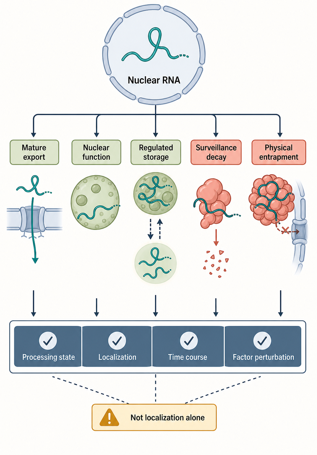
Figure 30.4. Nuclear retention decision tree. A nuclear RNA can follow several fates: export to the cytoplasm as a mature mRNP, retention for normal nuclear function as a lncRNA or chromatin-associated RNA, regulated storage for release in response to cell-state changes, routing to nuclear surveillance and decay, or physical entrapment because of size or assembly defects. Distinguishing these fates requires time-resolved measurement, compartmental localization, processing-state analysis, and factor perturbation rather than localization data alone, and the decision tree corrects the misconception that retention always means failed export.
Regulated release means that a retained RNA can later enter export or translation pathways. A useful example is intron retention in gene regulation. Some transcripts are held in the nucleus with retained introns and later spliced or released in response to cell state. Stress, differentiation, neuronal activation, immune signaling, or developmental transitions can alter the balance between retention, splicing, export, and decay. Evidence for regulated release requires time-resolved measurement: a static nuclear pool could represent newly transcribed RNA, failed processing, stable storage, or slow decay.
Recent primary studies show how retention can be regulated by protein modification and signaling. A 2025 study reported that phosphorylation of a nuclear condensate regulates cohesion and mRNA retention. A 2023 study found that EGFR promotes nuclear retention of ALKBH5 in glioblastoma, with consequences for m6A levels and ferroptosis resistance. These papers address different molecules and outcomes, but together they illustrate a broader point: retention is regulated through signaling, post-translational modification, RNP composition, and nuclear organization.
The common misconception is that nuclear retention equals defective export. Sometimes it does. But retention can also be a normal intermediate or a regulated endpoint. Another misconception is that a retained RNA must be noncoding. Protein-coding transcripts can be retained through introns, stress-responsive elements, repeat content, or RBP binding. Conversely, some noncoding RNAs are exported. The correct interpretation depends on the RNA class, molecular partners, processing state, and functional assay.
30.5. Large transcript export and nuclear-envelope constraints
Large transcript export is a logistics problem. The nuclear pore complex is large, but it is not infinitely permissive. An mRNP’s effective size depends on RNA length, folded structure, bound proteins, compaction, branching, and remodeling state. A transcript of tens or hundreds of kilobases cannot be imagined as a thin thread moving freely through a pore. It is a dynamic RNP that must be packaged, kept soluble, protected from surveillance when appropriate, and remodeled during transport.
Most large cellular mRNAs are still thought to use nuclear pore-mediated export after appropriate packaging. Polyadenylation and export are linked because 3′-end formation helps define a completed transcript and contributes to adaptor loading. Transcript homeostasis reviews emphasize that synthesis, processing, export, localization, translation, and decay together determine the observable abundance and location of each mRNA. For large RNAs, each step becomes more vulnerable to slow kinetics and partial failure.
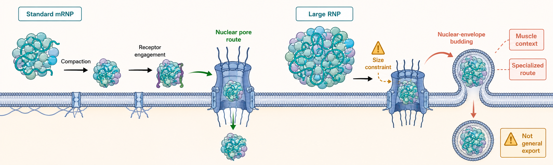
Figure 30.5. Large RNP logistics and alternative export routes. Most mRNPs reach the cytoplasm through nuclear pore-mediated export after compaction, receptor engagement, and cytoplasmic remodeling, but very large or unusually packaged transcripts face size and quality-control constraints that standard pore transit cannot easily accommodate. In muscle cells, nuclear-envelope budding has been reported as a specialized export route for large transcripts, representing a context-specific mechanism rather than a replacement for general pore-mediated transport.
Muscle cells provide an important boundary case. Muscle cells can express extremely large transcripts, including RNAs from giant genes whose products participate in specialized cellular architecture. A 2026 study reported that nuclear envelope budding enables export of large transcripts in muscle cells. Nuclear-envelope budding means that a nuclear-envelope membrane deformation encloses or transfers material in a route distinct from ordinary passage through a nuclear pore channel. This mechanism should be described as specialized and context-dependent. It does not overturn the central role of nuclear pores in bulk mRNA export.
Why might large RNPs need special routes? One reason is geometry: the central channel of a pore may be inefficient for bulky particles unless remodeling and compaction are sufficient. Another is quality control: very long transcripts have more opportunities for incomplete splicing, RBP misloading, damage, R-loop formation, or entanglement. A third is cell-type specialization: differentiated cells can produce transcripts and nuclear architectures unlike proliferating cultured cells. Large RNP logistics therefore sits at the intersection of RNA processing, nuclear mechanics, and cell biology.
Viral systems provide a second boundary case, though the biology is different. HIV-1 and related retroviruses must move large viral RNP or capsid-associated nucleic-acid complexes through nuclear and cytoplasmic environments. Reviews of HIV-1 nuclear capsid uncoating and reverse transcription emphasize that viral nucleic acid logistics can intersect with nuclear pores and host transport pathways. Viral examples should not be used as direct models for ordinary cellular mRNA export, but they reveal how pathogens exploit or challenge nuclear transport systems.
Retroelement transcripts blur the line between normal export and immune surveillance. A 2025 study reported that the mRNA export pathway can license viral mimicry responses and antitumor immunity by actively exporting nuclear retroelement transcripts. The key point is that export is not immunologically neutral. Which nuclear RNAs are allowed into the cytoplasm can influence innate immune detection, particularly when repeat-derived or double-stranded RNA-like species appear outside the nucleus. This topic connects Chapter 30 to chapters on repeat-derived RNAs, innate immune sensing, and RNA therapeutics.
Some references in the current bibliography for large transcript export are only indirect. Selection on synonymous sites and transcriptomic signatures in disease can be relevant to transcript homeostasis, but they do not directly establish mechanisms of large RNP export. A future curation pass should add more direct studies of Balbiani ring RNPs, giant cellular transcripts, nucleoplasmic viscosity, nuclear pore imaging, and nuclear-envelope mechanics. Until then, the chapter should state the large-transcript model as supported by selected direct examples and broader export principles rather than as a fully mapped universal pathway.
30.6. mRNA identity marks, packaging, and quality control
mRNA identity marks are molecular features that tell the cell what kind of RNA it is handling and what should happen next. The cap, cap-binding complex, splice status, EJC pattern, exon architecture, 3′-end formation, poly(A) tail, RNA modifications, RBP occupancy, export adaptors, and receptor contacts all contribute. These marks are not a barcode with one fixed decoder. They are interpreted by competing and cooperating factors in a specific cell state.
Packaging is the physical organization of the mRNA with proteins. Packaging protects RNA, compacts RNA, exposes or hides motifs, helps recruit export factors, and prevents inappropriate interactions. A highly structured RNA may require helicases or chaperones to become export competent. A repeat-rich transcript may be retained or surveilled. A properly spliced and polyadenylated mRNA may recruit adaptors that favor NXF1-NXT1 engagement. An aberrantly processed RNA may recruit nuclear decay or retention factors instead. This is why mRNP composition is often more informative than RNA abundance alone.
Quality control acts at several points. Before export, nuclear systems can test cap status, splice completion, 3′-end formation, poly(A) tail features, RNP composition, and retention signals. At the pore, improperly assembled mRNPs may fail to dock, traverse, or remodel. After export, cytoplasmic surveillance can detect premature termination codons, abnormal translation, deadenylation states, or immune-stimulatory RNA features. Nuclear and cytoplasmic quality control are connected because nuclear packaging determines what substrates reach the cytoplasm.
The link to therapeutic and engineered mRNA is conceptual but useful. Synthetic mRNAs delivered as vaccines or therapeutics bypass nuclear transcription and export because they are introduced into the cytoplasm or formulated for cytoplasmic delivery. Yet they still require identity features: cap structure, untranslated regions, coding sequence, modified nucleotides, poly(A) tail length, purity, and packaging in delivery vehicles influence translation, stability, and innate immune sensing. Engineered mRNA therefore highlights which identity marks can be supplied synthetically and which nuclear history marks are bypassed.
Box 30.3. Synthetic mRNA bypasses nuclear export
- mRNA vaccines and therapeutics are delivered directly to the cytoplasm or endosomes, so they never undergo nuclear transcription, co-transcriptional capping, splicing, or nuclear mRNP assembly and export.
- Identity features must be supplied synthetically: a 5′ cap analog, optimized 5′ and 3′ untranslated regions, modified nucleotides such as N1-methylpseudouridine to reduce innate immune detection, and a poly(A) tail of appropriate length.
- Purity matters because double-stranded RNA contaminants and uncapped species can activate innate immune sensors even when modified nucleotides are incorporated.
- Delivery vehicles—lipid nanoparticles, ionizable lipids, exosome-like particles—determine cellular uptake, endosomal escape, and cytoplasmic access but do not replicate the RNP packaging logic of nuclear mRNP assembly.
Exosome-mediated mRNA delivery is another comparison. Engineered exosomes can package mRNA for delivery, but this packaging is not the same as nuclear mRNP assembly. Nuclear export packages endogenous transcripts for pore transit and cytoplasmic handoff; delivery vesicles package RNA for extracellular stability, cell uptake, endosomal trafficking, and cytoplasmic release. The shared term “packaging” should not obscure the different physical route and quality-control logic.
Post-translational modification of nuclear pore proteins can tune export. A 2025 study reported that O-GlcNAc modulation of nuclear pore complexes orchestrates mRNA export efficiency. O-GlcNAc is a reversible sugar modification on serine or threonine residues of proteins. In this context, the modification is a regulatory input on the pore rather than a mark on the mRNA. This distinction matters: mRNP identity includes RNA marks and bound proteins, while export capacity also depends on the state of the transport machinery.
The evidence logic for quality control must separate processing defects from export defects. If a mutation causes nuclear RNA accumulation, the primary defect may be transcription elongation, splicing, 3′-end formation, adaptor recruitment, pore docking, pore traversal, cytoplasmic remodeling, or cytoplasmic decay. A useful experiment asks where the RNA accumulates, whether it is capped, whether introns remain, whether the 3′ end is correct, which proteins are bound, whether export receptors are recruited, whether acute rescue restores export, and whether total transcription has changed.
Biological Contexts Across RNA Classes and Organisms
Budding yeast has been central for defining mRNA export because genetic screens, live-cell imaging, and nuclear accumulation of poly(A)+ RNA revealed many export factors. Yeast genes are shorter and often have fewer introns than metazoan genes, so yeast models are powerful but incomplete. The yeast Mex67-Mtr2 receptor, TREX components, nuclear basket proteins, and Dbp5 remodeling logic illuminate core principles, while metazoan cells add extensive alternative splicing, longer genes, more specialized cell types, and additional adaptor diversity.
Metazoan mRNAs illustrate the coupling between splicing and export. Many mammalian genes contain multiple introns, and splicing creates EJC-marked exon-exon junctions. Alternative splicing changes the final exon architecture and can alter export, localization, stability, and translation. However, metazoan genomes also encode intronless genes, and viruses that infect metazoan cells often produce RNAs that must evade or redirect nuclear retention. Thus metazoan export biology is both more splicing-dependent and more adaptor-diverse than a simple rule would suggest.
Long noncoding RNAs and enhancer-associated RNAs expand the RNA sorting problem. Many are retained near chromatin or in nuclear bodies, and some are rapidly degraded by nuclear surveillance. Others are exported or processed into different RNA products. The distinction between lncRNA retention and mRNA retention is not absolute; rather, the RNA’s processing marks, sequence features, and binding proteins define its route.
Viral RNAs and retroelement RNAs show how export connects to immunity. Some viral RNAs use host mRNA export pathways, some encode their own export adaptors, and some rely on structured RNA elements. Retroelement-derived transcripts can become immune-stimulatory when exported and sensed in the cytoplasm. These examples are important because export decisions can influence disease, antiviral defense, and cancer immunotherapy.
Technology, Computational, Clinical, and Engineering Links
RNA localization assays are central to diagnosing export defects. RNA FISH can show nuclear accumulation, single-molecule FISH can quantify export kinetics for individual transcripts, and live-cell reporters can track transcription site release and cytoplasmic arrival. Fractionation followed by sequencing can survey transcriptome-wide nuclear-cytoplasmic distribution, but long RNA preparation and contamination controls are crucial. Crosslinking and immunoprecipitation can identify export-factor contacts, while proximity proteomics can map pore-associated states.
Computational interpretation usually starts with sequence and annotation: intron number, exon length, retained introns, 3′ UTR length, repeat content, RNA structure predictions, m6A motif enrichment, and RBP-binding motifs. These features can suggest retention or export mechanisms, but they are not proof. A predicted retained intron must be validated at the RNA isoform level. A predicted RBP motif must be connected to binding and function. A nuclear enrichment score must be interpreted with RNA stability and extraction bias in mind.
Clinically, export and retention defects can alter gene expression, immune signaling, cancer biology, neurobiology, and viral replication. Mutations or dysregulation in splicing factors, nuclear pore components, RNA helicases, and RNA modification enzymes can produce disease phenotypes through multiple routes. The ALKBH5 example in glioblastoma connects nuclear retention of a modification enzyme to RNA methylation and ferroptosis resistance. The retroelement export example connects export to antitumor immunity. In both cases, the causal path must be traced rather than inferred from localization alone.
Engineering applications include expression vector design, intron placement, UTR choice, codon and sequence optimization, RNA purification, and synthetic mRNA formulation. Nuclear expression constructs may benefit from introns or export elements that promote mRNP assembly. Cytoplasm-delivered mRNA therapeutics bypass nuclear export but still require cap, UTR, nucleotide, tail, and packaging choices. Viral vectors and oncolytic strategies can exploit export pathways, but export manipulation risks broad effects on host gene expression.
Recent Consensus
Current consensus supports six broad statements. First, mRNA export is coupled to transcription, capping, splicing, 3′-end formation, and RNP assembly rather than occurring as an isolated late step. Second, TREX-related factors, EJCs, and other adaptors help encode processing history into export competence. Third, NXF1-NXT1 or Mex67-Mtr2-like receptors mediate much bulk mRNA export through nuclear pore complexes, followed by cytoplasmic remodeling. Fourth, nuclear retention is both a quality-control mechanism and a regulated RNA fate. Fifth, large RNPs reveal physical and cell-type-specific constraints that can require specialized logistics. Sixth, mRNA identity is combinatorial: sequence, processing, modification, packaging, and transport-machinery state all matter.
Open Questions, Controversies, Deprecated Models, and Common Misconceptions
Open questions:
- How many distinct export routes exist for endogenous mammalian mRNAs?
- How do cells distinguish regulated intron retention from defective splicing?
- How are large RNPs selected for alternative nuclear-envelope routes, and how general are those routes across tissues?
Common misconceptions:
- “Nuclear RNA accumulation always means failed export.” Nuclear accumulation may instead reflect increased transcription, regulated retention, slow processing, nuclear function, altered decay, or technical fractionation artifacts.
Deprecated or weakened claims:
- The naked-mRNA model of export is deprecated. mRNAs are not usually made, checked, and exported as bare polymers; the mRNP state is the functional substrate.
- The model that splicing alone licenses export is oversimplified. Splicing is influential, but intronless transcripts and specialized RNAs show that multiple export-competence routes exist.