Chapter 89. tRNA Fragments, rRNA Fragments, Y RNA Fragments, snoRNA-Derived RNAs, and Emerging Small RNAs
Scope Note
This chapter covers small RNAs that are reproducibly detected as fragments of longer, abundant noncoding RNAs rather than as canonical microRNAs, small interfering RNAs, or PIWI-interacting RNAs. The main focus is transfer RNA-derived fragments, but ribosomal RNA fragments, Y RNA fragments, small nucleolar RNA-derived RNAs, vault RNA fragments, and newer fragment classes are included because they raise a shared interpretive problem: a short RNA read can represent regulated biogenesis, stress-induced cleavage, stable decay intermediate, extracellular cargo, technical bias, contamination, or ordinary degradation. The chapter treats fragment identity, nuclease specificity, biological function, extracellular biomarker claims, disease associations, and evidence standards for distinguishing functional small RNAs from degradation products.
Executive Summary
Fragments derived from abundant structured RNAs form a large and heterogeneous part of many small RNA sequencing datasets. Transfer RNAs are the best-studied source. Mature tRNAs and precursor tRNAs can be cleaved into 5′ tRNA halves, 3′ tRNA halves, shorter 5′ tRNA-derived fragments, 3′ CCA-containing fragments, leader-derived fragments, trailer-derived fragments, and internal fragments. Cleavage can occur at the anticodon loop, D loop, T loop, acceptor stem, or processing boundaries. The enzyme angiogenin is an important stress-responsive nuclease in mammalian tRNA-half production, but it is not the only nuclease involved. Dicer, RNase Z, RNase P-related processing, RNase T2-family enzymes, RNase L-linked pathways, Rny1-like enzymes, DIS3/exosome-associated turnover, and autophagic RNA degradation can contribute in specific systems. Nuclease identity should therefore be assigned by direct perturbation and cleavage-site mapping rather than by fragment class alone.
tRNA-derived fragments have credible regulatory roles in several contexts, including stress granule biology, translational repression, retrotransposon control, sperm-mediated intergenerational signaling, mitochondrial translation links, cancer phenotypes, renal stress responses, aging-associated metabolism, and plant abiotic stress. The strength of evidence varies widely. The most convincing cases combine stress-dependent biogenesis, defined nuclease or binding protein dependence, sequence-specific rescue or mutation, dose-relevant perturbation, and an endogenous target or pathway readout. Weaker claims rely only on differential abundance or transfection of high-dose synthetic fragments.
Fragments from rRNA, Y RNA, snoRNA, and vault RNA are increasingly reported in cells and extracellular samples. Some are reproducible, structured, protein-associated, or disease-enriched, and several small RNAs derived from snoRNAs or Y RNAs can enter Argonaute-containing complexes or affect gene expression in model systems. However, these classes are especially vulnerable to overinterpretation because parent molecules are abundant, structured, modified, and resistant to complete degradation. Recurrent sequencing reads can reflect protected degradation intermediates, preferential ligation, ribosome or RNP breakdown, sample handling, or biofluid contamination as well as regulated small RNA biogenesis.
Extracellular fragments are useful biological readouts when measured carefully. Plasma, serum, urine, saliva, cerebrospinal fluid, and extracellular vesicle preparations contain tRNA, Y RNA, rRNA, and other noncanonical small RNA fragments. These molecules can correlate with cancer, infection, inflammatory disease, renal injury, aging, pregnancy, or tissue damage. The clinical translation standard is higher than the discovery standard: a biomarker claim requires control of hemolysis, platelet activation, vesicle-isolation bias, batch effects, library bias, normalization, population structure, comorbidities, and independent validation. A disease-associated fragment is not automatically a causal regulator or a vesicle-delivered signal.
The chapter’s working consensus is conservative. Stable RNA fragments are real, often nonrandom, and sometimes functional. tRNA-derived fragments have the strongest mechanistic base. Other fragment classes are promising but unevenly proven. The field should treat fragment biology as a spectrum from purposeful processing to protected decay, with explicit evidence labels rather than a binary distinction between “functional RNA” and “trash RNA.”
Concept Inventory
- tRNA-derived fragment: a small RNA whose sequence maps to a transfer RNA gene, precursor tRNA, mature tRNA body, or tRNA trailer. The term includes multiple size and origin classes.
- tRNA half: an approximately 30-40 nucleotide fragment generated by cleavage near the anticodon loop, usually producing complementary 5′ and 3′ halves from a mature tRNA. In mammalian stress responses, angiogenin is a frequent contributor.
- tiRNA: stress-induced tRNA-derived RNA. This term is often used for tRNA halves generated during stress, but usage varies across papers.
- 5′ tRF: a shorter fragment from the 5′ end of mature tRNA, often ending in the D loop or anticodon region.
- 3′ CCA tRF: a fragment from the mature tRNA 3′ end that includes the post-transcriptionally added CCA tail.
- 3′ U tRF: a fragment derived from the trailer sequence of a precursor tRNA, often ending in uridines from transcription termination or processing.
- Internal tRF: a fragment mapping within the tRNA body without coinciding with the mature 5′ or 3′ end.
- rRNA-derived fragment: a small RNA mapping to mature rRNA or pre-rRNA sequences. Recurrent rRNA fragments can arise from regulated cleavage, ribosome turnover, protected decay, or contamination by abundant ribosomal material.
- Y RNA fragment: a small RNA derived from a Y RNA, a structured noncoding RNA that forms RNPs with RO60 and other proteins. Y RNA fragments are abundant in some biofluids and disease datasets.
- sdRNA: snoRNA-derived RNA, a small RNA derived from a small nucleolar RNA. Some sdRNAs behave like miRNA-like Argonaute-associated species, whereas others are better interpreted as stable processing or decay products.
- Vault RNA fragment: a small RNA derived from vault RNAs, polymerase III transcripts associated with vault particles and additional cellular functions.
- Protected degradation intermediate: a reproducible fragment whose accumulation reflects RNA structure or bound proteins protecting a piece of a parent RNA during decay, without necessarily implying a regulatory function.
- Functional evidence standard: the set of experiments required to infer a biological role, including endogenous abundance, biogenesis mechanism, target or effector binding, perturbation and rescue, specificity controls, and physiological context.
What to Know Before Reading This Chapter
The chapter assumes familiarity with canonical small RNA pathways, especially microRNAs and small interfering RNAs, but it does not assume that every short RNA read enters Argonaute or Dicer pathways. A microRNA is normally produced from a defined hairpin precursor and functions through Argonaute-guided target recognition. A tRNA-derived fragment or snoRNA-derived RNA may mimic some features of a microRNA, but the parent RNA, processing logic, chemical modifications, and evidence burden are different.
Readers should also know the basic architecture of tRNA and rRNA. Mature tRNAs are short, highly modified, L-shaped adaptor RNAs with acceptor stems, D loops, anticodon loops, variable loops, T loops, and a CCA end used for aminoacylation. rRNAs are much larger structural and catalytic RNAs embedded in ribosomes. Y RNAs, snoRNAs, and vault RNAs are stable RNP-associated RNAs. These parent RNAs are abundant and structured, so they can yield short, stable fragments during processing, stress, extraction, or decay.
Finally, readers should distinguish three questions that are often merged. The first question is whether a fragment is reproducibly present. The second is whether the fragment is produced by a regulated biogenesis pathway. The third is whether the fragment has an endogenous biological function. A fragment can satisfy the first question without satisfying the second or third.
89.1. Fragment biogenesis and nuclease specificity
Fragment Classes Are Defined by Parent RNA, Endpoint, Size, and Context
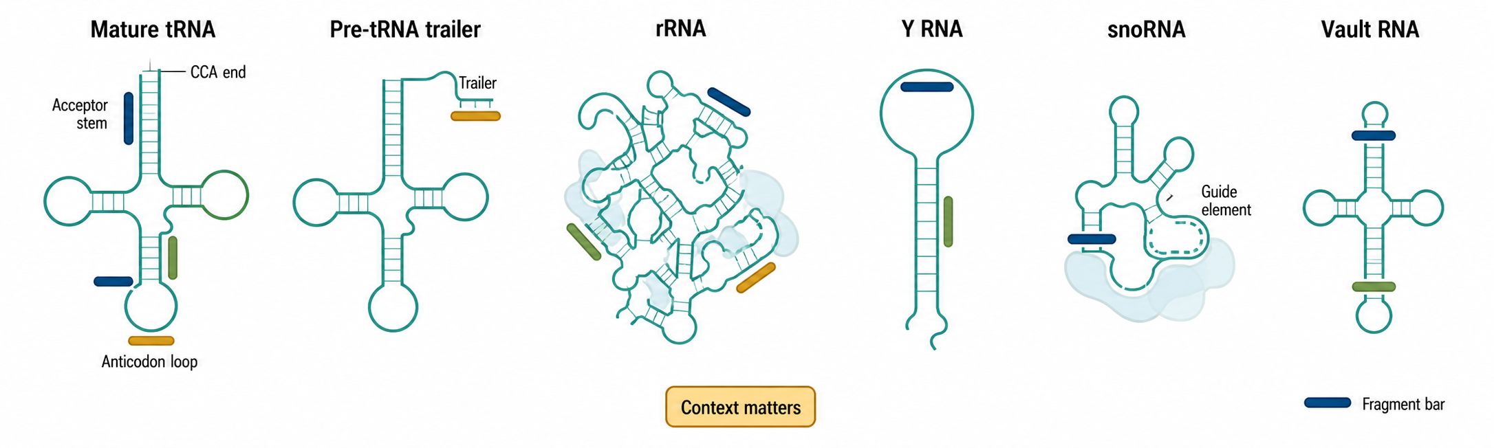
Figure 89.1. Fragment map across parent stable RNAs. “Stable-RNA fragment classes are best described by parent RNA, endpoint, size, terminal chemistry, and context. The same parent RNA can yield regulated cleavage products, protected decay intermediates, and technical artifacts.”
The phrase “tRNA-derived fragment” is useful only as a family name. It does not specify a single biogenesis pathway or function. A 5′ tRNA half from tRNA-Gly-GCC generated during oxidative stress is mechanistically different from a 3′ trailer fragment produced during pre-tRNA maturation, even though both map to tRNA loci. For that reason, tRNA fragments should be annotated by parent isodecoder when possible, genomic or transcript origin, start and end position, length, terminal chemistry, modification status, and experimental context.
The same principle applies to rRNA, Y RNA, snoRNA, and vault RNA fragments. A short read mapping to 28S rRNA may be a ribosome-protected decay piece, a nuclease cleavage product during stress, a contaminant from abundant ribosomes, or a library artifact. A Y RNA fragment from plasma may be biologically informative, but the first annotation should describe where it maps in the Y RNA, whether the endpoint is recurrent, whether it appears in vesicle, lipoprotein, or protein fractions, and whether the signal survives contamination controls. Classification by parent RNA alone is not enough.
Length also matters. Fragments in the 14-18 nucleotide range are often too short for unique genomic assignment, especially among multicopy tRNA, rRNA, and repetitive RNA genes. Fragments near 20-24 nucleotides can overlap with canonical small RNA sizes and may enter Argonaute complexes in some cases. Fragments near 30-40 nucleotides often represent tRNA halves or larger protected pieces that behave differently from microRNAs in library preparation and functional assays. Size alone, however, is not proof of a pathway.
Box 89.1. How to Describe a Stable-RNA Fragment Without Overclaiming
“A stable-RNA fragment name should describe what is known from the molecule and experiment, not what the investigator hopes the fragment does.”
tRNA Cleavage Reflects Structure, Modification, and Nuclease Access
Mature tRNAs are difficult substrates. They are compactly folded, heavily modified, aminoacylated or deacylated depending on cellular state, and bound by aminoacyl-tRNA synthetases, elongation factors, modification enzymes, quality-control proteins, and stress-associated factors. Cleavage therefore depends not only on the sequence of the tRNA but also on structure, modification, charging state, protein occupancy, compartment, and stress condition.
Anticodon-loop cleavage produces tRNA halves. In mammalian cells, angiogenin can be released or activated during stress and cleave exposed tRNA anticodon loops, generating 5′ and 3′ halves. The resulting 5′ halves from selected tRNAs can inhibit translation initiation and promote stress granule assembly in some systems. This mechanism is not universal. Yeast uses Rny1 in some stress conditions. Plants and parasites have their own nuclease repertoires and stress responses. Some tRNA fragments appear without angiogenin dependence, and some angiogenin-dependent cleavage events do not produce proven regulatory RNAs.
Shorter tRNA fragments arise at multiple positions. RNase Z processing of pre-tRNA trailers can generate 3′ U fragments. Dicer has been implicated in subsets of shorter tRNA fragments in some systems, but many tRNA fragments are Dicer-independent. RNase P defines mature 5′ tRNA ends and can indirectly affect leader-derived fragments. Exonucleases and endonucleases involved in RNA surveillance can create or eliminate fragments. Direct lysosomal RNA import has also been linked to tRNA-derived small RNA production in specific stress or tissue contexts. A hypoxia-responsive renal study provides one such example (Li et al. 2025), while DHX8 work supports a SIDT2-dependent RNA-uptake pathway in cultured mammalian cells (Sakai et al. 2025). These studies do not establish that all autophagy-dependent RNA turnover produces regulatory fragments. Chapter 37 owns the distinction among direct RNautophagy, ribophagy, lysosomal nuclease degradation, and RNA loss secondary to bulk organelle autophagy; this chapter retains ownership of fragment identity and function.
Table 89.1. Nucleases, pathways, and fragment classes. “Nuclease assignments require context-specific evidence. Fragment size and endpoint can suggest a pathway but should not replace perturbation, terminal chemistry, and rescue experiments.”
| Enzyme or pathway | Typical substrate context | Fragment class | Terminal clues | Evidence required | Key caveat |
|---|---|---|---|---|---|
Angiogenin (ANG) |
Mammalian stress-exposed mature tRNAs with accessible anticodon loops | 5′ and 3′ tRNA halves, often 30-40 nt | Cleavage near anticodon loop; nuclease-dependent endpoints | ANG perturbation, stress timing, endogenous fragment validation, rescue or cleavage-site evidence | Important mammalian mechanism, but not a universal source of all tRNA fragments |
Dicer (DICER1) |
Structured precursor-like RNAs or selected shorter stable-RNA fragments | Some 20-24 nt tRFs, sdRNAs, or other AGO-sized candidates | Dicer-size products with 5′ phosphate and 3′ hydroxyl are suggestive | Dicer loss-of-function, in vitro cleavage, AGO loading, seed-mutant reporter or target tests | Size alone does not prove Dicer biogenesis |
| RNase Z / ELAC enzymes | Pre-tRNA 3′ trailer processing before CCA addition | 3′ U tRFs / trailer-derived fragments | Trailer sequence, often uridine-rich Pol III termination tract | Mapping to pre-tRNA trailer, RNase Z dependence, distinction from mature CCA tRFs | Trailer fragments can be locus-specific but may not be regulatory products |
| RNase P-linked processing | Pre-tRNA 5′ leader maturation and mature-end definition | Leader-derived reads or indirectly altered 5′ tRF profiles | Mature tRNA 5′ boundary or upstream leader sequence | Processing-boundary mapping plus RNase P perturbation or precursor accumulation | RNase P primarily makes mature tRNA ends; downstream fragments need separate evidence |
| RNase T2 / Rny1-like enzymes | Stress, starvation, or compartment-specific RNA cleavage in fungi, plants, and animals | tRNA halves and mixed stable-RNA fragments | End chemistries compatible with acid RNases or stress nucleases | Enzyme perturbation, stress specificity, terminal chemistry, orthogonal blot validation | Nuclease repertoires differ across organisms and compartments |
| RNase L-linked pathways | Interferon or antiviral contexts with activated 2-5A/RNase L signaling | tRNA, rRNA, or other small RNA fragments during innate immune activation | RNase L-preferred cleavage patterns in activated cells | RNase L activation marker, genetic or pharmacologic perturbation, infection or dsRNA controls | Collateral RNA decay can be biologically informative without creating regulatory fragments |
| DIS3 / exosome-associated turnover | Nuclear or cytoplasmic RNA surveillance and decay of structured RNAs | Protected decay intermediates from stable RNAs | Heterogeneous decay endpoints near structured or protein-protected regions | Exosome-component perturbation, subcellular localization, decay kinetics | Reduced decay can increase fragments without implying purposeful biogenesis |
| Direct lysosomal RNA import (RNautophagy in the cited mammalian literature) | SIDT2/LAMP2C-linked lysosomal RNA uptake in specific stress or tissue contexts | tRNA-derived small RNAs and other degradation-linked fragments in selected systems | Compartment- and pathway-dependent fragment enrichment | Lysosomal uptake or flux assay, SIDT2/LAMP2C perturbation, compartment controls, and rescue | Not synonymous with ribophagy or bulk autophagy; Chapter 37 owns pathway mechanism, whereas this table asks whether a fragment is produced and functional |
Modification status is a major interpretive variable. tRNAs contain methylations, pseudouridine, queuosine, inosine, thiolations, and many other marks. These modifications can influence folding, cleavage sensitivity, reverse transcription, adapter ligation, and read-through in sequencing. A fragment may appear condition-specific because the parent tRNA is more cleavable, because a modification blocks cloning in one condition, or because a library protocol captures one terminal chemistry better than another. Modified bases therefore connect biology and measurement bias. A serious tRNA-fragment study should state how the library captures modified RNAs and whether demethylase treatment, specialized reverse transcriptase, terminal repair, or spike-ins were used.
Fragment Biogenesis Is Not Equivalent to Degradation
The field often frames fragments as either functional products or meaningless degradation. That binary is too simple. A cell can produce a fragment through ordinary RNA turnover, but the fragment can still be stable because it is protected by structure or proteins. Conversely, a regulated nuclease cleavage event can be part of destructive quality control rather than a signaling pathway. The more useful categories are purposeful processing, stress-triggered cleavage, protected decay intermediate, collateral degradation, and technical artifact. These categories can overlap.
Box 89.2. Protected Decay Is Not the Same as Meaningless Degradation
“Protected decay intermediates can be reproducible and biologically informative without being dedicated regulatory RNAs.”
For example, rRNA fragments may rise when ribosomes are damaged, when ribosome biogenesis is perturbed, or when ribophagy and lysosomal pathways turn over ribosomal material. Ribophagy refers to autophagic ribosome turnover and is not a synonym for direct RNA translocation across a lysosomal membrane. The resulting reads can be biologically meaningful as a footprint of ribosome turnover without being regulatory small RNAs. Similarly, snoRNA-derived fragments may reflect the stable core of a snoRNP protected by proteins after partial degradation. If that fragment binds Argonaute and represses a target at endogenous levels, it can also acquire a regulatory interpretation. The correct conclusion depends on the experiments, not on the parent RNA name; pathway mechanisms and evidence boundaries are developed in Chapter 37.
89.2. tRNA-derived fragments in stress, translation, and signaling
The clearest cellular roles for tRNA fragments involve stress and translation. During oxidative stress, heat shock, nutrient limitation, viral infection, or other insults, cells can change tRNA charging, activate ribonucleases, remodel translation initiation, and assemble stress granules. Selected tRNA halves are positioned within this logic: they arise rapidly, can interact with translation initiation factors or RNA-binding proteins, and can contribute to global translational repression or granule formation in specific systems. Reviews by Shen et al. (2018), Park and Kim (2018), and Chen and Zhou (2023) provide local reference anchors for these principles.
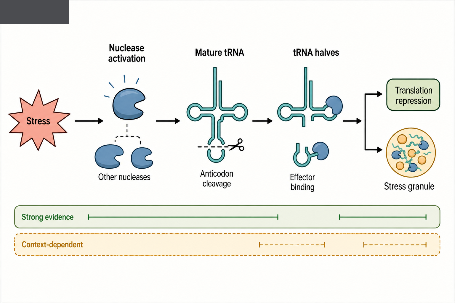
Figure 89.2. Stress-induced tRNA-half production and translation effects. “In selected mammalian stress responses, nuclease activation can cleave mature tRNAs near the anticodon loop. Some resulting tRNA halves bind effectors that alter translation initiation or stress granule assembly, but the nuclease and mechanism are context-specific.”
The mechanism is not simply “a tRNA fragment silences mRNAs.” Some tRNA fragments appear to act through protein binding rather than through base pairing. Others are reported to enter Argonaute complexes and behave more like guide RNAs. Still others influence ribosome assembly, mitochondrial translation, RNA stability, retroelement expression, or signaling pathways. The fragment’s length and terminal chemistry strongly affect which mechanisms are plausible. A 35-nucleotide tRNA half is unlikely to behave exactly like a 22-nucleotide microRNA even if both can be detected by small RNA sequencing.
Several disease and physiology studies now report specific tRNA fragments with stronger mechanistic claims. A hypoxia-responsive tRNA-derived small RNA was reported to protect kidney tissue through an RNA autophagy-linked mechanism (Li et al. 2025). An aging-induced tRNA-Glu-derived fragment was reported to impair glutamate biosynthesis by affecting mitochondrial translation-dependent cristae organization (Li et al. 2024). These examples illustrate the direction of the field: the strongest papers move beyond cataloging fragments and connect a defined fragment to a cellular pathway, a perturbation experiment, and a physiological readout. Even so, each case must be interpreted in its own experimental system rather than generalized to all tRNA fragments.
89.3. rRNA, Y RNA, snoRNA, and vault RNA fragments
rRNA, Y RNA, snoRNA, and Vault RNA Fragments Occupy a Broader Evidence Spectrum
rRNA-derived fragments are common in small RNA datasets because ribosomes are abundant and rRNA is structured. Some rRNA fragments show nonrandom endpoints and stress- or tissue-specific enrichment. In bacteria and eukaryotes, ribosome turnover and ribosome quality control can generate structured decay intermediates. In eukaryotic cells, perturbations in ribosome assembly, nucleolar stress, ribophagy, or cytoplasmic RNA decay can change rRNA fragment profiles. However, functional claims for rRNA fragments require especially strict controls because even small amounts of ribosomal RNA contamination can dominate small RNA libraries.
Y RNA fragments are prominent in extracellular RNA studies and some inflammatory or cancer contexts. Full-length Y RNAs are RO60-associated structured RNAs with roles in RNA quality control and chromatin replication-associated processes. Short Y RNA fragments can be abundant in plasma and extracellular vesicle preparations. Some studies report disease associations, immune links, or cell-state specificity. The key unresolved question is how often these fragments act as signals rather than stable cargo or breakdown products. Their reproducible presence is not in doubt; the causal mechanism is often less secure.
snoRNA-derived RNAs sit between stable RNP biology and small RNA regulation. Box C/D snoRNAs guide 2′-O-methylation, and box H/ACA snoRNAs guide pseudouridylation, mostly on rRNA and snRNA targets. Some snoRNAs also have noncanonical functions. When a small RNA maps to a snoRNA, it may represent a protected guide region, a processing product, an RNP decay intermediate, or a miRNA-like regulatory RNA. A subset of sdRNAs is Argonaute-associated and can repress reporter targets, but Argonaute occupancy alone is not sufficient; abundant RNAs can appear in immunoprecipitations through indirect or nonspecific routes unless controls are strong.
Vault RNA fragments derive from polymerase III-transcribed vault RNAs. Full-length vault RNAs associate with vault particles but also have vault-independent functions in drug response, innate immunity, and RNA-binding protein networks. Vault RNA-derived small RNAs have been reported in cancer and stress datasets, sometimes with Argonaute association or gene-regulatory effects. As with Y RNA and snoRNA fragments, the field has promising observations but fewer deeply mapped biogenesis pathways than for tRNA halves.
Table 89.2. Non-tRNA fragment classes and interpretation risks. “Fragments from rRNA, Y RNA, snoRNA, and vault RNA are reproducible in many datasets, but their evidence base differs by class and context.”
| Parent RNA | Normal biological role | Common fragment observations | Plausible mechanisms | Major artifact risks | Current evidence maturity |
|---|---|---|---|---|---|
| rRNA | Structural and catalytic core of ribosomes | Abundant reads from 18S, 28S, 5.8S, 5S, or pre-rRNA regions; sometimes stress- or tissue-enriched | Ribosome turnover, ribosome biogenesis defects, ribophagy, protected decay, stress cleavage | Ribosomal contamination, repetitive loci, extraction degradation, library domination by abundant rRNA | Reproducible detection is common; regulatory function usually needs stronger proof |
| Y RNA | RO60-associated structured RNP RNA involved in RNA quality-control and replication-linked contexts | Prominent Y RNA fragments in plasma, serum, extracellular vesicle preparations, inflammation, and cancer datasets | RNP-protected cleavage, extracellular packaging, cell injury release, stress-linked processing | Hemolysis, platelet activation, co-isolated protein/lipoprotein complexes, Y RNA pseudogene mapping | Strong as biomarker candidates in some datasets; signaling mechanisms remain uneven |
| snoRNA | Guide RNAs for rRNA/snRNA 2′-O-methylation or pseudouridylation; some noncanonical roles | sdRNAs from box C/D or H/ACA snoRNAs; occasional AGO association or reporter effects | Protected snoRNP decay, guide-region processing, noncanonical regulatory processing | Immunoprecipitation carryover, abundant RNP protection, confusing host-gene or intron signals | Moderate and context-dependent; selected sdRNAs may be functional |
| Vault RNA | Pol III transcript associated with vault particles and vault-independent RNP functions | vtRNA-derived small RNAs reported in stress, cancer, and AGO-associated datasets | Dicer- or non-Dicer cleavage, RBP-protected fragments, stress-responsive processing | Low nomenclature standardization, small family mapping ambiguity, correlation-heavy disease studies | Promising but citation-thin and less settled than tRNA halves |
| Other stable RNAs | SRP RNA, RNase P/MRP RNA, snRNA, scaRNA, and other RNP-associated RNAs | Occasional recurrent fragments in small RNA or extracellular RNA datasets | RNP-protected decay, processing byproducts, stress or surveillance cleavage | Misannotation, multimapping, incomplete parent-RNA annotation, sample handling | Treat as candidate fragmentomics signals until class-specific evidence accumulates |
Experimental Foundations and Evidence
Mapping Small RNA Reads to Fragment Classes Is Technically Hard
Fragment discovery often begins with small RNA sequencing, but the method was originally optimized around microRNAs and similar 5′ phosphate, 3′ hydroxyl molecules. Many fragments from tRNA, rRNA, snoRNA, and other stable RNAs have chemical ends that are not captured efficiently by standard adapter ligation. Some nucleases leave 2′,3′ cyclic phosphate, 3′ phosphate, or 5′ hydroxyl ends. Some parent RNAs contain modifications that terminate reverse transcription or cause misincorporation. A library can therefore undercount one fragment class and overcount another.
Mapping is another challenge. tRNA genes are multicopy, many tRNA isodecoders are nearly identical, and mature tRNAs include a CCA tail not genomically encoded for many nuclear tRNAs. Pre-tRNA leaders and trailers may map to unique genomic loci, but mature-body fragments often cannot be assigned to a single gene copy. rRNA genes are repetitive, multicopy, and sometimes incompletely represented in genome assemblies. Y RNA pseudogenes and repeats complicate assignment. A rigorous analysis should report whether reads are assigned to parent RNA families, isodecoders, individual loci, or transcript isoforms, and should avoid overclaiming gene-level specificity when the read sequence does not support it.
Validation Requires Orthogonal Assays
Northern blotting remains valuable because it can show the approximate size of an endogenous fragment and distinguish a discrete product from a smear of degradation. qRT-PCR can be sensitive but is vulnerable to primer specificity and modification-dependent reverse transcription. Synthetic spike-ins help with technical normalization but do not solve biological normalization in plasma or extracellular vesicle samples. Enzymatic treatments can reveal terminal chemistry, but they must be interpreted with controls because treatment efficiency varies.
Functional validation requires more than overexpression. A synthetic tRNA fragment transfected at high concentration can bind proteins, activate innate immune sensors, repress translation, or change cell physiology in ways that endogenous fragments do not. Better evidence includes loss of the nuclease that produces the fragment, mutation of the parent RNA cleavage site without disrupting the parent RNA’s essential function, antisense inhibition or target protection at dose-relevant levels, rescue by a fragment resistant to the perturbation, and direct target or effector measurement. Reporter assays are useful but incomplete unless paired with endogenous target data.
Table 89.3. Functional evidence standards and common artifacts. “Stable-RNA fragment function should be graded with explicit evidence standards. Detection and association are useful starting points but do not substitute for mechanism, specificity, and rescue.”
| Claim type | Minimal evidence | Stronger evidence | Common false positive | Example control |
|---|---|---|---|---|
| Reproducible fragment detection | Recurrent read with defined parent, endpoint, length, and protocol | Northern blot or orthogonal assay confirms discrete endogenous size | Library ligation bias or degraded abundant RNA | Spike-ins, replicate extraction, terminal-chemistry-aware library preparation |
| Regulated fragment formation | Abundance changes with stress, tissue, development, or disease state | Time course links stimulus, nuclease activity, and fragment appearance | Parent RNA abundance or sample composition changes | Measure parent RNA, cell composition, and degradation markers in parallel |
| Nuclease assignment | Fragment endpoint matches a plausible nuclease context | Enzyme perturbation, in vitro cleavage, terminal chemistry, and rescue agree | Inferring enzyme from size or pathway reputation | Knockout/knockdown plus rescue with nuclease-active and inactive alleles |
| Argonaute-like regulation | Fragment appears in AGO pulldown and has AGO-compatible size | Endogenous target repression, seed mutation, target-site mutation, and stoichiometry support guide function | Abundant RNA carryover in immunoprecipitation | AGO-IP controls, seed-swap reporter, endogenous target protection |
| Protein-binding mechanism | Fragment binds a candidate RBP or translation factor | Binding-site mutation, competition, endogenous complex stoichiometry, and pathway rescue | Nonspecific binding by transfected high-dose RNA | Dose-matched mutant fragment and endogenous RNP pull-down |
| Translation or stress-granule effect | Fragment perturbation changes translation markers or granule abundance | Polysome/ribosome profiling, factor binding, rescue, and stress-specific dependence | Innate immune activation or transfection toxicity | Chemically matched control RNA, immune-marker assay, dose-response curve |
| Extracellular biomarker | Fragment classifies cases and controls in discovery cohort | Independent cohort, pre-specified model, confounder control, and clinical comparator | Hemolysis, platelet activation, batch effect, or storage bias | Hemolysis markers, platelet depletion, randomized processing, locked validation set |
| Intercellular signaling | Fragment is detected in extracellular carrier fraction | Source cell, carrier topology, uptake route, recipient target, and physiological effect are shown | Co-isolated protein/lipoprotein RNA or dead-cell release | Protease/RNase protection, density separation, uptake blockade, source-cell perturbation |
| Disease mechanism | Fragment abundance correlates with disease phenotype | Loss- and gain-of-function, target or effector mechanism, rescue, and in vivo relevance | Biomarker mistaken for causal regulator | Fragment-specific inhibitor or mimic with rescue and tissue-source evidence |
Argonaute immunoprecipitation is informative only when interpreted carefully. If a fragment is consistently found in AGO complexes, has the expected size and end chemistry, shows seed-like target effects, and loses activity when its seed is mutated, then a miRNA-like mechanism becomes plausible. If an abundant fragment appears weakly in an AGO pulldown without stoichiometric or functional evidence, Argonaute association should be treated as a clue rather than a conclusion. The same logic applies to other RNA-binding proteins: binding must be connected to mechanism.
Nuclease Perturbation Is Necessary but Not Sufficient
Knocking down a nuclease can reduce a fragment and change a phenotype, but this does not prove the fragment mediates the phenotype. Ribonucleases often have many substrates. Angiogenin, RNase L, Dicer, DIS3, and lysosome-associated RNA decay factors can affect broad RNA populations. The strongest inference comes when fragment restoration rescues the phenotype after nuclease perturbation, and when cleavage-site mutations in the parent RNA selectively prevent fragment formation. These experiments are difficult for tRNAs because parent tRNA function is essential and multicopy genes complicate editing.
Nuclease specificity can be inferred from cleavage-site motifs, terminal chemistry, in vitro cleavage with purified enzyme, and cellular perturbation. No single line of evidence is enough. For instance, anticodon-loop cleavage during stress is consistent with angiogenin in mammalian cells, but the assignment should still be tested. Similarly, Dicer-sized fragments are not automatically Dicer products. The chapter treats enzyme labels as evidence-weighted, not as default annotations.
Biological Contexts
Stress Responses and Translation Control
Stress changes the balance between protein synthesis, RNA storage, RNA decay, and cellular survival. tRNA-derived fragments fit into this balance because tRNAs are direct substrates of translation and sensitive reporters of metabolic state. During oxidative stress or nutrient limitation, deacylated tRNAs accumulate, translation initiation can be inhibited, and stress granules can assemble. Specific tRNA halves can promote translational repression or granule formation, although the exact protein partners and sequence requirements differ among systems.
In plants, tRNA fragments have been linked to abiotic stress signal transduction, including responses to drought, salt, cold, and phosphate or nutrient stress (Park and Kim 2018). Plant evidence often includes stress-responsive abundance and target predictions, while mechanistic validation varies. Plant systems also illustrate an important principle: fragment biology may be conserved at the level of stress-responsive tRNA cleavage without preserving identical fragment sequences, targets, or nuclease proteins across kingdoms.
Development, Gametes, and Intergenerational Signaling
tRNA-derived fragments are abundant in sperm and have been implicated in paternal diet, metabolic inheritance, and early embryonic gene regulation in mammalian models. The proposed logic is that sperm carry small RNAs shaped by epididymal maturation and paternal environment; after fertilization, these RNAs can influence early developmental gene expression. This field has generated influential results, but it also faces strong evidence challenges. Sperm RNA preparations can be contaminated by somatic cells, bacteria, epididymal vesicles, or environmental RNA. Microinjection experiments can use doses that exceed physiological delivery. A convincing inheritance model must connect paternal exposure, endogenous sperm fragment changes, embryo delivery, target mechanism, and offspring phenotype with appropriate rescue or loss-of-function tests.
The broader developmental relevance of noncanonical fragments remains unsettled. Some fragments may be state markers rather than instructive regulators. Others may have true roles in cell fate, gametogenesis, or early embryonic transitions. The correct posture is neither dismissal nor broad acceptance; each claim should be placed on an evidence ladder.
Cancer, Metabolism, Aging, and Organ Injury
Cancer studies report many tRNA, Y RNA, snoRNA, and vault RNA fragments with altered abundance. Some fragments are associated with proliferation, invasion, metastasis, therapy response, or immune microenvironment features. Reviews of tRNA fragments in cancer progression (Lu et al. 2024) emphasize that the field is moving toward pathway-specific mechanisms, but many reported markers remain correlative. Cancer is also a setting where sample composition is a major confounder: tumor biopsies contain malignant cells, stromal cells, immune cells, necrotic regions, blood, extracellular vesicles, and variable RNA degradation.
Metabolic and aging contexts provide more mechanistic examples for tRNA fragments. The aging-associated tRNA-Glu-derived fragment reported by Li et al. (2024) links a defined fragment to mitochondrial translation and cristae organization. This kind of pathway connection is stronger than a simple age-associated differential abundance result. Kidney injury and hypoxia studies likewise show how a fragment can be connected to tissue protection rather than treated merely as a biomarker (Li et al. 2025).
89.4. Extracellular fragments, biomarkers, and disease claims
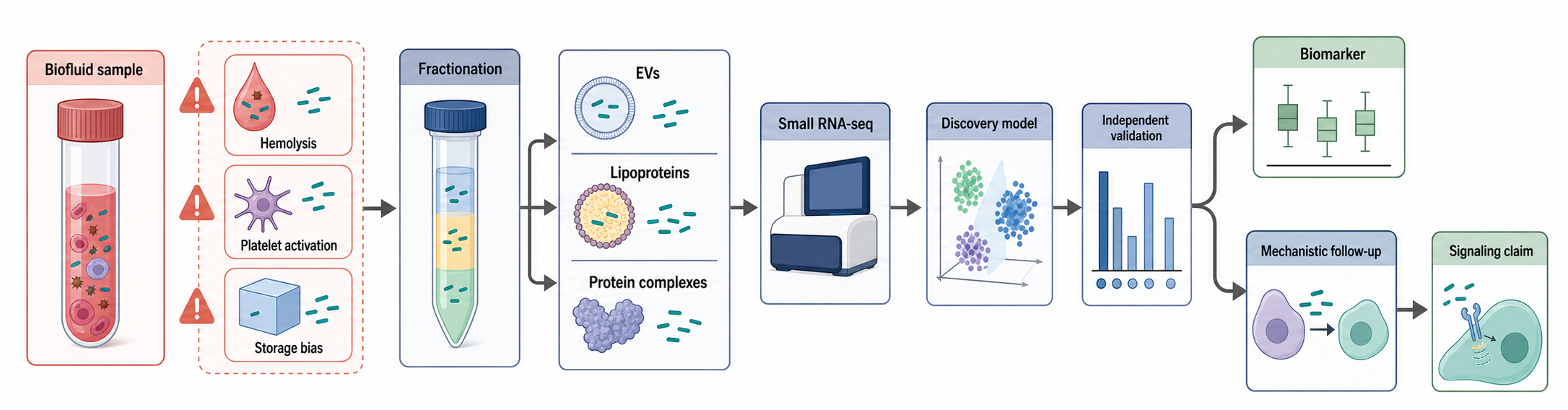
Figure 89.3. Extracellular fragment biomarker workflow and confounders. “Extracellular RNA fragment studies must separate discovery from clinical validation and mechanism. Biofluid fragments can originate from tissue injury, blood-cell release, extracellular vesicles, lipoproteins, protein complexes, or sample-handling artifacts.”
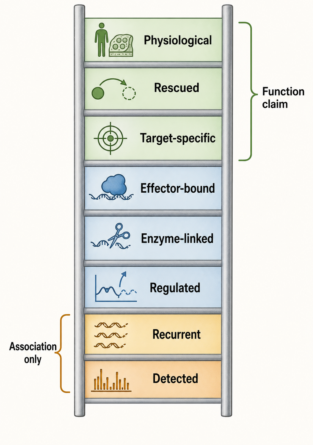
Figure 89.4. Evidence ladder for functional fragment claims. “Functional confidence increases from reproducible detection to regulated biogenesis, molecular effector binding, endogenous target effect, perturbation, rescue, and physiological relevance. Disease association or Argonaute association alone is not sufficient.”
Extracellular RNA samples contain fragments from many stable RNAs. These fragments may travel in extracellular vesicles, ribonucleoprotein particles, lipoproteins, apoptotic bodies, or protein complexes. The same small RNA species may appear in multiple carrier fractions depending on sample preparation. A plasma tRNA fragment or Y RNA fragment can therefore be a signal of tissue injury, immune activation, platelet release, hemolysis, vesicle secretion, or clearance biology.
Biomarker studies should separate discovery, validation, and mechanism. In discovery, it is acceptable to identify fragments that classify disease and control samples under a defined protocol. In validation, the same fragments should be tested in independent cohorts with pre-specified processing, normalization, and statistical models. In mechanism, researchers must show the source cell or tissue, carrier, recipient cell, uptake route, molecular target, and physiological consequence. A fragment can be a useful biomarker even if it has no causal function. Conversely, a functional intracellular fragment may be a poor biomarker if it is unstable, low abundance, or confounded by blood-cell composition.
Box 89.3. Biomarker Evidence Versus Intercellular Signaling Evidence
“An extracellular fragment can be a useful biomarker without being a vesicle-delivered signal.”
The local reference list includes a systematic review of extracellular vesicle clinical trials (Mizenko et al. 2024) and a primary study on noncanonical small noncoding RNAs in plasma extracellular vesicles as gastric cancer biomarkers (Yang et al. 2025). These sources support cautious discussion of clinical promise and validation burdens, but chapter-specific expert review should add more focused extracellular RNA standards and hemolysis-control references.
Technology, Computational, and Clinical Links
89.5. Contamination, degradation artifacts, and functional evidence standards
Fragmentomics Requires Purpose-Built Informatics
Small RNA fragment analysis is not a simple extension of microRNA analysis. Pipelines must handle multimapping reads, mature RNA features absent from the genome, CCA addition, tRNA isodecoders, RNA modifications, terminal heterogeneity, and short-read ambiguity. A useful report should include parent RNA class, fragment coordinates relative to mature and precursor RNA, length distribution, terminal sequence, multimapping policy, library protocol, and normalization strategy.
Fragment databases and naming schemes remain less standardized than microRNA nomenclature. Some studies use tRF-1, tRF-3, tRF-5, tiRNA, tRNA half, tRF-2, or internal tRF in overlapping ways. Others name fragments by tRNA amino acid and anticodon, but a short read may not distinguish isodecoders. For rRNA, Y RNA, snoRNA, and vault RNA fragments, nomenclature is even less settled. This chapter uses descriptive names rather than implying an official registry when none exists.
Clinical Translation Demands Preanalytic Control
Clinical fragment biomarkers face the same obstacles as other extracellular RNA markers, with additional complications from abundant stable RNAs. Blood draw tube type, time to processing, freeze-thaw history, centrifugation protocol, platelet depletion, hemolysis, anticoagulant, vesicle isolation method, RNA extraction kit, and library preparation can all change apparent fragment abundance. Disease cohorts also differ in age, medication, renal function, inflammation, tissue injury, and sample storage.
A clinically useful model must be robust under realistic collection and analysis conditions. The field should avoid claims that a fragment is a “liquid biopsy” marker until independent validation demonstrates performance beyond simpler clinical variables and known confounders. For cancer biomarkers, the comparator should include stage, tumor burden, inflammation, blood-cell counts, and established markers where available. For sepsis, renal injury, or inflammatory disease, the comparator should include severity, organ dysfunction, treatment timing, and cell-composition effects.
Therapeutic Possibilities Are Early
If a fragment has a protective or pathogenic function, it could in principle be mimicked, inhibited, or delivered. tRNA-fragment mimics might modulate translation or stress pathways, while antisense oligonucleotides could inhibit harmful fragments. However, therapeutic development faces multiple barriers: delivery, specificity, immune activation, off-target binding, parent RNA interference, dose control, and context dependence. The therapeutic maturity of tRNA or Y RNA fragments is far behind that of siRNAs, antisense oligonucleotides, and mRNA platforms. Claims about fragment therapeutics should therefore remain exploratory unless supported by pharmacology, delivery, toxicity, and efficacy data.
Recent Consensus
The strongest consensus is that tRNA-derived fragments and other stable-RNA fragments are not merely random sequencing noise. Many show reproducible endpoints, stress responsiveness, tissue specificity, protein association, extracellular enrichment, or disease correlation. The second consensus is that these observations do not automatically imply function. Parent RNAs are abundant, highly structured, and chemically modified; fragments can accumulate because of protection, cleavage bias, or technical capture.
For tRNA-derived fragments, the field accepts that stress-induced tRNA halves are real biological products in many organisms and that selected fragments can affect translation, stress granules, metabolism, or signaling. The exact nuclease, target mechanism, and physiological consequence must be defined case by case. For snoRNA-, Y RNA-, rRNA-, and vault RNA-derived fragments, the consensus is less mature. These fragments are real and often reproducible, but many claims still sit at the level of association or candidate mechanism.
Open Questions, Controversies, Deprecated Models, and Common Misconceptions
Open questions:
- How many fragments are produced by dedicated pathways? The word “biogenesis” can imply purposeful production analogous to microRNA maturation. For many stable-RNA fragments, “formation” may be the safer word until nuclease specificity, regulation, and function are established. The field needs better maps of enzyme-substrate relationships under defined stress and developmental conditions.
- How do fragments find targets? Some tRNA fragments use sequence complementarity, some bind proteins, some influence ribosome or mitochondrial biology, and some may alter RNA decay or translation indirectly. Target prediction based on microRNA seed rules is often inappropriate unless Argonaute loading and seed-like repression are demonstrated. For longer fragments, structural motifs and protein-binding surfaces may matter more than short seed matches.
- How do extracellular fragments move between cells? A fragment detected in extracellular vesicles may be inside vesicles, attached outside vesicles, co-isolated with protein complexes, or enriched in lipoprotein fractions. Uptake by recipient cells can be inefficient or cell-type-specific. Many extracellular studies prove that fragments are present outside cells, not that they mediate intercellular communication.
Common misconceptions:
- “Every recurrent short read from tRNA or rRNA is a functional regulatory RNA.” Recurrence may reflect nuclease preference, RNA structure, protein protection, or library bias.
- “Dicer-sized fragments are Dicer products.” Size is only circumstantial evidence; Dicer dependence requires perturbation and preferably in vitro or rescue evidence.
- “Disease association proves disease mechanism.” A fragment can be a marker of tissue damage, blood-cell contamination, inflammation, or sample processing without causing disease.
- “Removing all rRNA reads is always correct.” In mRNA-seq, rRNA reads are often treated as contamination, but in fragmentomics, rRNA-derived fragments may be the object of study. The correct approach is explicit annotation and controls, not automatic dismissal.