Chapter 38. RNA Stability in Stress, Development, Immunity, Infection, Aging, and Disease
Scope Note
This chapter explains how RNA half-life is tuned by sequence, RNA-binding proteins, ribonucleoprotein state, stress signaling, developmental programs, immunity, infection, aging, and disease. Chapter 32 introduces the enzymes that degrade RNA; Chapter 35 covers canonical eukaryotic mRNA decay pathways; Chapter 36 treats codon optimality and sequence-directed stability in detail. This chapter asks how those mechanisms are redeployed in physiological contexts where the cell must rapidly preserve, destroy, translate, silence, or expose specific RNA molecules.
Executive Summary
RNA stability is the probability that a particular RNA molecule persists for a given time in a defined biological context. RNA half-life is the time required for half of a defined RNA population to disappear after new synthesis is stopped or separated from older molecules. These definitions sound simple, but cellular interpretation is difficult because the abundance measured by RNA-seq, quantitative polymerase chain reaction, or imaging is not half-life alone. A transcript can become abundant because it is transcribed more, processed more efficiently, exported more rapidly, localized to a protected site, translated in a stabilizing state, or degraded more slowly. Conversely, a transcript can decline because synthesis stops, because processing fails, because the cells expressing it disappear, or because decay accelerates. For this reason, this chapter treats RNA stability as a kinetic and mechanistic property, not as a synonym for steady-state RNA level.
The first principle is that intrinsic sequence and ribonucleoprotein context create a stability grammar. An mRNA contains a 5′ cap, untranslated regions, a coding sequence, a poly(A) tail, structures, motifs, codon patterns, and binding sites for RNA-binding proteins and small RNAs. None of these features acts alone. Adenylate-uridylate-rich elements in a 3′ untranslated region can recruit destabilizing factors in an inflammatory cell, but the same transcript may be protected when a stabilizing protein occupies overlapping sequence or when translation and localization change. Codon optimality can influence decay through translation elongation and ribosome behavior, but codon effects must be interpreted together with open-reading-frame length, amino acid demand, tRNA pools, stress state, and decay pathway availability. Chapter 36 treats codon optimality in depth; this chapter places sequence rules inside biological programs.
The second principle is that stress changes both RNA decay and RNA protection. Heat shock, oxidative stress, nutrient limitation, endoplasmic reticulum stress, viral infection, and inflammatory signaling can repress bulk translation, remodel ribonucleoprotein particles, and redistribute RNAs into stress granules, processing bodies, endoplasmic reticulum-associated decay sites, or other compartments. Stress-regulated decay is not a single pathway. Some RNAs are destroyed to reset gene expression; some are stabilized so that protective proteins can be synthesized after stress; some are sequestered in a translation-repressed but potentially reversible state; and some damaged RNAs are routed into specialized quality-control pathways. Ottens et al. review decay pathways at the endoplasmic reticulum as one example of compartmental stress logic, and Zhou et al. provide a recent primary example in which DHX9-linked stress granules compartmentalize damaged RNA (Ottens et al. 2024; Zhou et al. 2024).
The third principle is that development and tissue identity use RNA stability as a timing mechanism. Embryos, germ cells, neurons, immune cells, and differentiating tissues do not rely only on transcriptional switches. Maternal mRNAs can be stored and later cleared; neuronal mRNAs can be transported and locally translated; germline specification can involve both transcription and RNA turnover; and tissue-specific RNA-binding proteins can stabilize or destabilize suites of transcripts. Aoi and Shilatifard emphasize transcriptional elongation control in development, aging, and disease, while Prashad and Gopal review RNA-binding proteins in neurological development and disease (Aoi and Shilatifard 2023; Prashad and Gopal 2021). The important synthesis is that transcriptional control and RNA half-life control are coupled layers rather than competing explanations.
The fourth principle is that immunity and infection make RNA turnover a defense and counter-defense system. Innate immune receptors sense unusual RNA features, including double-stranded RNA, 5′ triphosphate-bearing RNA, mislocalized mitochondrial RNA, and viral replication intermediates. Host cells can degrade viral RNA, stabilize cytokine and interferon-stimulated transcripts, or clear self RNAs that might otherwise trigger inflammation. Pathogens can redirect host RNA stability, protect viral genomes, encode RNA-binding proteins, or exploit host factors. Akira and Maeda provide a major review anchor for RNA stability in immunity, and Marques et al. place animal antiviral RNA immunity in evolutionary context (Akira and Maeda 2021; Marques et al. 2024).
The fifth principle is that aging and disease transcriptomes must be interpreted cautiously. Age-associated expression patterns can reflect changes in RNA synthesis, RNA polymerase behavior, RNA processing, RNA stability, cell composition, immune activation, mitochondrial stress, senescence, or tissue injury. Single-cell transcriptomics can separate some cell-state effects, but it does not directly measure half-life. Therapeutic RNA design also depends on stability: cap chemistry, untranslated regions, codon usage, modified nucleotides, tail length, purity, delivery, innate immune sensing, and tissue distribution all influence how long an RNA persists and what proteins or immune signals it produces. Chen et al. review RNA therapeutics for healthy aging, but the chapter bibliography still needs more direct expert-selected sources for disease-specific RNA stability mechanisms before final citation lock (Chen et al. 2025).
Concept Inventory
- RNA stability: the persistence of an RNA molecule or RNA population over time. Stability can be described by a half-life, but the half-life is meaningful only when the RNA population, compartment, cell type, condition, and measurement method are defined.
- RNA half-life: the time required for half of an RNA population to be lost. Half-life is usually estimated after blocking synthesis, labeling newly made RNA, or mathematically separating synthesis and decay. A half-life estimate is not the same thing as a steady-state abundance measurement.
- Steady-state RNA abundance: the amount of RNA present at a sampling time. It reflects transcription, processing, export, localization, translation, degradation, and cell composition. A high steady-state level can coexist with rapid turnover if synthesis is also high.
- RNA-binding protein: a protein that binds RNA directly or as part of a ribonucleoprotein complex. RBPs can stabilize, destabilize, localize, translate, store, or remodel RNA. The effect depends on the binding site, protein partners, modification state, and cellular condition.
- Ribonucleoprotein particle: an RNA-protein assembly. Messenger RNPs, stress granules, processing bodies, viral RNPs, spliceosomal particles, and ribosomal complexes are all RNP states. RNA stability is often determined by which RNP state an RNA enters.
- Stress-regulated decay: condition-dependent acceleration of RNA degradation during heat shock, oxidative stress, nutrient stress, unfolded protein stress, infection, inflammation, or other perturbations. The phrase includes targeted decay of specific RNAs and broader remodeling of decay pathways.
- Stress-regulated stabilization: condition-dependent protection of an RNA from degradation. Stabilization can preserve transcripts needed for survival, hold RNAs in translationally repressed granules, or maintain immune transcripts long enough to produce a response.
- Developmental RNA stability programs: coordinated changes in RNA half-life that help order developmental transitions. Examples include maternal mRNA clearance, germline specification, neuronal differentiation, tissue-specific RBP expression, and differentiation-associated decay of transcripts from an earlier cell state.
- Immune-linked RNA turnover: regulation of RNA decay during innate immunity, adaptive immunity, inflammation, and infection. It includes host control of cytokine transcripts, removal of immune-stimulatory self RNA, degradation of viral RNA, and pathogen manipulation of host RNA stability.
- RNA damage: chemical or physical alteration of RNA bases, ribose, phosphate backbone, or ends. Oxidation, alkylation, cleavage, and abnormal end chemistry can alter translation, recognition, and decay. RNA damage should be distinguished from regulated RNA modification.
What to Know Before Reading This Chapter
The central dogma summary that DNA makes RNA makes protein is too slow and too linear to explain many stress and developmental responses. A cell often needs to change protein production within minutes. Transcriptional induction can contribute, but regulating the lifetime and translation of existing RNAs is faster. If an inflammatory macrophage already contains a transcript with a short half-life, stabilizing that transcript can rapidly increase protein output. If a stressed cell must stop producing growth proteins, accelerating decay or sequestration of the relevant mRNAs can reduce protein synthesis before the genome-wide transcription program has fully changed.
Readers should distinguish three time scales. Transcriptional time is the rate at which new RNA is made. Processing and localization time is the period required for capping, splicing, cleavage, polyadenylation, export, transport, or RNP assembly. Decay time is the rate at which existing RNA molecules are removed or rendered undetectable. A single RNA-seq snapshot collapses all three. Methods that measure nascent RNA, newly synthesized RNA, or RNA age are therefore essential for strong stability claims.
Readers should also distinguish RNA sequence from RNA state. A transcript sequence contains potential regulatory information, but the molecule in the cell is not naked sequence. The RNA may be capped, tailed, modified, folded, bound by proteins, paired with a microRNA, engaged by ribosomes, localized to the endoplasmic reticulum, sequestered in a granule, or packaged in a viral replication complex. RNA stability emerges from these states.
Finally, do not treat decay as failure. RNA decay is a normal regulatory process. Short half-lives allow rapid transitions, prevent harmful persistence of inflammatory or developmental regulators, remove faulty transcripts, and recycle nucleotides. Stabilization is also not automatically beneficial. Persistently stabilized cytokine RNAs can promote inflammation; stabilized oncogenic transcripts can support cancer; stabilized viral RNAs can support infection.
38.1. Sequence and RNP determinants of RNA half-life
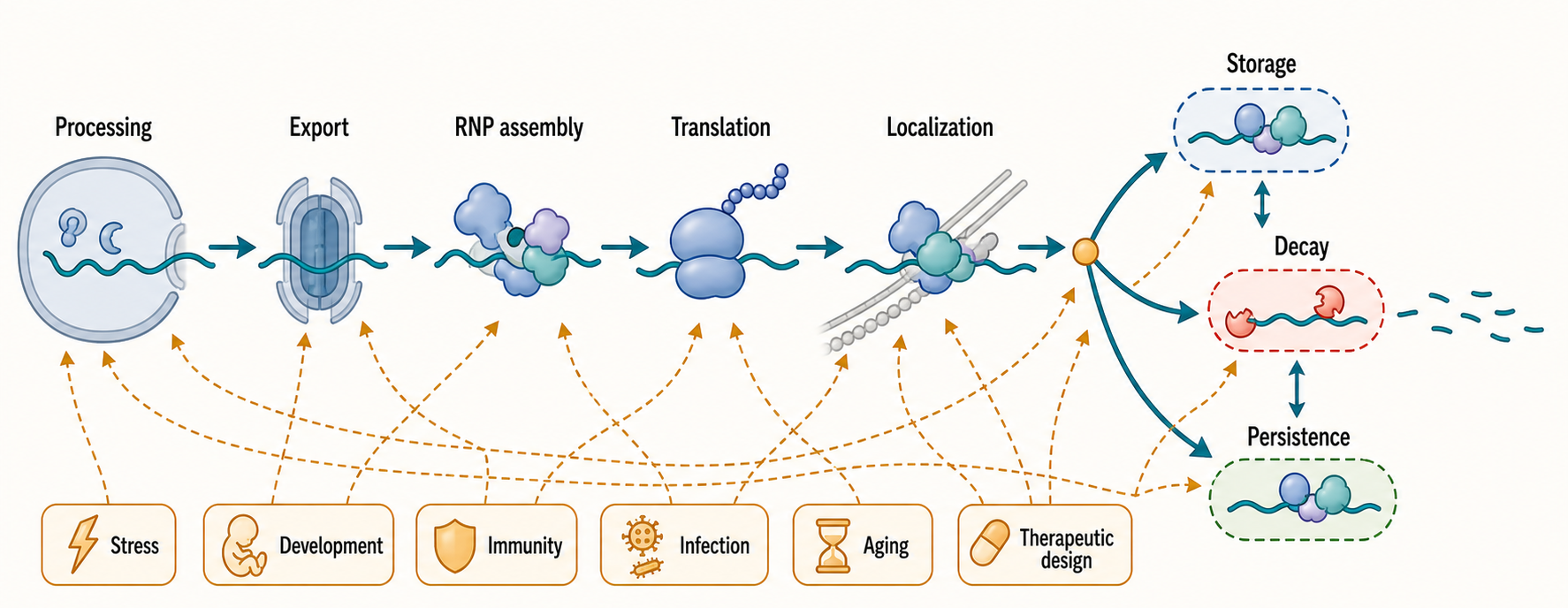
Figure 38.1. RNA Half-Life Decision Map. An mRNA produced by transcription encounters successive regulatory decision points — processing, export, RNP assembly, translation, localization, and potential storage — before it reaches decay or long-term persistence. This schematic traces those decision points and marks where stress signaling, developmental programs, immune activation, viral infection, aging, and therapeutic modification can alter the outcome. The key pedagogical lesson is that RNA half-life is not determined at a single step but emerges from integrated molecular decisions along the entire lifecycle of the transcript.
An RNA molecule’s half-life is shaped by cis-acting features, which are features encoded in the RNA molecule itself, and trans-acting factors, which are proteins or RNAs that bind and regulate it. For a protein-coding mRNA, the main cis-acting features include the 5′ cap, 5′ untranslated region, coding sequence, stop codon context, 3′ untranslated region, poly(A) tail, RNA structures, modification sites, and binding motifs for RBPs and small RNAs. The main trans-acting factors include cap-binding proteins, exon junction complex proteins, deadenylases, decapping factors, exonucleases, endonucleases, helicases, miRNA-loaded Argonaute complexes, and transcript-specific RBPs.
Table 38.1. Determinants of RNA Stability. Summary of the major cis- and trans-acting features that influence RNA half-life, with the molecular mechanism, a representative biological context, the evidence needed for a mechanistic claim, the main interpretive caveat, and the related chapter.
| Determinant | Molecular mechanism | Example context | Evidence needed | Main caveat | Related chapter |
|---|---|---|---|---|---|
| 5′ cap and cap-binding state | Cap protects the 5′ end from exonuclease attack; cap-binding proteins influence decapping rate and access by decay factors | mRNA therapeutics; decapping during P-body routing | Kinetic comparison of capped vs. uncapped reporter; decapping factor perturbation | Cap analogs differ in structure and immune-sensing properties; cannot isolate cap effect from translation effects | Chapter 26 |
| Poly(A) tail length and binding | Deadenylation is often the rate-limiting first step in mRNA decay; PABP occupancy affects deadenylase access | Cytokine mRNA shortening after decay-signal engagement | Tail-length profiling (e.g., PAL-seq) combined with half-life assay | Tail length also affects translation initiation; decay and translation effects must be separated | Chapter 29 |
| 3′ UTR AU-rich elements | Recruit TTP/ZFP36-family proteins and related factors that promote deadenylation and decapping | TNF and IL-6 mRNAs in macrophages after LPS stimulation | Element deletion or mutation in reporter; RBP binding confirmation; kinetic half-life assay | Same element may be stabilizing or destabilizing depending on competing RBP occupancy and signaling state | Chapter 36 |
| miRNA sites | miRNA-loaded Argonaute recruits GW182/TNRC6 and deadenylase complexes to target transcripts | Developmental gene silencing; miR-155 targeting in immune cells | Seed-match mutation; Argonaute binding confirmation; half-life after miRNA perturbation | miRNA effect magnitude varies; translation repression can occur without strong mRNA decay | Chapter 84 |
| Codon optimality | Suboptimal codons slow elongation, increase ribosome pausing, and promote decay through translation-linked pathways | Codon-recoded reporter stability in yeast and mammalian cells | Codon-recoded reporter series; tRNA pool measurement; elongation rate assay | Effect size depends on tRNA pool, stress state, and open-reading-frame context | Chapter 36 |
| RNA modification | m6A creates or weakens binding sites for reader proteins, altering decay or translation; other modifications change structure and RBP recognition | m6A on Xist RNA affecting NEXT-complex decay and X-inactivation dynamics | Writer, eraser, or reader perturbation; modification mapping; kinetic rescue with wild-type vs. binding-defective reader | Modification detection does not prove the modification controls half-life | Chapter 48 |
| RBP occupancy | Stabilizing RBPs (e.g., HuR) can occlude AU-rich elements; destabilizing RBPs recruit deadenylases or decapping enzymes | HuR stabilizing cytokine mRNAs after immune stimulation | Crosslinking immunoprecipitation; half-life change after RBP perturbation; binding-defective rescue | A bound RBP may have no net effect or context-dependent opposing effects | Chapter 35 |
| Translation state | Ribosome occupancy shields coding sequence from endonucleases; translation repression exposes decay sites and affects deadenylation | Polysome displacement during heat shock; ribosome stalling at NMD-triggering premature stop codons | Ribosome profiling combined with half-life measurement; initiation inhibitor experiments with controls | Translation inhibitors are pleiotropic and can alter many decay factors simultaneously | Chapter 35 |
| Subcellular localization | ER-localized ribosomes place mRNAs near ER-associated decay factors; stress granule or P-body entry removes mRNAs from the translating pool | ER-associated RNA decay during ER stress; neuronal dendritic transport granules | Fractionation with matched kinetic measurements; live-cell imaging of RNA fate after compartment entry | Localization correlation does not prove compartment-specific decay rate | Chapter 105 |
| RNA damage | Oxidized or nicked RNA can stall ribosomes, activate quality-control sensors, or be sorted into damage compartments by helicases such as DHX9 | DHX9 stress granule compartmentalization of damaged RNA during oxidative stress | Direct damage detection; kinetic comparison with undamaged counterpart; helicase or sensor perturbation | RNA damage must be distinguished from regulated RNA modification | Chapter 32 |
The simplest stability model would say that a destabilizing motif recruits a nuclease. Real RNP biology is more conditional. A 3′ untranslated region may contain an adenylate-uridylate-rich element, often called an AU-rich element, that can recruit decay-promoting proteins in immune cells. The same region may also contain stabilizing RBP sites, miRNA sites, secondary structures, and alternative polyadenylation choices that shorten or lengthen the regulatory region. A cytokine mRNA is therefore not intrinsically “unstable” in an absolute sense. It is unstable in cell states where destabilizing factors are present and active, and it can become stabilized when signaling modifies those factors, changes protein occupancy, or alters poly(A)-tail dynamics. Akira and Maeda review this kind of immune RNA-stability control as a central layer of inflammatory gene expression (Akira and Maeda 2021).
Table 38.2. Biological Programs Using RNA Stability. Representative biological programs in which regulated RNA half-life is a key component, with the stability goal, the RNA class involved, the main regulatory factors, the evidence standard required for mechanistic claims, and current citation status.
| Program | Stability goal | Representative RNA class | Main regulatory factors | Evidence standard | Citation status |
|---|---|---|---|---|---|
| Acute stress response | Degrade growth-related transcripts; preserve stress-adaptive and survival mRNAs | Heat-shock and antioxidant mRNAs; bulk ribosomal and cell-cycle mRNAs | Stress granules, P-bodies, translation repression, phosphorylated eIF2α, stress-activated RBPs | Time-course transcriptomics with synthesis-decay separation; kinetic metabolic labeling | Review-supported synthesis |
| ER stress | Reduce load of secretory-pathway mRNAs entering the ER; preserve UPR-adaptive transcripts | Secretory and membrane protein mRNAs; UPR target transcripts | ER-associated decay, IRE1-mediated RIDD, ATF6, translation attenuation | ER-stress induction with matched RNA decay measurement; IRE1 perturbation and rescue | Review-supported (Ottens et al. 2024) |
| Maternal-to-zygotic transition | Clear maternal mRNAs to allow zygotic program to take control | Maternal mRNAs encoding early developmental regulators and patterning factors | Embryo-specific deadenylases, zygotic decay factors, polyadenylation state changes | Embryo time-course RNA-seq with synthesis markers; perturbation of clearance factors | Synthesis; direct clearance sources pending |
| Neuronal local translation | Stabilize transported mRNAs; activate them locally at synapses after signaling | Synaptic protein mRNAs (e.g., actin, PSD-95, BDNF transcripts) | Transport RBPs (FMRP, HuD, G3BP), local translation activators, motor-protein complexes | Axon and dendrite fractionation; live mRNA imaging; local translation reporters | Review-supported (Prashad and Gopal 2021) |
| Cytokine induction and shutdown | Transiently stabilize cytokine mRNAs for immune activation; restore rapid decay to end the response | TNF, IL-6, IL-8, GM-CSF mRNAs | AU-rich elements, TTP/ZFP36 destabilization, HuR stabilization, MAP kinase and NF-κB signaling | Kinetic assays after stimulation and signal withdrawal; RBP perturbation and rescue | Review-supported (Akira and Maeda 2021) |
| Viral infection | Host degrades viral RNA; virus protects its genomic and messenger RNAs from host decay and immune sensing | Viral genomic RNA, viral mRNAs; interferon-stimulated host transcripts | RIG-I sensing, RNase L, interferon kinases; viral caps, UTR structures, replication compartments, RNP shielding | Host factor knockdown or viral mutant with matched decay measurement; kinetic infection time course | Review-supported (Marques et al. 2024; Sullivan et al. 2025) |
| Aging tissue remodeling | Expression shifts may reflect altered synthesis, RNA stability, cell composition, RNA polymerase behavior, or immune infiltration together | Senescence-associated transcripts; mitochondrial and inflammatory mRNAs | RNA polymerase stalling, RBP composition changes, senescence programs, immune cell infiltration | Single-cell RNA-seq with cell-type deconvolution; nascent RNA data; explicit half-life measurement | Review-supported (Huang et al. 2025; Gyenis et al. 2023) |
| Therapeutic RNA exposure | Design RNA persistence to achieve sufficient protein output without prolonged immune activation or toxicity | mRNA vaccines, therapeutic mRNAs, siRNAs, antisense oligonucleotides | Cap chemistry, UTR sequences, codon optimization, modified nucleotides, poly(A)-tail length, delivery formulation | In vivo pharmacokinetics; protein output time course; innate immune assays; safety and biodistribution studies | Review-supported (Chen et al. 2025) |
Table 38.3. RNA Stability Measurement Caveats. Each method used to assess RNA stability measures a distinct aspect of RNA kinetics and introduces characteristic artifacts; choosing or interpreting a method requires understanding both what it reveals and what it obscures.
| Method | What it directly measures | What it can infer | Common artifact | Strong follow-up |
|---|---|---|---|---|
| Steady-state RNA-seq | RNA abundance at one time point per condition | Relative expression differences between conditions | Confounds transcription rate, decay rate, processing, export, and cell-composition changes | Add time-course or metabolic labeling to separate synthesis from decay |
| Transcription shutoff | RNA remaining after new synthesis is pharmacologically blocked | Apparent half-life under inhibitor conditions | Blocking RNA polymerase activates stress responses and can alter decay-factor activity | Confirm with parallel metabolic labeling; verify inhibitor does not change decay-factor levels |
| Metabolic labeling | Newly synthesized RNA (labeled fraction) and remaining old RNA (unlabeled fraction) | Synthesis and decay rates as mathematically separable parameters | Nucleotide incorporation depends on metabolic state; labeling can perturb stressed or differentiating cells | Time-series labeling with parallel synthesis rate estimates and cell-viability controls |
| Nascent RNA profiling | RNA being actively transcribed at capture time (GRO-seq, TT-seq, or similar) | Transcription rate; paired with total RNA-seq it supports inference about relative stability | Gene length, pausing index, processing, and extraction variability introduce bias across conditions | Pair with total RNA-seq and metabolic labeling for full synthesis-decay decomposition |
| Reporter assay | Stability of a defined sequence feature placed in an artificial transcript | Whether a motif, codon set, or UTR element is sufficient to alter stability | Native transcript context (competing RBPs, alternative polyadenylation, chromatin, processing) is absent | Validate the finding in the endogenous transcript using perturbation and kinetic assay |
| Single-cell RNA-seq | Captured RNA abundance per cell at one time point | Cell-type composition, cell-state transitions, gene covariation | Dropout, capture bias, dissociation stress, and cell-state annotation confounding | Paired kinetic labeling in single cells (e.g., scSLAM-seq) for per-cell half-life estimation |
| Long-read RNA sequencing | Full-length transcript sequences including isoform, poly(A)-site choice, and modification context | Isoform-specific expression levels; alternative polyadenylation patterns | Capture efficiency, read-length bias, and RNA quality affect isoform representation | Quantify isoform-specific half-lives using long-read metabolic labeling |
| RBP interaction mapping | Physical contacts between an RBP and RNA under crosslinking conditions | Candidate regulatory interactions; binding-site location on target transcripts | Crosslinking efficiency variation, indirect binding, and antibody specificity can produce false positives | Perturb the RBP, measure half-life change, and test rescue with binding-defective mutant |
The coding sequence also contributes to stability. Codon optimality refers to the match between codon usage and the decoding capacity of the cell, including tRNA abundance, tRNA modification, and translation elongation behavior. In many systems, optimal codons tend to correlate with greater mRNA stability, while nonoptimal codons can promote decay through translation-linked pathways. The mechanism is not that the ribosome reads “good” or “bad” codons as labels. Instead, elongation kinetics, ribosome dwell time, recruitment of decay factors, and interactions between the poly(A)-binding protein and decay machinery can bias the transcript toward persistence or degradation. Chapter 36 treats this grammar in detail. In this chapter, the practical point is that codon-dependent stability changes during stress, development, infection, or therapeutic RNA design cannot be interpreted without measuring translation and tRNA state.
RNP composition can dominate sequence predictions. An RNA-binding protein can hide a decay motif, recruit a deadenylase, block an exonuclease, connect the transcript to translation initiation, or move the RNA into a compartment where decay enzymes are enriched or excluded. Neurons provide a useful example because long cellular processes require mRNAs to travel far from the nucleus. A neuronal mRNA transported into a dendrite or axon may be translationally repressed during transport, stabilized by neuronal RBPs, and then locally translated after synaptic signaling. Prashad and Gopal review many RBPs whose developmental and disease phenotypes arise from altered neuronal RNA metabolism (Prashad and Gopal 2021). The same general principle applies in germ cells, immune cells, and embryos: a transcript’s fate depends on its RNP itinerary.
RNA chemical modifications can also affect stability, but claims require care. A modified nucleotide such as N6-methyladenosine, often abbreviated m6A, can create or weaken binding sites for reader proteins, alter RNA structure, and influence decay or translation. However, a modification detected on an RNA does not prove that the modification controls half-life. Strong evidence requires perturbing the writer, eraser, or reader in a way that changes modification, RNA occupancy, and RNA kinetics, ideally with rescue. Wei et al. provide a recent example linking m6A and the NEXT complex to Xist RNA turnover and X-inactivation dynamics, illustrating how modification and nuclear decay can intersect in a transcript-specific developmental system (Wei et al. 2025). Chapter 48 treats m6A mechanisms more broadly, and Chapter 92 covers Xist biology.
Boundary cases matter. Circular RNAs lack the usual free 5′ and 3′ ends, which can make some circular RNAs unusually stable, but circularity does not make an RNA immortal. Endonucleases, immune pathways, and cellular dilution can still reduce circular RNA abundance. Long noncoding RNAs can be very unstable or very stable depending on processing, localization, RNP composition, and surveillance. Viral RNAs can carry caps, internal ribosome entry sites, protein shields, structured untranslated regions, or replication complexes that alter stability rules. Therefore a stability claim should specify RNA class, cell type, compartment, condition, and assay.
38.2. Stress-regulated decay and stabilization
Stress-regulated RNA turnover is the rapid remodeling of RNA decay and protection when cells encounter damaging or demanding conditions. Stress can include heat shock, oxidative damage, ultraviolet exposure, nutrient limitation, hypoxia, unfolded protein accumulation in the endoplasmic reticulum, osmotic change, viral infection, bacterial products, or inflammatory cytokines. The purpose of stress-regulated turnover is not simply to destroy RNA. It reallocates gene expression capacity by removing transcripts that are no longer useful, preserving transcripts needed for survival, preventing translation of damaged RNA, and reducing immune-stimulatory or proteotoxic products.
One common stress response is global translation repression. When initiation slows, many mRNAs leave polysomes, which are complexes of multiple ribosomes translating the same mRNA. Translation state affects stability because ribosomes can protect coding regions, expose or hide decay signals, and influence deadenylation and decapping. Some untranslated mRNAs move into stress granules, which are reversible RNP assemblies enriched for translation initiation factors, RBPs, and untranslated mRNAs. Some RNAs and decay factors are also found in processing bodies, or P-bodies, which are cytoplasmic RNP foci associated with translational repression and decay machinery. Chapter 105 treats these RNP bodies in depth. The stability lesson is that granule localization is not equivalent to decay or protection by itself. A transcript in a stress granule may later re-enter translation, remain stored, or be routed to decay depending on stress duration, RNP composition, and cell type.
The endoplasmic reticulum, abbreviated ER, illustrates compartment-specific stress turnover. The ER is the membrane network where secretory and membrane proteins are synthesized and folded. Many mRNAs encoding secreted or membrane proteins are translated on ER-associated ribosomes. During ER stress, cells activate the unfolded protein response and remodel synthesis of secretory-pathway proteins. ER-associated RNA decay pathways can reduce the burden of proteins entering the ER while selectively preserving or inducing stress-adaptive transcripts. Ottens et al. review RNA decay at the ER as a stress-responsive layer that connects RNA fate to protein-folding capacity (Ottens et al. 2024). The mechanistic chain is causal: unfolded proteins accumulate, signaling pathways alter translation and decay factors, particular ER-associated mRNAs are cleaved or destabilized, and the protein load entering the ER changes.
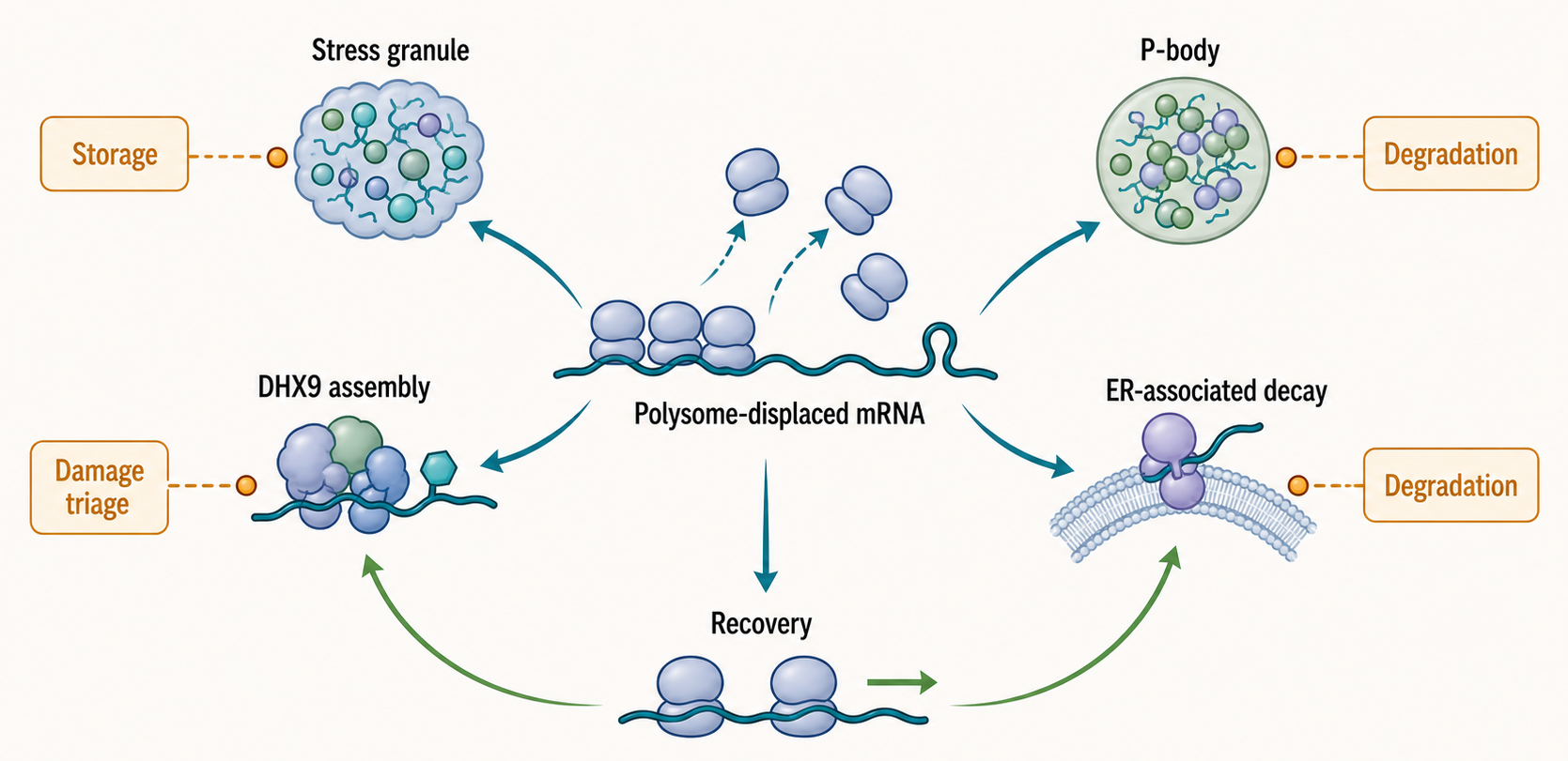
Figure 38.2. Stress Triage of mRNAs. During cellular stress, mRNAs are not uniformly destroyed. This diagram shows the major fates available to a polysome-displaced mRNA: entry into stress granules for temporary storage, routing to processing bodies, compartmentalization of damaged RNA by DHX9-linked assemblies, targeting to ER-associated decay, and eventual recovery into polysomes when stress resolves. Stress granule localization indicates that an mRNA has exited the translating pool, but it does not specify whether that mRNA will be stored, repaired, or degraded.
RNA damage introduces a second stress logic. Oxidized or otherwise damaged RNA can miscode, stall translation, bind abnormal proteins, or become difficult to process. Zhou et al. report that DHX9 stress granules compartmentalize RNA damage, placing a helicase-linked RNP assembly in the pathway that handles damaged RNA during stress (Zhou et al. 2024). This example is useful because it separates damage management from ordinary transcript regulation. A stress granule can be a location where damaged RNA is sorted, not merely a passive aggregate of untranslated mRNAs.
Stress can also stabilize selected transcripts. In immune cells, transcripts encoding cytokines, chemokines, and signaling proteins often have short half-lives under basal conditions. Short half-life prevents inappropriate inflammation. After stimulation, signaling pathways can inhibit destabilizing RBPs, recruit stabilizing RBPs, change deadenylation, or alter miRNA function so that inflammatory mRNAs persist long enough to be translated. Akira and Maeda frame this as a major immunological control layer rather than a minor postscript to transcription (Akira and Maeda 2021). The same design principle appears outside immunity: heat-shock transcripts, antioxidant-response transcripts, and repair factors may be stabilized while growth-related transcripts are destabilized.
The boundary case is chronic stress. Acute stabilization can be adaptive, but chronic stabilization of inflammatory, senescence-associated, or proteotoxic transcripts can contribute to disease. Acute degradation can be protective, but excessive decay can prevent recovery by eliminating RNAs needed after stress ends. A stability program is therefore judged by context and timing. A cell’s best response to a ten-minute stress may become harmful if sustained for months in a tissue.
38.3. Developmental and tissue-specific stability programs
Developmental RNA stability programs are coordinated changes in RNA half-life that help move a cell or organism from one state to another. Development is not only a sequence of transcriptional on-off switches. Many transitions require old RNAs to be removed, stored RNAs to be activated, and tissue-specific RNAs to be protected. The half-life of an RNA can therefore act as a timer.
Early embryos provide the clearest conceptual example, although detailed embryonic clearance mechanisms are covered in Chapter 102. Before zygotic transcription becomes fully active, many animals depend on maternal mRNAs deposited in the egg. These maternal RNAs encode proteins needed for early divisions and patterning. At the maternal-to-zygotic transition, a large subset of maternal RNAs is cleared while zygotic transcription begins. The causal logic is straightforward: stable maternal RNAs support early development when the embryo cannot yet rely on its own genome; then regulated decay removes maternal instructions so that zygotic programs can take control. final reference item notes: add chapter-specific maternal mRNA clearance review and primary sources before final release.
Germline specification illustrates coupling between transcription and RNA turnover. Germ cells must preserve reproductive potential while shutting down somatic programs. Tan and Wilkinson report that both transcription and RNA turnover contribute to germline specification, reinforcing the idea that cell fate emerges from synthesis and decay together rather than from transcription alone (Tan and Wilkinson 2022). A transcript that is no longer made may still persist if stable; a transcript that is actively transcribed may remain low if decay is rapid. Developmental interpretation therefore requires both production and destruction rates.
Neural development and neuronal function depend strongly on RBP-controlled stability. Neurons are polarized cells with axons and dendrites that can extend far from the nucleus. Local translation allows a synapse or growth cone to change protein composition without waiting for new transcripts to travel from the nucleus. For local translation to work, mRNAs must be stabilized during transport, repressed until needed, and then activated or degraded after use. RBPs can bind 3′ untranslated regions, assemble transport granules, interact with motor proteins, and tune decay. When these RBPs are mutated, mislocalized, aggregated, or expressed at the wrong level, neuronal development and maintenance can fail. Prashad and Gopal review this RBP-centered view of neurological development and disease (Prashad and Gopal 2021).
Tissue-specific stability programs also depend on alternative polyadenylation. Alternative polyadenylation chooses among different cleavage and polyadenylation sites, producing mRNA isoforms with longer or shorter 3′ untranslated regions. A shorter 3′ untranslated region may remove miRNA sites or RBP motifs and thereby increase stability or translation in a particular tissue. A longer 3′ untranslated region can add localization motifs or decay elements. Chapter 29 covers alternative polyadenylation; here the important point is that a tissue-specific isoform can change half-life without changing the protein sequence.
Developmental and tissue-specific stability programs interact with metabolism and environment. Nutritional state, hormone signaling, hypoxia, microbiome exposure, and inflammatory history can all alter RBP activity and decay-pathway capacity. Chavatte-Palmer et al. review developmental programming and nutrition in herbivores from an epigenetic perspective, and this chapter treats that source only as broad developmental context rather than direct RNA half-life evidence (Chavatte-Palmer et al. 2018). final reference item notes: add direct tissue-specific RNA stability atlases or metabolic-labeling studies for final release.
The common overgeneralization is that developmental expression equals transcriptional regulation. A developmental RNA-seq time course is an expression time course, not automatically a transcription time course. If a differentiation marker rises, it may be transcribed more, degraded more slowly, or retained in a growing cell population. If a stem-cell transcript falls, it may be transcriptionally silenced, actively degraded, diluted by cell division, or confined to a subpopulation that becomes rare. Strong developmental stability claims use kinetic RNA measurements, perturb specific RBPs or decay factors, and connect RNA half-life to developmental outcome.
38.4. Immune and infection-linked RNA turnover
Immune-linked RNA turnover is the regulation of RNA stability during host defense, inflammation, and immune-cell differentiation. Infection-linked RNA turnover includes both host regulation of its own RNAs and turnover of pathogen RNAs. These processes are inseparable because many immune receptors distinguish self from nonself partly by RNA features, location, and persistence.
Innate immune sensing detects molecular patterns associated with infection or damage. For RNA, these patterns can include long double-stranded RNA, 5′ triphosphate RNA, uncapped RNA, abnormal nucleotide composition, viral replication intermediates, or self RNA appearing in the wrong compartment. When such RNAs accumulate, receptors such as Toll-like receptors, RIG-I-like receptors, protein kinase R, and oligoadenylate synthetase/RNase L pathways can induce interferon and inflammatory responses. Chapter 108 covers these sensors in detail. This chapter focuses on the turnover logic: cells must remove or hide self RNAs that might trigger sensors, while preserving or amplifying immune transcripts that coordinate defense.
Cytokine mRNAs are a classic immune-stability problem. Cytokines are secreted signaling proteins that can recruit, activate, or polarize immune cells. Many cytokine mRNAs contain destabilizing 3′ untranslated-region elements so that accidental transcription does not produce prolonged inflammation. After stimulation by microbial products or cytokines, signaling pathways can stabilize selected cytokine transcripts. The result is a rapid but reversible burst of protein production. If decay is too fast, defense fails; if stabilization persists, chronic inflammation and tissue damage can follow. Akira and Maeda review this balance as a core feature of immune regulation (Akira and Maeda 2021).
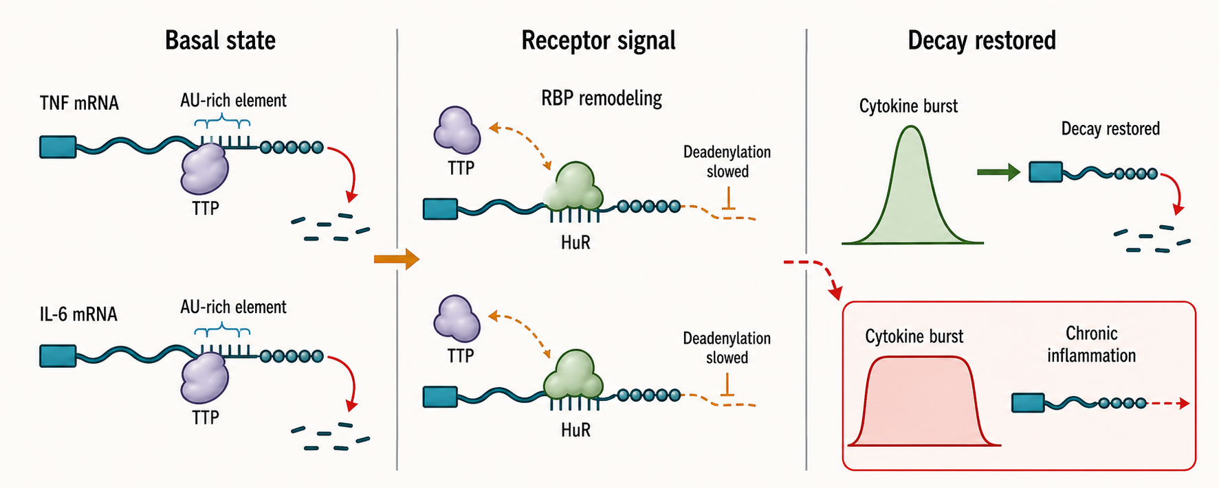
Figure 38.3. Immune RNA Turnover Balance. Cytokine mRNAs such as those encoding tumor necrosis factor and interleukin-6 are intrinsically short-lived under basal conditions, preventing inappropriate inflammation. Receptor stimulation triggers signaling cascades that remodel RBP occupancy and deadenylation, transiently stabilizing these transcripts and enabling a burst of cytokine protein production. Subsequent restoration of rapid decay shuts down the inflammatory signal; failure to restore decay can lead to chronic inflammation and tissue damage.
Viral infection adds an opposing evolutionary pressure. RNA viruses need their genomes and transcripts to persist long enough to replicate and translate, but host cells attempt to degrade or sense those RNAs. Viral RNAs may be capped, protein-linked, structured, chemically modified, shielded in replication compartments, or packaged in RNPs. Host pathways can cleave viral RNA, shut down host translation, induce RNase L activity, alter deadenylation, or change RBP availability. Marques et al. review innate antiviral immunity across animals, emphasizing that RNA defense systems evolve under persistent host-pathogen conflict (Marques et al. 2024). Li et al. provide a current antiviral-drug development perspective for RNA viruses, although drug mechanisms are treated more directly in Chapter 160 (Li et al. 2024).
Pathogens can also manipulate host RNA turnover. Some viral proteins degrade host mRNAs, block RNA decay, redirect RBPs, or change the translation efficiency of host and viral transcripts. Sullivan et al. report that IFIT3 RNA-binding activity can promote influenza A virus infection and translation efficiency, an example that cautions against assuming every interferon-stimulated RNA-binding activity is antiviral in every context (Sullivan et al. 2025). The evidence lesson is important: a host factor induced during immune activation can still be exploited by a virus, and the effect may depend on viral species, cell type, and timing.
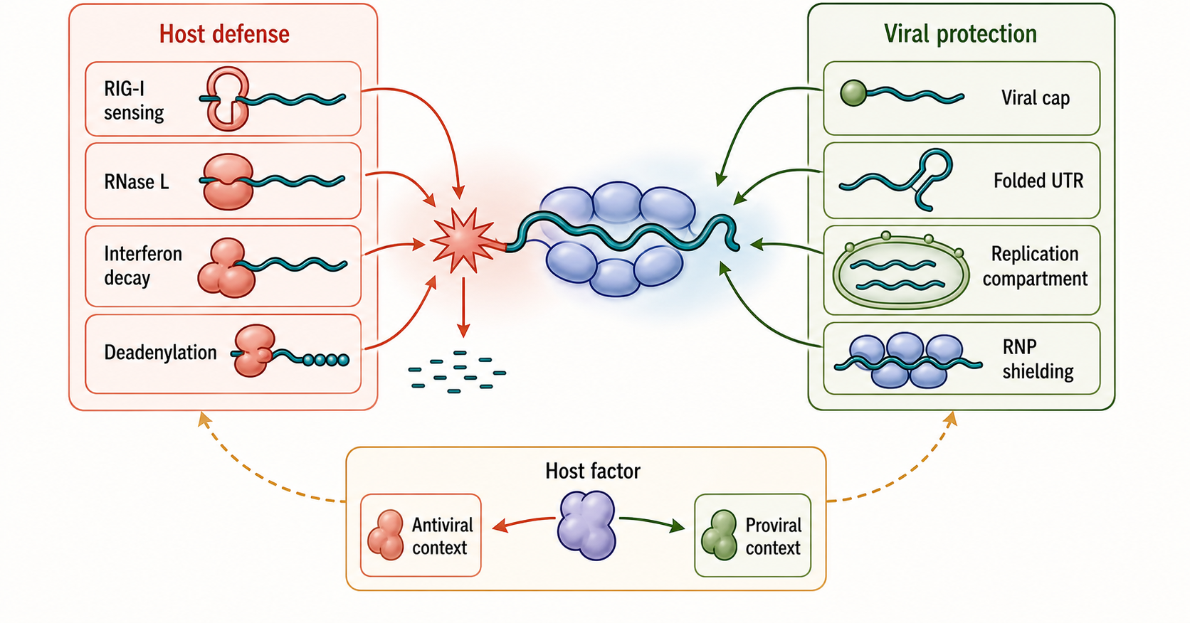
Figure 38.4. Host-Pathogen RNA Stability Conflict. Hosts and RNA viruses deploy opposing strategies to control viral RNA persistence. The host side uses RIG-I-like receptor sensing, RNase L activation, interferon-induced decay factors, and altered deadenylation to detect and degrade viral RNA. The viral side uses cap structures, highly folded untranslated regions, replication compartments, RNP shielding, and host-factor exploitation to protect viral genomes and mRNAs. A single host factor such as an interferon-stimulated RNA-binding protein can be antiviral in one context and exploited to enhance viral translation efficiency in another, illustrating why factor labels must specify biological context.
Noncoding RNAs participate in immune turnover networks. Long noncoding RNAs can scaffold immune complexes, influence chromatin, pair with mRNAs, or affect stability of immune transcripts. Khan et al. review long noncoding RNAs from an immune-cell perspective (Khan et al. 2021). In plants, Liu et al. describe a lncRNA that fine-tunes salicylic acid biosynthesis to balance plant immunity and growth, illustrating that immunity-linked RNA regulation is not limited to animals (Liu et al. 2022). tRNA fragments and stress-induced tRNA-derived RNAs can also participate in mucosal immunity, although detailed tRNA-fragment biology belongs in Chapter 41 (Chen and Shen 2021).
Immune turnover has boundary cases involving self RNA. Mitochondrial RNAs, endogenous retroelement transcripts, repeat-derived double-stranded RNAs, and damaged RNAs can become immune stimulatory when localization, modification, editing, or degradation fails. This does not mean these RNAs are always pathogenic. It means their normal compartmentalization and turnover help preserve self-nonself discrimination. Thomas et al. report that mitochondrial RNA degradation regulates differentiation, stemness, and immune sensitivity in acute myeloid leukemia, providing a disease-linked example where RNA degradation and immune sensitivity intersect (Thomas et al. 2026). Chapter 109 treats mitochondrial double-stranded RNA and interferon-stimulated pathways more fully.
38.5. Aging, disease, and therapeutic implications
Aging changes RNA biology at many layers. Aging tissues show altered transcription, RNA polymerase behavior, splicing, RNA modification, RBP abundance, mitochondrial function, immune activation, senescence, cell composition, and stress responses. RNA stability can contribute to these changes, but an age-associated expression signature is not automatically an age-associated half-life signature. Huang et al. review transcriptomic approaches to aging and emphasize the complexity of interpreting age-linked RNA data (Huang et al. 2025). Gyenis et al. provide primary evidence that RNA polymerase stalling shapes the transcriptome during aging, reminding readers that changes in RNA abundance can originate at transcriptional elongation as well as decay (Gyenis et al. 2023).
Single-cell studies sharpen aging interpretation but do not eliminate stability caveats. Campello et al. analyze aging mouse retina at single-cell resolution, Lin et al. analyze aging mouse liver, and Li et al. examine aging-associated changes in mammary epithelia and stroma (Campello et al. 2025; Lin et al. 2024; Li et al. 2020). Such studies can identify cell types, cell states, and tissue composition changes that bulk RNA-seq would mix together. However, ordinary single-cell RNA-seq usually measures captured RNA abundance, not RNA half-life. Dropout, capture bias, cell dissociation stress, nuclear versus cytoplasmic content, and cell-state annotation can all affect interpretation.
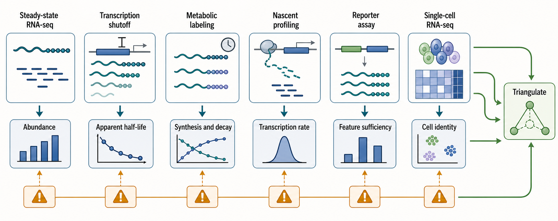
Figure 38.5. Measuring RNA Stability Without Confusing It with Abundance. Six common approaches to measuring RNA stability each capture a different slice of RNA kinetics and carry distinct artifacts. Steady-state RNA-seq measures abundance but cannot separate transcription from decay; transcription shutoff allows apparent half-life estimation but perturbs the cell; metabolic labeling separates synthesis and decay but is sensitive to nucleotide metabolism; nascent RNA profiling captures transcriptional rates with gene-length bias; reporter assays isolate features but remove native transcript context; and single-cell RNA-seq resolves cell identity without directly measuring half-life. Strong stability claims triangulate across at least two orthogonal approaches.
Disease mechanisms can involve either excessive RNA persistence or excessive RNA loss. In cancer, stabilized oncogenic mRNAs, altered alternative polyadenylation, mutant RBPs, impaired nonsense-mediated decay, and chronic stress granule states can support growth, immune evasion, or therapy resistance. In neurological disease, RBP mislocalization or aggregation can destabilize needed neuronal mRNAs and stabilize toxic or mislocalized RNAs. In inflammatory and autoimmune disease, failure to degrade immune-stimulatory self RNA can activate interferon pathways, while persistent stabilization of cytokine transcripts can maintain inflammation. Barta and Jantsch provide broad context for RNA in disease and development, but disease-specific stability mechanisms need more targeted references before final release (Barta and Jantsch 2017).
Infection can accelerate aging-like phenotypes in model systems through RNA-centered stress, immune, and tissue-damage pathways. González et al. report that enteric viral infections promote systemic accelerated aging in Drosophila (González et al. 2026). This does not prove that RNA stability alone causes organismal aging. It does show why infection, immunity, RNA turnover, and aging should not be studied as isolated themes. Persistent infection can alter transcription, RNA decay, translation, mitochondrial function, tissue composition, and inflammatory state together.
Therapeutic RNA design treats stability as both a pharmacological and immunological variable. An mRNA therapeutic must persist long enough to produce useful protein but not so long that expression becomes unsafe. A vaccine RNA must balance antigen expression with innate immune activation that supports immunity without excessive reactogenicity. An antisense oligonucleotide or small interfering RNA must resist nuclease degradation long enough to reach its target but avoid off-target binding and immune stimulation. Chen et al. review RNA therapeutics for healthy aging, and Chapter 153 covers mRNA therapeutics in greater depth (Chen et al. 2025).
Several design features influence therapeutic RNA stability. Cap analogs and cap methylation affect recognition by translation and immune factors. Untranslated regions alter translation and decay. Codon choices can influence translation-linked stability. Modified nucleotides can reduce innate immune sensing and alter RNA-protein interactions. Poly(A)-tail length and purity can affect translation and degradation. Manufacturing impurities, double-stranded RNA contaminants, delivery formulation, endosomal escape, and tissue biodistribution also shape apparent persistence. Chapter 156 covers lipid nanoparticle delivery, and Chapter 159 covers RNA manufacturing and analytical release testing.
The therapeutic boundary case is that greater stability is not always better. A stabilized therapeutic RNA may increase protein output, but it may also prolong immune stimulation, broaden tissue exposure, or make dose control harder. A less stable RNA may be safer or better matched to a transient therapeutic window. Stability design must therefore be evaluated with protein output, innate immune activation, biodistribution, toxicity, and clinical context, not with RNA half-life alone.
38.6. Measurement methods and interpretation caveats
RNA stability measurement asks how quickly an RNA population is lost. The most direct conceptual experiment is a pulse-chase: mark RNAs made during a pulse, follow those labeled RNAs during a chase, and estimate decay over time. In practice, researchers use metabolic labeling, transcription inhibition, inducible promoters, RNA export reporters, nascent RNA profiling, time-course RNA-seq, or mathematical modeling. Each approach measures a different aspect of RNA kinetics and carries different artifacts.
Transcription shutoff methods inhibit new RNA synthesis and monitor remaining RNA. The advantage is conceptual simplicity. The weakness is that inhibiting transcription is a major perturbation. Blocking RNA polymerase activity can alter stress pathways, RNA processing, chromatin state, and decay itself. Short-lived transcripts may be measured reasonably, but long-lived transcripts require long treatments that can create secondary effects. Therefore transcription shutoff is useful but should not be treated as artifact-free half-life measurement.
Metabolic labeling methods incorporate labeled nucleosides into newly synthesized RNA. Newly made RNA can be purified or detected, and old RNA can be modeled separately from new RNA. These methods can estimate synthesis and decay more directly than steady-state RNA-seq, but they have caveats: labeling efficiency varies by cell type, nucleotide metabolism changes under stress, labeling can perturb cells, and short time windows may miss slow decay. final reference item notes: add direct recent metabolic RNA labeling and half-life method reviews for final release.
Nascent and total RNA comparisons estimate whether expression changes arise from production or decay. If nascent RNA rises while total RNA is stable, decay or processing may buffer transcription. If total RNA rises without a matching nascent increase, stabilization may contribute. These inferences are useful, but they are model-dependent. Nascent RNA methods capture specific transcriptional states and can be biased by gene length, chromatin, RNA processing, and extraction. Aoi and Shilatifard’s review of transcriptional elongation control is relevant because elongation changes can mimic or mask stability effects in developmental, aging, and disease contexts (Aoi and Shilatifard 2023).
Reporter assays test whether an RNA feature is sufficient to alter stability. A researcher can place a 3′ untranslated region, motif, codon pattern, or modified sequence into a reporter transcript and measure reporter half-life or protein output. Reporters are powerful because they isolate features. Their weakness is that they remove the feature from its native transcript architecture, chromatin context, processing history, and RNP environment. A motif that destabilizes a reporter may be buffered in the endogenous transcript by neighboring proteins, alternative polyadenylation, localization, or translation.
Single-cell and clinical transcriptomics require special caution. Andreatta et al. discuss interpretation of T cell states from single-cell transcriptomics using reference atlases, and Thareja et al. discuss standardization and interpretation of RNA-seq for transplantation (Andreatta et al. 2021; Thareja et al. 2023). These sources are not half-life method papers, but they support a broader caution: expression signatures in complex samples reflect cell identity, activation state, sampling, processing, and computational annotation. A clinical expression change should not be called altered RNA stability unless kinetic or mechanistic evidence supports that claim.
Long-read and targeted RNA sequencing can reveal isoforms, retained introns, alternative polyadenylation, fusions, and disease-relevant transcript structures that short reads miss. Wang et al. report targeted long-read RNA sequencing for rare disease diagnosis and variant interpretation (Wang et al. 2026). Long reads can improve interpretation of which transcript isoform is present, but long-read abundance is still not automatically half-life. Isoform detection, capture efficiency, read length bias, and RNA quality all matter.
RNA-protein interaction mapping can connect stability changes to RBPs. Crosslinking, immunoprecipitation, interactome capture, and computational modeling can identify candidate RBP-RNA contacts. Zhu et al. describe HDRNet for interpreting protein-RNA interactions across cellular conditions (Zhu et al. 2023). Binding evidence is not regulation evidence by itself. A bound RBP may stabilize, destabilize, localize, translate, or have no measurable effect on an RNA. Strong claims combine binding with perturbation, kinetic measurement, rescue, and pathway evidence.
The strongest RNA stability studies triangulate. They measure RNA kinetics, perturb the candidate factor or motif, confirm binding or pathway engagement, control for transcription and cell-state changes, and show a biological consequence. The weakest studies infer stability from one steady-state RNA-seq comparison. The middle ground is common and useful, but should be labeled as inference rather than mechanism.
Experimental Foundations and Evidence
Evidence for RNA stability programs comes from several layers. Kinetic assays estimate synthesis and decay. Perturbation experiments test whether a motif, RBP, nuclease, signaling pathway, viral factor, or modification enzyme is required. Reporter assays test sufficiency of a sequence feature. RBP mapping identifies physical contacts. Imaging and fractionation show localization to granules, endoplasmic reticulum, mitochondria, nucleus, or viral compartments. Proteomics and ribosome profiling test whether RNA persistence changes protein output.
The evidence standard depends on the claim. A broad claim that immune stimulation changes RNA stability can be supported by time-course transcriptomics and synthesis-decay modeling. A mechanistic claim that a specific RBP stabilizes a cytokine mRNA requires direct or strongly supported binding, altered half-life after RBP perturbation, rescue by wild-type protein, loss of rescue by binding-defective or regulatory mutants, and controls for transcription. A disease claim requires linking the RNA stability defect to cellular phenotype, tissue state, or clinical features.
Artifact control is central. Stress treatments change transcription and decay simultaneously. Infection changes cell composition and viability. Development changes cell cycle, size, RNA content, and lineage proportions. Aging changes tissue structure and immune infiltration. Therapeutic RNA experiments depend on delivery and formulation. Therefore RNA stability claims should state the sampling time, cell type, RNA class, condition, and method.
Box 38.1. Do Not Equate Abundance with Stability
- RNA abundance at any sampling time reflects both synthesis and decay, not decay alone.
- A transcript can be highly abundant because it is transcribed rapidly, even if its half-life is short.
- A transcript can be rare because synthesis is slow, even if its half-life is long.
- Example: Transcript A is synthesized at ten times the rate of Transcript B but degraded ten times as fast; both can reach identical steady-state levels.
- A change in steady-state RNA level requires kinetic evidence — measuring synthesis and decay separately — before that change can be attributed to altered stability rather than altered transcription, processing, localization, or cell composition.
Biological Contexts Across Organisms and Systems
Mammalian immune cells use unstable transcripts to keep inflammation reversible. Many inflammatory mRNAs are short-lived until receptor signaling stabilizes them. This design allows rapid activation and shutdown, but it also creates disease risk when stabilization is chronic.
Neurons use stability and localization together. Long neuronal processes make local RNA control essential, and RBP defects can have outsized effects because a small stability change at many synapses can alter circuit development or maintenance.
Plants use RNA regulation to balance immunity and growth. The salicylic acid-linked lncRNA example from Liu et al. shows that RNA regulation can tune defense hormone pathways, but plant-specific mechanisms should be handled with plant literature rather than inferred from mammalian immunity (Liu et al. 2022).
RNA viruses provide both substrates and antagonists of RNA decay. Their genomes are RNA molecules that must survive host decay, immune sensing, and translation competition. Host-pathogen RNA turnover is therefore a molecular arms race.
Aging tissues combine many contexts at once. Aged tissues often contain stressed cells, senescent cells, immune cells, altered extracellular environments, and changed stem-cell compartments. RNA stability may be a cause, consequence, or compensatory response depending on the tissue and RNA.
Technology, Clinical, and Engineering Links
RNA stability engineering appears in therapeutic mRNA design, vaccine optimization, RNA interference, antisense oligonucleotide chemistry, and RNA-targeted small molecules. The engineering goal is not maximum RNA persistence. The goal is a useful exposure profile: enough RNA or RNA-targeting activity in the right cells for the right duration with acceptable immune activation and toxicity.
Clinical transcriptomics increasingly reports RNA signatures in transplantation, cancer, rare disease, infection, and aging. These signatures can guide diagnosis and stratification, but stability language should be used only when the assay supports it. A differential expression signature is not automatically a decay signature.
Computational models can help separate synthesis and decay when time-course or nascent RNA data are available. Models become fragile when sampling is sparse, cell mixtures are unresolved, or stress changes the labeling process itself. Computational output should therefore be treated as an estimate with assumptions, not as direct observation.
Box 38.2. Stress Granules Are Not a Fate Label
- Stress granule entry means an mRNA has left the translating polysome pool, not that it has a defined subsequent fate.
- Possible outcomes after stress granule localization:
- Return to active translation when stress resolves
- Continued storage if stress is prolonged or the granule is maintained
- Compartmentalization of damaged RNA for quality-control sorting (as seen with DHX9-linked assemblies)
- Transfer to processing bodies and eventual decay
- Remodeling into a different RNP state depending on stress type, duration, and cell identity
- A mechanistic claim about granule-linked RNA fate requires tracking individual transcripts through granule entry and exit, not just colocalization at a single time point.
Box 38.3. Therapeutic RNA Stability Is an Exposure Design Problem
- The goal is not maximum RNA persistence but an appropriate exposure profile: sufficient protein or targeting activity in the right cells for the right duration with acceptable immune activation and toxicity.
- Too little persistence: insufficient protein expression; vaccine or therapeutic effect fails to develop or is too brief.
- Useful persistence: protein is produced at therapeutic levels within the needed window; for vaccines, immune activation supports immunogenicity without excess reactogenicity.
- Excessive persistence: prolonged innate immune stimulation; off-target tissue expression; difficulty controlling dose; potential toxicity if the encoded protein or RNA-targeting activity extends beyond the therapeutic window.
- Design variables affecting persistence: cap chemistry, UTR sequences, codon optimization, modified nucleotides, poly(A)-tail length, manufacturing purity, delivery formulation, endosomal escape efficiency, and tissue biodistribution.
Box 38.4. Evidence Ladder for an RNA Stability Claim
- Level 1 (weakest): Steady-state RNA-seq comparison — establishes that levels differ but cannot assign cause to synthesis, decay, or cell composition.
- Level 2: Time-course expression data or nascent-RNA comparison — supports inference that synthesis or decay may differ between conditions.
- Level 3: Kinetic measurement (transcription shutoff, metabolic labeling, or pulse-chase) — directly estimates half-life under defined conditions and perturbations.
- Level 4: Motif deletion or factor perturbation combined with kinetic measurement — shows that the motif or factor is required for the observed stability effect.
- Level 5: Direct binding or pathway evidence (crosslinking immunoprecipitation, reporter rescue) — connects the factor mechanistically to the target transcript.
- Level 6 (strongest): Full rescue with wild-type protein but not a binding-defective or regulatory mutant, plus demonstration of a biological consequence — provides causal evidence that the stability change drives a phenotype.
Recent Consensus
Current consensus treats RNA stability as a regulated, context-dependent layer of gene expression rather than as background cleanup. Sequence features, codon use, untranslated regions, RNA modifications, and RBP binding create potential stability states. Stress, development, immunity, infection, aging, and disease choose among those states by changing signaling, translation, compartmentalization, and decay-factor availability. RNA stability must be interpreted alongside transcription, processing, translation, localization, and cell composition.
There is also consensus that measurement matters. Steady-state RNA abundance is insufficient for a half-life claim. Strong stability work uses kinetic measurements, perturbation, and orthogonal evidence. The field is moving toward richer time-resolved, single-cell, long-read, and interaction-aware approaches, but these methods still require careful controls.
Open Questions, Controversies, Deprecated Models, and Common Misconceptions
Open questions:
- What determines how cells assign RNAs to storage, translation, repair, or decay during stress? Stress granules and P-bodies contain many RNAs and proteins, but localization alone does not define fate. More live-cell kinetic and molecule-specific approaches are needed.
- How much aging-associated expression change is caused by altered RNA stability rather than transcriptional elongation, splicing, cell composition, or immune infiltration? Current transcriptomic data are rich, but direct half-life evidence in aging tissues remains comparatively thin.
- What determines how pathogens exploit host RNA stability factors? Some interferon-stimulated proteins are antiviral in one setting but can be used by a virus in another. Factor labels such as “host defense” or “viral proviral factor” should be context-specific.
Common misconceptions:
- “RNA abundance equals RNA stability.” Abundance is a pool size; stability is a kinetic property.
- “Stress granule localization means an RNA is protected.” Stress granule localization may indicate storage, triage, altered translation, damage compartmentalization, or later decay.
- “Inflammatory mRNAs are simply transcriptionally induced.” Many inflammatory transcripts are also controlled by rapid stabilization and decay.
- “Therapeutic RNA should always be made as stable as possible.” Therapeutic utility depends on dose, tissue, protein output, immune activation, safety, and desired duration.