Chapter 115. RNA Virus Genome Strategies, Taxonomy, Replication Cycles, and Host Range
Scope Note
This chapter introduces the foundational concepts that organize the study of RNA viruses: how their genomes are classified, replicated, expressed, and transmitted, and how these molecular strategies shape host range, ecology, and emergence. It is the entry point for Part 19 and provides the vocabulary and conceptual framework that chapters 116 through 120 deepen with mechanistic, structural, and evolutionary detail. The chapter covers Baltimore classification, genome polarity and segmentation, the replication cycle from entry to release, the ecological and zoonotic dimensions of RNA virus biology, and the reverse-genetics and surveillance tools that connect sequence to phenotype. Viruses with DNA genomes, retroviruses in their integrated proviral phase, and subviral agents such as viroids are mentioned only for boundary definition; their dedicated treatment appears in later chapters.
Executive Summary
RNA viruses are the most abundant and genetically diverse biological entities on Earth. Their genomes range from roughly 2,000 to 41,000 nucleotides and can be single-stranded or double-stranded, positive-sense or negative-sense, monopartite or segmented. Despite this diversity, all RNA viruses share a set of universal requirements: they must deliver a replication-competent RNA into a host cell, translate that RNA to produce an RNA-dependent RNA polymerase (RdRP), replicate the genome through an RNA intermediate, produce mRNAs that the host translation machinery can decode, assemble progeny genomes into particles, and exit the cell to begin the next cycle. The strategies that different virus families use to meet these requirements define the Baltimore classes and form the organizing principle of RNA virus taxonomy.
The Baltimore classification divides viruses into seven groups based on the pathway from genome to mRNA. RNA viruses occupy classes III (double-stranded RNA), IV (positive-sense single-stranded RNA), V (negative-sense single-stranded RNA), and VI (positive-sense single-stranded RNA with a DNA intermediate, the retroviruses). Class IV is the largest and most diverse, encompassing picornaviruses, flaviviruses, coronaviruses, togaviruses, and many plant and insect viruses. Class V includes the mononegaviruses (Rhabdoviridae, Paramyxoviridae, Filoviridae), the segmented negative-strand viruses (Orthomyxoviridae, Bunyavirales, Arenaviridae), and ambisense genome viruses in which some genes are encoded in the positive sense on a largely negative-sense template. Class III contains the double-stranded RNA viruses, including the medically important Reoviridae that cause rotavirus diarrhea and the bacteriophage phi6 model system.
Genome polarity determines the immediate fate of the viral RNA upon entry. Positive-sense genomes function directly as mRNA and are infectious as naked RNA; negative-sense and double-stranded genomes must carry a polymerase inside the virion and are not infectious as purified RNA. Segmentation, present in Orthomyxoviridae, Bunyavirales, Arenaviridae, and Reoviridae, allows reassortment and creates independent evolutionary trajectories for different genome segments, with packaging signals ensuring that each segment, or at least one copy of each, is incorporated into progeny particles. Coding strategies include polyprotein processing (picornaviruses, flaviviruses), subgenomic mRNA transcription (coronaviruses, togaviruses), ribosomal frameshifting and readthrough (retroviruses, some plant viruses), ambisense coding (arenaviruses, some bunyaviruses), and transcriptional gradients (mononegaviruses).
The replication cycle proceeds through entry, uncoating, translation, genome replication, assembly, and release. Each step involves specific RNA-protein and RNA-membrane interactions. Entry is mediated by viral surface proteins binding host receptors, followed by membrane fusion or endocytosis and uncoating. Replication occurs on host membranes in replication organelles that shield double-stranded RNA intermediates from innate immune sensors. Assembly couples genome packaging and particle formation, and release occurs by lysis, budding, or exocytosis. Host range is determined by the compatibility of viral proteins with host receptors, polymerases, translation machinery, innate immune effectors, and other cellular factors, and is a dynamic property that evolves through mutation, recombination, and reassortment.
RNA viruses are the dominant cause of emerging infectious diseases in humans, because their high mutation rates, large population sizes, and frequent recombination and reassortment generate the genetic diversity from which new host-range variants can arise. Zoonotic emergence typically involves a reservoir host (often a bat, rodent, or bird) in which the virus circulates without causing severe disease, an intermediate or amplifying host in which adaptation to human-like receptors can occur, and spillover into humans with subsequent human-to-human transmission. Ecological factors — land-use change, agricultural intensification, wildlife trade, climate change, and vector expansion — create the contact networks through which spillover occurs.
Reverse genetics and surveillance are the two pillars of modern RNA virus science and public health. Reverse genetics — the ability to recover infectious virus from cloned DNA — allows the experimental dissection of every nucleotide and amino acid in a viral genome, the development of attenuated vaccines, and the insertion of reporter genes. Surveillance by metagenomic sequencing, targeted PCR, and genomic epidemiology detects known viruses, discovers novel ones, tracks transmission chains, identifies variants of concern, and guides public health interventions. The convergence of these techniques has transformed virology from a descriptive science into an experimentally tractable, genomically informed, and predictively oriented discipline.
Concept Inventory
- Baltimore class: one of seven groups in David Baltimore’s 1971 classification of viruses by genome type and the pathway from genome to mRNA. RNA viruses span classes III (dsRNA), IV (+ssRNA), V (-ssRNA), and VI (+ssRNA with reverse transcription). The classification organizes viral diversity by a fundamental molecular property — how the genome becomes translatable mRNA — rather than by virion morphology or disease.
- Positive-sense RNA: single-stranded RNA whose sequence is identical to mRNA and can be directly translated by host ribosomes. Purified positive-sense genomic RNA is infectious when introduced into permissive cells. Aliases: plus-strand RNA, (+)RNA, mRNA-sense RNA. Boundary: positive-sense does not mean the RNA is capped or polyadenylated, though many positive-sense genomes are.
- Negative-sense RNA: single-stranded RNA complementary to mRNA. It cannot be translated directly; the virion must carry an RNA-dependent RNA polymerase that transcribes mRNA from the genomic template upon entry. Purified negative-sense RNA is not infectious. Aliases: minus-strand RNA, (-)RNA, anti-mRNA-sense. Boundary: ambisense genomes contain both positive-sense and negative-sense coding regions on the same RNA strand, making polarity classification more nuanced.
- Ambisense RNA: a genome strategy, found in Arenaviridae and some Bunyavirales, in which a single genomic RNA segment encodes one gene in the negative-sense orientation and another in the positive-sense orientation on the same strand. The positive-sense gene can be translated directly from the genomic RNA; the negative-sense gene requires transcription from the antigenomic template. This is distinct from the bipartite coding of some plant viruses, in which two genes on one strand are translated by different mechanisms.
- Genome segmentation: the division of the viral genome into two or more physically separate RNA molecules, each encapsidated separately or together in a single particle. Segmented viruses include Orthomyxoviridae (6-8 segments), Bunyavirales (2-3 segments), Arenaviridae (2 segments), and Reoviridae (10-12 dsRNA segments). Segmentation enables reassortment but imposes a packaging problem: each particle must contain a complete set of segments, or at least enough to establish infection. Packaging is governed by segment-specific packaging signals, often located in the terminal untranslated regions.
- Polyprotein: a single long polypeptide translated from a genomic RNA that is subsequently cleaved by viral and host proteases into multiple mature proteins. This strategy is common in positive-sense RNA viruses (picornaviruses, flaviviruses) and allows a single translation initiation event to produce a full set of replication proteins. The order of proteins in the polyprotein and the cleavage kinetics determine the timing and stoichiometry of protein production.
- Subgenomic mRNA: an RNA species transcribed from the negative-sense antigenomic template of a positive-strand RNA virus, representing only a portion of the genome, usually the 3′-proximal structural protein genes. It allows viruses with polyprotein-based replicase translation to produce structural proteins from separate mRNAs with distinct translation efficiencies. Coronaviruses and togaviruses are the classic examples, and the discontinuous transcription of coronaviruses adds a common leader sequence to each subgenomic species.
- Replication organelle: a host-membrane-derived compartment in which viral RNA replication occurs, sequestering double-stranded RNA intermediates from cytoplasmic innate immune sensors. Architectures include spherules (flaviviruses), double-membrane vesicles (coronaviruses, picornaviruses), and membranous webs (hepatitis C virus). Detailed treatment of replication organelle architecture and formation appears in Chapter 116.
- Packaging signal: an RNA sequence or structure in the viral genome that directs its selective incorporation into assembling virions. Packaging signals can be located at the genomic termini (the packaging sequence in many positive-strand viruses), distributed across the genome (as proposed for some negative-strand viruses), or segment-specific (as in influenza and other segmented viruses). The physical chemistry of packaging is treated in depth in Chapter 118.
- Host range: the set of host species, cell types, and tissues that a virus can infect. It is determined by the compatibility of viral attachment proteins with host receptors, the ability of the viral polymerase and accessory proteins to function in the host cellular environment, the capacity to evade or suppress host innate immune responses, and the availability of host factors required for replication, assembly, and release. Host range is not a fixed property; it evolves through mutation, recombination, and reassortment.
- Zoonosis: an infection transmissible from non-human animals to humans. In RNA virus biology, zoonotic emergence refers to the process by which an animal RNA virus acquires the ability to infect and transmit among humans, often through a series of adaptive mutations in receptor-binding proteins, polymerase subunits, and immune evasion factors. Spillover is the initial cross-species transmission event, which may or may not lead to sustained human-to-human transmission.
- Reverse genetics: the recovery of infectious virus from cloned DNA or RNA. For RNA viruses, this typically involves generating full-length cDNA copies of the viral genome, transcribing infectious RNA in vitro, and transfecting the RNA into permissive cells to initiate the replication cycle. Reverse genetics enables site-directed mutagenesis, reporter virus construction, and the molecular dissection of every cis- and trans-acting element.
- Infectious clone: a plasmid or set of plasmids containing the full-length cDNA of an RNA virus genome, typically under the control of a bacteriophage RNA polymerase promoter (T7 or SP6) or a eukaryotic RNA polymerase II promoter (CMV), allowing transcription of replication-competent viral RNA. Infectious clones are the foundational tool for reverse genetics.
- Replicon: a self-replicating subgenomic RNA derived from a viral genome, in which structural protein genes have been deleted and often replaced with a reporter gene. Replicons replicate in cells without producing infectious virus, making them useful for studying replication and screening antiviral drugs under biosafety conditions below those required for infectious virus.
- Minigenome: a short RNA containing the minimal cis-acting signals for replication and transcription, typically the genomic termini, together with a reporter gene. When expressed in cells that also provide the viral polymerase and nucleoprotein in trans, the minigenome replicates and transcribes, serving as a simplified system for studying polymerase function and cis-element requirements.
- Virus discovery by metagenomics: the identification of novel viruses through unbiased high-throughput sequencing of nucleic acids from environmental, clinical, or animal samples, followed by computational assembly and phylogenetic classification. This approach has dramatically expanded the known RNA virome and continues to identify novel virus families, genera, and species.
- Genomic epidemiology: the use of viral genome sequences, together with sampling dates, locations, and clinical metadata, to reconstruct transmission chains, estimate epidemiological parameters such as the effective reproduction number, identify the geographic origin and timing of outbreaks, and monitor the spread of variants of concern. It depends on sustained sequencing effort, open data sharing, and analytical methods that account for sampling bias and phylogenetic uncertainty.
What to Know Before Reading This Chapter
The reader should understand the central dogma at the level of a molecular biology textbook: DNA is transcribed into RNA, and mRNA is translated into protein. RNA viruses invert, subvert, or shortcut this flow. A positive-sense RNA virus genome is mRNA upon entry; a negative-sense RNA virus genome is the template for mRNA synthesis; a double-stranded RNA virus genome requires transcription of the negative strand within the particle; and a retrovirus genome is reverse-transcribed into DNA before integration and transcription to mRNA. These distinctions organize the Baltimore classes and determine which enzymatic activities must be present in the virion, the cytoplasm, or the nucleus.
The reader should also be comfortable with the concept that a viral genome is not merely a passive carrier of genetic information. Viral RNA genomes contain cis-acting signals — promoters, enhancers, packaging signals, and structural elements — embedded in the same molecule that encodes proteins. Furthermore, viral RNA is both genome and, during replication, a template for RNA synthesis that generates double-stranded RNA, a potent pathogen-associated molecular pattern recognized by innate immune sensors. These dual roles — genetic information and immune ligand — place conflicting constraints on viral RNA sequences and structures.
Several topics are intentionally separated from this chapter. The structural biology, catalytic mechanism, and fidelity of viral RNA-dependent RNA polymerases are covered in Chapter 116. The specific RNA structures that regulate viral translation, replication, and packaging — including internal ribosome entry sites, frameshift elements, and packaging signals — are treated in Chapter 117. The physics of RNA packaging, the material properties of viral ribonucleoprotein condensates, and the assembly of virus-like particles are covered in Chapter 118. Viroids, satellite RNAs, and other subviral RNA pathogens are treated in Chapter 119. Retrovirus-specific biology, including the reverse transcription mechanism, integration, and latency, is covered in Chapter 120. Innate immune sensing of viral RNA, including RIG-I, MDA5, TLRs, and PKR, is treated in Chapter 108. The pharmacology of antiviral polymerase inhibitors is covered in Chapter 160.
Throughout this chapter, “RNA virus” refers to viruses whose genome is RNA and whose replication does not involve a DNA intermediate in the canonical replication cycle, unless retroviruses are explicitly included in a comparative statement. The virosphere also includes reverse-transcribing DNA viruses (Hepadnaviridae) and single-stranded DNA viruses that replicate through RNA intermediates, but these are not the focus.
115.1. Baltimore classes and RNA virus taxonomy
David Baltimore published his classification of viruses by genome type and mRNA synthesis strategy in 1971. The insight was that every virus, regardless of virion morphology, host, or disease, must produce mRNA that the host ribosome can translate, and that the pathway from the viral genome to mRNA is a fundamental property that groups viruses into seven internally coherent classes. The classification has proven remarkably durable. It was incorporated into the International Committee on Taxonomy of Viruses (ICTV) framework, is taught in every virology course, and continues to guide the organization of viral diversity as metagenomic virus discovery adds thousands of new genomes each year.
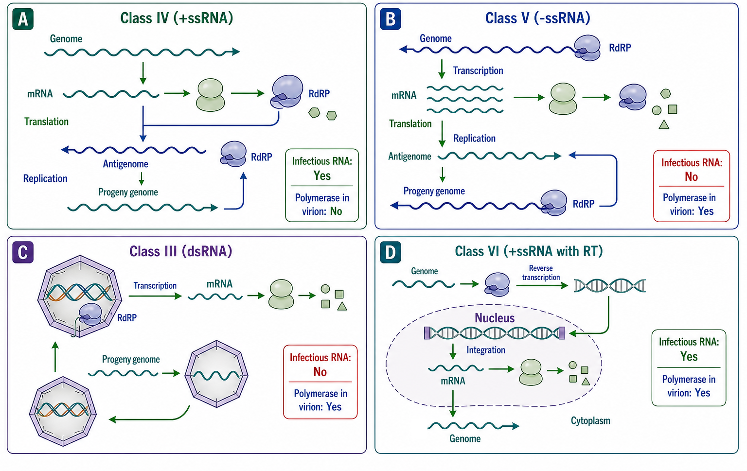
Figure 115.1. Baltimore Classification of RNA Viruses. The four Baltimore classes containing RNA viruses, distinguished by the pathway from the viral genome to translatable mRNA. Positive-sense RNA (class IV) is directly translated; negative-sense RNA (class V) requires virion-associated polymerase for transcription; double-stranded RNA (class III) is transcribed within the particle; and retrovirus RNA (class VI) is reverse-transcribed into DNA before mRNA production.
RNA viruses occupy four of the seven Baltimore classes. Class IV (positive-sense single-stranded RNA) is the largest and most diverse. The genomic RNA is mRNA-sense; upon entry into the cytoplasm, host ribosomes translate the genome directly, producing the viral replicase. The replicase then synthesizes a full-length negative-sense antigenomic RNA, which serves as template for new genomic RNA and, in many class IV viruses, for subgenomic mRNAs encoding structural proteins. Because the genome itself is infectious, class IV viruses do not need to package a polymerase in the virion. Families in class IV include Picornaviridae (poliovirus, rhinovirus, hepatitis A virus), Flaviviridae (dengue virus, Zika virus, hepatitis C virus, yellow fever virus), Coronaviridae (SARS-CoV, MERS-CoV, SARS-CoV-2), Togaviridae (chikungunya virus, Sindbis virus), Caliciviridae (norovirus), Hepeviridae (hepatitis E virus), and many plant and insect virus families. The genome size of class IV viruses ranges from approximately 7 kilobases (picornaviruses) to approximately 30-32 kilobases (coronaviruses), the largest known RNA genomes.
Class V (negative-sense single-stranded RNA) requires that the virion carry its own RNA-dependent RNA polymerase. The incoming genomic RNA is the template for mRNA synthesis, not the mRNA itself. The polymerase, together with the nucleoprotein that coats the genomic RNA, transcribes monocistronic or polycistronic mRNAs from the genomic template. Class V viruses are further divided into nonsegmented negative-strand RNA viruses (order Mononegavirales, including Rhabdoviridae, Paramyxoviridae, Filoviridae, Bornaviridae, and others) and segmented negative-strand RNA viruses (Orthomyxoviridae with 6-8 segments, Bunyavirales with 2-3 segments, Arenaviridae with 2 ambisense segments). The Mononegavirales share a common gene order and transcriptional strategy: the polymerase enters at a single 3′-terminal promoter and transcribes genes sequentially, with attenuation at each gene junction producing a transcription gradient in which promoter-proximal genes are expressed at higher levels than promoter-distal genes. This gradient is a form of gene regulation by genome position, and the conserved gene order — typically nucleoprotein, phosphoprotein, matrix protein, glycoprotein, polymerase — reflects functional constraints on relative expression levels.
Class III (double-stranded RNA) genomes are composed of 1 to 12 segments of double-stranded RNA enclosed within a characteristic multi-layered capsid. The virion-associated polymerase transcribes the negative strand of each genomic segment within the particle, extruding capped (but not polyadenylated) positive-sense mRNAs into the cytoplasm. The most medically important class III family is Reoviridae, which includes rotavirus (the leading cause of severe childhood diarrhea globally) and mammalian orthoreovirus. Other class III families include the bacteriophage Cystoviridae (phi6, an important model system) and families that infect fungi, plants, and insects. The double-stranded RNA genome is a potent innate immune agonist, and class III viruses have evolved strategies to keep the double-stranded RNA sequestered within the particle throughout the replication cycle, transcribing mRNA through pores in the capsid.
Class VI (positive-sense single-stranded RNA with a DNA intermediate) comprises the Retroviridae. The genomic RNA is positive-sense and carries two copies of the genome per particle. Upon entry, the virion-associated reverse transcriptase copies the RNA genome into double-stranded DNA, which is transported to the nucleus and integrated into the host chromosome by the viral integrase. Host RNA polymerase II then transcribes the integrated provirus into mRNA and new genomic RNA. The requirement for reverse transcription and integration places retroviruses in a distinct mechanistic category from other RNA viruses, although they share some genomic features with class IV and their quasispecies dynamics resemble those of other error-prone RNA viruses. Retrovirus biology is treated in detail in Chapter 120.
The ICTV taxonomy below the Baltimore class level arranges RNA viruses into orders, families, genera, and species based on genome organization, replication strategy, virion structure, and phylogenetic relationships. The advent of metagenomic sequencing has challenged this framework by revealing vast numbers of divergent RNA viruses that cannot be assigned to existing families. The ICTV has responded by creating new higher taxa and by accepting sequences as the basis for species demarcation in cases where no cultured isolate exists. A practical consequence is that the RNA virome is now understood to be far larger and more phylogenetically diverse than the set of viruses that have been cultured and studied in the laboratory. Taxonomic revisions are therefore frequent, and the reader should consult current ICTV releases for the latest classification.
Table 115.1. Genome Organization Across Representative RNA Virus Families. RNA virus families span a wide range of genome sizes, segment numbers, and coding strategies while conforming to a small number of Baltimore classes. Positive-sense viruses do not carry a polymerase; negative-sense and double-stranded viruses do.
| Virus family (example virus) | Baltimore class | Genome size | Polarity | Segments | Coding strategy | 5′ end feature | 3′ end feature | Virion polymerase |
|---|---|---|---|---|---|---|---|---|
| Picornaviridae (poliovirus) | IV | ~7.5 kb | +ssRNA | 1 | Single polyprotein | VPg | Poly(A) | None |
| Flaviviridae (dengue virus) | IV | ~11 kb | +ssRNA | 1 | Single polyprotein | Type 1 cap | No poly(A) | None |
| Coronaviridae (SARS-CoV-2) | IV | ~30 kb | +ssRNA | 1 | Polyprotein + subgenomic mRNAs | Type 1 cap | Poly(A) | None |
| Togaviridae (chikungunya virus) | IV | ~12 kb | +ssRNA | 1 | Genomic polyprotein + subgenomic mRNA | Type 0 cap | Poly(A) | None |
| Caliciviridae (norovirus) | IV | ~7.5 kb | +ssRNA | 1 | Polyprotein + subgenomic RNA | VPg | Poly(A) | None |
| Rhabdoviridae (VSV) | V | ~11 kb | −ssRNA | 1 | Transcription gradient | Type 1 cap | No poly(A) | Yes |
| Paramyxoviridae (measles virus) | V | ~15 kb | −ssRNA | 1 | Transcription gradient | Type 1 cap | No poly(A) | Yes |
| Filoviridae (Ebola virus) | V | ~19 kb | −ssRNA | 1 | Transcription gradient | Type 1 cap | No poly(A) | Yes |
| Orthomyxoviridae (influenza A) | V | ~13.5 kb | −ssRNA | 8 | Monocistronic per segment | Cap-snatched | No poly(A) | Yes |
| Bunyavirales (Lassa virus) | V | ~10.5 kb | Ambisense | 2 | Ambisense per segment | Cap-snatched | No poly(A) | Yes |
| Reoviridae (rotavirus) | III | ~18.5 kb | dsRNA | 11 | Monocistronic per segment | Type 1 cap | No poly(A) | Yes (inside particle) |
Table 115.1. Genome Organization Across Representative RNA Virus Families
| Virus family (example) | Baltimore class | Genome size | Polarity | Segments | Coding strategy | 5′ end feature | 3′ end feature | Virion polymerase |
|---|---|---|---|---|---|---|---|---|
| Picornaviridae (poliovirus) | IV | ~7.5 kb | +ssRNA | 1 | Single polyprotein | VPg | Poly(A) | None |
| Flaviviridae (dengue virus) | IV | ~11 kb | +ssRNA | 1 | Single polyprotein | Type 1 cap | No poly(A) | None |
| Coronaviridae (SARS-CoV-2) | IV | ~30 kb | +ssRNA | 1 | Polyprotein + subgenomic mRNAs | Type 1 cap | Poly(A) | None |
| Togaviridae (chikungunya virus) | IV | ~12 kb | +ssRNA | 1 | Genomic polyprotein + subgenomic mRNA | Type 0 cap | Poly(A) | None |
| Caliciviridae (norovirus) | IV | ~7.5 kb | +ssRNA | 1 | Polyprotein + subgenomic RNA | VPg | Poly(A) | None |
| Rhabdoviridae (VSV) | V | ~11 kb | −ssRNA | 1 | Transcription gradient | Type 1 cap | No poly(A) | Yes |
| Paramyxoviridae (measles virus) | V | ~15 kb | −ssRNA | 1 | Transcription gradient | Type 1 cap | No poly(A) | Yes |
| Filoviridae (Ebola virus) | V | ~19 kb | −ssRNA | 1 | Transcription gradient | Type 1 cap | No poly(A) | Yes |
| Orthomyxoviridae (influenza A) | V | ~13.5 kb | −ssRNA | 8 | Monocistronic per segment | Cap-snatched | No poly(A) | Yes |
| Bunyavirales (Lassa virus) | V | ~10.5 kb | Ambisense | 2 | Ambisense per segment | Cap-snatched | No poly(A) | Yes |
| Reoviridae (rotavirus) | III | ~18.5 kb | dsRNA | 11 | Monocistronic per segment | Type 1 cap | No poly(A) | Yes (inside particle) |
The Baltimore classification, like any scheme, has boundary cases. Ambisense genomes in Arenaviridae and some Bunyavirales have both positive-sense and negative-sense coding regions, making the assignment to class V technically correct but incomplete; students should learn that the class is defined by the mRNA synthesis pathway (the genomic RNA is the template for mRNA, not the mRNA itself) rather than by the polarity of every gene. Some positive-sense RNA viruses have a poly(A) tail (picornaviruses, coronaviruses); others have a 3′-terminal transfer RNA-like structure (some plant viruses). Some have a 5′ cap (flaviviruses, coronaviruses, togaviruses); others have a VPg protein covalently linked to the 5′ end (picornaviruses, caliciviruses). These variations in terminal modifications are compatible with the same Baltimore class because they do not change the fundamental pathway from genome to mRNA.
115.2. Genome polarity, segmentation, coding strategies, and packaging
The polarity of the viral genome — positive-sense, negative-sense, ambisense, or double-stranded — is the most fundamental distinction among RNA virus genome strategies. The polarity dictates the immediate steps after entry and determines which enzymatic activities the virion must carry. Beyond polarity, genomes differ in segmentation, in the strategy for producing multiple proteins from a single genomic RNA, and in the mechanism by which genomes are packaged into particles.
Positive-sense RNA genomes function as mRNA upon entry. This has several consequences. First, transfection of purified positive-sense genomic RNA into permissive cells is sufficient to initiate infection, making positive-sense RNA viruses experimentally tractable even before reverse genetics was available. Second, the infecting genome must contain all of the signals for translation initiation, including a 5′ cap or internal ribosome entry site (IRES) and often a 3′ poly(A) tail, or their functional equivalents. Third, because translation of the genome and replication of the genome occur on the same RNA molecule in opposite directions, a mechanism must exist to clear ribosomes from the template RNA before the polymerase can copy it, or to ensure that translation and replication are temporally or spatially separated. Fourth, the genome replicase proteins must be expressed first, from the genomic RNA, before replication can begin.
Coding strategies are the solutions that different RNA virus families have evolved to produce multiple proteins from a single genomic RNA. Eukaryotic ribosomes generally initiate at a single site per mRNA and translate one open reading frame, so viruses that need multiple proteins must adopt one of several strategies. Polyprotein processing is the most common strategy among class IV viruses: the genomic RNA is translated as a single long open reading frame, and the resulting polyprotein is cleaved by viral proteases (and sometimes host proteases) into individual functional proteins. Picornaviruses encode a single polyprotein that the viral 3C protease or its precursor 3CD cleaves into the structural proteins (VP1-4) and nonstructural proteins (2A-2C, 3A-3D). Flaviviruses encode a single polyprotein that is cleaved by the viral NS2B-NS3 serine protease and host signal peptidase into the structural proteins (C, prM, E) and nonstructural proteins (NS1-5). The order of proteins in the polyprotein and the kinetics of cleavage — with some sites cleaved rapidly and others slowly — provide a mechanism for controlling the timing and stoichiometry of viral protein accumulation.
Subgenomic mRNA transcription is a second strategy, used by coronaviruses, togaviruses, and other viruses with large positive-sense genomes. The replicase proteins are translated from the full-length genomic RNA as a polyprotein. The viral RdRP then synthesizes a full-length negative-sense antigenomic RNA, from which a nested set of subgenomic mRNAs is transcribed. These subgenomic mRNAs share a common 3′ end but have different lengths, each encoding one or a few proteins from the 3′-proximal portion of the genome (the structural protein genes). The coronaviruses use a discontinuous transcription strategy in which a common leader sequence from the 5′ end of the genome is fused to each subgenomic mRNA body, as detailed in Chapter 116. The subgenomic mRNA strategy separates the early expression of replicase proteins from the late expression of structural proteins and allows structural protein genes to be expressed at high levels without interference from the upstream replicase reading frame.
Ribosomal frameshifting and readthrough are strategies that produce two proteins from a single open reading frame by altering the normal decoding rules. In programmed -1 ribosomal frameshifting, the ribosome encounters a “slippery” heptanucleotide sequence and a downstream RNA pseudoknot or stem-loop that causes a fraction of ribosomes to shift one nucleotide backward and continue translating in a new reading frame. Retroviruses use -1 frameshifting to produce the Gag-Pol polyprotein, which supplies the protease, reverse transcriptase, and integrase at a defined ratio relative to Gag. In readthrough, the ribosome fails to terminate at a stop codon with a defined probability and continues translation into a downstream reading frame; some plant RNA viruses and alphaviruses use readthrough to produce C-terminal extensions of the replicase or coat protein. Frameshifting and readthrough are specialized forms of translational recoding whose structural biology and regulation are treated in Chapter 117.
The ambisense strategy, found in Arenaviridae and some Bunyavirales genera, encodes one gene in the negative-sense orientation and another in the positive-sense orientation on the same RNA segment. The S (small) segment of arenaviruses encodes the nucleoprotein in the negative sense and the glycoprotein precursor in the positive sense. This means that the nucleoprotein mRNA is transcribed from the genomic RNA by the virion polymerase early in infection, while the glycoprotein mRNA is transcribed from the antigenomic RNA after replication has produced sufficient antigenomic template. The ambisense strategy thus enforces a temporal order of gene expression without requiring a transcription gradient: the genomic-sense genes are expressed early, and the antigenomic-sense genes are expressed late.
Segmentation divides the viral genome across multiple RNA molecules. The selective advantage is reassortment: during co-infection, progeny virions can package segments from both parental viruses, generating novel genotypes without the requirement for template-switching recombination within a segment. The cost is a packaging problem, because assembling an infectious particle requires at least one copy of each segment. Influenza A virus (eight segments) uses segment-specific packaging signals located in the terminal coding and noncoding regions of each segment, and electron tomography of budding virions shows that the eight segments are arranged in a specific “7+1” pattern, with one central segment surrounded by seven others. Bunyaviruses (three segments) and arenaviruses (two segments) presumably use similar packaging signals, although the structural basis is less well characterized. The bacteriophage phi6 (three double-stranded RNA segments) packages its segments sequentially, with one segment entering the procapsid first and facilitating the entry of the others.
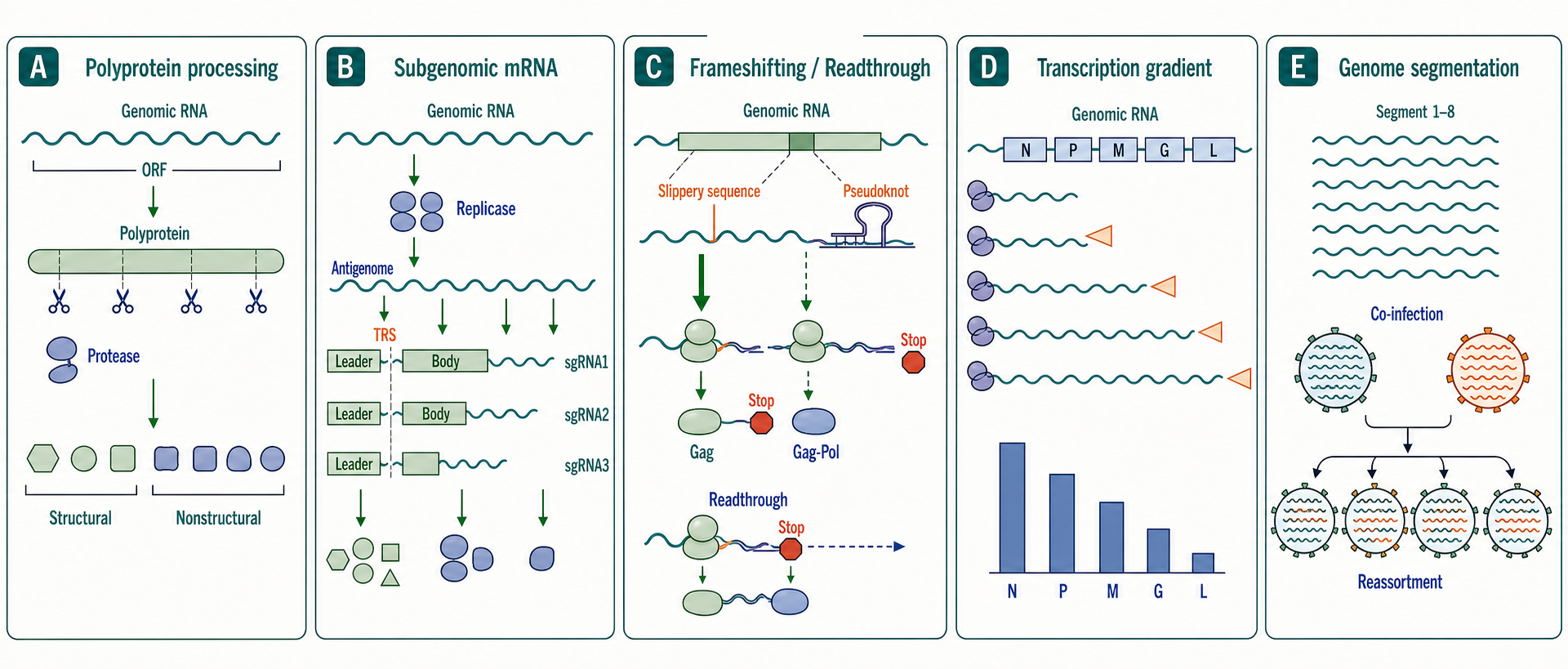
Figure 115.2. RNA Virus Genome Coding Strategies. RNA viruses use four major strategies to produce multiple proteins: polyprotein processing, subgenomic mRNA transcription, ribosomal frameshifting and readthrough, and transcription gradients. Genome segmentation provides a fifth dimension, distributing genes across physically separate RNA molecules that assort independently during co-infection.
Packaging is the process by which progeny genomes are incorporated into assembling virions. Genomes are selected for packaging through cis-acting packaging signals — RNA sequences or structures that are recognized by the viral nucleocapsid or capsid proteins. In many positive-sense RNA viruses, the packaging signal is located near the 5′ end of the genome, ensuring that only full-length genomic RNA is packaged and excluding subgenomic mRNAs and cellular RNAs. In retroviruses, the packaging signal (psi) is located in the 5′ untranslated region and is recognized by the nucleocapsid domain of Gag. In negative-sense RNA viruses, the nucleoprotein coats the genomic and antigenomic RNA, and the resulting helical nucleocapsid is recognized by the matrix protein during assembly. The material properties of packaged viral RNA, the role of phase separation in assembly, and the energetics of genome packaging are treated in Chapter 118.
A common misconception is that segmentation and polyprotein processing are alternative strategies for the same problem. They are not. Segmentation enables reassortment and allows genes to evolve semi-independently but creates a packaging problem. Polyprotein processing enables a single translation initiation event to produce multiple proteins with regulated stoichiometry but requires protease activity and precludes independent regulation of individual gene expression at the translation initiation step. A virus can use both strategies: arenaviruses have a segmented ambisense genome and produce the glycoprotein precursor as a polyprotein that is cleaved by a host protease.
115.3. Entry, replication, assembly, release, and host range
The RNA virus replication cycle is a chain of obligate steps, each requiring specific molecular interactions. The cycle begins with attachment and entry, proceeds through uncoating, translation, genome replication, assembly, and culminates in release. At every step, the viral RNA genome is the substrate, template, or product. Because replication errors occur at each cycle (see Chapter 116 for polymerase fidelity), the cycle is also the unit of evolution.
Entry begins when a viral surface protein binds a host cell receptor. For enveloped viruses, the surface protein is a transmembrane glycoprotein (or glycoprotein complex) embedded in the viral membrane. For non-enveloped viruses, the surface protein is part of the capsid. Receptor binding can be the sole trigger for entry (as in influenza virus, where hemagglutinin binding to sialic acid and the low pH of the endosome trigger fusion) or can require a co-receptor (HIV requires CD4 binding followed by binding to CCR5 or CXCR4). Receptor identity is a primary determinant of tissue tropism and host range. A virus that cannot bind a receptor on a particular species or cell type cannot enter, regardless of whether the intracellular environment is permissive. Angiotensin-converting enzyme 2 (ACE2) is the receptor for SARS-CoV and SARS-CoV-2; dipeptidyl peptidase 4 (DPP4) is the receptor for MERS-CoV; the poliovirus receptor (CD155) determines the neurotropism of poliovirus; the hepatitis C virus entry complex includes CD81, scavenger receptor class B type I, Claudin-1, and Occludin.
After receptor binding, the virus must deliver its genome across a membrane. Enveloped viruses fuse their membrane with a host membrane — either the plasma membrane at neutral pH or the endosomal membrane after endocytosis and acidification. Fusion is mediated by conformational changes in the viral glycoprotein (class I, II, or III fusion proteins) that expose a hydrophobic fusion peptide, insert it into the target membrane, and refold to bring the two membranes together. Non-enveloped viruses penetrate or disrupt the target membrane, often through conformational changes in the capsid triggered by receptor binding or low pH, exposing hydrophobic domains that form a pore or rupture the membrane. The result in all cases is delivery of the viral genome or nucleocapsid into the cytoplasm.
Uncoating is the release of the viral genome from the capsid or nucleocapsid so that it can be translated or transcribed. The mechanisms of uncoating are diverse and incompletely understood for many viruses. For picornaviruses, receptor binding triggers the release of the genomic RNA through a channel in the capsid. For influenza virus, the low pH of the endosome triggers fusion of the viral envelope with the endosomal membrane and the acidification also dissociates the M1 matrix protein from the ribonucleoprotein, allowing the viral RNPs to be transported into the nucleus. For reoviruses, the outer capsid is progressively removed by endosomal proteases, exposing the transcriptionally active core particle. In all cases, uncoating must balance the need to expose the genome for translation or transcription against the need to avoid premature degradation by cellular nucleases and detection by innate immune sensors.
Genome replication and transcription are the central biosynthetic events of the cycle. For positive-sense RNA viruses, the incoming genome is translated to produce the RdRP (and other nonstructural proteins), which then synthesizes a negative-sense antigenomic RNA that serves as template for new genomic RNA and, in many families, subgenomic mRNAs. Replication occurs on host membranes in replication organelles whose architecture and formation are treated in Chapter 116. For negative-sense RNA viruses, the virion-associated polymerase transcribes the genomic RNA into mRNAs immediately upon entry, and genome replication produces full-length positive-sense antigenomic RNA that serves as template for new genomic RNA. The polymerase must switch between transcription (producing capped and polyadenylated mRNAs) and replication (producing full-length, uncapped RNA), a switch that is regulated by the concentration of nucleoprotein in Mononegavirales. For double-stranded RNA viruses, the virion-associated polymerase transcribes mRNAs inside the particle throughout the cycle; genome replication produces progeny double-stranded RNA that is packaged into assembling particles.
The distinction between transcription and replication is crucial. During transcription, the polymerase produces messenger-sense RNA — capped, polyadenylated, and often shorter than the full genome — that is translated. During replication, the polymerase produces full-length RNA that is the template for more RNA synthesis or the genome packaged into progeny particles. In negative-sense RNA viruses, the same polymerase carries out both processes, but the RNA products differ in their 5′ ends, 3′ ends, and associated proteins. In positive-sense RNA viruses, the genomic RNA is both mRNA and the template for replication, and the transition from translation (ribosomes moving 5′ to 3′) to replication (polymerase moving 3′ to 5′ on the same template) requires a switch that clears ribosomes from the template. The mechanisms of this switch are virus-specific and often involve RNA structural elements and viral proteins that modulate the balance between translation and replication.
Assembly is the process of genome packaging and particle formation. For helical nucleocapsid viruses (most negative-sense RNA viruses), the nucleoprotein-coated genomic RNA forms a helical filament that is recognized by the matrix protein, which in turn interacts with the cytoplasmic tail of the envelope glycoprotein at the budding site. For icosahedral capsid viruses (picornaviruses, flaviviruses), the capsid protein assembles around the genomic RNA, or the genomic RNA is packaged into a preformed capsid or procapsid through a portal or packaging motor. For segmented viruses, assembly must incorporate at least one copy of each segment. For retroviruses, the Gag polyprotein assembles at the plasma membrane, the genomic RNA dimer is packaged through interactions between the Gag nucleocapsid domain and the psi packaging signal, and budding releases an immature particle that is subsequently cleaved by the viral protease to form the mature, infectious virion. The physics, material science, and structural biology of assembly are treated in Chapter 118.
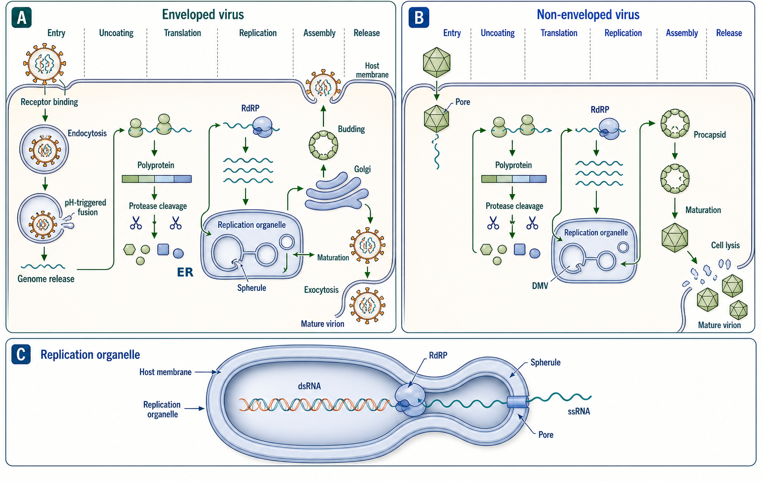
Figure 115.3. The RNA Virus Replication Cycle. The RNA virus replication cycle proceeds through entry, uncoating, translation, genome replication, assembly, and release. The strategy for each step varies by virus family, but the central role of the RNA genome — as mRNA, template, packaging substrate, and immune ligand — is universal.
Release completes the cycle. Non-enveloped viruses are typically released by cell lysis, which can be mediated by viral proteins that disrupt membrane integrity (picornavirus 2B, some adenovirus proteins). Enveloped viruses are released by budding from the plasma membrane (influenza, HIV) or by budding into intracellular compartments followed by exocytosis (coronaviruses, flaviviruses, bunyaviruses). Budding is driven by the interaction of the viral matrix or nucleocapsid protein with the cytoplasmic tail of the envelope glycoprotein and by host ESCRT (endosomal sorting complexes required for transport) machinery or viral late-domain motifs that recruit ESCRT components. The site of budding — apical versus basolateral plasma membrane, Golgi, endoplasmic reticulum, or endosome — determines the direction of virus release in polarized epithelial cells and influences pathogenesis and transmission.
Host range emerges from the sum of compatibilities — receptor, entry, uncoating, polymerase, translation, innate immune evasion, assembly, and release — required for productive infection. Incompatibility at any step restricts the host range. A virus may enter cells of a non-host species but fail to replicate because the polymerase does not recognize host factors. Alternatively, a virus may replicate but fail to assemble infectious particles because the glycoprotein cannot be properly processed or transported. The host range is therefore a multigenic trait, and adaptation to a new host generally requires multiple coordinated mutations. The evolutionary and ecological processes by which host-range mutations arise and spread are the subject of the next section.
A misconception to address is the idea that the viral replication cycle is a simple linear pathway with a fixed order of events. In reality, the steps overlap in time and space. Translation, replication, and assembly can occur simultaneously in different regions of the same infected cell. Replication can begin before uncoating is complete. Multiple rounds of assembly and release can occur in a single cell over many hours. The replication cycle is better described as a set of coupled processes than as a sequence of discrete steps, though the sequence is a useful pedagogical simplification.
Box 115.1. Receptor, Tropism, Host Range, and Vector Competence — Defining Four Related Terms
- Receptor: the specific host cell surface molecule (protein, carbohydrate, lipid) to which a viral attachment protein binds. The receptor is a molecular entity, not a cell type or species. A virus may use different receptors in different hosts, and receptor usage can evolve.
- Tissue tropism: the set of tissues and cell types within a host that support viral replication. Tropism is determined by receptor expression, but also by the presence of co-receptors, proteases for glycoprotein activation, the intracellular environment (nucleotide pools, translation machinery, innate immune state), and physical barriers (basement membrane, tight junctions). A virus may enter a cell but fail to replicate, a phenomenon called abortive infection that can be mistaken for true tropism if only entry is measured.
- Host range: the set of host species that a virus can infect productively. Host range is a multi-genic trait. Incompatibility at any step (receptor binding, entry, uncoating, polymerase function, innate immune evasion, assembly, release) restricts host range. Host range can be narrower than tissue tropism within the natural host; a virus that infects many tissues in bats may infect only one tissue in humans.
- Vector competence: the ability of an arthropod vector species (or population, or individual) to acquire, maintain, and transmit a pathogen. Vector competence is distinct from vectorial capacity, which includes ecological factors such as vector density, biting rate, and lifespan. A mosquito may be competent for a virus in the laboratory but contribute little to transmission in the field because of low abundance or feeding preference.
- Distinction rule of thumb: receptor is a molecule; tropism is within a species; host range is across species; vector competence is specific to the arthropod-virus pair and is a component of vectorial capacity.
- Practical example: Aedes aegypti is a competent vector for dengue virus (vector competence), which enters human cells via the DC-SIGN receptor on dendritic cells and other receptors on hepatocytes and endothelial cells (receptor), replicates in monocytes, macrophages, liver, and spleen (tissue tropism in humans), and can also infect non-human primates (host range).
Include review coverage of class I, II, and III fusion proteins, ESCRT-dependent and ESCRT-independent budding, and the molecular determinants of tissue tropism and species specificity.
115.4. RNA virus ecology, zoonosis, and emergence
RNA viruses cause the majority of emerging infectious diseases in humans. The list of RNA virus emergences since 2000 alone — SARS-CoV, MERS-CoV, H1N1pdm09 influenza, Ebola virus in West Africa, Zika virus in the Americas, SARS-CoV-2, and successive avian influenza spillovers — underscores the ongoing epidemiological significance of these pathogens. The biological properties that make RNA viruses prone to emergence include high mutation rates (which generate the variation for host-range adaptation), large population sizes within infected hosts (which ensure that rare adaptive mutations are present), recombination and reassortment (which create novel genotypes from existing diversity), and the existence of vast animal reservoirs (which supply genetically diverse viral populations for spillover).
The ecological framework for RNA virus emergence includes reservoir hosts, intermediate or amplifying hosts, spillover, and human-to-human transmission. A reservoir host is a species in which the virus circulates endemically, often without causing severe disease. Bats are reservoirs for many high-profile emerging viruses, including henipaviruses (Nipah, Hendra), filoviruses (Ebola, Marburg), and coronaviruses (SARS-CoV, MERS-CoV, SARS-CoV-2 and related sarbecoviruses). Bats appear to be particularly permissive reservoir hosts, a property that has been attributed to constitutive interferon pathway components, dampened inflammasome activation, and mechanisms of tolerance to viral replication that allow persistent infection without pathology. Rodents are reservoirs for arenaviruses (Lassa, Junin, Machupo) and hantaviruses. Birds are reservoirs for influenza A virus and for several arboviruses with avian amplification cycles, including West Nile virus and Japanese encephalitis virus.
An intermediate or amplifying host is a species that acquires the virus from the reservoir and in which mutations that increase the virus’s ability to infect humans can arise. For SARS-CoV, masked palm civets served as the intermediate host between horseshoe bats and humans. For MERS-CoV, dromedary camels are the source of most human infections. In human infections of MERS-CoV, the virus isolated from patients is genetically nearly identical to the virus circulating in camels, suggesting direct camel-to-human transmission without substantial human adaptation. For SARS-CoV-2, the evolutionary pathway from bat sarbecoviruses is less clear: a direct bat origin, an intermediate host (pangolins have been proposed but not confirmed), or a progenitor virus circulating undetected in humans or an animal population before the first recognized cases are all consistent with the available sequence data.
Spillover is the cross-species transmission event that initiates human infection. For spillover to occur, an infected reservoir or intermediate host must shed virus, a human must be exposed to a sufficient dose through a compatible route (respiratory, fecal-oral, direct contact, or vector-borne), and the virus must be capable of replicating in human cells. Most spillover events do not lead to outbreaks because the virus either fails to replicate efficiently in humans or is not transmitted efficiently from human to human. The factors that determine whether a spillover event initiates a chain of human-to-human transmission are the subject of intensive study and include the ability of the virus to replicate in the upper respiratory tract (for respiratory transmission), the infectious dose, the rate of contact between infectious and susceptible individuals, and the basic reproduction number (R0) of the virus in the human population.
Vector-borne RNA viruses add an arthropod dimension to emergence ecology. Arboviruses (arthropod-borne viruses) alternate between vertebrate hosts and hematophagous arthropod vectors, most commonly mosquitoes and ticks, but also sandflies, biting midges, and other vectors. The distribution of an arbovirus is constrained by the distribution of competent vectors. The expansion of Aedes aegypti and Aedes albopictus mosquitoes has expanded the geographic range at risk for dengue, chikungunya, and Zika virus epidemics. Climate change is projected to further expand the range of vector species and the length of the transmission season in temperate regions. The biology of vector infection, dissemination, and transmission — including the antiviral RNA interference and other RNA-based defenses in vectors — is treated in Chapter 114.
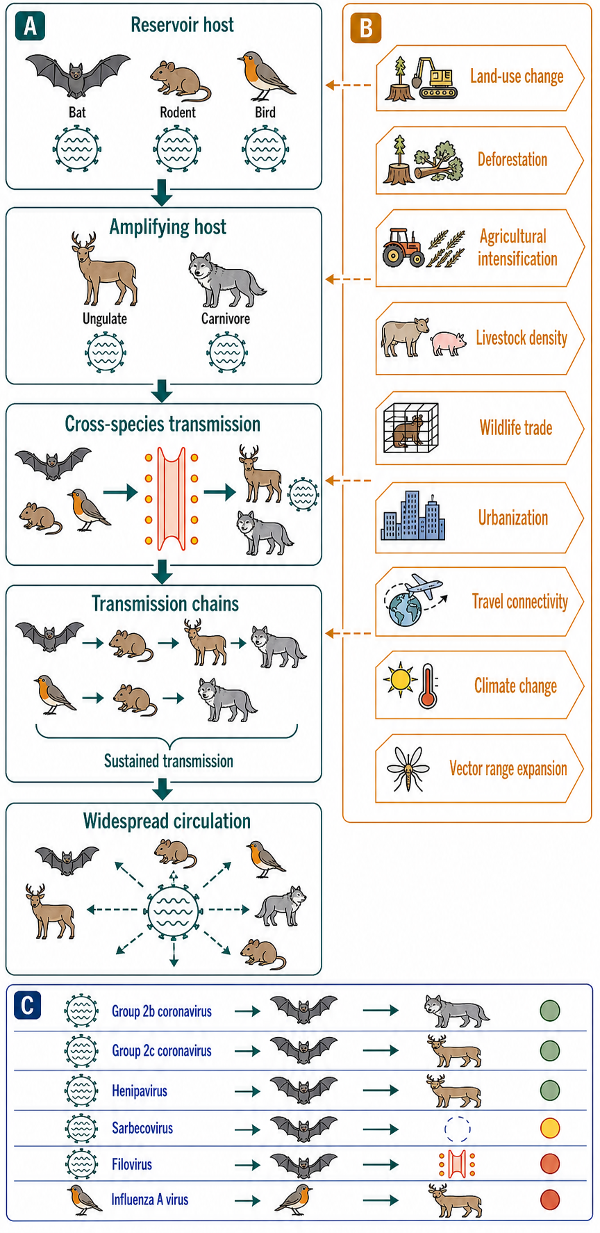
Figure 115.4. The Ecological Chain of Zoonotic RNA Virus Emergence. Zoonotic RNA virus emergence is a multi-stage ecological process. The virus circulates in a reservoir host, may adapt in an intermediate host, spills over into humans through defined exposure routes, and may achieve sustained human-to-human transmission. Ecological changes — land use, agriculture, wildlife trade, urbanization, and climate — drive each transition.
Ecological drivers of emergence include land-use change and deforestation, which bring humans into contact with reservoir host populations; agricultural intensification and livestock density, which create amplification opportunities for zoonotic viruses; wildlife trade and consumption, which provide direct human contact with infected animals and facilitate virus transport over long distances; urbanization and population density, which enhance human-to-human transmission once a virus is established; and climate change, which alters the distribution of vectors and reservoir hosts. These drivers are not mutually exclusive and often coincide; the emergence of Nipah virus in Malaysia involved bat reservoirs, pig amplification hosts, agricultural expansion into bat habitat, and dense pig farming. A reductionist focus on the virus alone, without the ecological context in which spillover occurs, misses essential determinants of emergence.
The concept of the “virosphere” — the totality of viral genetic diversity on Earth — has been transformed by metagenomic sequencing. Surveys of animal tissues, feces, environmental samples, and invertebrate transcriptomes have revealed thousands of novel RNA viruses, many in deeply branching lineages that may represent new families or orders. The discovery of enormous RNA virus diversity in apparently healthy animals suggests that most animal RNA viruses are not pathogenic in their natural hosts and that pathogenicity is the exception rather than the rule. The functional significance of this “virosphere dark matter” is largely unknown, but it constitutes the raw material for future emergence and is an active area of virus discovery.
A common misconception is that “emerging virus” means “newly evolved virus.” Many emerging viruses are ancient in their reservoir hosts and emerge because of changes in human ecology and behavior that increase exposure, not because of recent evolution. SARS-CoV-2 probably existed in bat populations long before it infected humans. The emergence event reflects a change in host range that may involve relatively few adaptive mutations in the virus combined with a change in the opportunity for transmission. A second misconception is that all zoonotic RNA viruses that enter humans will eventually adapt to efficient human-to-human transmission. Many zoonotic viruses infect humans sporadically without sustained transmission; H5N1 and H7N9 avian influenza viruses have caused hundreds of human cases with high case fatality rates but have not achieved sustained human-to-human transmission despite decades of opportunity. The barriers to human adaptation are substantial and varied across virus families.
Include reviews on the bat immune system and its role in reservoir competence, the ecology of pandemic influenza emergence, and the environmental and anthropogenic drivers of zoonotic spillover.
115.5. Reverse genetics, surveillance, and annotation
The ability to manipulate RNA virus genomes at will — to introduce mutations, deletions, insertions, and reporter genes, and to recover the modified virus — transformed virology from a descriptive to an experimental science. Reverse genetics for RNA viruses was first achieved for poliovirus in 1981, when Vincent Racaniello and David Baltimore transcribed infectious poliovirus RNA from a full-length cDNA clone and showed that the recovered virus had the expected phenotype. The experiment demonstrated that a purely synthetic RNA copy of a DNA plasmid could launch a complete, authentic viral replication cycle, and it opened the way to systematic structure-function analysis of every nucleotide and amino acid in the poliovirus genome.
The general workflow for positive-sense RNA virus reverse genetics is conceptually straightforward because the genomic RNA is itself infectious. A full-length cDNA copy of the viral genome is cloned into a plasmid under the control of a bacteriophage T7 or SP6 RNA polymerase promoter. The plasmid is linearized at the 3′ end of the viral sequence, in vitro transcription produces full-length genomic RNA with the correct 5′ and 3′ ends, and the RNA is transfected into permissive cells. If the viral genome carries a genetic marker that distinguishes it from wild-type virus, the recovered virus can be verified. The approach works for picornaviruses, flaviviruses, togaviruses, coronaviruses, and many other positive-sense RNA viruses, although the technical difficulty and efficiency vary. Coronavirus reverse genetics is particularly challenging because of the large genome size (approximately 30 kilobases), which makes full-length cDNA cloning difficult, and because of toxic sequences in the replicase gene that kill bacteria during plasmid propagation. These problems have been overcome by using bacterial artificial chromosomes, in vitro ligation of subgenomic cDNA fragments, or vaccinia virus vectors as intermediates.
Negative-sense RNA virus reverse genetics requires reconstitution of the viral ribonucleoprotein complex, because naked negative-sense RNA is not infectious and does not produce mRNA. The breakthrough was the development of systems that co-express the viral genomic or antigenomic RNA together with the viral polymerase (L protein), the nucleoprotein (N or NP), and in some cases the phosphoprotein (P) from plasmid DNAs. When cells are transfected with these plasmids, the polymerase and nucleoprotein assemble on the expressed RNA, forming a functional RNP that can transcribe mRNA and replicate the genome, producing infectious virus. The first such system was developed for influenza virus by Peter Palese’s group and for rabies virus by Karl-Klaus Conzelmann’s group. Influenza virus reverse genetics is now highly efficient; the eight genomic segments are cloned into bidirectional plasmids that express both the negative-sense viral RNA and the positive-sense mRNA from the same template, allowing virus recovery with high efficiency from transfected cells. This system is used for annual vaccine strain development and for generating candidate pandemic vaccine viruses.
For double-stranded RNA viruses, reverse genetics has been achieved for members of the Reoviridae and for bacteriophage phi6. The general approach involves expressing the viral polymerase and core proteins to generate transcriptionally active core particles into which in vitro synthesized positive-sense RNAs are packaged and replicated to produce double-stranded RNA segments. Rotavirus reverse genetics, achieved in 2017 using a plasmid-based system, has enabled the functional analysis of rotavirus gene segments that was previously possible only by reassortment with characterized laboratory strains.
Replicon and minigenome systems are simpler tools that do not require the recovery of infectious virus. A replicon is a self-replicating subgenomic RNA in which the structural protein genes have been deleted and replaced with a selectable marker or reporter gene. Replicon-bearing cells can be passaged under antibiotic selection, and the RNA replication level is measured by reporter activity. Because no infectious virus is produced, replicons can be studied under lower biosafety levels and are the workhorse of antiviral drug screening, particularly for hepatitis C virus, for which replicon systems enabled the discovery of direct-acting antivirals years before a complete cell-culture infectious system was available. A minigenome contains only the minimal cis-acting replication and transcription signals, typically the genomic termini, together with a reporter, and is replicated by the polymerase and nucleoprotein provided in trans. Minigenomes are used to dissect the cis-acting elements required for replication and transcription, to study polymerase function, and to screen polymerase inhibitors.
Virus-like particles (VLPs) are assembled from viral structural proteins without a viral genome. VLPs mimic the antigenic surface of the authentic virion and are the basis of the hepatitis B and human papillomavirus vaccines. For RNA viruses, VLPs are used to study assembly, to map neutralizing antibody epitopes, and as vaccine candidates. VLPs can package cargo, including reporter RNAs or heterologous proteins, for delivery applications. The assembly of VLPs, and the relationship between genome packaging and VLP assembly, is treated in Chapter 118.
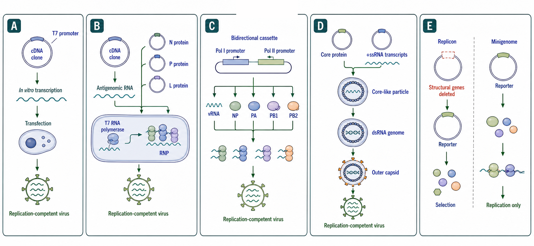
Figure 115.5. Reverse Genetics Workflows for RNA Viruses. Reverse genetics for RNA viruses requires different strategies depending on whether the naked genomic RNA is infectious (positive-sense), requires co-expressed viral proteins (negative-sense), or requires assembly of a transcriptionally active particle (double-stranded RNA). Replicon and minigenome systems provide simpler tools for studying replication and screening antivirals.
Virus surveillance has entered the genomic era. For known viruses, targeted RT-PCR and Sanger sequencing have been supplemented or replaced by amplicon-based next-generation sequencing, probe-capture enrichment sequencing, and metagenomic sequencing. Influenza surveillance by the Global Influenza Surveillance and Response System (GISRS) sequences thousands of isolates annually to monitor antigenic drift and to recommend vaccine strain updates. SARS-CoV-2 genomic surveillance during the COVID-19 pandemic achieved an unprecedented scale, with millions of genomes sequenced and shared through the GISAID platform, enabling near-real-time tracking of variant emergence, spread, and association with clinical outcomes. The combination of genomic data with epidemiological metadata — sampling date, location, patient age, vaccination status, and disease severity — has made genomic epidemiology a standard public health tool.
Virus discovery by metagenomics involves unbiased library preparation and deep sequencing of nucleic acids from clinical, animal, or environmental samples, followed by computational removal of host reads and classification of the remaining reads by sequence similarity to known viruses. Viruses with little or no similarity to known reference sequences require de novo assembly and inference of viral origin from properties such as RNA-dependent RNA polymerase motifs, genome organization, codon usage, or association with disease. This approach has identified hundreds of novel RNA viruses in recent years, including the highly divergent Jingmenvirus group of segmented flavivirus-like viruses, numerous insect-specific viruses, and deep-branching lineages that may represent new families. The identification of a novel virus sequence is only the first step; establishing whether it is a human pathogen, an animal pathogen, or a benign component of the virome requires additional epidemiological, serological, and experimental evidence.
Annotation of RNA virus genomes involves identifying open reading frames, predicting cleavage sites in polyproteins, annotating cis-acting RNA elements, and mapping transcription regulatory sequences, packaging signals, and other noncoding features. Automated annotation pipelines can handle known virus families reasonably well but struggle with divergent viruses and with noncanonical coding features such as ribosomal frameshifting, alternative initiation, and overlapping reading frames. Expert curation remains essential, and efforts such as the ViralZone resource and the RefSeq viral genome collection provide manually annotated reference genomes that serve as the basis for computational annotation.
A misconception to guard against is that reverse genetics recovers “the same virus” as the wild-type. Every step — PCR amplification, cloning, in vitro transcription, transfection, and cell-culture passage — can introduce sequence changes. The recovered virus should be fully sequenced to confirm that it matches the intended sequence. Even then, cell-culture adaptation can select mutations that differ from the original clinical isolate. A second common error is assuming that a virus detected by metagenomic sequencing is the cause of a disease in the host from which the sample was taken. Association requires epidemiological evidence, and causation requires fulfillment of Koch’s postulates or their molecular equivalents, typically involving the recovery of infectious virus and reproduction of disease in an animal model or fulfillment of the molecular Koch’s postulates with specific viral determinants.
This chapter provides the framework for understanding RNA viruses as biological entities with definable genomes, replication cycles, host ranges, and evolutionary trajectories. The subsequent chapters in Part 19 deepen each dimension: Chapter 116 treats the enzymatic and biophysical core of RNA synthesis, fidelity, and the population-genetic consequences; Chapter 117 examines the RNA structures that regulate each step of the viral life cycle; Chapter 118 addresses the material and physical principles that govern genome packaging and particle assembly; Chapter 119 covers the minimalist RNA pathogens — viroids and satellite RNAs — that exist at the boundary of life; and Chapter 120 treats the retroviruses and the biology of reverse transcription. The common thread across all of Part 19 is that RNA is not merely a carrier of viral genetic information; it is an active participant in every step of the viral life cycle, and understanding its roles requires integrating virology, biochemistry, structural biology, biophysics, immunology, ecology, and evolutionary biology.
Include historical context for the development of reverse genetics and modern applications to vaccine development and antiviral screening.
Experimental Foundations and Evidence Standards
The evidence for RNA virus biology spans a wide range of methodologies, and the appropriate evidence standard depends on the claim. Virion structure and genome organization are established by electron microscopy, X-ray crystallography, cryo-electron microscopy, and sequencing. The assignment of a virus to a Baltimore class is based on genome sequence and the demonstration of mRNA production pathway, typically by Northern blotting or RNA sequencing of infected cells. The identification of a receptor is established by demonstrating that the purified or expressed protein binds the virus, that expression of the receptor in non-permissive cells confers susceptibility, and that knockout or blockade of the receptor abolishes infection. The assignment of a function to a viral protein is established by reverse genetics: mutation of the protein-coding sequence and demonstration of a specific defect in the viral life cycle.
Evidence for spillover and emergence combines phylogenetics, epidemiology, and ecology. The phylogenetic evidence for a zoonotic origin is that human viruses nest within animal virus diversity; the epidemiological evidence is that human cases are associated with animal contact; the ecological evidence is that the animal host and humans overlap in space and time. The evidence for human-to-human transmission requires showing that the viral sequences from epidemiologically linked cases are more closely related than sequences from unlinked cases, and that the timing of symptom onset is consistent with person-to-person spread.
The main interpretive hazards include: confusing the detection of viral RNA by RT-PCR with the presence of infectious virus (RNA can persist after infectivity is lost, and RNA from defective particles or environmental contamination can be PCR-positive); overinterpreting cell-culture phenotypes as in vivo phenotypes (a mutation that attenuates virus in a tumor cell line may have no effect in the relevant primary cell or tissue); assuming that a virus that replicates in a cell line can replicate in the corresponding tissue in vivo (cell lines often lack innate immune pathways present in primary cells); and inferring ancestral states from phylogenetic trees without adequate sampling of the diversity (missing taxa can invert inferred relationships). Reverse genetics provides the most rigorous standard: a phenotype is attributed to a specific sequence change only when the change is introduced into an isogenic background and the phenotype is recapitulated.
Biological Contexts and Cross-Chapter Boundaries
This chapter is the integrator for Part 19. Chapter 116 will examine the polymerase that carries out the replication and transcription described here. Chapter 117 will examine the RNA structures — promoters, packaging signals, IRESs, frameshift elements, and others — that are described functionally in this chapter. Chapter 118 will treat the biophysics of the assembly, packaging, and condensation processes introduced here. Chapters 119 and 120 will treat the subviral RNA agents and the retroviruses, respectively, that are mentioned only for boundary definition here.
Beyond Part 19, this chapter connects to Chapter 108 (innate immune sensing of viral RNA) because the replication intermediates and products described here — especially double-stranded RNA and 5′-triphosphate RNA — are the ligands for RIG-I, MDA5, TLR3, TLR7/8, PKR, and OAS. It connects to Chapter 111 (host-pathogen RNA interactions) because the host proteins and RNAs that participate in viral replication, assembly, and restriction are part of a larger network of host-pathogen molecular conflict. It connects to Chapter 160 (antiviral ribonucleoside analogs) because the polymerase and the replication cycle described here are the targets of these drugs. It connects to Chapter 110 (RNA in adaptive immunity) because the antigens that are the targets of neutralizing antibodies and cytotoxic T cells are the protein products of the viral RNA genomes described here.
The most important boundary is between the genome-strategy focus of this chapter and the polymerase-mechanism focus of Chapter 116. This chapter says what the genome looks like, how it is expressed, and how it is replicated in outline; Chapter 116 says how the polymerase does the chemistry, how fidelity and proofreading work, and how quasispecies genetics follows from polymerase error rates. The reader should understand the genome strategies before the polymerase details; the two chapters are designed to be read sequentially.
Recent Consensus
The Baltimore classification remains the standard framework for organizing viral genome diversity and is incorporated into ICTV taxonomy. The division of single-stranded RNA viruses into positive-sense, negative-sense, and ambisense is universally accepted. The general architecture of the replication cycle — entry, uncoating, translation or transcription, replication, assembly, release — is established for every well-studied RNA virus family, although the molecular mechanisms differ.
The role of bats, rodents, and birds as major reservoir hosts for zoonotic RNA viruses is established. The principle that emerging RNA virus diseases result from ecological changes bringing reservoir hosts, vectors, and humans into new contact patterns is the consensus view among infectious disease ecologists. The importance of metagenomic sequencing for virus discovery and of genomic epidemiology for outbreak response is established and was demonstrated at scale during the COVID-19 pandemic.
Reverse genetics for most major RNA virus families has been achieved and is a standard tool. The principle that reverse genetics provides the gold standard for attributing phenotype to genotype in virology is universally accepted.
Open Questions, Controversies, Deprecated Models, and Common Misconceptions
Open questions:
- What determines how broad RNA virus diversity in nature is? Metagenomic studies continue to discover deep-branching lineages that suggest we have sampled only a small fraction of existing RNA virus diversity. Whether these deeply divergent viruses have fundamentally different genome strategies from known viruses is unknown.
- The determinants of host range at the molecular level are incompletely understood for most viruses. The fact that a bat coronavirus such as RaTG13, which is approximately 96 percent identical to SARS-CoV-2
- At the nucleotide level, does not use human ACE2 efficiently, while SARS-CoV-2 does, demonstrates that even small differences can alter host range. Predicting host range from sequence alone remains unreliable.
Common misconceptions:
- “The non-pathogenic animal RNA virosphere has well-defined ecological functions.” The existence of many animal RNA viruses without known disease association is recognized, but whether these viruses regulate host populations, transfer genes, or modulate host immunity remains functionally unexplored.
- “Positive-sense RNA viruses are simpler than negative-sense RNA viruses.” Positive-sense RNA viruses do not need to package a polymerase, but their genomes must serve as mRNA, replication template, and packaging substrate simultaneously, placing conflicting constraints on RNA sequence and structure. Negative-sense RNA viruses package a polymerase but can separate the transcriptome (mRNAs) from the antigenomic template and the genomic template, allowing distinct structures for distinct functions. Complexity is differently distributed, not absent.
- “The Baltimore class tells you everything about a virus’s replication strategy.” Viruses within the same Baltimore class can differ profoundly. Flaviviruses and coronaviruses are both class IV, but flaviviruses use a single polyprotein and replicate in spherules on the endoplasmic reticulum, while coronaviruses use ribosomal frameshifting, discontinuous subgenomic mRNA transcription, and replicate in double-membrane vesicles. The Baltimore class defines the pathway from genome to mRNA, not the details of the replication cycle.
- “If a virus is detected by metagenomic sequencing in a sick patient, it causes the disease.” Metagenomic detection of a viral sequence is not evidence of causation. Many healthy individuals carry RNA viruses without disease. Establishing causation requires epidemiological association, plausibility, and ideally recovery of infectious virus and reproduction of disease in an animal model.
- “Reverse genetics is now routine for all RNA viruses.” Reverse genetics has been achieved for representatives of most major families, but efficiency varies widely, and some viruses still lack tractable reverse genetics systems. Coronaviruses remain challenging because of genome size. Many recently discovered metagenomic viruses have not been cultured, let alone made into infectious clones.