Chapter 71. Ribosome Collisions, Ribosome Quality Control, Ribotoxic Stress, and Proteostasis Coupling
Scope Note
Ribosomes are not only protein-synthesis machines. They are also moving sensors of messenger RNA integrity, nascent-chain behavior, translation-factor availability, cellular stress, and infection. This chapter explains how stalled or slowed ribosomes form collisions, how cells recognize collided ribosome states, how ribosome quality-control pathways dispose of damaged nascent chains and defective mRNAs, how ribotoxic stress signaling converts translation damage into inflammatory and stress responses, and how these pathways connect to proteostasis, stress granules, viruses, bacteria, organelles, disease, and therapy.
Executive Summary
Ribosome collision occurs when a trailing ribosome catches up to a leading ribosome that has slowed, paused, or stopped on the same mRNA. A single slow ribosome can reflect normal elongation dynamics, codon usage, nascent-chain folding, targeting, or regulatory pausing. A collided ribosome array is different: the geometry of adjacent ribosomes creates composite surfaces that recruit surveillance factors. The central principle is that cells read ribosome state, not merely mRNA sequence. Reviews of ribosome-state signaling and no-go decay describe collisions as a conserved way to distinguish transient translation heterogeneity from potentially harmful defects in mRNA, nascent protein, or ribosome function (D’Orazio and Green 2021; De and Mühlemann 2022; Alagar Boopathy et al. 2023).
In eukaryotes, collision sensing is coupled to ribosome quality control (RQC). Yeast Hel2 and mammalian ZNF598 ubiquitinate small-subunit proteins on collided ribosomes. GCN1 can also bind collided ribosomes and connect collision load to integrated stress signaling. Splitting factors including Pelota, Hbs1, and ABCE1 separate ribosomal subunits when a ribosome cannot terminate normally. The large subunit retains the peptidyl-tRNA-linked nascent chain and recruits RQC factors such as LTN1/Listerin and NEMF/Rqc2, which promote ubiquitination, extraction, and proteasomal degradation. When RQC is incomplete, NEMF/Rqc2-mediated C-terminal alanine/threonine additions, often called CAT tails, can alter aggregation and stress signaling in model systems. The same collision can therefore produce several outputs: rescue of ribosomal subunits, degradation of the mRNA, degradation or modification of the nascent chain, and activation of stress pathways.
No-go decay and related pathways remove mRNAs that repeatedly cause translation arrest. The exact nuclease identities, cleavage positions, and decay routes vary by organism and context. Some mRNAs are cleaved near stalled ribosomes and then degraded by exonucleases; other defective messages are decapped, deadenylated, or routed through endoplasmic-reticulum-associated decay pathways. A key boundary case is that ribosome pausing is not automatically pathological. Regulated pauses can assist cotranslational folding, targeting, recoding, or localization. Experimental interpretation must therefore distinguish a normal pause, a translation bottleneck, a collision, and a quality-control-triggering collision.
Ribotoxic stress is an overlapping but not identical response. Ribosome damage or collision-prone translation can activate ZAK-alpha, a ribosome-associated kinase isoform that senses perturbed ribosomal states and initiates p38 and JNK mitogen-activated protein kinase signaling. Recent primary work links ZAK-alpha-dependent ribotoxic stress to RNase L cleavage products and UV-mediated cell death, showing that translation damage can connect RNA surveillance, innate immunity, and inflammatory stress (Xi et al. 2024; Sinha et al. 2024). The chapter uses ZAK-alpha for the protein name and ZAKα as a common symbol variant.
Proteostasis coupling is the reason collision biology matters beyond RNA surveillance. A failed translation event produces a damaged mRNA, a stuck ribosome, and a partially synthesized protein that may misfold or aggregate. RQC therefore intersects with chaperones, the ubiquitin-proteasome system, autophagy, stress granules, P-bodies, and organelle quality control. Stress granules are dynamic ribonucleoprotein condensates enriched in untranslated mRNAs and proteins such as G3BP1; they do not simply store every stressed mRNA. Recent evidence indicates that ribosome association can prevent some stress-induced mRNAs from entering stress granules, emphasizing that translation status governs RNA partitioning (Helton et al. 2025). Viral infection, bacterial translation stress, and organellar translation expand the field beyond the canonical cytosolic eukaryotic pathway, but many mechanistic details remain less settled than the yeast and mammalian RQC core.
Concept Inventory
- Ribosome collision: a physical state in which one ribosome encounters another ribosome stalled or slowed ahead on the same mRNA. Collisions can involve two ribosomes, called a disome, or larger arrays.
- Ribosome quality control: mechanisms that recognize aberrant ribosome states, rescue ribosomal subunits, degrade incomplete nascent chains, and often remove the problematic mRNA.
- No-go decay: mRNA surveillance triggered by ribosome stalling or collisions on an mRNA that cannot be productively translated.
- Non-stop decay: surveillance of mRNAs lacking a functional stop codon, causing ribosomes to translate into poly(A) tails or 3′ extensions.
- Ribotoxic stress response: stress signaling triggered by ribosome damage or collision-prone translation, prominently involving ZAK-alpha and downstream p38/JNK signaling in mammalian cells.
- Proteostasis: maintenance of a functional proteome through folding, chaperone action, degradation, trafficking, and stress responses.
- Stress granule: a non-membrane-bound cytoplasmic ribonucleoprotein assembly that forms when initiation is inhibited and untranslated mRNAs accumulate with RNA-binding proteins.
- Disome profiling: sequencing footprints from two ribosomes protected together on an mRNA, used to infer collision positions.
- CAT tail: a C-terminal alanine/threonine-rich extension added by NEMF/Rqc2-like factors to some stalled nascent chains in RQC contexts.
- Artifact-prone boundary: a high ribosome-profiling footprint peak is not by itself proof of collision; it may reflect nuclease bias, drug treatment, initiation pileup, RNA structure, slow elongation, or protected non-ribosomal complexes.
What to Know Before Reading This Chapter
Translation elongation moves ribosomes along coding sequences in the 5′ to 3′ direction while each ribosome reads codons, recruits aminoacyl-tRNAs, forms peptide bonds, and translocates. Chapter 68 explains this cycle and the normal causes of pausing, including codon usage, tRNA availability, nascent-chain folding, and targeting. Chapter 69 explains termination and recycling, because many quality-control states arise when termination or recycling fails. Chapter 70 explains regulatory translation features such as upstream open reading frames and internal ribosome entry sites, which can change ribosome density without necessarily causing damage.
The reader should separate four terms. A pause is a temporary slow point in elongation. A stall is a longer arrest that may prevent completion of translation. A collision is a physical encounter between ribosomes. A surveillance trigger is a molecular state that recruits quality-control factors. These terms often overlap but are not interchangeable. For example, a regulatory pause in a secretory protein can assist cotranslational targeting without inducing RQC, whereas a defective mRNA with a strong stem-loop in the coding sequence can generate repeated collisions that recruit ubiquitin ligases and mRNA decay factors.
The main running example is a mammalian cytosolic mRNA containing an obstacle in its coding region. The leading ribosome slows at the obstacle. A following ribosome catches up, producing a collided disome. Collision-sensing factors bind the composite ribosome surface, ubiquitinate ribosomal proteins, recruit splitting and decay factors, and route the nascent chain and mRNA into degradation or signaling pathways. Variants of this example appear in yeast, bacteria, viral infection, and organellar translation, but the exact factors differ.
71.1. Ribosome collision formation and sensing
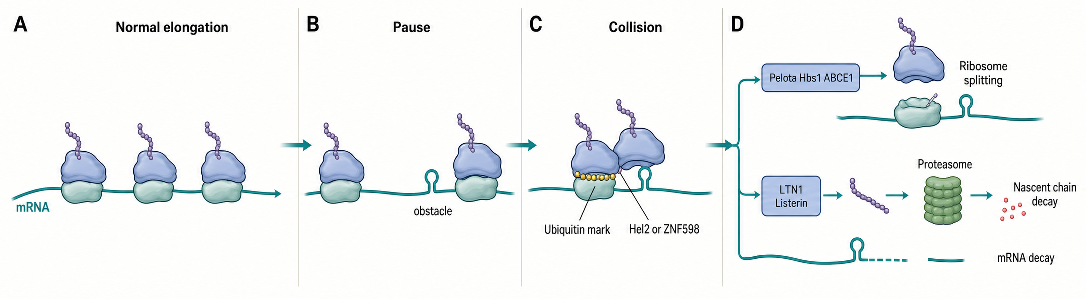
Figure 71.1. From Pause to Collision to RQC. A transient pause becomes a quality-control signal when continued initiation and elongation create a collided ribosome state. The collision exposes a composite molecular surface that recruits surveillance factors including Hel2 in yeast and ZNF598 in mammals, leading to small-subunit ubiquitination, ribosome rescue by Pelota–Hbs1–ABCE1, nascent-chain degradation by LTN1/Listerin and the proteasome, and decay of the problematic mRNA.
Ribosome collision formation begins with an imbalance between initiation and elongation. If initiation continues while elongation slows at a downstream site, ribosomes accumulate upstream of the obstacle. A leading ribosome may slow because of a stable RNA structure, a damaged nucleotide, a rare codon cluster, a depleted charged tRNA, a problematic nascent peptide, a polybasic sequence, a missing stop codon, an antibiotic, an amino-acid limitation, or physical obstruction by an RNA-binding protein. A trailing ribosome then translocates into the space normally occupied by the leading ribosome and forms a collided state. The collision is not just crowding. Adjacent ribosomes generate a particular three-dimensional surface that can be recognized by surveillance proteins.
Table 71.1. Comparative Collision Biology. Collision sensing and rescue strategies differ across translating systems; eukaryotic cytosol, bacteria, organelles, and viral-infection contexts are compared by sensors, rescue factors, and the fates of the nascent chain and mRNA.
| System | Main stall or collision problem | Known or likely sensors | Rescue factors | Nascent-chain fate | mRNA fate |
|---|---|---|---|---|---|
| Eukaryotic cytosol | Stable RNA structures, rare codons, polybasic sequences, missing stop codon | ZNF598, Hel2, GCN1, RACK1 | Pelota, Hbs1, ABCE1 | Ubiquitination by LTN1/Listerin; proteasomal degradation; CAT tailing by NEMF/Rqc2 | No-go decay; endonucleolytic cleavage; decapping or deadenylation |
| Bacteria | Truncated mRNAs, rare codons, antibiotic-induced stalls, damaged nucleotides | MutS-family paralogue | tmRNA–SmpB, alternative rescue factors | tmRNA-mediated C-terminal tagging; protease degradation | Degradation following rescue; Rho-dependent termination |
| Mitochondria | Stalled organellar ribosomes; unusual mRNA leaders and editing patterns | Not well established; bacterial-derived mechanisms | Bacterial-like rescue factors; less defined | Not well established; likely protease-mediated | Not well established |
| Chloroplasts | Stalled organellar ribosomes; unusual mRNA features from plastid editing | Not well established | Bacterial-like rescue factors; less defined | Not well established | Not well established |
| Viral infection | Viral RNA structures, frameshift elements, IRES; host RNase L cleavage of RNA | ZAK-alpha activated by cleavage products; host RQC factors | Host Pelota–Hbs1–ABCE1; viral evasion possible | Host RQC; viral manipulation of degradation possible | RNase L cleavage; host decay pathways; viral protection at some elements |
This geometry helps solve a specificity problem. Translation is naturally uneven. Ribosomes pause during decoding, peptide-bond formation, translocation, nascent-chain folding, and membrane targeting. If every pause triggered degradation, cells would destroy many normal mRNAs and nascent proteins. Collision-dependent sensing allows cells to tolerate isolated pauses while responding to persistent obstacles that create arrays of ribosomes. Reviews emphasize this ribosome-state logic: quality-control factors often recognize the shape and composition of stalled or collided ribosomes rather than a single defective RNA sequence feature (D’Orazio and Green 2021; De and Mühlemann 2022).
Sensing is best understood as a tiered system. At low collision load, local quality-control factors can resolve individual problematic translation events. At higher collision load, global stress pathways are engaged. In yeast and mammals, collision-responsive ubiquitin ligases decorate ribosomal proteins on the small subunit. In yeast, Hel2 is a central collision-associated E3 ligase. In mammals, ZNF598 has an analogous role in ubiquitinating small-subunit proteins such as uS10 and eS10 in many model systems. GCN1, historically associated with amino-acid starvation signaling through GCN2, can also bind collided ribosomes and connect collision states to integrated stress signaling. These factor names should not be treated as a single universal pathway. Collision-sensing modules differ across organisms and stresses, and several factors compete for or cooperate on overlapping ribosomal surfaces.
Table 71.2. Experimental Readouts and Artifact Controls. Each experimental approach for studying ribosome collisions and quality control supports specific conclusions but cannot alone prove collision-specific pathway activation; pairing multiple readouts with appropriate controls is required for strong causal claims.
| Readout | What it supports | What it cannot prove alone | Key controls |
|---|---|---|---|
| Monosome ribosome profiling | Genome-wide ribosome positions; high-occupancy sites | Collision (peaks may reflect pausing, nuclease bias, drug artifact, RNA structure, or non-ribosomal protection) | Drug-free or alternate-inhibitor protocols; disome profiling for comparison |
| Disome profiling | Physical adjacency of two ribosomes; collision-enriched positions transcriptome-wide | That detected collisions are pathological or activate quality control | Collision sensor mutants; perturbation of stall source; monosome comparison |
| Ubiquitinated ribosomal protein detection | Collision-associated small-subunit ubiquitination by ZNF598 or Hel2 | Downstream RQC activation or mRNA decay | E3 ligase mutants; site-specific mass spectrometry; proteasome inhibitor controls |
| Reporter protein output | Stall-induced reduction in protein output from a defined reporter construct | That endogenous transcripts behave identically | Stall-sequence mutants; parallel mRNA abundance measurement; factor depletion |
| mRNA abundance or cleavage fragments | mRNA decay or cleavage associated with translation stress | That decay is collision-specific or proceeds through no-go decay | Rescue factor knockouts (Pelota, Hbs1); northern blotting with decay-factor mutants |
| Stress kinase phosphorylation (p38, JNK) | Activation of MAP kinase signaling during translation stress | That activation is ribosome-collision-specific or ZAK-alpha-dependent | ZAK-alpha depletion or inhibition; eIF2α phosphorylation measurement to distinguish ISR |
| Stress-granule imaging | Formation of cytoplasmic G3BP1-positive foci under stress | That foci contain specific mRNAs or functionally repress translation | Multiple markers (G3BP1, TIA-1); mRNA identity in granules; avoid overexpression artifacts |
| Genetic knockout or depletion | Requirement of a factor for a pathway step | Direct mechanistic role; single-factor deletion can reveal redundancy rather than linear dependence | Epistasis experiments; rescue by re-expression; comparison of multiple mutant combinations |
Ribosome profiling changed the field by letting investigators map ribosome positions transcriptome-wide. Standard ribosome profiling sequences monosome-protected mRNA fragments, whereas disome profiling isolates longer fragments protected by two adjacent ribosomes. A strong monosome peak can show pausing, but disome footprints provide more direct evidence that ribosomes are physically adjacent. Han et al. (2020) developed genome-wide collision survey approaches that helped distinguish collision-prone regions from ordinary ribosome occupancy. Even so, interpretation requires care. Cycloheximide and other inhibitors can create or redistribute ribosome footprints. Nuclease digestion conditions can bias footprint size. Highly translated mRNAs can have high ribosome density without pathological collisions. A collision map is therefore strongest when combined with perturbation of collision sensors, rescue experiments, reporter assays, and biochemical or structural evidence.
Box 71.1. Why a Ribosome Profiling Peak Is Not Enough
- A peak in monosome ribosome profiling can reflect ribosome pausing, nuclease-digestion bias, drug-induced redistribution, RNA secondary structure, or protection by non-ribosomal complexes — not necessarily a collision.
- Disome footprints, which capture two physically adjacent ribosomes, provide stronger evidence of a collision but still cannot confirm that quality-control pathways are activated.
- Causal interpretation requires genetic perturbation of collision sensors, rescue of the translation obstacle, and orthogonal measurements such as ubiquitination assays or detection of split-ribosome intermediates.
Concrete examples illustrate why context matters. A polybasic tract can slow elongation because positively charged amino acids interact with the negatively charged exit tunnel and because the encoding mRNA may include codons sensitive to tRNA supply. If initiation is high, polybasic slowing can generate collisions and RQC activation. By contrast, a programmed ribosomal frameshift signal in a virus or retroelement can deliberately pause ribosomes to promote recoding, and this regulated pause may be balanced by viral RNA structure and host factors. Chapter 69 discusses recoding in detail; the important point here is that the same physical event, ribosomal pausing, can be regulatory or damaging depending on duration, initiation pressure, cellular state, and surveillance engagement.
71.2. Ribosome quality-control complexes
Ribosome quality control is the collection of reactions that follow persistent ribosome arrest. The core tasks are to recognize the aberrant ribosome state, split or recycle ribosomal subunits, release the mRNA for decay or repair, and dispose of the incomplete nascent chain. RQC is therefore not a single linear pathway. It is a coordinated response at the intersection of translation, ubiquitin biology, mRNA decay, and proteostasis.
In a simplified eukaryotic collision model, the collided small-subunit surface recruits an E3 ubiquitin ligase. Yeast Hel2 and mammalian ZNF598 attach ubiquitin to proteins of the 40S subunit. Ubiquitin is a small protein modifier that often marks substrates for downstream recognition, but in this setting the immediate purpose is not simply to degrade the ribosome. Ubiquitination creates a signal for ribosome rescue and quality-control factor recruitment. The ASCC complex, including the helicase ASCC3 in mammals, participates in later steps that remodel collided ribosomes and promote resolution. The exact sequence of recruitment can depend on the reporter, stress, and organism, and several reviews caution that genetic knockouts may reveal pathway redundancy rather than one obligatory order of events (D’Orazio and Green 2021; Alagar Boopathy et al. 2023).
When a stalled ribosome cannot terminate normally, splitting factors separate the ribosomal subunits. Pelota, the eukaryotic homolog of bacterial Dom34-like factors, resembles a release factor but lacks the catalytic motif needed for normal peptide release. Hbs1 is a GTPase partner, and ABCE1 uses ATP hydrolysis to split ribosomal subunits. After splitting, the small subunit and mRNA can be recycled, while the large subunit may remain attached to a peptidyl-tRNA-linked nascent chain. This large-subunit-nascent-chain complex is the substrate for ribosome-associated protein quality control.
The large-subunit RQC complex includes LTN1, also known as Listerin, an E3 ubiquitin ligase that ubiquitinates stalled nascent chains. NEMF in mammals and Rqc2 in yeast bind the 60S-nascent-chain complex and help recruit or position other factors. The AAA ATPase p97/VCP and its cofactors can extract ubiquitinated nascent chains so that the proteasome can degrade them. This pathway protects the cell from incomplete polypeptides that might expose hydrophobic surfaces, lack targeting sequences, or contain abnormal C termini. The review by Alagar Boopathy et al. (2023) emphasizes that RQC and no-go decay function as proteostasis pathways, not merely as RNA cleanup.
An important mechanistic feature is CAT tailing. In yeast and mammalian systems, Rqc2/NEMF-like factors can add alanine and threonine residues to the C terminus of certain stalled nascent chains without using an mRNA template in the ordinary way. These C-terminal alanine/threonine additions, called CAT tails, can promote downstream degradation in some contexts but can also increase aggregation or stress when RQC is impaired. The field has not reduced CAT tailing to one universal function. CAT tails are best treated as context-dependent modifications of stalled nascent chains whose consequences depend on substrate, degradation capacity, chaperones, and cellular stress state.
RQC complexes also include or intersect with ribosome-associated chaperones, deubiquitinases, and mRNA decay factors. A nascent chain emerging from the ribosomal exit tunnel is not a free peptide; it is physically linked to a ribosome, nearby chaperones, targeting factors, and sometimes a membrane translocon. ER-associated translation adds another layer because a stalled secretory or membrane protein may be connected to translocation, ER-associated mRNA decay, ER-associated protein degradation, and unfolded-protein responses. Ottens et al. (2024) review RNA decay pathways at the ER, where translation, membrane targeting, and RNA surveillance overlap.
The boundary between RQC and ordinary translation control is not always sharp. Ribosomes can be rescued after transient stalls without visible stress. Conversely, severe collision loads can activate kinase signaling before all mRNAs are degraded. Genetic experiments that delete a single RQC factor can produce secondary changes in translation, proteasome load, and stress signaling, making epistasis essential. A strong mechanistic claim should ideally show collision formation, factor recruitment, substrate modification, pathway dependence, and rescue by restoring the missing factor or removing the translation obstacle.
71.3. Nascent-chain and mRNA decay outputs
A collided ribosome state creates at least three molecular problems: the ribosome is unavailable for normal translation, the mRNA may contain a harmful feature, and the nascent chain may be defective. Cells therefore route the products of collision through coupled but separable outputs.
The nascent-chain output is degradation or modification of the incomplete protein. If a ribosome stalls before termination, the nascent chain may lack domains needed for folding, localization, complex assembly, or degradation signals that would normally appear at the C terminus. LTN1/Listerin-mediated ubiquitination marks many stalled nascent chains for proteasomal degradation. When ubiquitination, extraction, or proteasome capacity is compromised, incomplete products can accumulate and aggregate. This is one reason RQC is considered a proteostasis pathway. It prevents translation errors from becoming protein-folding damage.
The mRNA output is decay, often grouped under no-go decay when elongation is blocked. No-go decay can involve endonucleolytic cleavage near the stalled ribosome followed by exonucleolytic degradation of the fragments. Depending on the organism and context, decay can also involve decapping, deadenylation, or specialized ER-associated routes. The principle is to reduce further initiation on a problematic message and to remove RNA fragments that could continue producing collisions. No-go decay overlaps conceptually with non-stop decay, which targets mRNAs lacking a functional stop codon, and nonsense-mediated decay, which targets many mRNAs with premature termination codons. Chapter 91 and Chapter 92 treat mRNA decay systems in greater depth.
The relationship between nascent-chain fate and mRNA fate is causal but not perfectly synchronized. A nascent chain can be ubiquitinated before the mRNA is fully degraded. An mRNA can be cleaved while ribosome rescue is still incomplete. Some substrates strongly activate protein quality control but weakly activate mRNA decay, whereas others are mainly RNA surveillance substrates. Höpfler and Hegde (2023) discuss how encoded nascent polypeptides influence mRNA fate, a useful reminder that the translated peptide can regulate the RNA that encodes it.
Reporter assays have been central to this field. Investigators insert a stall sequence, stable stem-loop, poly(A) translation region, or problematic peptide motif into a coding sequence and measure protein output, mRNA abundance, ribosome occupancy, ubiquitination, and factor dependence. These reporters allow causal tests, but they can exaggerate features rarely present in endogenous transcripts. A strong artificial stall can create collisions that overwhelm natural buffering systems. Conversely, endogenous collision signals may be subtle and distributed across many mRNAs. Genome-wide methods and reporters therefore answer different questions: reporters define mechanism, while profiling defines physiological scope.
Collision-triggered decay also intersects with codon optimality. Chapter 66 explains that codon usage can influence elongation and mRNA stability. A transcript enriched for nonoptimal codons may have slower elongation and altered decay, but slow translation is not automatically no-go decay. Codon optimality-mediated decay can occur without obvious hard stalls, while no-go decay can be triggered by physical obstacles independent of codon optimality. The shared feature is that translation dynamics feed information into RNA stability pathways.
Therapeutic and biotechnology contexts sharpen this distinction. Synthetic mRNAs used for vaccines or protein replacement are designed to be translated efficiently, avoid excessive innate immune activation, and maintain suitable stability. Very high initiation, rare codon clusters, structured coding regions, or problematic nascent peptides could in principle increase collision load and reduce expression. The supplied reference list includes a broad mRNA vaccine review (Leong et al. 2025), but chapter-specific evidence for collision engineering in therapeutic mRNAs remains limited in this bibliography; verified primary and review citations on collision-aware codon design and therapeutic mRNA translation stress remain final-reference-expansion items.
71.4. ZAK-alpha, ribotoxic stress, and inflammation
Ribotoxic stress is the cellular response to damage or perturbation of translating ribosomes. The term historically included stress caused by ribosome-targeting toxins and antibiotics, but current usage includes a broader set of ribosome perturbations that activate stress kinases. ZAK-alpha is a long isoform of the ZAK kinase that associates with ribosomes and can initiate downstream p38 and JNK mitogen-activated protein kinase signaling. ZAK-alpha is often written ZAKα in the literature; this chapter uses ZAK-alpha in prose and keeps ZAKα as a synonym.
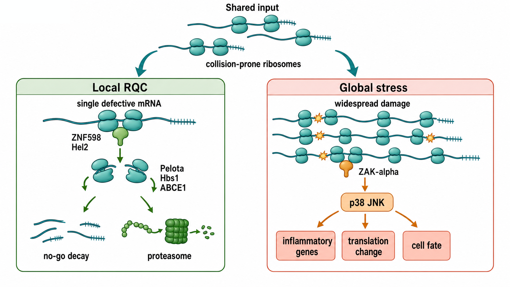
Figure 71.2. Local Quality Control Versus Ribotoxic Stress. Ribosome perturbation can be resolved locally by quality-control pathways or interpreted globally as ribotoxic stress. When a single defective mRNA generates collisions, RQC and no-go decay degrade the nascent chain and mRNA while sparing the rest of the transcriptome. When collisions or ribosome damage are widespread, ZAK-alpha activates p38 and JNK signaling, driving inflammatory gene expression, altered translation, and cell death decisions. These outputs share upstream collision-prone ribosome states but diverge in sensors, effectors, and cellular consequences.
The key concept is that ribosomes can signal before or alongside quality-control disposal. If a cell experiences widespread UV damage, nuclease-generated RNA breaks, toxin exposure, or severe translation stress, many ribosomes may pause or collide. ZAK-alpha senses ribosome-associated perturbations and activates kinase cascades that alter transcription, translation, apoptosis, inflammation, and repair programs. This output is different from degrading one bad mRNA. It is a cell-state decision that asks whether the translation apparatus as a whole is under attack.
The upstream ownership boundary is equally important. Chapter 70 explains how stress kinases regulate translation initiation through eIF2α phosphorylation and eIF2B, and how mTORC1 regulates cap-dependent recruitment through 4E-BPs and eIF4F. This chapter begins when elongating ribosomes pause, collide, or suffer damage and follows GCN1, ZNF598, ZAK-alpha, rescue, decay, and nascent-chain quality control. The pathways can converge because reduced initiation can limit new collision formation and because collisions can activate GCN2-linked integrated stress signaling, but eIF2α phosphorylation is not evidence for ZAK-alpha activation, and p38/JNK activation is not evidence for a specific initiation defect.
Recent primary studies in the supplied bibliography illustrate the range of signals. Xi et al. (2024) reported that RNase L, an innate-immunity endoribonuclease activated during antiviral responses, can generate conditions that initiate ZAK-alpha-dependent ribotoxic stress. RNase L cleaves RNA during antiviral defense; cleavage products and interrupted translation can create ribosome states that are interpreted as ribotoxic. Sinha et al. (2024) linked the ribotoxic stress response to UV-mediated cell death, connecting ribosome perturbation with genotoxic or phototoxic stress outcomes. These studies show why ribotoxic stress belongs in an RNA chapter: RNA cleavage, mRNA translation, ribosome state, and inflammatory signaling are mechanistically connected.
Inflammation enters through several routes. First, innate immune pathways can produce RNA damage or translation inhibition that activates ribotoxic signaling. Second, ribotoxic signaling can induce inflammatory gene-expression programs. Third, unresolved translation stress can promote cell death, releasing damage-associated signals. Macrophages and other immune cells interpret such signals in tissue-specific ways. The references available for macrophage inflammation and tumor microenvironment are broad inflammation reviews rather than ribosome-specific anchors (Malainou et al. 2023; Niu and Zhou 2023; Li et al. 2018). They are useful for biological context but should not be treated as direct support for detailed ZAK-alpha mechanisms; recent ZAK-alpha-focused reviews and primary structural papers remain final-reference-expansion items before final publication.
Ribotoxic stress should not be conflated with the integrated stress response. The integrated stress response centers on eIF2-alpha phosphorylation by kinases such as GCN2, PKR, PERK, and HRI, reducing global initiation while allowing selective translation of stress-responsive mRNAs. Ribotoxic stress often signals through ZAK-alpha and p38/JNK. The pathways can interact because collision load, amino-acid limitation, viral double-stranded RNA, ER stress, and oxidative stress can occur together, but the sensors and outputs are not identical. A student misconception is to treat “translation stress” as one pathway. In practice, translation stress is a family of sensor-output modules.
Experimental interpretation is complicated by translation inhibitors. Some drugs freeze ribosomes, some cause miscoding, some block translocation, and some damage ribosomal RNA or ribosome-associated factors. Different inhibitors can produce similar reductions in protein synthesis but distinct collision patterns and kinase outputs. Therefore, evidence for ribotoxic stress should include pathway dependence on ZAK-alpha or downstream kinases, measurement of ribosome perturbation, and controls distinguishing global translation inhibition from collision-specific signaling.
71.5. Proteostasis coupling and stress granules
Proteostasis is the maintenance of protein homeostasis. It includes accurate synthesis, cotranslational folding, chaperone buffering, organelle targeting, complex assembly, ubiquitin-proteasome degradation, autophagy, and stress responses. Ribosome collisions threaten proteostasis because they generate incomplete nascent chains and reduce the availability of ribosomes for normal protein production. RQC connects the translation apparatus to the proteostasis network by converting stalled nascent chains into degradation substrates.
The connection is easiest to see at the ribosomal exit tunnel. A nascent chain emerging from the tunnel may begin folding before translation is complete. Chaperones can bind the nascent chain, membrane-targeting factors can recognize signal sequences, and modifying enzymes can act cotranslationally. If the ribosome stalls, the nascent chain remains tethered in a partially synthesized state. Some incomplete products are harmless after degradation, but others can aggregate, insert incorrectly into membranes, or sequester chaperones. RQC therefore protects not only the mRNA and ribosome but also the protein-folding environment.
Stress granules add a second layer of coupling. Stress granules form when translation initiation is inhibited and untranslated messenger ribonucleoproteins accumulate with RNA-binding proteins. G3BP1 is a central stress-granule nucleator in many mammalian contexts. The G3BP family binds RNA and protein partners, and RNA-induced conformational changes and clustering can drive stress-granule assembly (Guillén-Boixet et al. 2020). Stress granules are dynamic assemblies rather than static storage depots. Their composition changes with stress type, cell type, time, and measurement method. Reviews by Hofmann et al. (2021), Youn et al. (2019), and Kiebler and Bauer (2024) emphasize that assembly, disassembly, and disease relevance depend on regulated multivalent interactions.
The relationship between ribosome collisions and stress granules is not simply that collided mRNAs enter granules. Ribosomes and stress granules usually mark different translation states. Stress granules are enriched for mRNAs not actively engaged in elongating ribosomes, whereas collided mRNAs are ribosome-associated. Helton et al. (2025) reported that ribosome association can inhibit stress-induced gene mRNA localization to stress granules. This finding gives a useful rule of thumb: an mRNA’s ribosome occupancy influences whether it is routed to translation, surveillance, decay, or granule partitioning.
P-bodies, which are cytoplasmic ribonucleoprotein assemblies enriched in decay factors and translationally repressed mRNAs, also interact with stress granules. DDX6 can modulate P-body and stress-granule assembly, composition, and docking (Ripin et al. 2024). These observations support a view in which mRNAs move through multiple ribonucleoprotein states rather than a binary translated-versus-degraded fate. A stressed mRNA may be repressed, stored, decapped, cleaved, returned to translation, or degraded, depending on binding proteins, poly(A)-tail status, modifications, localization, and ribosome state.
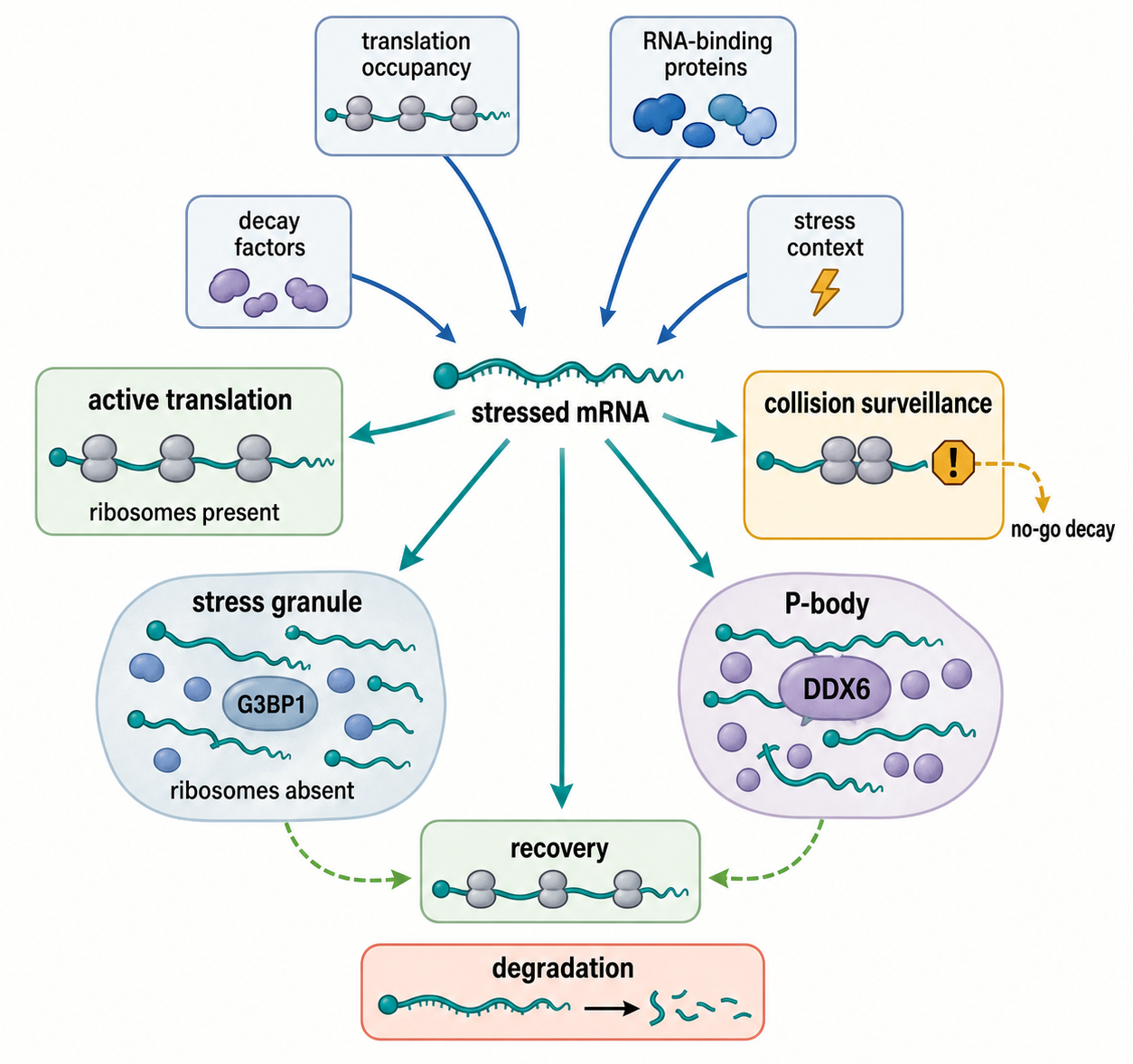
Figure 71.3. mRNA Routing During Translation Stress. Translation status, RNA-binding proteins, decay factors, and stress context collectively determine whether an mRNA is actively translated, subjected to collision surveillance and no-go decay, stored in a stress granule, targeted to a P-body, or degraded. Ribosome association can prevent some stress-induced mRNAs from entering stress granules, so an mRNA’s routing depends not only on the nature of the stress but also on its current translation occupancy and bound protein partners.
Proteostasis coupling also matters in aging. Lee et al. (2025) reported that Pelota-mediated ribosome-associated quality control counteracts aging and age-associated pathologies across species. The broad implication is that the ability to rescue ribosomes and clear aberrant translation products may decline or become limiting in aging tissues. Because aging involves many changes in proteasome activity, autophagy, mitochondrial function, inflammation, and translation, RQC should be treated as one component of a network rather than a single aging cause.
Artifact control is especially important in granule studies. Overexpression of fluorescent RNA-binding proteins can nucleate nonphysiological assemblies. Fixation can alter condensate appearance. Arsenite, heat shock, viral infection, and proteasome inhibition create different stress states. A punctum containing G3BP1 is not automatically a canonical stress granule with a defined functional output. Likewise, loss of a visible stress granule does not prove loss of translational repression or mRNA decay. Strong claims require orthogonal measurements: imaging, biochemical fractionation, translation status, RNA identity, perturbation, and recovery dynamics.
71.6. Viral, bacterial, and organellar collision biology
Viruses create unusual translation environments. Viral RNAs can contain structured elements, programmed frameshift sites, internal ribosome entry sites, overlapping open reading frames, and replication-associated RNA structures. Host antiviral responses can also cleave RNA, inhibit initiation, deplete tRNAs, activate PKR, or alter ribosome availability. These conditions can promote ribosome pausing or collision, but viruses may exploit some pauses for recoding or evade quality control. Chapter 69 covers viral recoding, and Chapter 141 covers RNA viruses in detail. In this chapter, the important point is that viral infection can both create collision substrates and activate host pathways that interpret ribosome stress as innate immune danger.
The available references include broad antiviral and innate-immunity reviews (Leite et al. 2022; Marques et al. 2024; Cui et al. 2024), but they are not sufficient for detailed claims about specific viral collision mechanisms. The RNase L and ZAK-alpha study by Xi et al. (2024) provides a stronger bridge because it directly connects an antiviral endoribonuclease to ribotoxic stress. Virus-specific primary references on ribosome collisions at frameshift elements, viral antagonism of RQC, and host collision sensing during infection remain final-reference-expansion items.
Bacterial collision biology is related but uses different molecular players. Bacteria have rescue systems such as trans-translation by transfer-messenger RNA and SmpB, alternative rescue factors, and ribosome hibernation factors. These systems release stalled ribosomes and tag incomplete proteins for degradation. Bacterial cells also sense collided ribosomes through factors that are not direct copies of eukaryotic RQC components. Cerullo et al. (2022) identified bacterial ribosome collision sensing by a MutS DNA repair ATPase paralogue, showing that collision sensing is not exclusively eukaryotic and can be coupled to bacterial stress physiology. This finding is conceptually important because MutS-family proteins are better known for DNA mismatch repair; a paralogue recognizing collided ribosomes demonstrates evolutionary repurposing of ATPase modules for translation surveillance.
Bacterial translation differs from eukaryotic cytosolic translation in ways that shape collision biology. Transcription and translation are coupled in many bacteria, so a stalled ribosome can influence RNA polymerase behavior, mRNA structure, and Rho-dependent termination. Bacterial mRNAs are often polycistronic, so a problem in one open reading frame can affect downstream translation. Antibiotics can create ribosome stalls with drug-specific signatures. These differences mean that “RQC” should not be used as if the mammalian LTN1-NEMF pathway exists unchanged in bacteria. The conserved problem is stalled ribosome rescue; the factors and outputs differ.
Organelles add another boundary case. Mitochondria and chloroplasts have translation systems derived from bacteria but reshaped by endosymbiosis, genome reduction, organelle-specific ribosomes, and nuclear-encoded factors. Organellar mRNAs often have unusual leaders, editing patterns, polyadenylation behavior, and translation factors. Stalled organellar ribosomes could threaten respiratory or photosynthetic complex assembly, but the detailed collision-sensing pathways are less well established in the provided bibliography. Chapter 17 covers organellar RNA biology, and Chapter 75 covers organelle-associated translation. Organellar references on mitochondrial and chloroplast ribosome rescue, collision detection, and nascent-chain quality control remain final-reference-expansion items.
The comparative lesson is that ribosome collision is a physical problem shared by translating systems, while the quality-control solution is lineage-specific. Eukaryotic cytosol emphasizes ubiquitin, Pelota/Hbs1/ABCE1, LTN1, NEMF, and proteasomal degradation. Bacteria emphasize tmRNA-SmpB and other rescue systems, with newly appreciated collision sensors. Organelles use bacterial-like ribosomes but eukaryotic cellular integration. Viruses create and manipulate all of these settings.
71.7. Disease links and therapeutic implications
Disease links arise when collision formation is excessive, collision resolution is defective, or stress signaling is miscalibrated. Because RQC intersects with proteostasis, neurons are a recurring concern. Neurons are long-lived, polarized cells with high demands for local translation, proteome maintenance, and stress recovery. Defects in ribosome rescue or nascent-chain degradation could contribute to accumulation of incomplete or aggregation-prone proteins. The supplied bibliography includes broad reviews of RNA helicases and neurological RNA-binding proteins (Bohnsack et al. 2023; Tilliole et al. 2024), but it lacks a full disease-focused RQC review. Disease claims in this chapter should therefore remain mechanistic and cautious unless supported by added references.
Ribosome dysfunction is also relevant to developmental disorders, ribosomopathies, cancer, and inflammatory disease. A cell with impaired ribosome biogenesis, altered tRNA pools, oncogenic translation programs, or high secretory load may operate closer to collision thresholds. Cancer cells often have high translation demand and proteotoxic stress, making translation quality control a potential vulnerability. However, broad cancer inflammation references do not prove that a particular tumor depends on RQC. Therapeutic claims need direct evidence from genetic dependency, drug sensitivity, or patient data. Chatterjee et al. (2024) provides a concrete example: RQC mitigates cytotoxicity from ribosome collisions induced by 5-fluorouracil, a widely used chemotherapeutic. This suggests that drug-induced translation stress and RQC capacity can influence treatment response or toxicity.
Ribotoxic stress has therapeutic implications because ZAK-alpha and downstream p38/JNK signaling can shape cell death, inflammation, and tissue injury. Inhibiting ribotoxic signaling might reduce harmful inflammation or toxicity in some settings, but it could also blunt protective responses to infection or damaged translation. Conversely, increasing ribosome collision or disabling RQC might sensitize cancer cells to chemotherapy, but such strategies risk harming normal tissues with high translation demand. The right therapeutic question is not whether collisions are good or bad. The question is which cell type, stress, pathway node, and time window should be modulated.
Antibiotics and antiviral therapies create additional possibilities. Many antibiotics target bacterial ribosomes and can induce stalls, miscoding, or rescue stress. Understanding bacterial collision sensing may help explain drug synergy, persistence, or stress adaptation. Viral therapies and antiviral immunity can produce RNA cleavage, translation inhibition, and ribotoxic signaling. Oncolytic viruses and RNA-targeting innate immune pathways may therefore intersect with collision biology, but the current chapter bibliography is too broad for detailed therapeutic design claims. Targeted antibacterial and antiviral collision-biology references should be added before expanding this subsection.
mRNA therapeutics provide a forward-looking application. Codon optimization, nucleotide modification, untranslated-region design, poly(A)-tail length, purity, and delivery all affect translation. A collision-aware design framework would avoid extreme initiation-elongation imbalance, strong coding-region obstacles, problematic nascent peptides, and immune-triggering RNA damage. Yet maximal protein output is not the only goal. A therapeutic mRNA may require controlled duration, tissue specificity, low innate immune activation, and acceptable manufacturing properties. Collision biology should therefore be integrated with the broader mRNA architecture principles in Chapter 72 and therapeutic design principles in later RNA therapeutics chapters.
Box 71.2. Therapeutic Opportunities and Risks
- Increasing ribosome collision load — for example by codon deoptimization or translation-stalling drugs — may sensitize cancer cells to chemotherapy, but high-translation normal tissues such as intestinal epithelium face similar risk.
- Reducing ribotoxic stress by inhibiting ZAK-alpha or downstream p38/JNK signaling might limit inflammatory tissue injury; however, the same pathway contributes to antiviral defense and detection of genotoxic damage.
- Collision-aware design of therapeutic mRNAs, avoiding extreme initiation–elongation imbalance and problematic nascent peptides, is a rational goal, but direct engineering evidence linking codon choices to collision burden in therapeutic contexts remains limited.
Recent Consensus
The current consensus is that ribosome collisions are biologically meaningful signals rather than incidental traffic jams. Collided ribosomes expose composite surfaces that recruit surveillance factors, and collision sensing helps cells distinguish persistent translation defects from ordinary pauses. Eukaryotic RQC couples small-subunit collision sensing to large-subunit nascent-chain quality control, mRNA decay, and proteostasis. No-go decay, non-stop decay, and RQC overlap but should not be collapsed into one pathway. ZAK-alpha-dependent ribotoxic stress is now recognized as a major bridge between ribosome perturbation, stress kinase signaling, cell death, and inflammation. Stress granules and P-bodies are dynamic mRNP states whose relationship to collision biology depends on ribosome occupancy and stress context.
The consensus is strongest for yeast and mammalian cytosolic RQC mechanisms, for collision-dependent ribosome ubiquitination, for the importance of Pelota/Hbs1/ABCE1-like rescue, and for the broad proteostasis role of nascent-chain degradation. The consensus is weaker for exact endogenous substrate repertoires, organism-wide collision thresholds, organellar collision pathways, viral manipulation of RQC, and therapeutic exploitation.
Open Questions, Controversies, Deprecated Models, and Common Misconceptions
Open questions:
- What determines how cells set collision thresholds? A single transcript with strong collisions may be handled locally, whereas a global increase in collisions can activate stress signaling. The molecular parameters that determine this switch include initiation rate, stall duration, ribosome density, sensor abundance, ubiquitination dynamics, mRNA localization, and proteasome capacity.
- What determines how endogenous substrates differ from artificial reporters? Reporters have revealed the pathway, but natural mRNAs may produce smaller, transient, or context-specific collision signals. Endogenous substrates may also be enriched in particular locations, such as the endoplasmic reticulum, neuronal processes, viral replication compartments, or organelle surfaces.
Controversies:
- A third controversy concerns the functions of CAT tails and related nascent-chain modifications. CAT tailing can promote degradation, but it can also create aggregation-prone products when downstream quality control fails. The safest statement is context-dependent rather than universal.
Common misconceptions:
- “A ribosome profiling peak proves a ribosome collision.” Peaks can reflect pauses, queues, nuclease bias, mapping ambiguity, inhibitor artifacts, or altered initiation; collision claims require pathway-consistent evidence.
- “Every translational pause is pathological.” Pauses can be programmed, neutral, regulatory, or artifact-prone; pathology requires evidence for harmful consequence.
- “Stress granules are generic containers for all stressed mRNAs.” Stress granule membership is selective, dynamic, and condition-dependent.
- “Ribotoxic stress is identical to the integrated stress response.” These pathways can intersect, but they have distinct sensors, triggers, and outputs.
- “Bacterial rescue systems are mammalian RQC with different names.” Bacterial rescue and mammalian ribosome quality control solve related problems with different factors, substrates, and evolutionary histories.
- “Reduced translation means ribosome quality control is activated.” Translation can fall through initiation control, elongation slowing, stress signaling, toxicity, or RNA loss without activating a specific quality-control pathway.