Chapter 122. RNA Extraction, Preservation, Fractionation, Quantification, Spike-Ins, and Sample QC
Scope Note
This chapter explains how RNA samples are collected, stabilized, purified, separated into useful fractions, measured, controlled for contamination, and qualified for downstream assays. Sample handling is part of RNA biology rather than a neutral prelude to measurement. The practical goal is fit-for-purpose RNA: RNA whose composition, contaminants, size distribution, controls, and metadata fit the biological question and assay chemistry.
Executive Summary
RNA experiments measure a sample history as well as a biological state. After a specimen is removed from its native environment, transcriptional responses, nuclease activity, cell rupture, ischemia, fixation chemistry, freeze-thaw damage, adsorption to plastic, and extraction bias can alter which molecules are recovered. A high-quality RNA experiment begins with a controlled transition from biological state to molecular stabilization.
RNA integrity means that the molecules remain usable for the intended assay, not that every RNA in the sample is full-length. An RNA integrity number (RIN) or related electrophoretic score summarizes ribosomal RNA peak patterns in many total RNA samples, but this metric is not universal. Degraded formalin-fixed paraffin-embedded (FFPE) RNA may be acceptable for short hybrid-capture or amplicon assays when its DV200, the fraction of RNA fragments longer than 200 nucleotides, meets assay requirements. Conversely, an apparently intact total RNA trace can still fail a small RNA, long-read isoform, direct RNA sequencing, or RNA modification workflow if the relevant molecules are lost, modified by handling, contaminated, or chemically incompatible with library preparation.
Extraction chemistry is a controlled partitioning problem. Chaotropic salts and detergents disrupt cells and ribonucleoprotein particles, denature proteins, and suppress ribonucleases. Acid phenol-chloroform workflows separate RNA from many proteins, lipids, and DNA through phase behavior. Silica columns and magnetic beads bind nucleic acids under high-salt and alcohol-rich conditions, then release them during elution. These workflows are not neutral. They differ in recovery of short RNAs, structured RNAs, modified RNAs, extracellular carrier-associated RNAs, inhibitors, DNA, phenol, salts, and low-abundance targets.
Fractionation, enrichment, and depletion define the RNA population being measured. Poly(A) selection enriches many mature eukaryotic mRNAs but loses nonpolyadenylated RNAs, many bacterial transcripts, some viral RNAs, and degraded molecules lacking accessible tails. Ribosomal RNA depletion enables broader total-RNA profiling but depends on probe design, organism, fragmentation state, and sample mixture. Size selection can enrich miRNAs or other short RNAs, but it also captures degradation fragments and excludes molecules whose termini, modifications, or structures are incompatible with adapters or enzymes.
Quantification and QC are multidimensional. Absorbance can reveal some contaminants but overestimates RNA in impure or low-input samples. Fluorescent assays are usually more RNA-specific but do not describe size or sequence representation. Capillary electrophoresis shows fragment distribution, but summary scores must be matched to the assay. Spike-ins, DNase treatment, extraction blanks, no-template controls, no-reverse-transcriptase controls, replicate extractions, and batch metadata are complementary safeguards, not interchangeable rituals.
Concept Inventory
- Sample pre-analytics: the collection, handling, preservation, storage, transport, thawing, and processing variables that occur before analytical measurement. Pre-analytics can change RNA abundance, integrity, chemical state, and extractability.
- Preservation: stabilization of RNA composition by freezing, direct lysis, chemical stabilization, dehydration, or fixation. Preservation slows change but can also introduce assay-specific artifacts.
- RNA integrity: the retention of useful RNA length and chemical compatibility for a defined assay. Integrity is distinct from concentration, purity, and absence of DNA.
- RIN: RNA integrity number, an algorithmic score derived from electrophoretic features of many total RNA traces, especially ribosomal RNA peaks. RIN is useful in compatible contexts but not a universal quality score.
- DV200: the percentage of RNA fragments longer than 200 nucleotides. DV200 is often useful for degraded and FFPE RNA because many library and targeted assays require fragments above a practical length threshold.
- Extraction chemistry: the combined lysis, denaturation, partitioning, binding, washing, elution, cleanup, and concentration steps used to separate RNA from biological matrix and contaminants.
- Fractionation: deliberate separation of RNA-containing material by compartment, size, molecular partner, biochemical feature, or functional state.
- Enrichment and depletion: selective increase or reduction of a target RNA class, such as poly(A) RNA enrichment, rRNA depletion, small RNA selection, circular RNA enrichment, or cap-dependent selection.
- Spike-in: an exogenous molecule added at a known amount to monitor recovery, inhibition, calibration, dynamic range, or batch behavior. A spike-in reports only on steps that occur after addition and only approximates targets with similar behavior.
- Extraction blank: a no-biological-input sample processed through extraction. Extraction blanks detect reagent, plasticware, environmental, aerosol, and handling contaminants.
- No-reverse-transcriptase control: a control reaction lacking reverse transcriptase, used to test whether signal can arise from DNA rather than cDNA.
- Batch effect: technical variation associated with processing date, operator, lot, instrument, storage time, extraction workflow, library preparation, sequencing run, or other nonbiological variable.
What to Know Before Reading This Chapter
RNA is a negatively charged polymer with a ribose-phosphate backbone, chemically diverse bases, frequent protein partners, and often strong secondary or tertiary structure. Many RNAs are not naked molecules in cells. mRNAs assemble with cap-binding proteins, exon junction complexes, poly(A)-binding proteins, translation factors, decay factors, and ribosomes. rRNAs and tRNAs are highly structured and modified. Small RNAs may be protein-bound or chemically modified at their termini. Viral RNAs may be packaged in virions or replication complexes. Extracellular RNAs may be inside vesicles, bound to ribonucleoproteins, associated with lipoproteins, or adsorbed to surfaces.
The running examples in this chapter are a fresh frozen tumor RNA-seq study, a cultured-cell qRT-PCR experiment, a bacterial transcriptome experiment, a plasma extracellular RNA biomarker study, an FFPE targeted panel, and a long-read isoform sequencing project. These examples show why the same phrase, “good RNA,” is incomplete. A short qRT-PCR amplicon may tolerate partial fragmentation, while full-length isoform sequencing requires long molecules. A plasma miRNA assay may be dominated by hemolysis or reagent background, while a bacterial RNA-seq assay may fail because the extraction did not lyse the relevant taxa or because a poly(A)-based workflow excluded most bacterial mRNAs.
Formal reference curation is required before the chapter is reference-complete.
122.1. Sample Collection and Preservation
Sample collection is the first experimental perturbation in an RNA study. A specimen has a biological state in its living context, and then it has a handling history. The interval between those states is not empty time. In an excised tissue, oxygen tension, nutrient supply, temperature, mechanical stress, immune signaling, and nuclease exposure change rapidly. In a blood tube, leukocytes can activate, platelets can release material, erythrocytes can lyse, and extracellular vesicle populations can shift. In a bacterial culture, centrifugation, temperature change, and time in spent medium can alter stress-response transcripts. Preservation is the attempt to stop, slow, or standardize these changes before measurement.
The main vocabulary is pre-analytical variation. Pre-analytical variables include the specimen source, time of collection, dissection or sampling method, warm ischemia time, cold ischemia time, stabilization reagent, storage temperature, freeze-thaw count, tube type, anticoagulant, transport conditions, and lysis delay. Warm ischemia is the interval in which a tissue remains at physiological or room temperature after blood supply or native growth conditions are interrupted. Cold ischemia begins when the specimen is cooled but not yet fully stabilized. These intervals can matter because cellular enzymes and stress pathways do not all stop at the same moment.
The strongest general preservation principle is rapid nuclease inactivation or rapid cooling. Direct lysis in a chaotropic reagent can be ideal for cultured cells when the reagent volume, cell number, and downstream extraction chemistry are compatible. Chaotropic reagents such as guanidinium thiocyanate denature many proteins, including many ribonucleases, and disrupt ribonucleoprotein assemblies. Snap freezing in liquid nitrogen or on a deeply chilled surface is common for tissues because it arrests many biochemical processes while preserving material for later extraction. Small pieces freeze and stabilize faster than large pieces, so specimen thickness and surface area affect preservation quality.
Chemical stabilization reagents provide a field- and clinic-friendly alternative when immediate freezing or extraction is not possible. These reagents generally work by permeating tissue, denaturing proteins, reducing water activity, or changing chemical conditions so RNA turnover and RNase activity slow. They are not universal preservatives. A reagent optimized for cellular transcript abundance may not be appropriate for extracellular vesicle RNA, RNA-protein interaction mapping, native ribonucleoprotein recovery, or direct RNA sequencing. Stabilization reagents can also impose extraction constraints, because salts, polymers, alcohols, or proprietary components may need removal before binding, reverse transcription, ligation, or sequencing.
Fixation is preservation by chemical immobilization rather than by keeping RNA native. Formalin fixation and paraffin embedding preserve histology and allow retrospective molecular studies from pathology archives. The tradeoff is that fixation fragments RNA, creates crosslinks, can modify bases, and makes extraction more difficult. FFPE RNA often supports targeted assays, short-fragment RNA-seq, fusion detection, and expression panels when the assay is designed for degraded input. It is a poor substitute for fresh frozen RNA when the question requires full-length transcripts, accurate RNA termini, native RNA-protein complexes, or long-read molecule continuity.
Blood, plasma, serum, and extracellular fluid samples deserve special caution. Whole blood contains abundant cellular RNA, and even small degrees of hemolysis can add erythrocyte-associated RNA species to plasma or serum. Serum is not simply plasma without anticoagulant; clotting changes the extracellular RNA environment by activating platelets and coagulation pathways. Anticoagulants, tube additives, centrifugation delay, residual cells, platelet carryover, and storage temperature can all change measured RNA profiles. A plasma miRNA biomarker study that ignores hemolysis and platelet handling may measure phlebotomy and processing rather than disease biology.
Plant, microbial, and environmental specimens introduce matrix-specific preservation problems. Plant tissues may contain polyphenols, polysaccharides, pigments, and secondary metabolites that oxidize or co-purify with RNA. Bacteria and fungi differ in envelope strength, so some cells remain intact during mild handling while others lyse and release nucleases. Stool, soil, marine, and biofilm samples contain inhibitors and mixed taxa whose activity may continue after sampling. For mixed communities, preservation is ecological sampling: the workflow must prevent some organisms from changing or lysing more than others.
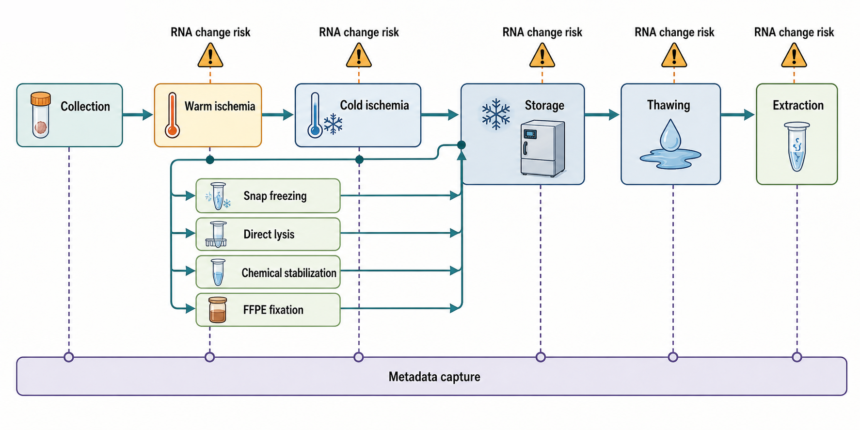
Figure 122.1. Sample Handling Timeline and Preservation Decision Points. Makes pre-analytical handling visible as an experimental variable rather than an administrative note.
The evidence basis for preservation claims comes from matched-sample comparisons, time-course handling studies, biobank validation, and downstream assay performance. A well-designed preservation comparison uses adjacent tissue pieces or matched aliquots, varies one handling parameter, and measures both global RNA quality and specific transcript changes. Common weaknesses are small sample numbers, one tissue type generalized to all tissues, and metrics limited to total yield or RIN. A useful preservation protocol therefore records enough metadata to interpret failures: collection time, stabilization time, temperature history, storage duration, thawing method, freeze-thaw count, tissue mass, reagent ratio, and protocol deviations.
Do not overgeneralize labels such as “fresh,” “frozen,” “stabilized,” or “archival.” A fresh sample can be transcriptionally perturbed if it sat warm for an hour. A frozen sample can be poor if freezing was slow or thawing occurred repeatedly. A stabilized sample can be incompatible with native complex recovery. An archival FFPE block can be scientifically valuable if the assay and QC thresholds match the fragmented material. Preservation quality is a relationship between handling history, RNA class, and assay design.
122.2. RNA Integrity and Degradation Assessment
RNA degradation is the shortening, chemical alteration, or loss of RNA molecules after their biological state of interest. Degradation can occur through endogenous RNases, alkaline hydrolysis, metal-catalyzed cleavage, heat, oxidative damage, fixation chemistry, mechanical shearing, and repeated freeze-thaw cycles. The vulnerable feature of RNA chemistry is the 2′ hydroxyl group on ribose, which can participate in backbone cleavage under unfavorable chemical conditions. In biological specimens, however, enzymatic ribonuclease activity and handling history usually dominate over spontaneous chemical hydrolysis.
RNA integrity is not the same as purity. A preparation can contain intact RNA and still be contaminated with DNA, protein, phenol, salts, ethanol, guanidinium, heme, polysaccharides, humic acids, or carrier polymer. RNA integrity is also not the same as biological quality. A sample may preserve long rRNAs while a stress response altered mRNA abundance before extraction. Conversely, a partially fragmented sample can still be useful when the assay reads short regions or uses random priming. The term “RNA quality” should therefore be unpacked into integrity, concentration, purity, inhibitor burden, DNA contamination, representation of target RNA classes, and fit for the intended assay.
Electrophoretic profiling is the most common way to assess RNA size distribution. In capillary electrophoresis or microfluidic electrophoresis, RNA fragments migrate by size and generate an electropherogram. For many eukaryotic total RNA preparations, strong 18S and 28S ribosomal RNA peaks, a low baseline of small degradation products, and a consistent size distribution suggest relatively intact bulk RNA. RIN and related instrument-specific metrics convert features of the electropherogram into a summary score. This score is convenient because it is reproducible within compatible sample types and easy to use in acceptance criteria.
The limitation is that RIN is trained on a particular view of RNA integrity. It is most interpretable for total RNA from organisms and preparations with recognizable rRNA profiles. It becomes less meaningful after rRNA depletion, poly(A) selection, small RNA enrichment, deliberate fragmentation, FFPE extraction, extracellular RNA isolation, or strong size selection. Bacterial RNA, plant RNA, organellar RNA, nonmodel organism RNA, and viral RNA preparations can produce traces that do not map neatly to mammalian total RNA expectations. A low RIN may be acceptable for an FFPE targeted panel; a high RIN does not guarantee that miRNAs, full-length mRNAs, modified tRNAs, or rare viral RNAs were recovered.
DV200 is a different kind of metric. It asks what fraction of RNA signal comes from fragments longer than 200 nucleotides. This is useful for degraded and FFPE samples because many library preparation and hybrid-capture workflows have practical fragment-length requirements. A high DV200 does not prove that specific transcripts are represented, that RNA termini are usable, or that inhibitors are absent. It simply provides a fragment-size qualification that is often better matched to degraded-input assays than ribosomal peak integrity.
Table 122.1. RNA Integrity and Degradation Metrics. Helps readers avoid using one metric as a universal RNA quality score.
| Metric | What it measures | Best-fit sample types | Misleading contexts | Downstream decisions it can support |
|---|---|---|---|---|
| RIN | Electrophoretic summary of ribosomal peak and degradation features. | Compatible total RNA with recognizable rRNA profiles. | FFPE, depleted, selected, small RNA, extracellular, bacterial, nonmodel, or fragmented input. | Set intact-input thresholds and flag handling or degradation risk. |
| RQN-like scores | Instrument-specific electrophoretic quality score. | Within-platform total RNA QC and internal acceptance criteria. | Cross-platform comparison or samples with nonstandard rRNA traces. | Track run consistency and apply platform-specific QC cutoffs. |
| Electropherogram inspection | Full fragment-size profile, rRNA peaks, smear, and library artifacts. | Total RNA, degraded RNA, small RNA fractions, and libraries. | Interpreted without assay context or below instrument sensitivity. | Diagnose degradation pattern, adapter dimers, unexpected fragments, or size-selection failure. |
| DV200 | Fraction of RNA signal above 200 nucleotides. | FFPE, degraded RNA, targeted panels, and hybrid-capture workflows. | Specific transcript survival, inhibitors, DNA, termini, or long-read suitability. | Choose degraded-input library strategy and practical target or insert size. |
| Gene-body bias | Positional coverage skew across transcript bodies after sequencing. | RNA-seq libraries where coverage should span transcripts. | 3′ tag designs, annotation artifacts, priming bias, or isoform shifts. | Identify degradation, reverse-transcription bias, or library-construction failure. |
| Target amplicon success | Amplification of a defined RNA-derived target region. | qRT-PCR, clinical panels, FFPE assays, and pathogen tests. | Poor primer design, absent target, DNA contamination, or regulation mistaken for integrity. | Confirm amplifiable length, inhibitor control behavior, and target-specific input fitness. |
| Library fragment distribution | Insert-size and artifact profile after library construction. | Sequencing libraries before pooling or sequencing. | Treated as original RNA integrity rather than library QC. | Decide cleanup, pooling, size selection, and adapter-dimer exclusion. |
Box 122.1. When an Integrity Score Is the Wrong Question
Key rule: use an integrity metric as a hypothesis about assay performance, not as a verdict. Ask first what molecules the assay must read. If the assay depends on full-length polyadenylated mRNAs, then fragment distribution, gene-body coverage, and gentle long-molecule handling matter. If the assay reads short FFPE amplicons, DV200 and target amplicon success may be more relevant than ribosomal peak shape. If the assay measures small RNAs, extracellular RNAs, modified tRNAs, or deliberately fragmented libraries, a ribosomal-RNA-based score can be weak or irrelevant. Interpret each metric with three linked checks: the physical size range required by the assay, the RNA class represented by the metric, and downstream performance evidence. A high score should not override failed controls, inhibition, DNA contamination, or loss of the target fraction. A low score should not automatically discard a sample when the intended readout uses short, validated targets.
Integrity interpretation should be assay-specific. A short-amplicon qRT-PCR assay may only require that the target region survives and that inhibitors and DNA are controlled. A 3′ tag RNA-seq assay may tolerate degradation if poly(A) tails and nearby sequence are intact enough for capture. A full-length cDNA or long-read direct RNA sequencing assay requires much longer molecules and is vulnerable to shearing, freeze-thaw, and extraction conditions that reduce molecule length. RNA structure probing, RNA modification mapping, and RNA-protein interaction workflows may care about chemical state and native context as much as length.
Degradation can also be nonuniform. Some transcripts are more stable because of protein binding, structure, translation state, subcellular localization, or sequence features. rRNA can appear intact while labile mRNAs have changed abundance. In blood and tissue, degradation and stress responses can produce apparent biological signatures that correlate with ischemia time or handling site. In extracellular RNA, the protected fraction associated with vesicles or proteins may persist while naked RNA disappears. A degradation metric based on bulk RNA mass may miss these selective effects.
Evidence for integrity metrics comes from correlations between pre-library QC and downstream assay performance. Strong validation links a metric to library yield, mapping rate, gene-body coverage, transcript detection, fusion detection, amplicon success, or clinical assay reproducibility. Weak validation reports only that one sample has a higher score than another. For a rigorous RNA study, integrity metrics should be used as predictors of the actual assay, not as ceremonial thresholds copied from unrelated protocols.
A single number does not certify RNA quality. RIN, DV200, concentration, absorbance ratio, and library yield each report a different dimension. A fit-for-purpose decision asks whether the RNA preparation contains the intended molecules in a usable physical and chemical state, whether known contaminants are below harmful levels, whether controls behave as expected, and whether the metadata allow technical variation to be modeled.
122.3. Extraction Chemistries and Phase or Column Workflows
RNA extraction converts a complex specimen into an analyte solution. The process has five linked tasks: disrupt the sample, inactivate ribonucleases, release RNA from cells or carriers, separate RNA from unwanted molecules, and elute or resuspend RNA in a buffer compatible with the next step. Every task can introduce bias. A lysis step that fails to break Gram-positive bacteria under-represents those bacteria. A column that excludes short RNAs loses miRNAs and tRNA fragments. A harsh homogenization step can improve tissue disruption while shortening long molecules needed for isoform sequencing.
Lysis starts with the physical nature of the sample. Mammalian cultured cells often lyse readily in detergent and chaotropic buffer. Fibrous tissue, cartilage, plant tissue, fungal cells, bacterial spores, biofilms, and environmental particles require stronger disruption such as grinding, rotor-stator homogenization, bead beating, enzymatic wall digestion, or cryopulverization. Mechanical disruption must be matched to heat control because friction can warm samples. Incomplete lysis is not only lower yield; it is differential recovery of compartments, cell types, or organisms.
RNase inactivation is the defining requirement for RNA extraction. Ribonucleases are abundant, stable, and easily introduced from cells, skin, dust, contaminated reagents, or previous samples. Chaotropic salts such as guanidinium thiocyanate disrupt protein folding and hydrogen bonding, reducing many RNase activities. Detergents solubilize membranes and help release ribonucleoprotein complexes. Reducing agents, chelators, and low-temperature handling may contribute depending on protocol. These steps suppress degradation but do not make sloppy handling harmless. Dilution of chaotrope, residual aqueous pockets in tissue, or delayed mixing can leave active nucleases in contact with RNA.
Acid phenol-chloroform extraction is a classic broad RNA isolation strategy. In acid guanidinium phenol workflows, cells are lysed in a strongly denaturing solution, chloroform is added, and centrifugation separates the mixture into aqueous, interphase, and organic phases. Under acidic conditions, much RNA remains in the aqueous phase, while many proteins and lipids partition into the organic phase and much DNA is reduced in the aqueous phase relative to neutral extraction. RNA is then precipitated or further purified. This approach can recover diverse RNA classes and handle difficult matrices, but it is operator-sensitive. Disturbing the interphase increases DNA and protein carryover. Residual phenol inhibits enzymes and affects absorbance. Low-input samples may lose material during precipitation.
Silica column workflows use a different principle. Under high-salt and alcohol-rich conditions, nucleic acids bind to silica surfaces. The sample is lysed, mixed with binding buffer and alcohol, passed through a membrane, washed, and eluted in water or low-salt buffer. Columns are convenient, reproducible, and easy to combine with on-column DNase digestion. The binding conditions, membrane properties, and wash design determine what is retained. Some kits are optimized for total RNA including small RNAs, while others intentionally remove small molecules and short RNAs. Overloaded columns can clog or bind poorly. Residual ethanol, guanidinium, or salts can inhibit reverse transcriptase, polymerase, ligase, and sequencing library enzymes.
Magnetic bead workflows use particles that bind nucleic acids under defined buffer conditions or through sequence-specific capture. They are well suited to automation because magnets replace centrifugation. Bead chemistry can support total RNA purification, cleanup after enzymatic reactions, size selection, poly(A) capture, or depletion workflows. Bead ratio and buffer composition can impose size selection, especially in solid-phase reversible immobilization-like cleanups. Under-dried beads can carry ethanol into reactions; over-dried beads can reduce elution. In low-input assays, adsorption to tube walls, bead loss, and incomplete resuspension become important sources of variability.
Precipitation methods concentrate RNA or remove selected contaminants. Ethanol or isopropanol precipitation uses salt and alcohol to reduce nucleic acid solubility. Glycogen or linear acrylamide carriers can improve recovery from dilute samples, but carriers can complicate mass measurement and downstream chemistry. Lithium chloride precipitation preferentially precipitates many longer RNAs while leaving some DNA, protein, carbohydrates, and small molecules in solution; it can under-recover small RNAs. Precipitation is therefore a fractionation step as well as a cleanup step.
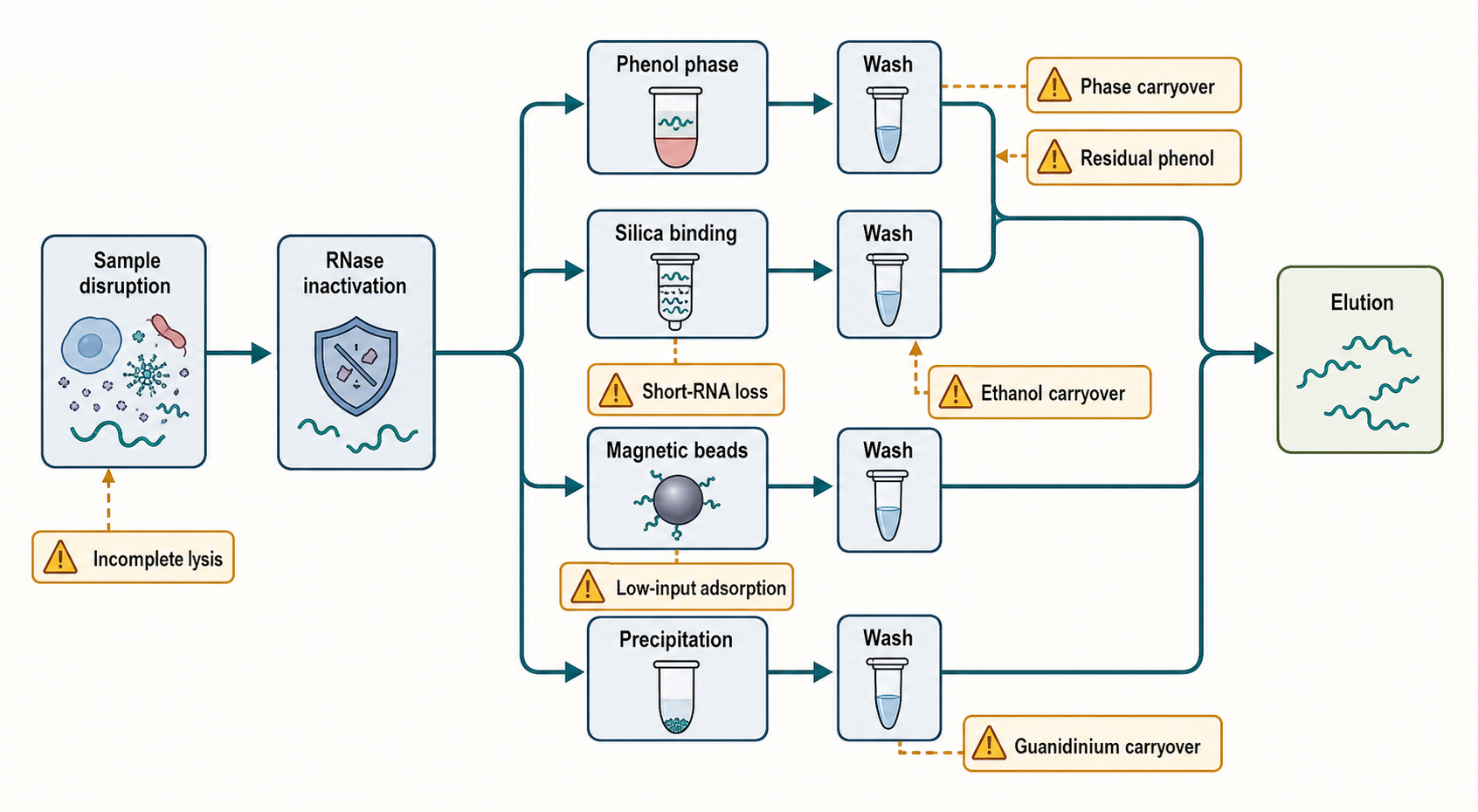
Figure 122.2. Extraction Chemistry Workflow and Failure Points. Helps readers connect chemistry to artifacts and downstream assay failure.
Extraction workflows must also remove inhibitors. Phenol, ethanol, guanidinium, heme, humic substances, polysaccharides, polyphenols, detergents, salts, and carryover proteins can reduce reverse transcription, ligation, amplification, or nanopore sequencing performance. A sample with high RNA concentration can still fail if inhibitors persist. Dilution sometimes reduces inhibition but also reduces sensitivity. Cleanup columns, beads, precipitation, or additional washes can help, but each cleanup can lose RNA or shift size distribution.
The evidence basis for extraction choices includes side-by-side protocol comparisons, recovery of synthetic standards, replicate extractions, inhibitor-spiking experiments, sequencing metrics, qRT-PCR inhibition tests, and analysis of size profiles. The most useful comparisons evaluate the intended target class rather than only total yield. For example, a protocol that maximizes total RNA from plasma may mostly recover carrier or contaminant nucleic acids, while a lower-yield workflow may better preserve reproducible miRNA signal. A long-read study should evaluate molecule length and read length distribution, not only nanograms recovered.
Table 122.2. Extraction Chemistry Comparison. Supports method choice based on target RNA and matrix rather than laboratory habit.
| Method | Chemical or physical principle | Strengths | Common losses or biases | Contaminants to monitor | Best-fit applications | Poor-fit applications |
|---|---|---|---|---|---|---|
| Acid phenol-chloroform | Acid guanidinium lysis followed by organic phase partitioning. | Broad RNA recovery and useful for difficult matrices. | Operator-sensitive interphase carryover, precipitation loss, and low-input variability. | Phenol, chloroform, guanidinium, DNA, protein, and salts. | Diverse total RNA, tissues, plants, and samples needing strong denaturation. | Enzyme-sensitive workflows without cleanup and high-throughput automation. |
| Silica column | High-salt and alcohol binding to silica, washing, and low-salt elution. | Convenient, reproducible, and compatible with on-column DNase. | Kit-dependent short-RNA loss, overload, clogging, and membrane retention. | Ethanol, guanidinium, salts, DNA, and wash carryover. | Routine total RNA for qRT-PCR, RNA-seq, and clinical workflows. | Very short RNA, ultra-long RNA, or inhibitor-rich matrices unless optimized. |
| Magnetic beads | Functionalized particles capture RNA under defined buffer conditions. | Automation-friendly, scalable, and useful for cleanup or size selection. | Bead-ratio size bias, bead loss, drying effects, and adsorption variability. | Ethanol, PEG or salt, bead carryover, and residual wash buffer. | High-throughput extraction, low-volume cleanup, poly(A) capture, and library cleanup. | Difficult lysis alone or workflows requiring size-neutral recovery without tuning. |
| Alcohol precipitation | Salt and ethanol or isopropanol reduce nucleic acid solubility. | Concentrates RNA and works as inexpensive broad cleanup. | Dilute or short RNA loss, variable pellets, and carrier-dependent recovery. | Salt, alcohol, carrier polymer, DNA, and co-precipitated inhibitors. | Concentrating moderate-input RNA after extraction or cleanup. | Ultra-low-input quantification and enzyme-sensitive reactions without thorough washing. |
| Lithium chloride precipitation | Lithium chloride preferentially precipitates many longer RNAs. | Removes some DNA, protein, carbohydrates, and small molecules. | Under-recovers small RNAs, short fragments, and degraded input. | Lithium salts, residual carbohydrates, and incomplete resuspension. | Long-RNA cleanup, plant matrices, and polysaccharide-rich samples. | miRNA, tRNA fragments, degraded FFPE RNA, and total small-RNA representation. |
| Direct lysis for qRT-PCR | Sample lysis in RT-compatible buffer without full purification. | Fast, low handling, and suitable for screening many high-input samples. | Matrix inhibition, DNA carryover, and no broad size or purity assessment. | Detergents, salts, genomic DNA, heme, media, and cell debris. | Cultured-cell qRT-PCR screens with validated controls. | RNA-seq, long-read sequencing, low-biomass samples, or inhibitor-rich tissues. |
| FFPE extraction | Deparaffinization, protease digestion, crosslink mitigation, and cleanup. | Enables archival pathology and short-fragment molecular assays. | Fragmentation, crosslinks, base damage, and fixation-age bias. | Paraffin or solvent residues, protein, salts, DNA, and inhibitors. | FFPE targeted RNA-seq, fusion panels, expression panels, and qRT-PCR. | Full-length isoforms, native RNPs, termini mapping, modifications, and long-read continuity. |
Boundary cases are common. Direct lysis without purification can work for some high-input qRT-PCR assays but leaves inhibitors and matrix effects that would be unacceptable for sequencing. FFPE extraction requires deparaffinization, reversal or mitigation of crosslinks, and acceptance of fragmentation. Viral RNA extraction may prioritize inhibitor removal and process controls over broad host RNA recovery. Microbial community extraction must balance strong lysis against shearing and inhibitor carryover. The right extraction method is the one whose known biases are compatible with the biological question and recorded in the metadata.
122.4. Fractionation, Enrichment, and Depletion Strategies
Fractionation is the deliberate narrowing of the measured RNA population. The denominator changes from “all RNA in the specimen” to “RNA that passes this physical, biochemical, or enzymatic selection.” This is often necessary because rRNA dominates mass, mRNAs may be rare, small RNAs need special handling, and subcellular localization can be central to the question. Fractionation becomes dangerous when the selected population is mislabeled as total RNA or when selection failure is treated as biology.
Subcellular fractionation separates compartments before extraction or during lysis. Nuclear, cytoplasmic, mitochondrial, chromatin-associated, ribosome-associated, membrane-associated, and extracellular fractions each require a lysis strength that releases one compartment while preserving another. Marker RNAs and proteins are essential. A nuclear fraction can be checked for pre-mRNA, small nuclear RNA, or chromatin markers, and for cytoplasmic contamination such as mature cytosolic mRNA or ribosomal components. A cytoplasmic fraction can be checked for nuclear leakage. Fraction purity is rarely absolute, so the claim should be proportional: enriched for a compartment, not a pure compartment unless convincingly demonstrated.
Poly(A) selection enriches RNAs with accessible polyadenylated tails by hybridization to oligo(dT). In mammalian and many other eukaryotic samples, this captures many mature mRNAs and reduces rRNA signal, making it efficient for coding-gene expression and isoform studies. The background concept is that many mature eukaryotic mRNAs receive a 3′ poly(A) tail during processing. The boundary is just as important: many noncoding RNAs, histone mRNAs, bacterial mRNAs, some viral RNAs, preprocessed fragments, deadenylated transcripts, and degraded molecules lacking tails are under-represented. Poly(A) selection therefore measures polyadenylated RNA, not the whole transcriptome.
Ribosomal RNA depletion removes abundant rRNAs with complementary probes, enzymatic digestion, hybrid capture, or related methods. It is often preferred for total RNA-seq, degraded RNA, bacterial RNA, noncoding RNA discovery, pre-mRNA analysis, and samples where poly(A) biology is not the target. Depletion depends on probe design and hybridization accessibility. A probe set designed for human cytoplasmic rRNAs may not remove bacterial, fungal, plant, mitochondrial, chloroplast, or divergent nonmodel-organism rRNAs efficiently. Mixed samples such as microbiomes may require broad or custom depletion. Off-target depletion can occur when probes bind unintended transcripts.
Small RNA enrichment typically combines size selection with adapter-compatible library chemistry. A microRNA workflow might select roughly 18 to 30 nucleotide molecules, while other protocols target broader short-RNA ranges. Size is not equivalent to identity. miRNAs, piRNAs, tRNA fragments, snoRNA fragments, Y RNA fragments, degradation products, and synthetic contaminants can overlap. Termini matter: many library methods require a 5′ phosphate and 3′ hydroxyl for adapter ligation. Capped RNAs, 5′ hydroxyl RNAs, 3′ phosphate RNAs, strongly modified RNAs, and protein-bound RNAs can be under-captured without pretreatment.
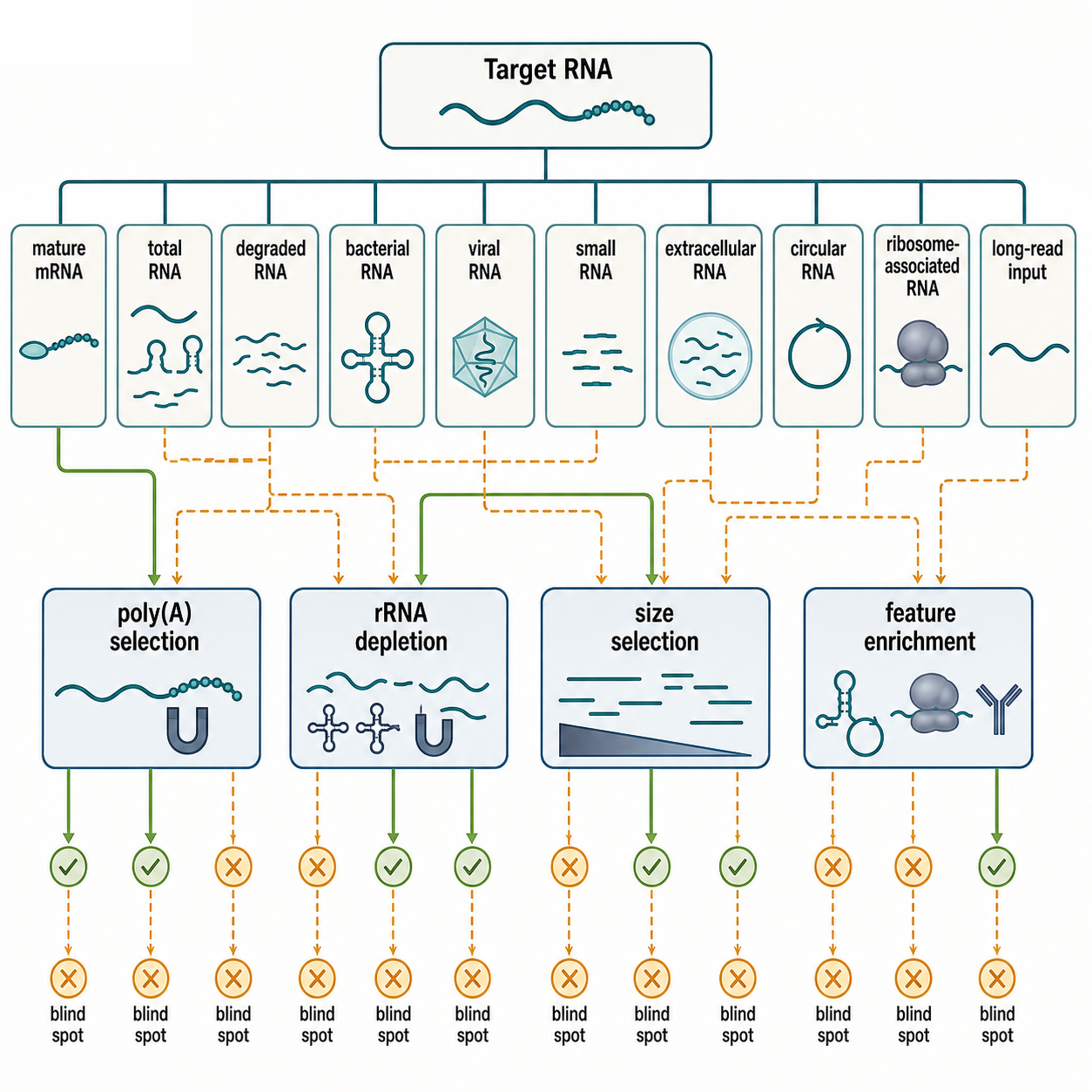
Figure 122.3. RNA Target Class to Fractionation Strategy Decision Tree. Prevents treating poly(A) selection, rRNA depletion, or size selection as neutral transcriptome methods.
Box 122.2. Selection Changes the Denominator
Do not describe a selected library as if it were total RNA. Poly(A) selection changes the denominator to RNAs with accessible polyadenylated tails. rRNA depletion changes the denominator to RNAs that remain after probe-dependent removal of targeted rRNAs and possible off-target molecules. Small-RNA size selection changes the denominator to molecules in a chosen length window with compatible termini and library chemistry. Circular RNA enrichment, cap capture, ribosome association, and extracellular vesicle isolation impose their own biochemical filters. The correct interpretation is therefore comparative: “within the selected RNA population, this signal changed.” A result becomes misleading when the selection boundary is hidden, such as calling a poly(A)-selected dataset a whole-transcriptome profile or treating depleted rRNA fraction as proof that all non-rRNA classes were preserved. State the selected population, expected losses, controls for selection performance, and whether the biological question concerns the retained or discarded molecules.
Other enrichment strategies target special RNA features. Cap-dependent capture enriches capped RNAs and can help identify transcription start sites or mature mRNA populations, but decapped or damaged molecules are lost. Circular RNA enrichment often uses exonuclease treatment to reduce linear RNAs, yet resistant structured linear RNAs and incomplete digestion complicate interpretation. Ribosome or polysome fractionation enriches translating or ribosome-associated RNAs, but sedimentation and affinity capture can co-purify stalled, nonproductive, or contaminating complexes. Extracellular vesicle isolation enriches vesicle-associated RNA, but vesicle preparations can contain lipoproteins, ribonucleoproteins, platelet material, and co-isolated contaminants.
Fractionation needs positive and negative controls. A fraction is convincing when expected marker molecules increase and expected contaminants decrease. For poly(A) selection, rRNA reduction and recovery of known polyadenylated transcripts should be checked. For rRNA depletion, residual rRNA fraction and off-target loss should be examined. For extracellular vesicle RNA, particle markers, protein contaminants, lipoprotein markers, hemolysis markers, and extraction blanks can be relevant. For bacterial or nonmodel samples, organism-specific rRNA depletion performance should be assessed rather than assumed.
The evidence basis for enrichment strategies includes hybridization chemistry, depletion efficiency, sequencing read composition, coverage bias, spike-in recovery, and comparison of matched total, poly(A)-selected, and rRNA-depleted libraries. The strongest studies make the selection boundary visible. For example, a comparison that reports both coding transcript detection and noncoding RNA loss teaches more than one that reports only total mapping rate. A good methods section states not only “RNA-seq was performed” but whether RNA was total, poly(A)-selected, rRNA-depleted, size-selected, cap-enriched, or otherwise fractionated.
Do not overgeneralize enrichment as improvement. Enrichment improves signal for a defined target class by discarding or reducing other classes. That tradeoff is desirable when explicit and harmful when hidden. A mature mRNA differential expression study may benefit from poly(A) capture. A viral, bacterial, circular RNA, pre-mRNA, or lncRNA study may require another strategy. A small RNA library is not a miniature total RNA library; it is a preparation shaped by size, termini, modifications, and adapter chemistry.
122.5. Quantification, Size Profiling, Spike-Ins, and Standards
RNA quantification asks how much RNA is present, but different instruments define “amount” differently. Absorbance at 260 nm estimates nucleic acid based on ultraviolet absorption. It is fast and nondestructive, but it is not RNA-specific. DNA, free nucleotides, phenol, and some contaminants absorb near 260 nm. The 260/280 ratio can warn about protein or phenol contamination, while the 260/230 ratio can warn about phenol, guanidinium, carbohydrates, salts, or other carryover. These ratios are useful screening signals, not proof of purity. They are especially unreliable at low concentration where baseline errors dominate.
Fluorescent RNA assays use dyes that preferentially bind RNA or nucleic acids under defined conditions. They are generally more sensitive and more specific than absorbance for low-input RNA, but they still do not reveal fragment size, sequence distribution, DNA contamination unless the chemistry discriminates well, or inhibitor burden. A fluorometric concentration can be accurate for input mass while the sample remains unusable because it is too fragmented, contaminated, or biased against the target RNA class.
Size profiling complements concentration. Capillary electrophoresis, microfluidic electrophoresis, gel electrophoresis, and some fragment analysis systems estimate RNA length distributions. These profiles reveal degradation, small RNA enrichment, adapter dimers in libraries, ribosomal peaks, and fragment sizes. For sequencing libraries, size profiling after library construction is also critical because adapter dimers, primer dimers, over-amplified products, and unexpected fragment distributions can consume sequencing capacity or distort quantification.
qRT-PCR and digital PCR can quantify specific RNA targets or process controls. qRT-PCR converts RNA to cDNA and amplifies a target region, reporting signal relative to cycle threshold or standard curve. Digital PCR partitions reactions so absolute molecule counts can be estimated from positive partitions. These methods are target-specific and can detect inhibition through dilution series or internal controls. Their limitations include primer specificity, reverse-transcription bias, DNA contamination, amplicon length sensitivity, and dependence on reference genes or standards. Chapter 123 owns the deeper amplification model, calibration, reference-gene, detection-limit, and orthogonal-validation treatment; this chapter retains sample-quality and point-of-addition control logic.
Spike-ins are exogenous molecules added at known amounts. Their most important property is point of addition. A spike-in added before lysis can monitor lysis, extraction, cleanup, and downstream steps if it experiences the matrix similarly to target RNA. A spike-in added after extraction cannot report extraction loss. A spike-in added before reverse transcription monitors reverse transcription and later steps but not sample preservation or purification. A sequencing spike-in added at pooling can monitor sequencing behavior but not library construction. The same molecule can be a useful control or a misleading normalization factor depending on when it enters the workflow.
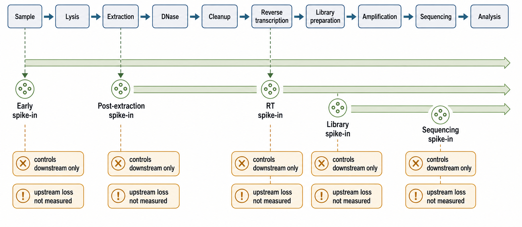
Figure 122.4. Spike-In Points of Addition and Interpretability. Prevents false claims that a late spike-in normalizes upstream degradation or extraction loss.
Box 122.3. Spike-In Placement Audit
Before using a spike-in for normalization, write an audit sentence: “This molecule was added at this step, at this amount, to represent this target class, and it can therefore test these workflow steps.” A pre-lysis spike-in can report lysis, extraction, cleanup, reverse transcription, library preparation, and sequencing only if it contacts the same matrix and survives the same losses as the target. A post-extraction spike-in can test reverse transcription or library chemistry but cannot correct preservation failure or extraction loss. A sequencing spike-in can monitor run behavior but cannot rescue a poor library. Molecular similarity is the second audit. Length, structure, modifications, termini, carrier state, and concentration range determine whether the spike-in resembles a long mRNA, a vesicle-associated miRNA, a modified tRNA, or an FFPE fragment. Use spike-ins to reveal technical behavior; do not let them erase sample biology, matrix effects, or failed negative controls.
Molecular similarity matters. A short unmodified synthetic RNA does not behave like a long structured mRNA, a modified tRNA, a protein-bound miRNA, a viral genome segment, or a crosslinked FFPE fragment. Spike-ins can differ from endogenous RNAs in length, sequence complexity, secondary structure, modification state, termini, carrier association, and susceptibility to extraction losses. A good spike-in panel spans concentrations relevant to the assay and avoids sequences that cross-hybridize or map ambiguously. Spike-ins should be stored, thawed, diluted, and added reproducibly because small pipetting errors at low concentrations can dominate interpretation.
Standards and reference materials serve different roles. A calibration standard supports quantitative conversion from signal to amount. A process control tests whether the workflow worked. A negative control estimates background. A reference RNA sample can compare library preparation or sequencing runs. A size ladder calibrates electrophoresis. A molecular weight marker supports gel interpretation. These should not be collapsed into one category. For example, an external RNA controls consortium-style spike-in mixture can help detect dynamic range and technical variation, but it cannot automatically correct for global shifts in endogenous RNA content per cell or changes in extraction efficiency if added after extraction.
Normalization with spike-ins is especially difficult when biology changes total RNA content. If a treatment doubles RNA per cell, equal-mass input and equal-cell input answer different questions. If spike-ins are added per sample volume rather than per cell or per tissue mass, they may report technical recovery but not biological concentration. If extraction efficiency differs by sample matrix, a post-extraction spike-in may hide the problem. These limitations do not make spike-ins useless; they make experimental design explicit.
Table 122.3. Quantification, Size Profiling, and Standards. Separates concentration, size, calibration, process control, and normalization roles.
| Tool or standard | Output | Strengths | Limitations | Good use | Common misuse |
|---|---|---|---|---|---|
| Absorbance | A260 estimate and 260/280 or 260/230 ratios. | Fast, nondestructive, and flags some contaminants. | Not RNA-specific and unreliable at low concentration. | Screen moderate-concentration RNA for phenol, protein, salt, or guanidinium warnings. | Treating purity ratios as proof of downstream usability. |
| Fluorometry | Dye-based RNA concentration. | Sensitive and usually more RNA-specific than absorbance. | Does not report size, sequence representation, inhibitors, or complete DNA absence. | Set input mass for library preparation or qRT-PCR. | Assuming concentration alone certifies RNA quality. |
| Capillary electrophoresis | Electropherogram, size distribution, RIN-like score, or DV200. | Shows degradation, enrichment range, and library fragment artifacts. | Summary scores are sample-class and assay dependent. | Qualify integrity, FFPE fragment length, small-RNA ranges, and library insert size. | Applying one RIN-style threshold to all RNA workflows. |
| qRT-PCR | Target-specific Ct or standard-curve signal. | Sensitive and useful for target, process-control, or inhibition checks. | Primer specificity, RT bias, DNA contamination, and reference-gene dependence. | Test amplicon survival, inhibition, or defined RNA target abundance. | Extrapolating one target to whole-transcriptome integrity. |
| Digital PCR | Partition-based absolute count estimate for defined targets. | Good precision for low-copy targets and calibration. | Limited target scope; RT bias and DNA contamination still matter. | Quantify standards, low-abundance targets, or diagnostic thresholds. | Treating partition counts as extraction-wide recovery. |
| Extraction spike-in | Recovery signal from pre-lysis or early-process addition. | Can sample lysis, extraction, cleanup, and downstream steps. | Only models endogenous RNA if matrix exposure and molecular behavior are similar. | Diagnose extraction loss, inhibition, and batch drift. | Using a non-matrix-matched spike-in for global normalization. |
| Reverse-transcription spike-in | Signal from RT and later assay steps. | Tests reverse transcription, amplification, or library chemistry. | Cannot report preservation, lysis, extraction loss, or cleanup bias. | Diagnose RT inhibition or cDNA/library preparation performance. | Normalizing away upstream degradation or extraction failure. |
| Sequencing spike-in | Reads from material added near library pooling or sequencing. | Tracks sequencing run behavior, dynamic range, or mapping calibration. | Cannot assess extraction, RT, or library construction if added late. | Monitor lane, run, basecalling, or alignment behavior. | Claiming it corrects sample preparation losses. |
| Reference RNA | Reproducible reference material or pooled RNA profile. | Supports cross-run library and pipeline comparison. | May not match sample matrix, target biology, or extraction difficulty. | Benchmark library preparation, sequencing, and analysis reproducibility. | Using it as an internal normalizer for every specimen. |
| Size ladder | Fragment-size calibration standard. | Anchors electrophoresis or gel sizing. | Does not measure sample purity, sequence content, or inhibitors. | Estimate RNA, cDNA, or library fragment sizes. | Treating a valid ladder as proof that sample QC passed. |
A practical quantification workflow uses multiple orthogonal measurements. For a bulk RNA-seq sample, one might use fluorometry for concentration, capillary electrophoresis for size and integrity, absorbance ratios for contaminant warnings when concentration is sufficient, and qRT-PCR controls for a sensitive target if needed. For plasma extracellular RNA, fluorometry may be near the detection limit, so process spike-ins, extraction blanks, hemolysis markers, and targeted assays become more informative. For long-read RNA sequencing, mass, molecule length, poly(A) status, and inhibitor absence matter more than a single concentration value.
A spike-in corrects everything. A spike-in corrects or diagnoses only the portion of the workflow that it samples. A spike-in added after RNA purification cannot reveal degradation during collection. A spike-in lacking modifications cannot model modified tRNA recovery. A spike-in that does not enter vesicles cannot model extracellular vesicle lysis. A spike-in panel can reveal technical variation, but it does not eliminate the need for balanced design, negative controls, and assay-specific QC.
122.6. DNase Treatment, Contamination Control, and Negative Controls
DNA contamination is a persistent problem because extraction workflows often recover some genomic DNA, plasmid DNA, mitochondrial DNA, microbial DNA, or residual template DNA. DNA can inflate absorbance, distort fluorometric measurements if the dye is not RNA-specific, and create false signal in qRT-PCR or RNA-seq. The problem is severe when primers amplify intronless genes, pseudogenes, mitochondrial transcripts, viral sequences, bacterial transcripts without introns, or transgenes. In RNA-seq, DNA contamination can contribute reads that map to intronic, intergenic, or promoter regions and can mislead nascent RNA or low-expression analyses.
DNase treatment uses deoxyribonuclease enzymes to degrade DNA before downstream assays. DNase can be applied on a silica column, in solution after extraction, or during specialized cleanup. The mechanism is straightforward: DNase hydrolyzes DNA phosphodiester bonds under conditions that preserve RNA. The practical details are less simple. DNase requires compatible ions and buffer, enough enzyme for the DNA burden, adequate mixing, and complete inactivation or removal before downstream steps. Some DNases can introduce salts, divalent cations, or protein contaminants; heat inactivation conditions may not be compatible with all RNA preparations.
DNase treatment reduces DNA contamination but does not prove absence of DNA. Residual DNA can persist if the sample is viscous, overloaded, poorly mixed, protected by proteins, or present in high copy number. DNA can also be reintroduced after DNase treatment through aerosols, contaminated water, primers, plasticware, or carryover from previous PCR products. For sensitive qRT-PCR, a no-reverse-transcriptase control is essential. This control omits reverse transcriptase, so amplification indicates DNA-dependent signal or another non-cDNA source. It is especially important when primers cannot span exon-exon junctions or when the target lacks introns.
Negative controls map different contamination routes. A no-template control contains reaction reagents but no nucleic acid template; it detects contamination in primers, water, master mix, or the amplification environment. An extraction blank is carried through extraction without biological input; it detects contaminants introduced by extraction reagents, plasticware, columns, beads, water, aerosols, and handling. A no-reverse-transcriptase control detects DNA-dependent amplification in a sample-derived nucleic acid preparation. A reagent blank tests a specific reagent. A matrix blank includes a sample-like matrix known or expected to lack the target, which can reveal matrix-specific contamination or inhibition.
Table 122.4. Negative Controls and Contamination Routes. Clarifies why DNase treatment and different negative controls are complementary rather than interchangeable.
| Control | Workflow step tested | Detects | Does not detect | Interpretation notes |
|---|---|---|---|---|
| Extraction blank | Full extraction and downstream assay without biological input. | Reagent, plasticware, column, bead, aerosol, water, and handling background. | Sample-specific DNA, inhibition, or matrix effects unless matrix is included. | Process beside samples; essential for low-input and low-biomass RNA. |
| No-template control | Amplification or library setup with no intended template. | Primer, water, master-mix, aerosol, or post-amplification contamination. | Contamination introduced during extraction or sample-derived DNA. | A clean NTC does not clear extraction-derived contamination. |
| No-reverse-transcriptase control | Sample reaction lacking reverse transcriptase. | DNA-dependent amplification or another non-cDNA template source. | RNA contaminants that still require reverse transcription. | Critical for intronless, bacterial, viral, pseudogene, or transgene targets. |
| Reagent blank | A specific reagent, lot, or buffer carried through testing. | Nucleic acid or inhibitor background from one reagent source. | Handling background, sample matrix effects, or cross-sample carryover. | Useful for lot comparisons and troubleshooting a suspect component. |
| Matrix blank | Target-negative sample-like material processed with the workflow. | Matrix-specific contamination, inhibition, or background signal. | True biological heterogeneity if the matrix is not genuinely target-negative. | Interpret with documented target-negative status and paired extraction blanks. |
| Replicate extraction | Independent extraction from the same specimen or matched aliquot. | Stochastic recovery, lysis variability, sporadic contamination, and low-input instability. | Shared reagent contamination or systematic method bias. | Discordance can flag matrix problems or unstable low-copy measurements. |
| Index or barcode control | Sequencing assignment, indexing, and lane or pool behavior. | Index hopping, barcode swapping, cross-sample reads, and misassignment. | Extraction contamination, DNA carryover, or RT failure. | Pair with balanced indexing and negative libraries when signal is near background. |
| Positive process control | Known target or reference material through workflow. | Gross extraction, RT, amplification, library, or inhibition failure. | False positives, cross-contamination, or endogenous representation. | Keep separate from samples and choose a matrix-appropriate control. |
Low-input and low-biomass experiments require extraction blanks because background molecules can be comparable to true signal. Plasma extracellular RNA, cerebrospinal fluid RNA, microbiome RNA from low microbial load sites, ancient or archival samples, single-cell or few-cell inputs, and pathogen detection near the limit of detection are especially vulnerable. Reagent-derived nucleic acids, index cross-talk, barcode swapping, environmental aerosols, and carryover from high-concentration samples can create plausible-looking signals. In such settings, absence of a no-template control signal is not enough; contamination may enter before amplification.
Cross-sample contamination is another route. Aerosols from amplified products, splashes during phase separation, shared homogenizers, bead carryover on automation decks, index hopping during sequencing, barcode misassignment, and sample swaps can all introduce reads or amplicons from another specimen. Physical separation of pre- and post-amplification areas, unidirectional workflow, sealed plates, filtered tips, decontamination, careful indexing, unique dual indexes where appropriate, and process controls reduce risk. For extraction workflows, cleaning the bench is not enough if homogenizer probes, centrifuge rotors, racks, or automated liquid handlers carry material between samples.
Contamination control also includes avoiding over-cleaning assumptions. RNase-free does not mean DNA-free. DNA-free does not mean RNA-free. Sterile does not mean nuclease-free. A reagent certified for one application may contain nucleic acid traces irrelevant to that use but important for ultrasensitive sequencing. Commercial kits can have lot effects. Water, carrier RNA, glycogen, enzymes, columns, and beads can all introduce background or inhibitors. For pathogen RNA assays, internal amplification controls help identify inhibition, but they do not prove that extraction recovered the pathogen if added too late.
Evidence for control design comes from contamination investigations, low-biomass sequencing studies, diagnostic validation, qRT-PCR assay development, and routine laboratory failure analysis. The strongest control evidence includes blanks processed alongside samples, barcode-aware sequencing analysis, replicate extractions, known-negative matrix controls, dilution tests for inhibition, and independent confirmation by a method with different contamination risks. A convincing RNA result near the detection limit should show that the signal is stronger, more reproducible, and biologically patterned compared with process blanks and negative controls.
Misconception note: controls are not interchangeable. A no-template control cannot detect contamination introduced during extraction. An extraction blank cannot test sample-specific genomic DNA contamination unless the same sample nucleic acid is present. A no-reverse-transcriptase control cannot reveal RNA contaminants that require reverse transcription. DNase treatment cannot replace a minus-RT control in sensitive assays. A good control panel is designed from the possible failure routes of the workflow.
122.7. QC Metrics, Batch Effects, and Protocol Metadata
Sample QC integrates measurements into a decision. The decision should be stated as fit for a specified assay, not as universal pass or fail. Typical pre-library RNA QC includes concentration, total yield, size distribution, RIN or related score when appropriate, DV200 for degraded input, purity ratios if absorbance is reliable, evidence of inhibitor absence, DNA contamination assessment, and sample identity checks. Library QC adds fragment size, adapter dimer level, library concentration, amplification cycle count, duplication risk, and sometimes qPCR-based library quantification. Sequencing QC adds read depth, mapping rate, rRNA fraction, mitochondrial fraction, gene-body coverage, strandedness, duplication, insert size, spike-in behavior, and negative-control signal.
Batch effects arise when technical variation aligns with biological labels. A batch can be a collection site, operator, date, tissue-processing queue, storage duration, extraction kit lot, homogenizer, column lot, automation deck, DNase batch, depletion kit, library preparation plate, PCR cycle number, index set, sequencer lane, or analysis pipeline version. If all controls are processed with one kit lot and all disease samples with another, differential expression can reflect kit chemistry. If all tumor samples have longer ischemia time than adjacent normal samples, stress-response and degradation signatures can masquerade as cancer biology.
The first defense is design. Randomize biological groups across collection, extraction, library preparation, and sequencing. Block samples so each processing unit contains a balanced representation of groups when randomization is limited. Include replicates and controls that travel through the workflow. Avoid processing all low-input samples on one day and all high-input samples on another. Record enough metadata to test whether principal components, outliers, or QC metrics correlate with handling variables.
Computational QC is diagnostic but not magic. Principal-component analysis, hierarchical clustering, sample correlation, surrogate-variable methods, batch-aware models, and control-feature analysis can reveal technical structure. These tools work best when batch is not perfectly confounded with biology. If disease status and extraction date are identical, no algorithm can confidently know which signal is disease and which is extraction. Removing batch-associated signal can remove true biology, while preserving biology can preserve artifact. The answer is balanced design plus transparent metadata, with computational correction used cautiously and reported clearly.
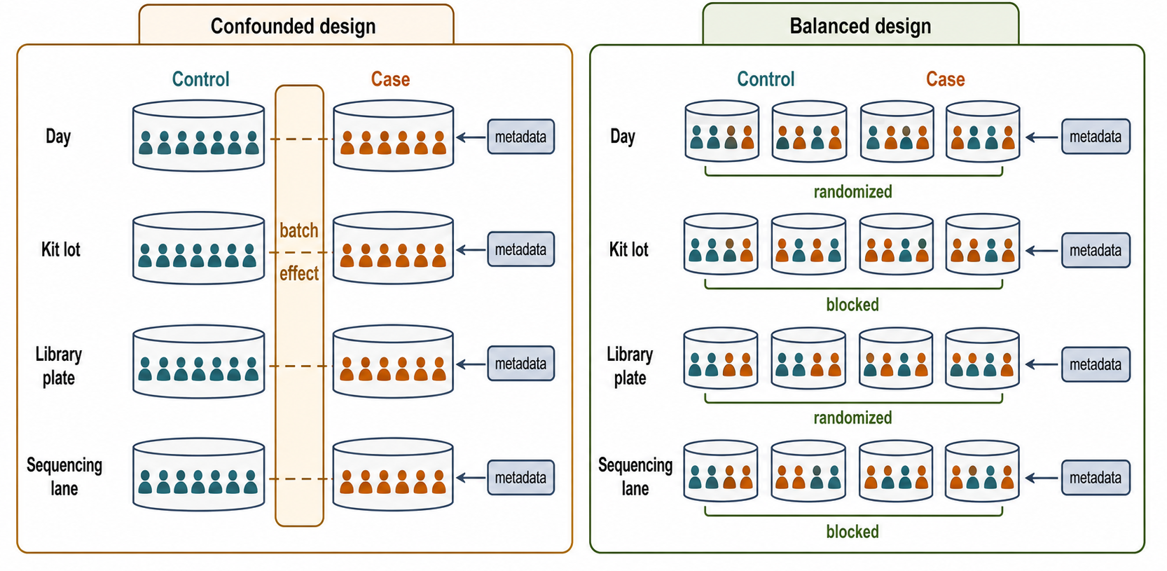
Figure 122.5. Batch-Effect Design and Randomization Schematic. Makes statistical identifiability concrete for RNA sample workflows.
Protocol metadata are the memory of the experiment. A minimal RNA extraction record should include sample identifier, organism, tissue or cell type, disease or treatment state, collection method, time to stabilization, preservation method, storage temperature and duration, freeze-thaw count, input mass or cell number, lysis and disruption method, extraction protocol or kit version, lot numbers when available, DNase strategy, cleanup method, enrichment or depletion method, elution volume, concentration method, QC instrument and kit, spike-in identity and point of addition, negative controls, operator or automation platform, processing date, and deviations. Clinical or regulated workflows also require chain of custody, acceptance criteria, calibration, and documented rejection rules.
Metadata should be structured enough to support analysis. Free-text notes are valuable for unexpected events, but controlled fields allow batch testing. “RNA extracted normally” is not sufficient. Useful metadata state that 25 mg of tissue was cryopulverized under liquid nitrogen, lysed in a defined guanidinium buffer within a defined time window, processed with a specified kit and lot, treated with DNase on-column, eluted in 30 microliters, quantified by a specific fluorometric assay, and profiled by a specific electrophoresis kit. This level of detail can seem excessive until a batch effect appears.
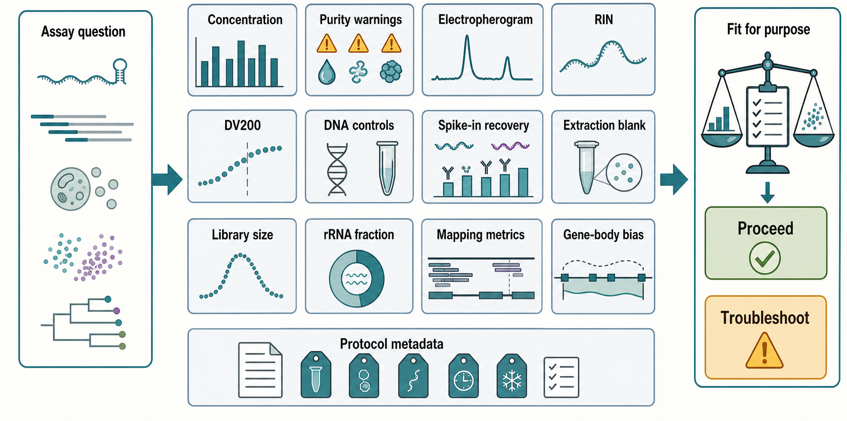
Figure 122.6. Fit-for-Purpose RNA QC Dashboard. Teaches that RNA quality is multidimensional and assay-specific.
The cross-chapter handoff is direct. Chapter 125 builds on these decisions for bulk and targeted RNA-seq library construction. Chapter 130 treats dissociation, nuclei preparation, ambient RNA, and single-cell QC. Chapter 128 extends integrity and molecule-length logic to long-read and direct RNA sequencing. Chapter 130 also treats fixation, permeabilization, and spatial RNA preservation. Chapter 139 applies standards and reproducibility logic to targeted assays and method comparisons. Later method chapters revisit extraction and preservation whenever the RNA feature being measured is structure, modification, translation, protein binding, localization, or therapeutic product quality.
Recent Consensus
Consensus in current RNA practice is that total yield and a single integrity score are insufficient. Fit-for-purpose assessment uses metrics matched to the assay. Fresh frozen bulk RNA-seq commonly cares about integrity, concentration, depletion or selection performance, DNA contamination, and library complexity. FFPE targeted assays commonly care about fragment length, amplifiable target size, inhibitors, and tumor content. Single-cell and single-nucleus workflows care about dissociation or nuclei isolation, ambient RNA, viability or nuclear quality, doublets, mitochondrial fraction, and batch. Long-read workflows care about molecule length, input mass, poly(A) status, and gentle handling. Extracellular RNA workflows care about low-input controls, hemolysis, carrier state, and process blanks.
Open Questions, Controversies, Deprecated Models, and Common Misconceptions
Open questions:
- How should spike-in normalization be handled when total RNA content per cell changes or when the spike-in does not share the sample matrix?
- How can extracellular RNA carrier states be preserved and fractionated without perturbation?
- How should low-biomass RNA measurements distinguish biological signal from reagent and handling background?
- How should nonmodel organisms and mixed communities be handled when validated depletion probes, extraction comparisons, and integrity standards are unavailable?
- Which preparation requirements for direct RNA sequencing and long-read methods were hidden by shorter-read workflows?
Common misconceptions:
- “High yield implies high quality.” Yield does not measure integrity, purity, end chemistry, contaminants, inhibition, or suitability for the downstream assay.
- “High RIN implies all RNA classes were recovered.” RIN mainly reflects abundant rRNA integrity and can miss small RNAs, long RNAs, organellar RNAs, extracellular RNAs, or chemically modified species.
- “Poly(A) selection is total transcriptome profiling.” Poly(A) selection enriches polyadenylated RNAs and underrepresents nonpolyadenylated, degraded, bacterial, organellar, and many regulatory RNAs.
- “Ribosomal RNA depletion is unbiased when probes do not match the sample.” Probe mismatch can distort recovery, leave rRNA background, and bias apparent transcript abundance.
- “DNase treatment proves DNA absence.” DNase treatment reduces DNA contamination but requires controls because residual DNA, inaccessible DNA, or reagent carryover can remain.
- “A spike-in added late can normalize early loss.” Late spike-ins cannot correct extraction, storage, lysis, or fractionation losses that occurred before addition.
- “An extraction blank is optional in low-input work.” Low-input RNA workflows are especially vulnerable to environmental, reagent, and index contamination, so blanks are essential controls.
- “Batch correction can repair a design in which technical processing is inseparable from biology.” Confounded designs cannot be fully rescued statistically because the model cannot distinguish technical and biological causes.