Chapter 42. rRNA Transcription, Processing, Modification, and Ribosome Biogenesis
Scope Note
This chapter follows ribosomal RNA from genomic rDNA repeats through transcription, precursor processing, chemical modification, ribonucleoprotein assembly, quality control, nuclear export, disease, stress signaling, and drug targeting. Ribosomal RNA is not only a structural scaffold for ribosomes. It is the catalytic and architectural core of translation, and each mature rRNA molecule is produced through a coordinated pathway that couples transcription rate, RNA folding, small nucleolar RNA guidance, nuclease processing, assembly-factor remodeling, surveillance, and cellular growth control. Chapter 21 covers RNA polymerase I and III transcription mechanisms in more detail; Chapter 25 covers co-transcriptional RNA folding and processing; Chapter 43 compares ribosome assembly pathways across bacteria, archaea, eukaryotes, mitochondria, and chloroplasts; Chapters 66-69 cover translation by mature ribosomes; Chapters 46-52 cover RNA modification chemistry more broadly.
Executive Summary
Ribosomal RNA is the RNA core of the ribosome. In all cellular life, rRNAs form the decoding center, peptidyl transferase center, subunit interfaces, and many of the binding surfaces used by messenger RNA, transfer RNAs, translation factors, and ribosomal proteins. Making rRNA is therefore one of the most expensive and tightly regulated RNA-production tasks in the cell. Rapidly growing cells devote large fractions of transcriptional and metabolic capacity to rRNA synthesis, while nutrient limitation, DNA damage, viral infection, developmental transitions, and oncogenic signaling can reshape or suppress ribosome biogenesis.
The first principle is that rRNA genes are organized for high output but not in one universal way. Bacteria commonly encode 16S, 23S, and 5S rRNAs in polycistronic operons, often with tRNAs embedded in the same transcription units. Eukaryotic nuclei usually separate the major RNA polymerase I transcript from the RNA polymerase III-transcribed 5S rRNA. In mammals, RNA polymerase I transcribes a long precursor containing 18S, 5.8S, and 28S rRNA regions separated by external and internal transcribed spacers; RNA polymerase III transcribes 5S rRNA from separate genes. Mitochondria and chloroplasts use organelle-specific polymerases, transcript organizations, processing landmarks, and ribosome compositions. Chapter 43 expands the comparative assembly logic; this chapter focuses on the production and maturation pathway.
The second principle is that mature rRNAs are not made by simply trimming the ends of a finished transcript. Pre-rRNA processing is a choreographed series of cleavage, exonucleolytic trimming, folding, modification, and assembly events. External transcribed spacers and internal transcribed spacers are not mature rRNA; they are precursor segments that help organize processing but must be removed. In eukaryotes, early cleavages commit the pre-rRNA to small-subunit or large-subunit maturation routes, and many processing steps occur while transcription is still ongoing. The classic “Christmas tree” view of nascent rRNA transcription units is useful because it makes the co-transcriptional nature of ribosome biogenesis visible, but modern interpretation requires molecular detail: nascent transcripts fold, recruit snoRNPs and assembly factors, undergo local modification, and enter surveillance before the full precursor is complete (Kos and Tollervey 2010; Rodgers and Woodson 2021).
The third principle is that rRNA modification is targeted and functional, but modification claims need precision. The two most abundant guided rRNA modifications in eukaryotes are 2′-O-methylation of ribose and pseudouridylation of uridine. Box C/D snoRNPs guide 2′-O-methylation, and box H/ACA snoRNPs guide pseudouridylation. The guide RNA base-pairs with the pre-rRNA near the target site, positioning a catalytic protein such as fibrillarin for methylation or dyskerin/Cbf5-family pseudouridine synthase for uridine isomerization. These modifications cluster in functionally important ribosome regions, including decoding and peptidyl-transferase neighborhoods, but a modification’s presence does not automatically prove a regulatory program. Stoichiometry, site identity, developmental context, and functional rescue matter. Mitochondrial rRNAs and bacterial rRNAs also carry modifications, many installed by stand-alone enzymes rather than snoRNA guides (Lopez Sanchez et al. 2020).
The fourth principle is that ribosome biogenesis uses many transient factors that are not part of the mature ribosome. Assembly factors stabilize intermediates, block premature functional sites, remodel RNA-protein contacts, license processing, and couple maturation to nuclear export. RNA helicases and G-patch proteins help rearrange RNA or RNP states; ATPases and GTPases drive directional transitions; export adaptors move pre-40S and pre-60S particles through nuclear pores; surveillance factors prevent immature subunits from entering translation. The final mature ribosome contains rRNAs and ribosomal proteins, but the pathway that creates it passes through many particles whose compositions change over time (Karika et al. 2026; McCool et al. 2022; Ni and Buszczak 2023).
The fifth principle is that defects in rRNA production are not merely “low protein synthesis.” Ribosome biogenesis defects can activate nucleolar stress, alter p53-dependent or p53-independent cell-death pathways, change translation of selected mRNAs, produce developmental disease, and create therapeutic vulnerabilities in cancer or infection. Ribosomopathies are disorders caused by defects in ribosomal proteins or biogenesis factors, often with tissue-specific phenotypes despite the general requirement for ribosomes. Cancer cells frequently increase rRNA transcription and ribosome production, making polymerase I transcription, nucleolar organization, rRNA modification enzymes, and assembly checkpoints attractive but toxicity-prone therapeutic targets. Viral infection can also perturb rRNA biogenesis, as shown for SARS-CoV-2 effects on ribosomal RNA biogenesis (Yerlici et al. 2024).
Concept Inventory
- Ribosomal RNA: the RNA component of the ribosome. Mature rRNAs form the structural and catalytic core of ribosomal subunits. In bacteria, the small subunit contains 16S rRNA and the large subunit contains 23S and 5S rRNAs. In human cytosol, the small subunit contains 18S rRNA and the large subunit contains 28S, 5.8S, and 5S rRNAs.
- Ribosomal DNA: the genomic DNA that encodes rRNAs. rDNA is often repeated many times because cells need high rRNA output. Repeats can be transcriptionally active, silent, damaged, recombining, or epigenetically distinct; a repeat count alone does not specify rRNA transcription rate.
- Pre-rRNA: an rRNA precursor transcript that includes mature rRNA segments plus extra spacer sequences or incomplete ends. In mammals, the long polymerase I precursor is often called 47S or 45S pre-rRNA depending on processing state and convention. In budding yeast, the analogous long precursor is commonly called 35S pre-rRNA.
- External transcribed spacers: transcribed regions outside the mature rRNA sequences at the ends of a pre-rRNA. Internal transcribed spacers are transcribed regions between mature rRNA sequences. Spacers are removed during processing. Their detection usually marks a precursor or processing intermediate, not mature ribosomal RNA.
- Nucleolus: a nuclear body organized around active rDNA transcription and ribosome biogenesis. It is not membrane-bounded. It contains rDNA transcription machinery, nascent pre-rRNA, snoRNPs, ribosomal proteins, processing factors, assembly intermediates, and many stress-responsive proteins. Nucleolar localization is suggestive but not proof of direct rRNA-processing function.
- Small nucleolar RNAs: guide or scaffold RNAs that act mainly in ribosome biogenesis and other stable-RNA maturation pathways. Box C/D snoRNAs usually guide 2′-O-methylation. Box H/ACA snoRNAs usually guide pseudouridylation. Some snoRNAs participate in pre-rRNA cleavage rather than, or in addition to, modification.
- 2′-O-methylation: methylation of the ribose 2′ hydroxyl. Pseudouridylation converts uridine into pseudouridine, an isomer in which the base is attached to ribose through a carbon-carbon bond rather than the usual nitrogen-carbon glycosidic bond. Both modifications can influence local rRNA structure, hydration, protein binding, and ribosome function.
- Ribosome biogenesis factors: transient proteins or RNPs required to assemble, process, remodel, inspect, or export ribosomal subunits. They are distinct from mature ribosomal proteins, although some proteins can have maturation and mature-subunit roles.
- Nucleolar stress: a cellular stress state caused by impaired ribosome biogenesis, rDNA damage, nucleolar disruption, or imbalance in ribosomal protein and rRNA production. It often signals through ribosomal proteins and p53 in mammalian cells, but p53-independent outputs also exist.
- Ribosomopathies: diseases caused by defects in ribosomal proteins, rRNA processing, modification, assembly, or related biogenesis factors. They illustrate that a pathway needed in every proliferating cell can produce tissue-selective developmental phenotypes.
What to Know Before Reading This Chapter
The reader should know that RNA molecules are synthesized 5′ to 3′ and that the 5′ and 3′ ends have different chemistry. Pre-rRNA processing uses this polarity. A cleavage that separates a future 18S rRNA segment from a spacer is not the same event as exonucleolytic trimming of a 3′ end, and neither is the same as degradation of a defective transcript.
The reader should also know the basic organization of the ribosome. A ribosome has a small subunit that decodes mRNA and a large subunit that catalyzes peptide-bond formation. The names 30S, 50S, 40S, and 60S are sedimentation coefficients, not simple molecular weights. Bacterial 70S ribosomes contain 30S and 50S subunits, while eukaryotic cytosolic 80S ribosomes contain 40S and 60S subunits. Chapters 66-69 treat mature ribosome function; here the question is how the RNA core of those subunits is made.
A third prerequisite is the distinction between transcription, processing, modification, and assembly. Transcription produces RNA from DNA. Processing removes spacer sequences and creates mature ends. Modification changes nucleotide chemistry after transcription. Assembly adds ribosomal proteins and transient factors to build a ribonucleoprotein particle. These steps overlap in time, but they are not synonyms. A change in mature rRNA abundance can reflect altered transcription, altered processing, altered degradation, altered export, altered subunit stability, or altered cell growth.
Finally, this chapter uses eukaryotic nuclear ribosome biogenesis as the main teaching example because the pathway is especially elaborate and clinically important. Bacterial, archaeal, mitochondrial, and chloroplast examples appear as boundary cases. Chapter 43 gives those comparative systems a fuller treatment.
42.1. rRNA gene organization and transcription systems
rRNA transcription is the synthesis of ribosomal RNA precursor molecules from rDNA templates. The biological purpose is simple: generate enough rRNA to assemble ribosomes at a rate compatible with cell growth. The implementation differs by domain and compartment. A bacterial cell, a yeast nucleus, a human nucleolus, a mitochondrion, and a chloroplast do not use one interchangeable rRNA gene design.
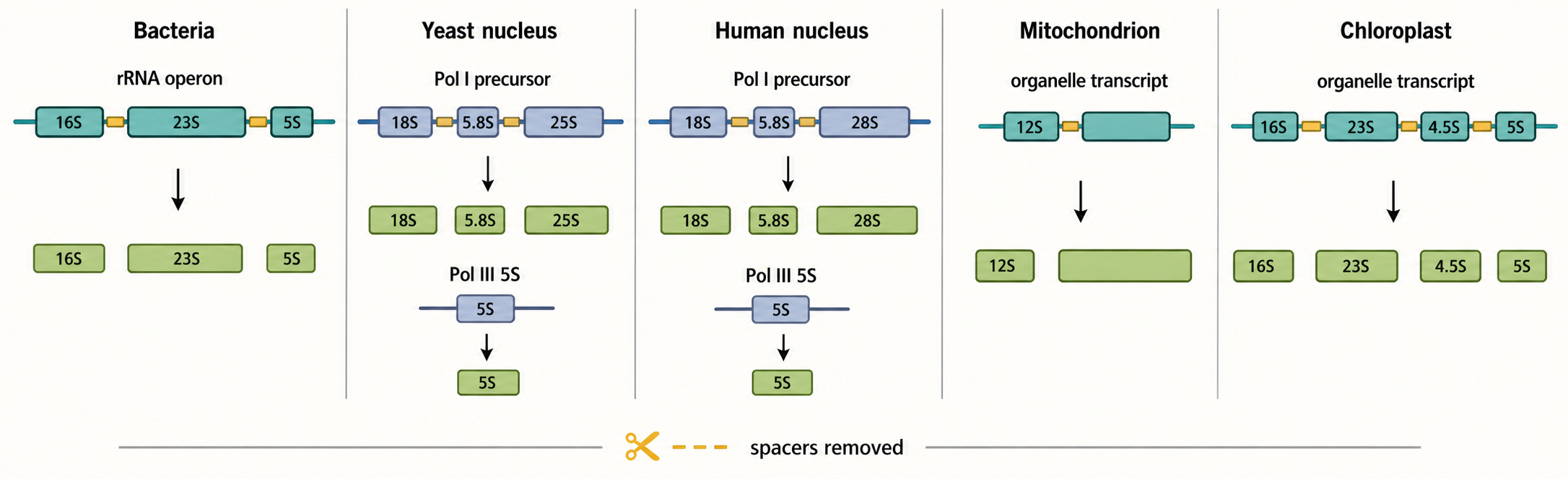
Figure 42.1. rRNA Gene Organization and Transcription Systems. rRNA production relies on different transcription systems depending on domain and cellular compartment. Bacteria typically encode 16S, 23S, and 5S rRNAs in a single polycistronic operon, often with interspersed tRNAs, allowing growth-regulated transcription to produce multiple ribosomal components together. Eukaryotic nuclei separate RNA polymerase I transcription of a long precursor containing 18S, 5.8S, and 28S rRNA regions from RNA polymerase III transcription of 5S rRNA at distinct loci, while mitochondria and chloroplasts use organelle-specific polymerases and transcript organizations. Spacer-containing precursors at every level are production intermediates that must be cleaved, trimmed, and removed before mature rRNA is assembled into ribosomes.
Table 42.1. Major rRNA Precursors and Mature rRNAs by System. Overview of rRNA transcription units, mature products, and processing highlights across five cellular systems, illustrating why nomenclature and pathway logic should not be generalized from one system to another.
| System | Transcription unit | Mature rRNAs produced | Main polymerase | Processing highlights | Caveats |
|---|---|---|---|---|---|
| Bacterial cytosol | Polycistronic operon (~5–7 kb) | 16S, 23S, 5S | Bacterial RNA polymerase | Co-transcriptional cleavage; tRNAs may be embedded; assembly begins on nascent rRNA | Operon copy number and tRNA content vary across species |
| Budding yeast nucleus | 35S pre-rRNA (Pol I); 5S genes (Pol III) | 18S, 5.8S, 25S (Pol I); 5S (Pol III) | RNA polymerase I + III | Multiple endonucleolytic and exonucleolytic steps; many assembly factors characterised genetically | 35S nomenclature is yeast-specific; spacer positions differ from mammalian |
| Human nucleus | 47S/45S pre-rRNA (Pol I); 5S genes (Pol III) | 18S, 5.8S, 28S (Pol I); 5S (Pol III) | RNA polymerase I + III | 5′ ETS and 3′ ETS removal, ITS1 and ITS2 removal; many intermediates detectable by northern blot | 47S vs. 45S naming reflects incomplete 3′ processing state, not separate genes |
| Mammalian mitochondrion | Long mt-RNA transcript | 12S mt-rRNA, 16S mt-rRNA | Mitochondrial RNA polymerase | tRNA punctuation model separates rRNAs; stand-alone methyltransferases modify rRNA | mt-rRNA nomenclature is distinct from cytosolic; sizes are much smaller |
| Plant chloroplast | Operon-like precursor | 16S, 23S, 4.5S, 5S | Phage-type chloroplast polymerase | Bacterial-derived processing logic; many maturation factors are nuclear-encoded | Plastid-specific factors often differ from expected bacterial homologs |
Table 42.2. Evidence Ladder for rRNA Biogenesis Claims. Standards of evidence required to support common rRNA biogenesis claims, with examples of strong evidence, weak or ambiguous evidence, typical artifacts, and recommended follow-up assays.
| Claim type | Strong evidence | Weak or ambiguous evidence | Common artifact | Useful follow-up |
|---|---|---|---|---|
| rRNA transcription rate | Pulse labeling of nascent rRNA; polymerase I ChIP-seq occupancy | Steady-state mature rRNA abundance | Mature rRNA is long-lived; levels change slowly and do not reflect transcription rate | Nascent RNA run-on or EU-RNA metabolic labeling |
| Processing-site use | Primer extension mapping; end-focused long-read sequencing; endonuclease mapping | Presence of a precursor band by northern blot | Precursor accumulation can reflect faster transcription or blocked degradation, not a processing defect | Pulse-chase with time-resolved sampling of intermediates |
| Direct nuclease substrate | In vitro cleavage by purified enzyme; depletion plus substrate recovery and rescue | Co-localization of nuclease with pre-rRNA | Indirect effect through upstream assembly defect leaving substrate inaccessible | Depletion rescue comparing nuclease-active versus catalytically inactive alleles |
| rRNA modification site | Site-specific primer-extension stop; mass spectrometry; RiboMeth-seq | snoRNA guide predicted to base-pair near a site | RT stops can reflect RNA structure or other modifications rather than the intended chemistry | Guide deletion or enzyme mutation to confirm modification dependency |
| Modification stoichiometry | Quantitative RiboMeth-seq; nanopore direct RNA quantification | Qualitative signal presence or absence | Signal can vary with extraction efficiency or library preparation | Multi-method quantification at the same site with normalization controls |
| Assembly-factor checkpoint | Depletion shifts precursor pattern plus rescue; cryo-EM of defined intermediate | Factor co-purification with pre-ribosomal particle | Essential-factor depletion causes global stress that indirectly alters processing | Time-resolved depletion and sampling before secondary stress accumulates |
| Nuclear export block | Subcellular fractionation; export-factor depletion; FISH for pre-ribosomal marker RNA | Apparent perinuclear accumulation by fluorescence imaging | Fixation and permeabilization artifacts can misreport nuclear retention | Live-cell tracking with tagged pre-ribosomal subunit component |
| Nucleolar stress pathway | p53 stabilization measurement; MDM2 interaction assay; marker colocalization | Nucleolar morphology change alone | Many stimuli alter nucleolar shape without a specific ribosome-biogenesis defect | Factor-specific rescue and measurement of p53-independent output |
| Therapeutic target engagement | Target-binding assay; cellular potency; rescue by target overexpression | Antiproliferative activity in ribosome-rich cells | Many cytotoxic drugs affect multiple targets simultaneously | On-target resistant mutations and direct pharmacodynamic marker measurement |
Table 42.3. rRNA Modification Systems. Principal rRNA modification classes, their guide or enzyme systems, representative cellular compartments, detection methods, and key interpretation caveats.
| Modification class | Typical guide/enzyme system | Example compartment | Evidence method | Interpretation caveat |
|---|---|---|---|---|
| 2′-O-methylation | Box C/D snoRNA + fibrillarin (Nop1 in yeast) | Eukaryotic nucleus | RiboMeth-seq, primer-extension stop, mass spectrometry | Guide-pairing predictions must be confirmed by guide deletion or enzyme catalytic mutation |
| Pseudouridylation | Box H/ACA snoRNA + dyskerin/Cbf5-family synthase | Eukaryotic nucleus | Pseudo-seq, CMCT-based primer extension, nanopore direct RNA signal | Pseudouridine is RT-transparent without chemical derivatization; signals need orthogonal validation |
| Base methylation | Stand-alone methyltransferases (e.g., KsgA/Dim1 for m6,6A) | Bacterial cytosol; eukaryotic nucleus | Mass spectrometry, genetic deletion of enzyme gene | Many methyltransferases are poorly characterized; enzyme-to-site assignments require in vitro reconstitution |
| Mitochondrial rRNA methylation | Mitochondria-specific methyltransferases (e.g., MRM1, MRM2, ERAL1) | Mammalian mitochondrion | Mass spectrometry, genetic perturbation of mt-methyltransferase | Disease variants can affect mt-rRNA modification without clear stoichiometry measurements |
| Bacterial stand-alone enzyme modifications | Protein enzymes recognizing rRNA structure or assembly intermediates | Bacterial cytosol | Enzyme gene deletion, in vitro methylation with purified enzyme | Assembly state of rRNA can gate enzyme access; in vitro conditions may not replicate in vivo order |
Bacteria commonly organize rRNA genes in operons. A typical bacterial rRNA operon encodes 16S rRNA, one or more tRNAs in some cases, 23S rRNA, and 5S rRNA in a single long transcript. The transcript is then processed into separate mature RNAs. This organization lets growth-regulated transcription produce several ribosomal components together. It also means that processing of rRNA and tRNA can be physically linked in the same precursor transcript. A bacterial rRNA operon should not be interpreted as a mature RNA molecule; it is a production unit that must be cut, modified, folded, and assembled.
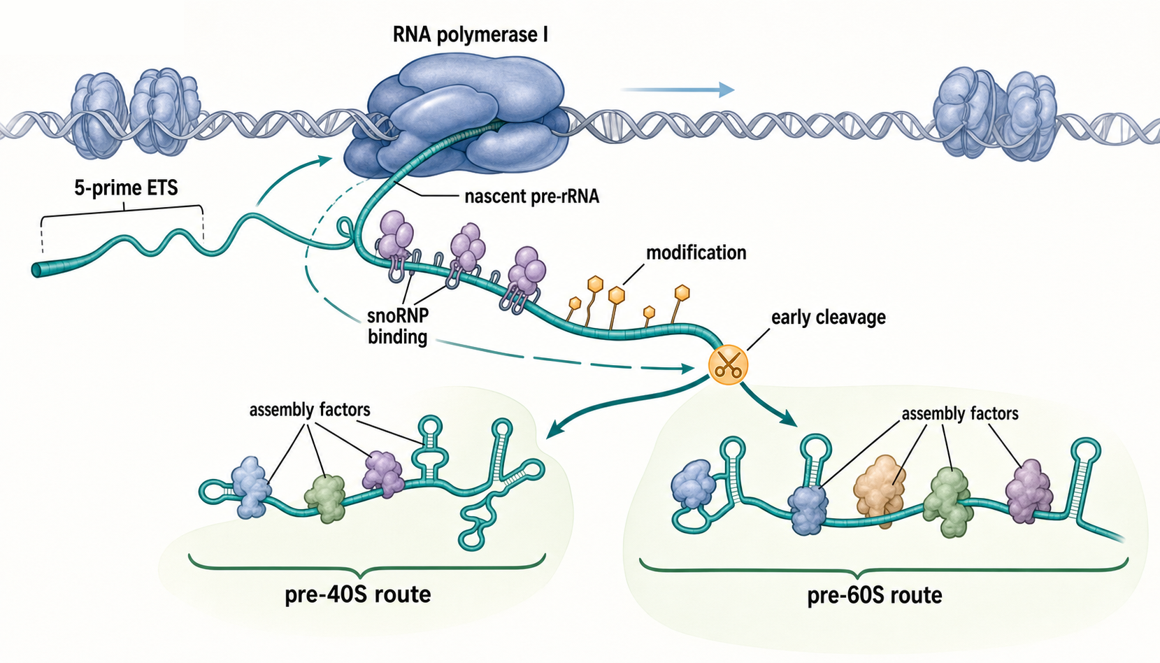
Figure 42.2. Cotranscriptional Pre-rRNA Processing and Assembly. Eukaryotic pre-rRNA maturation begins while RNA polymerase I is still elongating the nascent transcript. The 5′ external transcribed spacer folds rapidly after synthesis and recruits U3 snoRNP together with early small-subunit processome components, snoRNAs guide site-specific 2′-O-methylation and pseudouridylation co-transcriptionally, and early endonucleolytic cleavages separate the future pre-40S branch from the pre-60S maturation route before the full precursor is complete. Folding, modification, cleavage, and assembly-factor recruitment are therefore temporally coupled rather than cleanly sequential stages.
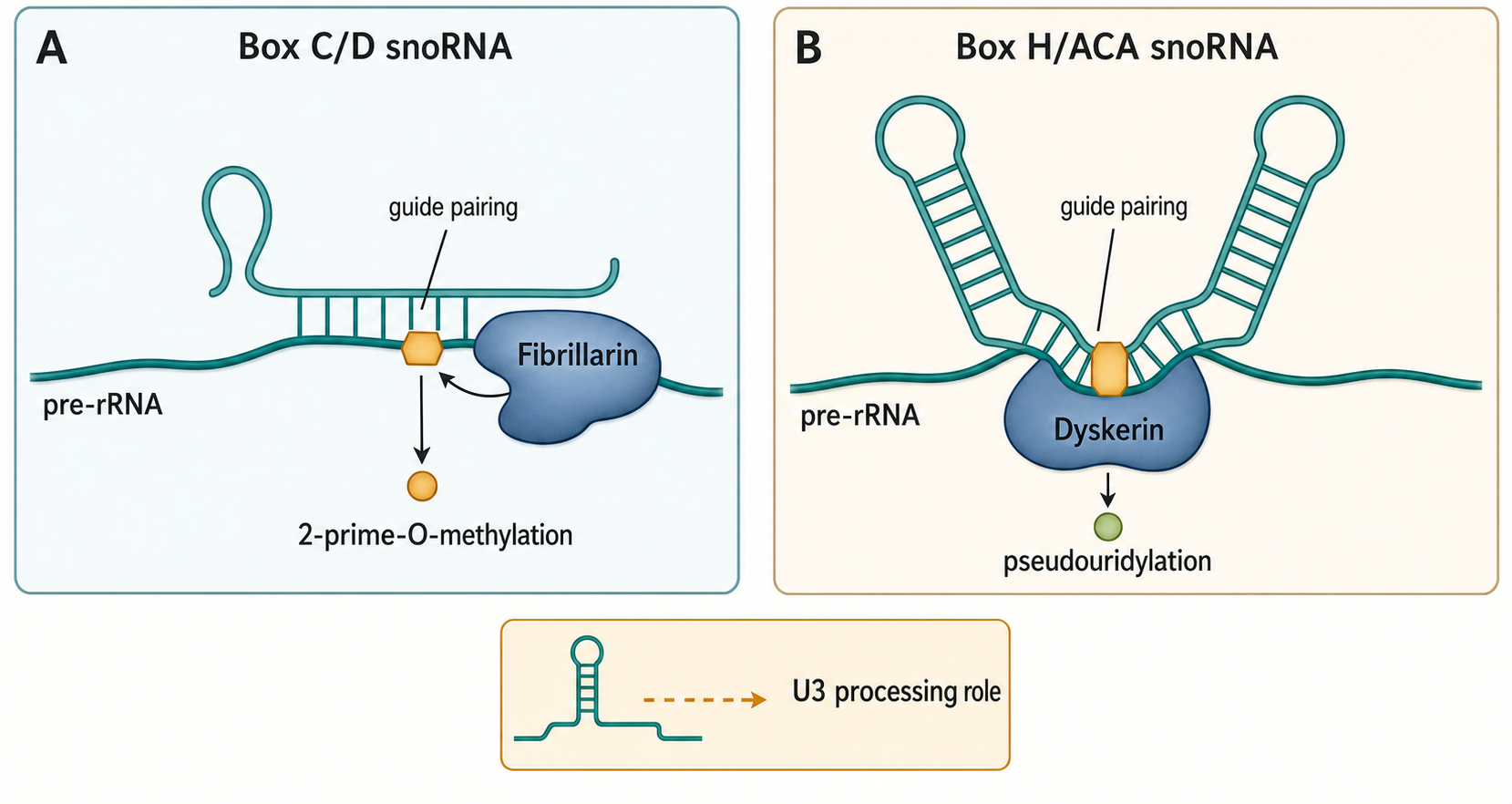
Figure 42.3. snoRNA-Guided rRNA Modification. Small nucleolar RNAs act as address systems that base-pair with pre-rRNA to position specific nucleotides for catalysis. Box C/D snoRNAs contain conserved C and D motifs flanking guide sequences that direct fibrillarin (Nop1 in yeast) to 2′-O-methylate the ribose hydroxyl of the nucleotide positioned five residues upstream of the D motif, while box H/ACA snoRNAs fold into a hairpin-hinge-hairpin-tail architecture that positions a uridine in a pseudouridylation pocket for isomerization by a dyskerin/Cbf5-family enzyme. Some snoRNAs, such as U3, act instead in early pre-rRNA cleavage and processome organization rather than targeting a single modification site.
Eukaryotic nuclear cells split rRNA transcription between polymerase systems. RNA polymerase I transcribes the large rDNA repeat unit that contains the future 18S, 5.8S, and 28S rRNAs in mammals or 18S, 5.8S, and 25S rRNAs in budding yeast. RNA polymerase III transcribes 5S rRNA from separate genes. This division matters because polymerase I and polymerase III use different promoter architectures, transcription factors, chromatin environments, inhibitors, and stress responses. A perturbation that blocks polymerase I transcription does not necessarily block 5S rRNA transcription in the same way, and an imbalance between 5S rRNA, ribosomal proteins, and the large polymerase I transcript can contribute to surveillance signaling.
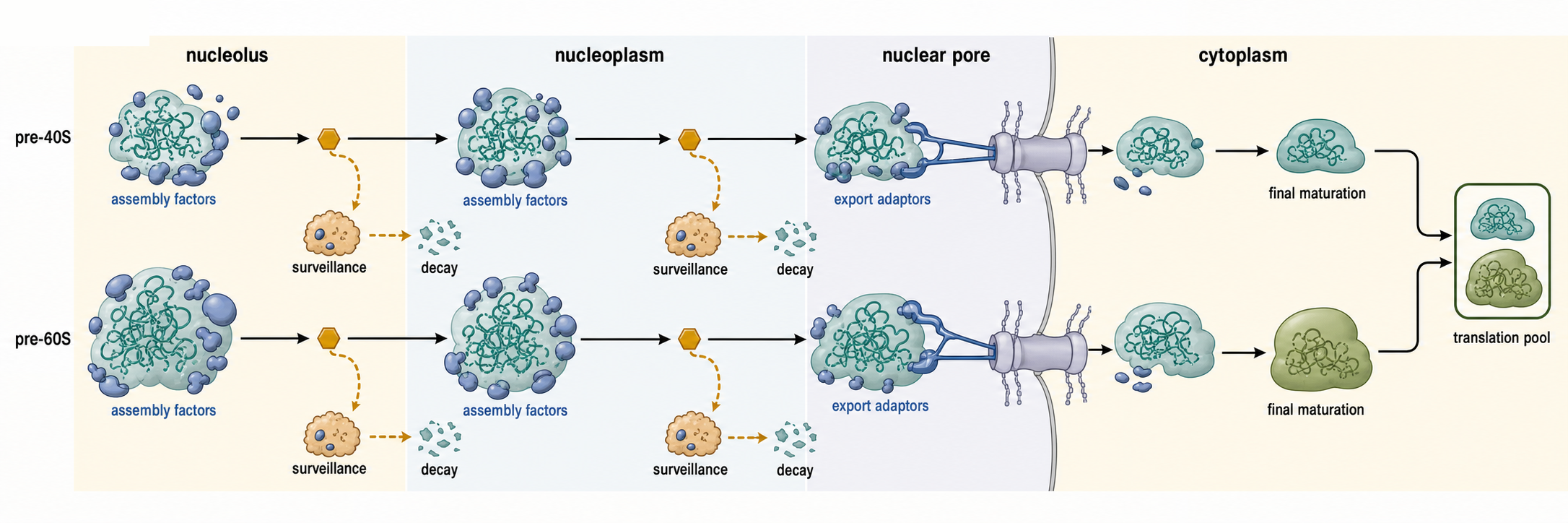
Figure 42.4. Checkpoints and Nuclear Export in Eukaryotic Ribosome Biogenesis. Pre-ribosomal particles traverse nucleolus, nucleoplasm, nuclear pore complex, and cytoplasm before joining the translating pool. At each compartmental stage, assembly factors stabilize intermediates and block premature activation of functional rRNA sites, export adaptors mediate the energy-dependent transit of pre-40S and pre-60S particles through nuclear pores, and cytoplasmic maturation steps complete subunit activation including final processing events and release of anti-association factors. A surveillance and decay branch at multiple checkpoints eliminates defective particles before they can enter translation, ensuring that only functional subunits reach the translating ribosome pool.
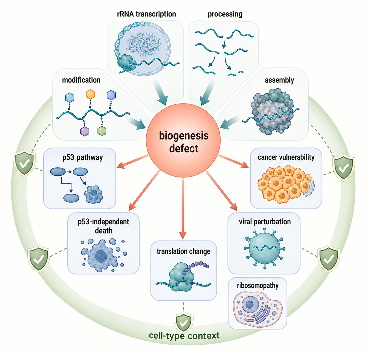
Figure 42.5. Ribosome Biogenesis Stress Outputs. Disruptions in rRNA transcription, processing, modification, or assembly produce stress outputs that extend beyond a simple reduction in total protein synthesis. Imbalanced production of ribosomal proteins relative to rRNA can stabilize p53 through MDM2 sequestration, but p53-independent apoptotic pathways also respond to ribosome-biogenesis defects. Cancer cells frequently depend on elevated ribosome-biogenesis output, creating therapeutic vulnerabilities when rRNA transcription or assembly is inhibited, while viral infection and inherited ribosomopathies illustrate how defects in a universally required pathway can yield tissue-selective phenotypes determined by cell-type proliferation demand, p53 threshold, and compensatory capacity.
Table 42.4. Ribosome Biogenesis Disease and Stress Categories. Ribosome biogenesis disruptions linked to disease and cellular stress, with molecular entry points, example phenotypes, and evidence requirements for each category.
| Category | Molecular entry point | Example phenotype or context | Evidence needed | Citation status |
|---|---|---|---|---|
| Inherited ribosomopathy | Ribosomal protein or biogenesis-factor mutation | Bone marrow failure, craniofacial anomalies, or growth defects (e.g., Diamond-Blackfan anemia, Treacher Collins syndrome) | Genetic lesion mapping, precursor accumulation, cellular rescue with wild-type allele, disease-variant functional data | Direct reviews pending; established clinical literature exists |
| Nucleolar stress | Impaired rDNA transcription or assembly imbalance leaving free ribosomal proteins | p53 stabilization, cell-cycle arrest, senescence, or apoptosis | MDM2-binding assay, p53 induction, rescue by p53 knockdown or MDM2 competition | Ni and Buszczak 2023; Ogawa et al. 2025 |
| Viral perturbation | Disruption of rDNA transcription or nucleolar organization by viral protein | Reduced host translation capacity, altered immune signaling | Direct pre-rRNA precursor analysis in infected cells, viral protein localization, processing-intermediate quantification | Yerlici et al. 2024 |
| Cancer-associated high ribosome output | Oncogenic signaling stimulates Pol I transcription and ribosomal protein expression | Elevated nucleolar activity, increased ribosome number, translational growth advantage | Pol I ChIP-seq, pre-rRNA quantification, functional rescue of growth phenotype | Yang et al. 2024; McCool et al. 2022 |
| Drug-induced Pol I or processing inhibition | Small-molecule inhibitor of Pol I transcription or a processing factor | Antiproliferative effect, nucleolar stress, selective sensitivity in high-output cancer cells | Target engagement, on-target resistance mutation, comparison of cancer versus normal cell toxicity | Xiao et al. 2023; direct Pol I inhibitor clinical sources pending |
| Mitochondrial rRNA modification defect | Loss or impairment of a mitochondrial rRNA methyltransferase | Mitochondrial translation impairment, multisystem energy disorder | mt-rRNA modification mapping, respiratory-chain functional assay, genetic complementation | Lopez Sanchez et al. 2020 |
The rDNA repeat is more than a coding sequence. A eukaryotic repeat contains promoter regions, transcribed spacers, mature rRNA segments, terminator regions, and intergenic spacer DNA. In mammals, the transcribed precursor includes a 5′ external transcribed spacer, the 18S rRNA region, internal transcribed spacer 1, the 5.8S rRNA region, internal transcribed spacer 2, the 28S rRNA region, and a 3′ external transcribed spacer. The spacers are useful in biogenesis because they provide binding platforms, folding constraints, and processing landmarks, but they are not retained in the mature ribosome.
The nucleolus is the cellular site where eukaryotic rDNA transcription and much ribosome biogenesis occur. It forms around active rDNA repeats and is often described as having fibrillar centers, dense fibrillar components, and granular components. These terms describe nucleolar subregions enriched for different stages of rRNA production and assembly. The key conceptual point is that the nucleolus is a process-organizing body rather than a storage compartment. Active rDNA transcription, nascent pre-rRNA folding, snoRNP-guided modification, early assembly, and later pre-ribosomal particle maturation occur in an ordered but dynamic environment. Recent work on snoRNP domains and nucleolar compaction reinforces that nucleolar material properties can affect rRNA modification and processing, but phase separation language should be tied to molecular mechanism and perturbation evidence (Dominique et al. 2024; Yang et al. 2024).
rRNA transcription rate is regulated by growth signals, nutrient availability, chromatin state, cell cycle state, DNA damage, and stress pathways. In a proliferating mammalian cell, high polymerase I activity supports biomass accumulation and ribosome production. During nutrient limitation or stress, reducing rRNA transcription saves energy and prevents accumulation of incomplete ribosome precursors. Ni and Buszczak describe ribosome biogenesis as a homeostatically controlled process rather than a constitutive background pathway. The term “housekeeping” is therefore misleading if it implies unregulated or biologically uninteresting expression (Ni and Buszczak 2023).
Organelles illustrate boundary cases. Mammalian mitochondria encode mitochondrial rRNAs in compact mitochondrial DNA and transcribe them with a mitochondrial RNA polymerase system. Plant chloroplasts have bacterial-derived ribosome biogenesis logic, but chloroplast rRNA maturation uses chloroplast-specific and nuclear-encoded factors, many of which differ from bacterial homolog expectations. Schmid and colleagues review chloroplast ribosome biogenesis factors and show that an endosymbiotic origin does not make plastid ribosome biogenesis identical to bacterial assembly (Schmid et al. 2024). Mitochondrial rRNA methylation and processing also have specialized enzymes and disease relevance (Lopez Sanchez et al. 2020).
The evidence for rRNA gene organization and transcription includes genome assemblies, rDNA copy-number assays, fluorescence in situ hybridization, chromatin immunoprecipitation of polymerase I or transcription factors, nascent RNA labeling, metabolic pulse-chase experiments, electron microscopy of active rDNA transcription units, sequencing of precursor ends, and perturbation of transcription factors. Each method has caveats. rDNA repeats are repetitive and difficult to assemble. Steady-state rRNA abundance is not a direct transcription-rate measurement because mature rRNAs are stable. Nucleolar enlargement can correlate with ribosome biogenesis but does not by itself identify the limiting step. Strong evidence connects rDNA occupancy, nascent transcript production, precursor processing, and downstream ribosome assembly.
42.2. Pre-rRNA processing pathways and nucleolytic enzymes
Pre-rRNA processing is the conversion of a long precursor transcript into mature rRNA molecules with correct ends and assembly-compatible structure. The word “processing” can sound like simple trimming, but rRNA processing is a multi-step pathway. The precursor must be cleaved at specific sites, spacers must be removed, ends must be trimmed by exonucleases, and the RNA must remain competent for folding and protein assembly. If an early step fails, later intermediates may never form.
In eukaryotic nuclear systems, the long polymerase I transcript contains mature rRNA regions separated by external and internal transcribed spacers. Processing begins before or soon after the transcript is complete. Some events occur co-transcriptionally, meaning while RNA polymerase I is still transcribing the same rDNA repeat. Kos and Tollervey provided landmark evidence in yeast that pre-rRNA processing and modification occur co-transcriptionally, and Rodgers and Woodson synthesize how transcription, folding, and assembly are coupled (Kos and Tollervey 2010; Rodgers and Woodson 2021).
A useful teaching model is to follow the future small subunit and large subunit rRNAs. The future 18S rRNA enters a small-subunit pathway that includes early processome assembly and cleavage events that separate pre-40S precursors from the rest of the transcript. The future 5.8S and 28S or 25S rRNAs enter a large-subunit pathway that requires internal spacer removal, end trimming, and progressive assembly with large-subunit ribosomal proteins. These routes are coordinated because both subunits must be made in appropriate stoichiometry, but they are not identical. Depleting a small-subunit processome factor may produce a different precursor pattern than depleting a large-subunit processing factor.
Nucleolytic enzymes are enzymes that cleave or degrade nucleic acids. Endonucleases cut internally, while exonucleases trim from an end. rRNA biogenesis uses both. RNase MRP, the exosome, Rat1/Xrn-family exonucleases, Las1, Nob1, and other nucleases are standard examples in eukaryotic model systems, but this chapter’s current bibliography is thin for direct enzyme-specific provenance. The initial draft therefore emphasizes pathway logic and cites available co-transcriptional and processing-map sources rather than pretending that the present reference file fully covers every nuclease.
Spacer removal has two linked purposes. First, it removes RNA that is not part of the mature ribosome. Second, it creates ordered maturation intermediates that can be checked. An internal transcribed spacer may prevent premature formation of a mature-like rRNA junction until the correct proteins and factors have assembled. Removal of that spacer can therefore be a licensing step, not merely cleanup. Conversely, accumulation of a spacer-containing intermediate can mark a blocked pathway stage. Northern blotting, primer extension, end-focused sequencing, and long-read approaches can distinguish precursor forms if the assay is designed around known processing sites.
Long-read sequencing is increasingly useful because rRNA precursors are long and processing intermediates can contain combinations of sites that short reads may not connect. Pastore and colleagues report single-nucleotide-resolution mapping of human pre-rRNA processing and modification using long-read nanopore sequencing. The value of such approaches is that they can join processing state and modification information on the same molecule more directly than many fragmented short-read assays. The caveat is that nanopore signal interpretation for modified nucleotides, structured RNA, and closely related precursor isoforms requires careful calibration and orthogonal validation (Pastore et al. 2026).
Pre-rRNA processing is also coupled to ribosomal protein binding. A ribosomal protein can stabilize a local rRNA fold, recruit a processing factor, or block an inappropriate cleavage. Conversely, a processing event can create the binding surface for later proteins. McCool and colleagues show that human pre-60S assembly factors can link rRNA transcription to pre-rRNA processing, illustrating that large-subunit maturation is not a post-transcriptional afterthought (McCool et al. 2022). The causal interpretation is important: assembly factors can influence processing efficiency, and processing defects can indirectly change transcription or nucleolar organization.
Processing evidence has common failure modes. A pulse-chase experiment can show precursor-product relationships, but only if the chase is fast enough and the intermediates are resolved. A steady-state increase in a precursor may result from faster transcription, slower processing, blocked degradation, or altered cell-cycle composition. A nuclease depletion phenotype may reflect direct substrate cleavage or an upstream assembly defect that makes the substrate inaccessible. Strong claims combine substrate mapping, factor perturbation, rescue, biochemical interaction, localization, and when possible in vitro cleavage or reconstitution.
42.3. snoRNA-guided methylation, pseudouridylation, and other modifications
rRNA modifications are post-transcriptional chemical changes in rRNA nucleotides. They are abundant in mature ribosomes and enriched near functionally important sites. The main guided eukaryotic rRNA modifications are 2′-O-methylation and pseudouridylation. A 2′-O-methyl group changes the ribose, often increasing local conformational stability and nuclease resistance. Pseudouridine changes the uridine base-ribose linkage and can alter hydrogen bonding, stacking, hydration, and local RNA architecture. These chemical effects are local, but ribosomes are allosteric machines, so local changes can influence larger functional neighborhoods.
Small nucleolar RNAs guide many eukaryotic rRNA modifications. A box C/D snoRNA contains conserved sequence motifs and guide regions that base-pair with pre-rRNA so that the target nucleotide is positioned for 2′-O-methylation by the methyltransferase fibrillarin, called Nop1 in budding yeast. A box H/ACA snoRNA folds into hairpin-hinge-hairpin-tail architecture and positions a uridine for pseudouridylation by a dyskerin/Cbf5-family pseudouridine synthase. The snoRNA guide is therefore an address system: it does not merely bind rRNA; it specifies a nucleotide neighborhood for chemistry.
Some snoRNAs have processing roles rather than simple one-site modification roles. U3 snoRNA is the standard teaching example for early small-subunit processing because it base-pairs with pre-rRNA and helps organize early cleavages and folding transitions. This distinction prevents a common misconception: “snoRNA” does not mean “modification guide only.” A snoRNA can guide modification, scaffold an RNP, chaperone folding, or participate in cleavage-site selection depending on the molecule and system.
Modification occurs in a crowded maturation context. The target nucleotide is part of a nascent or partially folded pre-rRNA, ribosomal proteins may be binding nearby, and assembly factors may hide or expose the guide-pairing region. Co-transcriptional modification means that the order and speed of transcription can influence guide access. Kos and Tollervey’s yeast work and the Rodgers-Woodson synthesis support the idea that folding, modification, and assembly are temporally linked rather than cleanly separated stages (Kos and Tollervey 2010; Rodgers and Woodson 2021).
The degree of modification can vary by site and context. Some rRNA sites appear nearly constitutively modified in a given cell type, while others may be substoichiometric or responsive to development, stress, mutation, or disease. The term “specialized ribosome” is sometimes invoked when rRNA modification patterns differ, but that claim requires careful evidence. A detectable difference in modification stoichiometry is not enough. The investigator should show which ribosome population carries the modification state, whether the state changes translation or ribosome stability, whether the effect is direct, and whether changes in growth or cell composition explain the observation. Chapter 44 treats ribosome heterogeneity and specialized ribosome claims in more depth.
Other rRNA modifications are installed by stand-alone enzymes. Bacterial rRNAs have multiple base and ribose modifications, often installed by protein enzymes that recognize rRNA structure or assembly intermediates. Mitochondrial rRNAs also contain modifications installed by mitochondrial enzymes, and methylation of mitochondrial rRNA has disease and translation implications. Lopez Sanchez and colleagues review mitochondrial rRNA methylation, providing a useful reminder that snoRNA-guided nuclear logic is not universal for every ribosome (Lopez Sanchez et al. 2020). Chloroplast and archaeal systems also mix conserved and lineage-specific modification strategies.
Nucleolar material organization can affect modification. Dominique and colleagues report that disordered lysine-rich domains of snoRNPs have roles in rRNA modification and nucleolar compaction, while Yang and colleagues link phase-separation-competent fibrillarin to early pre-rRNA processing and translation in acute myeloid leukemia (Dominique et al. 2024; Yang et al. 2024). These studies support a mechanistic view in which local concentration, RNP domain properties, and nucleolar organization influence rRNA maturation. The caveat is that phase separation should not be used as a vague explanation. A strong claim needs perturbations that separate condensation behavior from catalytic activity, expression level, localization, and global nucleolar disruption.
Modification mapping uses several method families. Classical primer-extension stops, chemical derivatization, mass spectrometry, RiboMeth-seq-like approaches, pseudo-seq-like approaches, nanopore direct RNA signal analysis, and genetic perturbation of guide RNAs or enzymes all provide different evidence. Each method has artifacts. Reverse transcription stops can reflect structure or other modifications, not only the target chemistry. Mass spectrometry is chemically direct but may lose sequence context unless coupled to digestion strategies. Nanopore signal can be powerful but model-dependent. A modification claim is strongest when site-specific detection, enzyme or guide dependence, stoichiometry, and functional consequence agree.
42.4. Ribosome biogenesis factors, checkpoints, and nuclear export
Ribosome biogenesis factors are transient participants in ribosome assembly. They include RNA helicases, GTPases, ATPases, scaffold proteins, chaperones, nucleases, modification enzymes, export adaptors, and quality-control factors. They are not necessarily present in the mature ribosome. Their job is to make assembly directional and selective: stabilize the right intermediate, remodel an incorrect or immature conformation, recruit the next factor, block premature activity, or mark a particle for export or decay.
Early eukaryotic small-subunit biogenesis centers on the small-subunit processome, a large assembly of pre-rRNA, U3 snoRNA, ribosomal proteins, and many assembly factors. The processome helps organize the future 18S rRNA, coordinates early cleavage events, and prevents premature formation of a mature decoding center. Bao and colleagues describe a UTP3-dependent nucleolar translocation pathway that facilitates pre-rRNA 5′ external transcribed spacer processing, illustrating that spatial movement within the nucleolus can be coupled to a defined processing step (Bao et al. 2024). The broader lesson is that the location of an intermediate can be part of its maturation state.
Large-subunit biogenesis proceeds through pre-60S particles that change composition as they move from nucleolus to nucleoplasm and eventually to cytoplasm. These particles must assemble 5.8S, 28S or 25S, and 5S rRNAs with many ribosomal proteins while removing internal spacer RNA and releasing assembly factors. McCool and colleagues’ work on human pre-60S assembly factors linking rRNA transcription to processing supports a coupled model in which transcription, large-subunit maturation, and factor recruitment influence each other (McCool et al. 2022).
RNA helicases are especially common in ribosome biogenesis because rRNA must form many local and long-range interactions, and snoRNAs or spacer regions must be released after doing their jobs. A helicase does not simply “unwind RNA” in a generic sense. In an RNP pathway, helicases can remodel RNA-RNA contacts, RNA-protein contacts, snoRNA-pre-rRNA pairing, or assembly-factor states. G-patch proteins are cofactors for some DEAH/RHA-family helicases and can tune substrate engagement or enzymatic activity. Karika and colleagues review G-patch proteins as regulators of pre-mRNA splicing and ribosome biogenesis, making them a useful bridge between RNA-processing systems (Karika et al. 2026).
Checkpoints in ribosome biogenesis are not always single on-off switches. They are distributed decision points where a particle either advances, waits, is remodeled, or is degraded. A checkpoint can involve a cleavage site becoming accessible, an assembly factor being released, an export adaptor binding, an immature functional site being blocked, or a surveillance nuclease recognizing a defective intermediate. The purpose is to prevent immature subunits from entering translation. Translation with an immature decoding center or peptidyl-transferase center could produce widespread errors, so cells invest heavily in delayed activation and inspection.
Nuclear export is the movement of pre-ribosomal subunits from nucleus to cytoplasm through nuclear pore complexes. In eukaryotes, ribosome subunits are assembled in the nucleus but function in the cytoplasm. Pre-40S and pre-60S particles are too large and complex to diffuse passively; they use export receptors and adaptor proteins. A common large-subunit export example is Nmd3-dependent Crm1/exportin-mediated export in model systems, but this initial bibliography lacks direct ribosome-export references.
Export is not the end of maturation. Cytoplasmic pre-ribosomal particles undergo final processing and quality-control steps before joining the translating pool. Some final small-subunit events include late cleavage and release of assembly factors; large-subunit events include release of anti-association and export factors and maturation of functional centers. Chapter 43 covers these assembly intermediates in more comparative detail. The important point here is that a particle can have left the nucleus and still be immature.
The evidence for biogenesis factors and checkpoints comes from genetic depletion, auxin degron or other rapid-depletion systems, immunoprecipitation of pre-ribosomal particles, cryo-electron microscopy of intermediates, mass spectrometry, pulse-chase labeling, northern blot precursor patterns, fluorescence localization, ribosome profiling, polysome analysis, and functional translation assays. Interpretation requires matching the assay to the claim. Cryo-EM can reveal an intermediate structure but not alone prove the order of events. Depletion of an essential factor can create indirect stress. Ribosome profiling detects translation consequences but does not identify the blocked assembly step by itself. Strong checkpoint models connect molecular state, factor activity, processing pattern, localization, and functional output.
42.5. Ribosomopathies, stress responses, and therapeutic targets
A ribosomopathy is a disease caused by a defect in ribosome production or ribosome function. The term is often applied to inherited disorders involving ribosomal proteins or biogenesis factors, but the conceptual scope includes defects in rRNA processing, rRNA modification, assembly, export, and surveillance. The paradox of ribosomopathies is that ribosomes are required in every cell, yet clinical phenotypes can be tissue-selective. Bone marrow failure, craniofacial anomalies, growth defects, cancer predisposition, and neurodevelopmental features can arise from broadly expressed ribosome-biogenesis genes.
Nucleolar stress is one major mechanistic bridge from ribosome-biogenesis disruption to cell fate. In mammalian cells, impaired rRNA transcription or assembly can leave some ribosomal proteins unincorporated. Several ribosomal proteins can bind MDM2, reducing MDM2-mediated p53 degradation and allowing p53 accumulation. p53 can then induce cell-cycle arrest, senescence, or apoptosis. This is an established teaching model, but it is not the only output. p53-independent apoptosis and other stress responses also occur. Ogawa and colleagues report that SLFN11-mediated ribosome biogenesis impairment induces TP53-independent apoptosis, underscoring that nucleolar or ribosome-biogenesis stress should not be reduced to p53 alone (Ogawa et al. 2025).
Stress responses can target multiple steps. DNA damage can inhibit polymerase I transcription. Nutrient limitation can reduce rRNA synthesis through growth signaling pathways. Proteotoxic stress can alter nucleolar composition. Viral infection can redirect host ribosome production or damage nucleolar organization. Yerlici and colleagues report that SARS-CoV-2 targets ribosomal RNA biogenesis, providing a current example of infection-linked disruption (Yerlici et al. 2024). The biological consequence may include reduced host translation capacity, altered immune signaling, or stress-induced cell fate changes, but each consequence needs direct evidence rather than inference from nucleolar localization.
Cancer cells often depend on high ribosome-biogenesis output. Oncogenic signaling can stimulate polymerase I transcription, ribosomal protein production, nucleotide metabolism, and nucleolar growth. This creates therapeutic opportunities because blocking rRNA transcription or assembly can preferentially affect cells with high growth demand. However, ribosome biogenesis is also essential in normal proliferating tissues such as bone marrow and intestinal epithelium, so selectivity is difficult. Drugs that target polymerase I transcription, rRNA processing, modification enzymes, or nucleolar organization must be evaluated for both anticancer efficacy and normal tissue toxicity.
rRNA modification enzymes can also become disease or therapy nodes. Fibrillarin, dyskerin, and other modification factors affect rRNA maturation and ribosome function, and altered expression or localization can be associated with cancer or developmental disease. Yang and colleagues link fibrillarin phase-separation competence to early pre-rRNA processing and translation in acute myeloid leukemia, suggesting that rRNA-modification machinery can contribute to malignant growth programs (Yang et al. 2024). The caution is that enzymes such as fibrillarin have catalytic, structural, localization, and condensate-related properties; a therapeutic interpretation should identify which property is causal.
Ribosome biogenesis is also a biomarker space. Nucleolar size, pre-rRNA levels, processing intermediates, snoRNA abundance, rRNA modification patterns, and expression of biogenesis factors can report growth state or stress. Yet biomarkers are not mechanisms by default. A high pre-rRNA signal can reflect increased transcription or blocked processing. Reduced mature rRNA can reflect less synthesis, more degradation, fewer cells in growth phases, or extraction bias. Clinical or translational biomarker claims should specify sample type, assay, normalization, cell composition, and whether the marker predicts a therapeutic response or merely correlates with proliferation.
Therapeutic targeting can be direct or indirect. Direct strategies inhibit polymerase I, disrupt rDNA transcription factors, block a processing enzyme, inhibit an assembly factor, or alter rRNA modification. Indirect strategies exploit the stress produced by ribosome-biogenesis defects, for example by combining nucleolar stress with DNA-damage response inhibitors or apoptosis sensitizers. RNA-targeting small-molecule discovery may eventually include structured rRNA or pre-rRNA elements, but ribosomes are abundant and essential, so target specificity and toxicity are major barriers (Xiao et al. 2023). A strong therapeutic claim names the target, shows target engagement, separates cytostatic from cytotoxic effects, tests rescue or resistance mechanisms, and evaluates normal-cell sensitivity.
Experimental Foundations and Evidence
rRNA biogenesis is studied with methods that capture different time scales. Metabolic labeling with radioactive or nucleotide-analog pulses can follow precursor-product conversion. Northern blots and primer extension can resolve specific processing intermediates. RT-qPCR can measure spacer-containing precursors, but primer placement and normalization are critical. Long-read RNA sequencing can connect multiple processing and modification states across a single precursor molecule (Pastore et al. 2026). Imaging can show nucleolar morphology, rDNA localization, or assembly-factor movement, but localization needs biochemical or genetic support before it becomes a mechanism.
Structural methods transformed the field by revealing pre-ribosomal intermediates. Cryo-electron microscopy can show where assembly factors bind, which rRNA regions are folded, and which mature ribosomal sites are blocked. The limitation is that cryo-EM snapshots must be ordered using kinetics, perturbation, composition, or biochemical logic. A structure with an assembly factor bound does not alone prove whether the factor arrived early, acts late, or marks a dead-end intermediate.
Proteomic and genetic methods identify factors. Affinity purification of tagged assembly factors can isolate pre-ribosomal particles; mass spectrometry reveals associated proteins; genetic depletion or mutation reveals precursor accumulation patterns. These approaches are powerful but prone to indirect effects because ribosome biogenesis is essential and highly connected. Rapid depletion, partial loss-of-function alleles, rescue constructs, and time-resolved sampling help distinguish direct maturation defects from secondary stress responses.
Modification mapping must distinguish chemistry from enzyme occupancy. A snoRNP bound near a site does not prove the modification occurred. A modification signal does not prove which guide or enzyme installed it unless guide deletion, enzyme perturbation, or reconstitution supports the assignment. Stoichiometry matters because a modification present on 20 percent of ribosomes may have different biological implications than a constitutive modification present on nearly all ribosomes.
Functional evidence connects biogenesis to translation. Polysome profiles, subunit ratios, ribosome profiling, reporter translation, mistranslation assays, and growth measurements show consequences of rRNA maturation defects. However, translation changes can arise from stress signaling, mRNA abundance changes, ribosome number, altered ribosome composition, or direct catalytic defects. Strong studies connect a defined rRNA biogenesis lesion to a defined ribosome population and a defined translation output.
Box 42.1. Do Not Overinterpret Pre-rRNA Accumulation
- A precursor band on a northern blot or a spacer-targeted qPCR signal has multiple possible causes; none alone identifies a direct processing block.
- Increased transcription rate produces more precursor without any processing defect.
- Direct processing block prevents conversion of a specific precursor to its downstream intermediate.
- Impaired exonucleolytic trimming allows endonuclease products to persist at elevated steady-state levels.
- Blocked degradation stabilizes defective or stalled transcripts that would normally be eliminated by surveillance.
- Cell-cycle shift or altered growth state changes the ratio of cycling to post-mitotic cells, changing baseline precursor levels.
- Non-specific nucleolar disruption from heat, toxin, or osmotic stress can indirectly slow processing without a direct enzyme defect.
- Extraction conditions can selectively recover one precursor form over another.
- Best practice: combine pulse-chase labeling to track flux, site-specific end mapping to identify the blocked cleavage, factor-rescue experiments to confirm direct involvement, and direct substrate tests in vitro where possible.
Biological Contexts Across Systems
Bacterial rRNA biogenesis is fast and tightly tied to growth. rRNA operons, ribosomal protein synthesis, nucleotide availability, and assembly factors respond to nutrient state. Many bacterial rRNA modifications are installed by stand-alone enzymes, and bacterial ribosome assembly can proceed co-transcriptionally as rRNA emerges from RNA polymerase. Chapter 43 treats bacterial assembly maps and cofactors in more detail.
Eukaryotic nuclear ribosome biogenesis is spatially distributed within the nucleus and cytoplasm. Transcription and early processing occur in the nucleolus, later maturation steps occur in the nucleoplasm and cytoplasm, and export connects nuclear assembly to cytoplasmic function. This compartmentalization creates checkpoints that bacteria do not have in the same form.
Plants combine nuclear, mitochondrial, and chloroplast ribosome biogenesis. Chloroplast ribosome biogenesis uses bacterial-derived rRNAs but depends on many nuclear-encoded factors and plant-specific regulatory inputs. Schmid and colleagues emphasize that chloroplast ribosome biogenesis factors are central to plastid gene expression and plant development (Schmid et al. 2024). Plant nuclear rRNA transcription and processing also respond to development, environment, and genome organization, but this initial draft leaves detailed plant nucleolar biology for later expansion.
Mitochondrial ribosome biogenesis is specialized because mitoribosomes differ substantially from bacterial and cytosolic ribosomes in RNA-protein ratio, rRNA size, and protein composition. Mitochondrial rRNA transcription, processing, and modification are embedded in mitochondrial gene-expression programs. Mitochondrial rRNA methylation is an example where a small number of modifications can have large translation consequences (Lopez Sanchez et al. 2020). Disease discussions should coordinate with Chapter 151 because mitochondrial translation defects often present as multisystem energy disorders.
Box 42.2. When Is an rRNA Modification Regulatory?
- Many rRNA modifications are constitutive structural features of assembled ribosomes, not dynamically regulated switches; their presence does not imply regulation.
- Reserve the term “regulatory” for cases with evidence that installation, removal, or stoichiometry changes in a controlled, context-dependent manner and produces a functional consequence.
- Evidence ladder: site detection → stoichiometry quantification → guide RNA or enzyme assignment → assignment to a specific ribosome population → demonstration of altered translation output → rescue with correctly modified rRNA.
- A modification enriched in cancer cells or under stress does not by itself demonstrate a regulatory program; differences in cell composition, ribosome abundance, and extraction efficiency must be ruled out.
- Substoichiometric modifications require especially careful interpretation because a 20 percent modified site may have different functional implications than a nearly constitutive one.
- See Chapter 44 for the evidence standards applied to specialized ribosome claims.
Box 42.3. Ribosomopathy Paradox
- Ribosomes are required in every proliferating cell, yet mutations in ribosomal proteins or biogenesis factors often produce tissue-selective developmental disease rather than uniform lethality.
- Mechanistic hypotheses for tissue selectivity:
- Rapidly proliferating lineages such as erythroid progenitors and craniofacial neural crest cells have high ribosome demand and reduced reserve capacity.
- Some cell lineages have lineage-specific sensitivity to changes in ribosome availability for translation of particular mRNAs.
- p53 activation thresholds differ by cell type, so nucleolar stress triggers apoptosis in some tissues but not others.
- Developmental timing creates windows of vulnerability when a lineage depends on high ribosome output and has limited compensatory mechanisms.
- Some ribosomal proteins or biogenesis factors have extra-ribosomal roles in specific tissues that are disrupted independently of ribosome number.
- Compensatory mechanisms including paralog expression or alternative assembly pathways may rescue some tissues but not others.
- Clinical examples include Diamond-Blackfan anemia, Shwachman-Diamond syndrome, Treacher Collins syndrome, dyskeratosis congenita, and 5q- myelodysplastic syndrome.
Box 42.4. Nucleolar Stress Is Not Only p53
- Canonical mammalian nucleolar stress model: impaired rRNA biogenesis → unincorporated ribosomal proteins (including RPL5 and RPL11) accumulate → bind MDM2 → MDM2-mediated p53 degradation is reduced → p53 stabilizes → cell-cycle arrest, senescence, or apoptosis.
- p53-independent stress outputs also occur: SLFN11-mediated ribosome biogenesis impairment can induce apoptosis in a TP53-independent manner, demonstrating that nucleolar stress has parallel effector branches (Ogawa et al. 2025).
- Do not reduce nucleolar stress to p53 alone; additional outputs include direct activation of apoptotic signaling, altered translation selectivity, and immune pathway activation.
- Experimental caution: failure to detect p53 stabilization after a ribosome-biogenesis perturbation does not mean nucleolar stress is absent; p53-independent pathways may still be activated.
- The blocked step, the magnitude of imbalance, the cell type, and the p53 status all influence which stress output predominates.
Recent Consensus
Current consensus treats ribosome biogenesis as a coupled pathway rather than a linear assembly line. rRNA transcription, folding, processing, modification, ribosomal protein binding, assembly-factor remodeling, and surveillance overlap in time and influence one another. Co-transcriptional events are especially important for early pre-rRNA folding and processing (Kos and Tollervey 2010; Rodgers and Woodson 2021).
There is also broad agreement that the nucleolus is dynamically organized and stress-responsive. Nucleolar bodies concentrate rDNA transcription and ribosome-biogenesis machinery, but nucleolar localization alone is not sufficient evidence for a direct function. The strongest recent work connects material organization, enzyme activity, RNA substrates, and defined processing outcomes (Dominique et al. 2024; Bao et al. 2024; Yang et al. 2024).
Another consensus point is that ribosome biogenesis is regulated homeostatically. Cells adjust rRNA production and assembly capacity to growth state and stress rather than maintaining a fixed constitutive output. Perturbations can trigger surveillance and stress responses, including but not limited to p53-dependent nucleolar stress (Ni and Buszczak 2023; Ogawa et al. 2025).
Open Questions, Controversies, Deprecated Models, and Common Misconceptions
Open questions:
- How many rRNA modification changes are instructive rather than correlative? Some sites are clearly important for ribosome function, but many context-dependent changes in modification stoichiometry still need causal testing. The field needs more studies that connect a specific modification state on a defined ribosome population to translation of defined mRNAs or cellular phenotypes.
- What determines how nucleolar material properties shape enzyme specificity and processing order? Condensation and phase separation can help explain concentration and spatial organization, but they can also become vague labels. Mechanistic tests should separate catalytic activity, RNA binding, multivalent interaction, localization, and global stress.
- A final caution is that “rRNA modification” should not be treated as synonymous with “epitranscriptomic regulation.” Many rRNA modifications are constitutive structural features of ribosomes. Others may vary. The word regulation should be reserved for cases with evidence that installation, removal, or stoichiometry changes in a controlled way and produces a functional consequence.
Common misconceptions:
- “Mature rRNA abundance measures rRNA transcription.” Mature rRNAs are stable and abundant. Their levels change slowly and can mask processing defects. Spacer-containing pre-rRNAs, nascent transcription assays, and pulse-chase approaches are better matched to transcription and processing questions.
- “Ribosome biogenesis defects simply reduce total translation.” Some defects reduce ribosome number, some alter subunit balance, some produce immature particles, some activate stress pathways, and some may change translation selectivity. The phenotype depends on the blocked step, cell type, stress state, and surveillance response.