Chapter 161. RNA-Targeting Small-Molecule Drugs: Approved Anchors, Clinical Pipelines, and Therapeutic Boundaries
Scope Note
This chapter explains how small molecules can act through RNA, RNA-rich ribonucleoprotein complexes, or RNA-processing pathways. The chapter treats ribosomal RNA antibiotics and splice-modifying drugs as approved anchors, then uses those anchors to evaluate emerging direct RNA targets such as riboswitches, repeat-expansion RNAs, viral RNA structures, microRNA precursors, long noncoding RNAs, and structured untranslated regions. The emphasis is pharmacological: what the target physically is, how binding or pathway modulation changes RNA function, how target engagement is measured, why selectivity is difficult, and where clinical claims exceed the evidence.
Executive Summary
Small-molecule RNA pharmacology is not a single modality. It includes direct ligands that bind folded RNA motifs, drugs that bind RNA-protein machines whose functional center is RNA-rich, and pathway-adjacent compounds that change RNA processing, translation, stability, or surveillance without binding a therapeutic RNA target as the main pharmacological event. Ribosome antibiotics and SMN2 splicing modifiers are the clearest approved anchors. Ribosome antibiotics demonstrate that structured RNA can present druggable pockets with high biological leverage, but they also show how resistance mutations, methyltransferase protection, efflux, permeability, and host toxicity shape clinical utility. Risdiplam-like splicing modifiers demonstrate that a small molecule can stabilize a sequence- and structure-specific pre-mRNA-splicing-factor arrangement, producing therapeutic exon inclusion rather than simple inhibition.
Direct RNA targets beyond these anchors are plausible but unevenly validated. Riboswitches contain natural metabolite-binding aptamer domains and therefore provide the strongest conceptual precedent for direct RNA ligandability in bacteria. Repeat-expansion RNAs can form disease-linked structures and recruit proteins, but cellular toxicity often involves both RNA and protein pathways. Viral RNAs contain conserved structural elements, yet viral mutation, host-RNA mimicry, intracellular exposure, and target-engagement measurement create hard development problems. MicroRNA precursors and long noncoding RNAs can be modulated in cells, but claims require evidence that binding to the intended RNA, not an upstream stress response or indirect transcriptional change, explains the phenotype.
The central pharmacological questions are ordinary drug questions applied to unusual targets: Can the ligand reach the compartment where the RNA exists? Is the target conformation present at sufficient abundance and lifetime? Does binding change a causal RNA function? Is the biomarker proximal to the binding event? Does selectivity hold across the transcriptome, proteome, microbiome, mitochondria, and disease-relevant tissues? A mature RNA-directed drug program therefore needs orthogonal evidence: chemistry, structure or mutational mapping, cellular transcriptome and proteome profiling, dose-response pharmacology, resistant-target alleles or bypass mutations, exposure-response modeling, and clinically meaningful biomarkers.
Concept Inventory
- Direct RNA binder: a small molecule whose therapeutically relevant binding event is interaction with an RNA molecule. The RNA may be free, protein-associated, nascent, mature, viral, bacterial, or host-derived. Direct binding alone is not sufficient for a drug claim; the binding event must alter a disease-relevant RNA function at achievable exposure.
- RNA-rich RNP modulator: a drug whose binding site or functional effect lies in a ribonucleoprotein complex, such as the ribosome or spliceosome, where RNA and protein form a joint pharmacological surface. Many ribosome antibiotics bind rRNA-dominated functional centers, whereas splicing modifiers often stabilize a pre-mRNA sequence in the context of spliceosomal or splicing-regulatory proteins.
- Pathway-adjacent RNA drug: a compound that changes an RNA output by targeting a protein enzyme, transporter, signaling pathway, or metabolic state rather than directly binding the disease-associated RNA. Antiviral ribonucleoside analogs, polymerase inhibitors, and many kinase-dependent splicing changes belong near RNA pharmacology but are not the same as direct RNA targeting.
- Target engagement: evidence that a drug binds or functionally occupies the intended target in the relevant biological context. For RNA, target engagement can be inferred from resistant mutations in the RNA target, structure-probing protection, chemical crosslinking, pull-down enrichment, dose-dependent correction of a proximal splicing or translation event, or loss of activity when the target motif is disrupted. Each method has artifacts.
- Therapeutic window: the exposure range in which desired RNA modulation occurs before unacceptable toxicity or off-target biology dominates. For RNA-targeted compounds, the window can be compressed by transcriptome-wide motif recurrence, mitochondrial translation effects, innate immune activation, microbiome effects, and general stress responses.
What to Know Before Reading This Chapter
RNA molecules are not merely linear strings of bases. RNA folds into helices, bulges, internal loops, junctions, pseudoknots, triplexes, G-quadruplexes, and RNP surfaces. A small molecule can recognize an RNA pocket by stacking on bases, forming hydrogen bonds with edges exposed in noncanonical pairs, contacting the phosphate backbone through cationic groups, displacing water or ions, or stabilizing one conformation in an ensemble. The same chemical features that help a ligand bind RNA can also create selectivity problems because many RNAs share similar backbone charge, base-stacking surfaces, and recurrent motifs.
Splicing is the removal of introns from precursor messenger RNA by the spliceosome. Exon inclusion depends on splice-site sequences, branch-point and polypyrimidine-tract recognition, exonic and intronic enhancers or silencers, RNA secondary structure, and cell-type-specific RNA-binding proteins. A splicing modifier can act like a molecular glue: rather than blocking every spliceosome, the compound can stabilize a weak local interaction that favors one splice decision.
The bacterial ribosome is a large RNP machine. Its catalytic and decoding centers are dominated by ribosomal RNA, even though ribosomal proteins stabilize architecture and contribute surfaces for assembly, regulation, and antibiotic binding. Many antibiotics exploit the evolutionary distance between bacterial and eukaryotic cytosolic ribosomes, but mitochondrial ribosomes and bacterial resistance mechanisms limit selectivity.
161.1. Therapeutic Taxonomy: Direct RNA Binders, RNA-Rich RNP Modulators, and Pathway-Adjacent RNA Drugs
The first difficulty in RNA-directed pharmacology is vocabulary. A compound can be called an “RNA drug” because the drug is an RNA molecule, because the drug acts on an RNA target, because the drug changes an RNA-processing event, or because the disease mechanism involves RNA. This chapter uses a narrower pharmacological taxonomy. A direct RNA binder has a therapeutically relevant binding interaction with an RNA target. An RNA-rich RNP modulator acts on a machine in which RNA and protein jointly define the functional center. A pathway-adjacent RNA drug changes RNA biology but primarily targets a protein, nucleotide pathway, signaling state, or polymerase process. This distinction matters because each class has different proof standards.
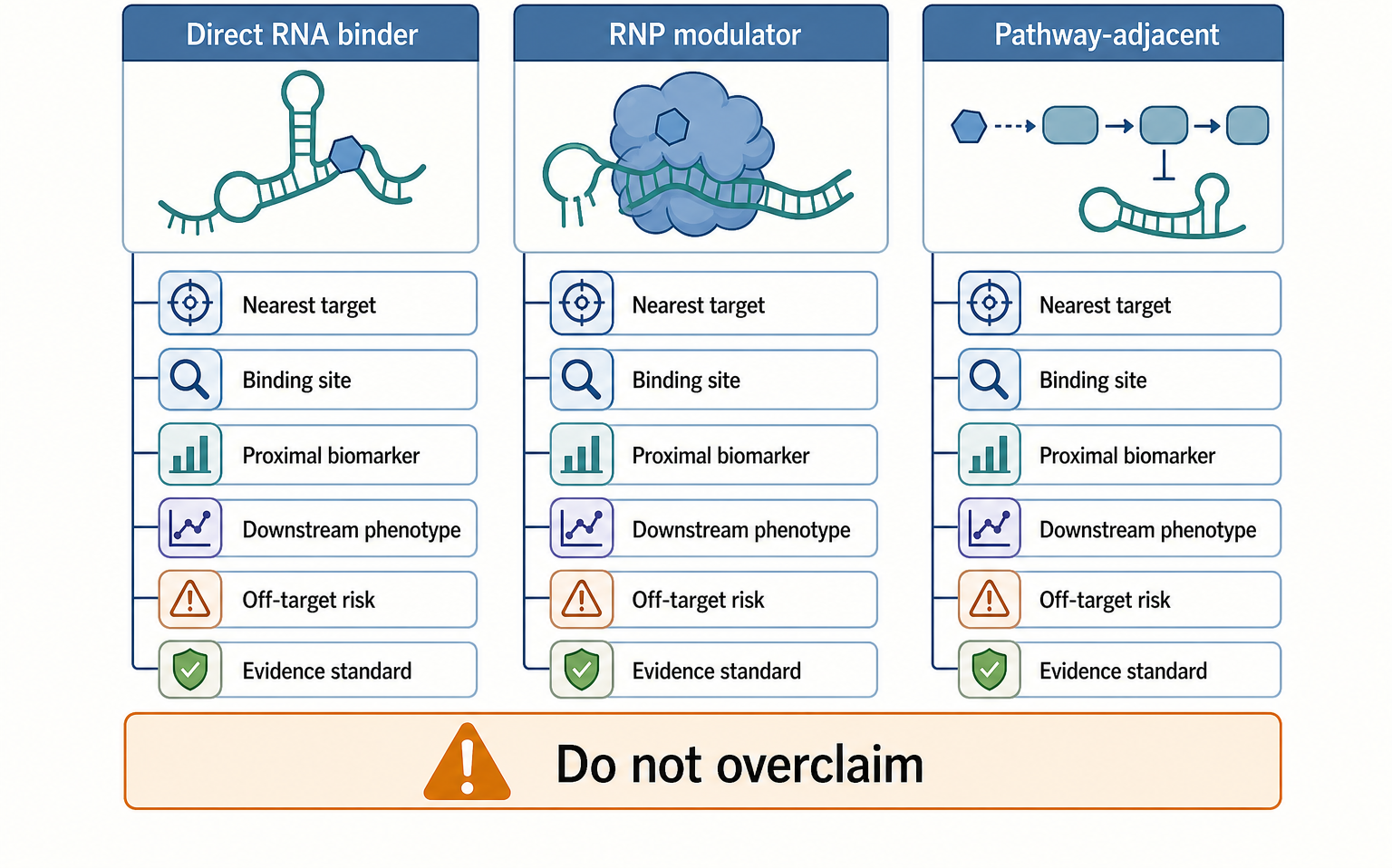
Figure 161.1. Taxonomy of Small-Molecule RNA Pharmacology. Small-molecule RNA pharmacology spans direct RNA binding, modulation of RNA-rich RNP machines, and pathway-adjacent effects on RNA biology. The evidence required for each class differs because binding site, proximal biomarker, and off-target risks differ.
The most mature examples are not the newest ones. Ribosome antibiotics show that a small molecule can bind an RNA-rich target and change a central biological process. Aminoglycosides, macrolides, tetracyclines, oxazolidinones, pleuromutilins, chloramphenicol-like drugs, and related classes occupy distinct sites near the decoding center, peptide exit tunnel, aminoacyl-tRNA accommodation site, or peptidyl transferase center. These are not all “direct RNA binders” in the same simple way. Some bind rRNA almost entirely; some contact both rRNA and ribosomal proteins; some bind conformational states that exist only in the assembled ribosome. Their shared lesson is that druggability can emerge from RNA architecture embedded in a large machine.
Splicing modifiers create a second anchor. Risdiplam, the best-known systemic small-molecule splicing modifier for spinal muscular atrophy, changes inclusion of exon 7 in SMN2 pre-mRNA. The disease context is important. Humans have SMN1 and SMN2 genes. Loss of functional SMN1 causes spinal muscular atrophy, while SMN2 mostly produces transcripts that skip exon 7 because a single-nucleotide difference weakens the splicing grammar. A drug that increases SMN2 exon 7 inclusion can increase full-length survival motor neuron protein. This is RNA pharmacology, but the drug does not simply coat all SMN2 RNA. The pharmacological event is local stabilization of a productive pre-mRNA-protein arrangement at a weak exon-definition site.
Pathway-adjacent drugs require a boundary line. A protein kinase inhibitor that changes alternative splicing through phosphorylation of splicing factors is relevant to RNA biology but is not a direct RNA-targeting drug. A ribonucleoside analog that is incorporated by a viral RNA-dependent RNA polymerase acts through RNA synthesis and viral RNA mutagenesis or chain termination, but its immediate target class is the polymerase-substrate pathway; Chapter 160 treats that modality. A drug that inhibits an RNA methyltransferase, decapping enzyme, ribonuclease, or helicase may be an RNA-pathway drug without being an RNA binder. The boundary is not meant to diminish those drugs. It prevents mechanistic overclaiming.
Box 161.1. Mechanistic Labels Should Follow the Nearest Target
When classifying an RNA-related small molecule, ask what physical event is closest to the causal pharmacology. If the active compound binds a defined RNA motif and loss of that motif removes the response, the term direct RNA binder is appropriate. If the compound acts only in an assembled ribosome, spliceosome, or other ribonucleoprotein, call it an RNA-rich RNP modulator and describe the RNA and protein surfaces together. If the compound inhibits a kinase, polymerase, nuclease, methyltransferase, transporter, or metabolic pathway that later changes RNA abundance or splicing, call it pathway-adjacent RNA pharmacology. Downstream RNA sequencing, reporter changes, viral-load decline, or altered protein expression can support pharmacodynamics, but they do not by themselves identify the nearest target. This vocabulary discipline protects useful pathway drugs from being mislabeled and prevents weak direct-binding claims from gaining authority through RNA-associated outcomes.
Table 161.1. Evidence Standards by Modality Class. Mechanistic labels should follow evidence. A cellular RNA phenotype does not by itself prove direct RNA binding.
| Modality class | Target entity | Example | Proximal evidence | Common overclaim | Key safety or selectivity concern |
|---|---|---|---|---|---|
| Direct RNA binder | Folded RNA motif or transcript domain whose binding event is causal. | Riboswitch ligand, viral RNA-structure ligand, or repeat RNA binder. | Defined RNA binding plus motif mutation, cellular occupancy, and proximal RNA output. | Treating in vitro affinity or reporter change as proof of therapeutic target engagement. | Transcriptome motif recurrence, nonspecific nucleic acid binding, and compartment access. |
| RNA-rich RNP modulator | Assembled RNP machine with joint RNA-protein pharmacological surface. | Ribosome antibiotic or risdiplam-like SMN2 exon 7 modifier. | Structural or resistance mapping, RNP-context binding, and target-dependent functional shift. | Describing RNP action as naked-RNA binding or global splicing activation. | Microbial resistance, mitochondrial translation toxicity, or off-target splice changes. |
| Pathway-adjacent RNA drug | Protein enzyme, nucleotide pathway, or signaling state that changes RNA output. | Viral polymerase-pathway analog, kinase-linked splice change, or RNA enzyme inhibitor. | Enzyme inhibition, substrate or pathway biomarker, and downstream RNA profile. | Calling any RNA phenotype a direct RNA-targeting mechanism. | Pleiotropic pathway effects and transcriptomic changes dominated by stress. |
| Oligonucleotide comparator | Sequence-defined RNA region recognized by base pairing. | Antisense oligonucleotide, siRNA, or splice-switching oligo. | Hybridization-dependent knockdown, splice switching, or RNase H/RISC activity. | Assuming small molecules can use the same sequence-information standard. | Delivery, innate immune activation, chemistry-specific toxicity, and off-target pairing. |
| Chemical biology probe | RNA or RNP feature used to test mechanism rather than treat disease. | Micromolar RNA binder, pull-down ligand, or inactive analog pair. | Reproducible assay perturbation, structure-activity relationship, and orthogonal control. | Presenting a useful probe as a drug lead without exposure or safety evidence. | Partial selectivity, assay artifacts, cellular accumulation, and limited therapeutic window. |
The taxonomy also clarifies evidence. A direct RNA binder should be supported by biochemical binding to a defined RNA, structure-activity relationships that track RNA affinity and functional activity, loss of activity when the target motif is mutated, cellular target engagement at relevant concentrations, and a proximal RNA output that changes before downstream phenotypes. An RNP modulator may be supported by cryogenic electron microscopy, crosslinking, toeprinting, resistant ribosomal mutations, splicing reporter alleles, or genetic rescue. A pathway-adjacent drug may be supported by enzyme inhibition, metabolite changes, and transcriptomic consequences. Problems arise when a cellular phenotype is used as if it proved direct RNA binding. Cytotoxic stress, translation slowdown, transcriptional repression, unfolded-protein responses, and innate immune activation can all reshape RNA readouts.
RNA-targeting small molecules also differ from oligonucleotide therapeutics. Antisense oligonucleotides, siRNAs, and splice-switching oligos use base pairing as their recognition principle and often require delivery technologies discussed in Chapters 149 to 158. Small molecules are usually shorter, more diffusible, and more compatible with oral dosing, but they have less sequence information. A 20-mer oligonucleotide can distinguish a near-unique transcript region by Watson-Crick pairing. A 500-Da small molecule usually recognizes shape, electrostatics, and a handful of base edges. Direct RNA small-molecule selectivity therefore depends on rare three-dimensional motifs, cooperative RNP context, local concentration, kinetic residence time, or disease-specific exposure of a structure.
The field sometimes uses “ligandability” as if every folded RNA has a drug pocket. A more cautious definition is needed. A ligandable RNA site has a recurrent conformation, a binding pocket or surface with enough chemical information for selective recognition, sufficient occupancy at safe drug exposure, and a causal relationship between binding and the desired biological output. A structured RNA may be detectable by chemical probing yet pharmacologically poor if it is transient, inaccessible in RNP context, too similar to many other RNAs, or downstream of the true disease driver. Conversely, an RNA site with modest affinity can be therapeutically useful if occupancy changes a highly amplified process, such as translation or splicing, and if the required exposure is tolerated.
The practical taxonomy for this chapter is therefore evidence based, not marketing based. Direct RNA pharmacology is strongest when the target is known, the binding mode is plausible, and the cellular response is proximal. RNP pharmacology is strongest when the RNA-containing machine is structurally defined and resistance maps to the intended site. Pathway-adjacent pharmacology is strongest when the protein or metabolic target is explicit and RNA changes are interpreted as downstream pharmacodynamic effects. The next sections apply this framework to approved anchors, emerging target classes, and clinical boundaries.
161.2. Approved Anchors: rRNA-Binding Antibiotics and Ribosome Functional Centers
Ribosome antibiotics are the clearest proof that RNA-rich structures can be drugged by small molecules. The bacterial ribosome contains the small 30S subunit, where messenger RNA decoding is coupled to transfer RNA anticodon selection, and the large 50S subunit, where peptide-bond formation and nascent-chain passage occur. Ribosomal RNA forms much of the active architecture. The decoding center in 16S rRNA monitors codon-anticodon geometry. The peptidyl transferase center in 23S rRNA positions the aminoacyl and peptidyl substrates. The exit tunnel is lined substantially by 23S rRNA and shapes nascent-peptide progression. These are functional centers, not passive scaffolds.
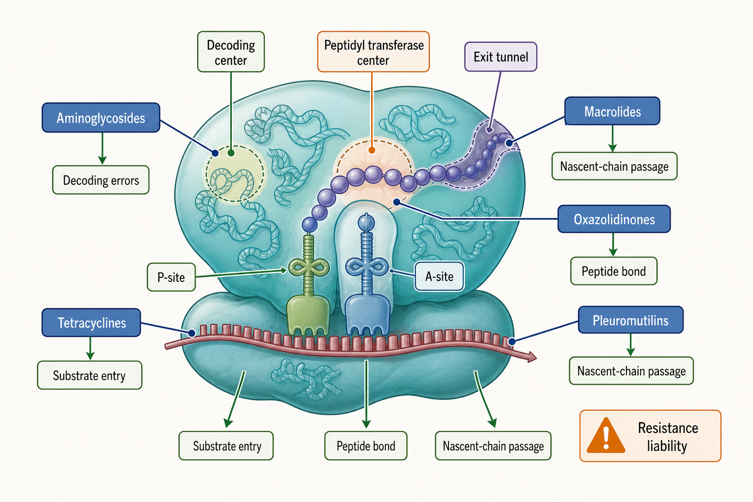
Figure 161.2. Ribosome Functional Centers Targeted by Antibiotics. Ribosome antibiotics demonstrate that RNA-rich functional centers can be drugged, but each class affects a different step in translation and has distinct resistance and toxicity liabilities.
Different antibiotic classes teach different principles. Aminoglycosides bind near the decoding center and can promote misreading, inhibit translocation, and perturb initiation depending on drug and organism. Tetracyclines bind near the A site and interfere with aminoacyl-tRNA accommodation. Macrolides bind in the peptide exit tunnel and can block elongation in a sequence-dependent manner rather than acting as a simple cork for every nascent peptide. Oxazolidinones act near the peptidyl transferase center and interfere with initiation or early elongation. Pleuromutilins bind the peptidyl transferase center and affect substrate positioning. Chloramphenicol-like drugs also target peptidyl transferase chemistry.
The ribosome example prevents two common overgeneralizations. First, RNA-targeted small molecules do not need to bind isolated RNA in solution to be useful. Many ribosome inhibitors recognize the assembled ribosome, a defined RNP state that presents the pocket. Isolated rRNA fragments may reproduce some binding features for certain drugs, but the therapeutic target is the cellular ribosome. Second, a drug can bind an RNA-dominated site and still have protein-dependent selectivity. Ribosomal proteins, rRNA modifications, local ions, subunit assembly, and species-specific sequence differences shape pocket geometry and resistance.
The evidence base for ribosome antibiotics is unusually strong because structural biology, genetics, biochemistry, microbiology, and clinical pharmacology converge. High-resolution structures locate binding sites. In vitro translation assays show functional inhibition. Toeprinting and ribosome profiling can reveal where ribosomes stall or mistranslate. Resistance mutations map to rRNA nucleotides, ribosomal proteins, methyltransferase-protected residues, or drug-modifying enzymes. Microbiological minimum inhibitory concentrations connect molecular activity to bacterial growth. Clinical experience then adds absorption, distribution, safety, microbiome, and resistance-selection constraints. This multi-layer evidence standard is a model for newer RNA-directed programs.
Resistance is not an afterthought; it is part of the mechanism. A bacterium can become resistant by mutating the rRNA binding site, changing ribosomal proteins that shape the pocket, methylating a key rRNA nucleotide, enzymatically modifying the antibiotic, reducing uptake, increasing efflux, or bypassing the affected process. For macrolides, methylation of 23S rRNA near the exit tunnel can block binding for a broad group of drugs. For aminoglycosides, drug-modifying enzymes and uptake barriers are common concerns. For linezolid-like oxazolidinones, rRNA mutations and methyltransferase-mediated resistance can emerge.
Selectivity has several layers. Bacterial cytosolic ribosomes differ from eukaryotic cytosolic ribosomes enough to create useful antibacterial windows for many drugs. However, human mitochondria descend from bacteria and contain mitoribosomes that retain translation machinery with bacterial ancestry. Some ribosome-targeting antibiotics can therefore impair mitochondrial translation, especially in susceptible tissues or genetic contexts. Ototoxicity, nephrotoxicity, marrow toxicity, neuropathy, and drug-drug interactions differ by antibiotic class and cannot be explained solely by rRNA binding. A pharmacological account must combine target conservation, tissue exposure, transport, metabolism, and patient risk factors.
Ribosome antibiotics also show why “translation inhibitor” is too coarse a label. A drug that causes codon misreading, a drug that blocks aminoacyl-tRNA entry, and a drug that stalls selected nascent peptide contexts all reduce protein synthesis but create different downstream biology. Sequence-dependent stalling can change bacterial stress responses and resistance phenotypes. Misreading can produce toxic proteins. Initiation inhibition affects proteome synthesis from the beginning of open reading frames. These distinctions matter when comparing antibacterial potency, resistance selection, synergy, and host toxicity.
For direct RNA pharmacology, the central lesson is that rRNA pockets are druggable because they are conserved, structured, functionally important, and exposed in a high-copy cellular machine. The same features are not guaranteed for a disease-associated lncRNA or viral UTR. The ribosome is abundant, essential, and structurally constrained. Many proposed RNA targets are lower abundance, more dynamic, more cell-type specific, and less structurally characterized. Ribosome antibiotics establish possibility, not general ease.
161.3. Splicing Modifiers: Risdiplam-Like Mechanisms and SMN2 Exon Inclusion
Small-molecule splicing modifiers provide a different kind of RNA pharmacology. Splicing is a decision process made by precursor mRNA sequence elements, RNA secondary structure, small nuclear ribonucleoproteins, and regulatory proteins. An exon is included when the spliceosome successfully recognizes the upstream and downstream splice sites and assembles a productive catalytic complex. Many disease alleles weaken splice sites or regulatory elements without eliminating the gene. A small molecule can be therapeutic if it shifts the local probability of exon inclusion, exon skipping, or poison-exon usage in the desired direction.
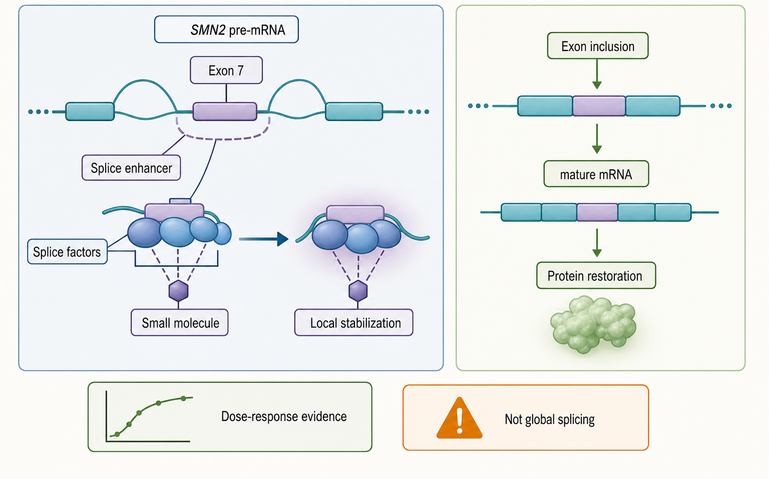
Figure 161.3. SMN2 Exon 7 Splice Correction by a Small Molecule. Splice-correcting small molecules can act as local stabilizers of a productive pre-mRNA-splicing-factor arrangement. The therapeutic claim is strongest when exon inclusion, protein restoration, and clinical response are connected by dose-response evidence.
The SMN2 example is the clearest approved anchor. Spinal muscular atrophy results from insufficient survival motor neuron protein. SMN1 loss removes the main source of functional protein. SMN2 is a paralog that could compensate, but most SMN2 transcripts skip exon 7 and encode unstable or less functional products. Increasing exon 7 inclusion raises full-length SMN protein. A risdiplam-like drug acts by stabilizing recognition of the exon 7 region in a way that favors productive splicing. The useful mental model is molecular stabilization of a weak exon-definition interface, not global spliceosome activation.
Mechanistically, a splice-modifying small molecule can operate through several routes. It may bind the pre-mRNA near a splice site and change local structure. It may stabilize the interaction between a pre-mRNA sequence and a splicing factor. It may recruit or block an RNA-binding protein. It may alter spliceosome assembly kinetics. It may inhibit a kinase or enzyme that controls splicing-factor state, although that would be pathway-adjacent rather than direct RNA engagement. In the strongest RNA-proximal cases, sequence changes in the target pre-mRNA alter drug activity, and structural or biochemical experiments show that the compound helps form a productive RNA-protein complex.
Table 161.2. Splicing Modifier Evidence and Biomarker Hierarchy. Splicing-modifier programs require evidence from local RNA-protein mechanism through disease-relevant protein and clinical response.
| Evidence layer | Example measurement | What it supports | Limitation | Preferred control |
|---|---|---|---|---|
| In vitro RNA/RNP binding | Binding to SMN2 exon 7 RNA or pre-mRNA-splicing-factor complex. | Local RNA-proximal mechanism and possible molecular-glue behavior. | Purified fragments may miss nuclear RNP context and spliceosome kinetics. | Target-sequence variants, inactive analogs, and reconstituted RNP assays. |
| Minigene reporter | Percent-spliced-in change from an exon 7 reporter construct. | Direction and dose dependence of the local splice decision. | Plasmids lack chromatin, transcription kinetics, and endogenous factor stoichiometry. | Matched mutant reporter and comparison with endogenous transcript response. |
| Endogenous exon inclusion | RT-PCR or RNA-seq measurement of SMN2 exon 7 inclusion. | Proximal pharmacodynamic correction in native transcript context. | Bulk assays may miss rare tissues, nascent RNA timing, or low-abundance isoforms. | Time course, dose response, motif disruption, and tissue-relevant sampling. |
| Full-length protein | SMN protein restoration by immunoassay or proteomics. | Translation of splice correction into disease-relevant protein rescue. | Protein level integrates turnover, cell type, and post-transcriptional regulation. | Exon-inclusion linkage, genetic rescue logic, and tissue-specific protein readout. |
| Transcriptome-wide off-target splicing | RNA-seq isoform analysis after active exposure. | Selectivity window and unintended exon or poison-exon changes. | Stress, cell-cycle shifts, and cell-state changes can dominate splice profiles. | Inactive analog, matched exposure, short time course, and stress-marker panel. |
| Clinical outcome | Motor milestone, survival, respiratory support, or functional scale. | Patient-level benefit for a genetic-disease splice modifier. | Outcome depends on disease stage, supportive care, tissue exposure, and genotype. | Biomarker-linked exposure response and prespecified genotype or severity strata. |
Splicing pharmacology is unusually sensitive to dose. Too little drug may leave the disease exon mostly skipped. Too much drug may affect additional splice events, stress pathways, or developmentally sensitive transcripts. A therapeutic splice change is usually measured as percent spliced in, exon-inclusion ratio, full-length transcript abundance, full-length protein, or downstream clinical biomarker. The causal chain matters. A change in percent spliced in is proximal. Increased protein is closer to disease correction. Motor function, survival, or hospitalization endpoints are clinical outcomes shaped by disease stage, tissue exposure, and supportive care.
Target engagement is difficult because the binding event occurs on a transient precursor mRNA in the nucleus. Mature mRNA measurements can miss the timing and location of drug action. Bulk RNA sequencing can detect global splice changes but may under-sample rare cell types, nascent transcripts, and low-abundance poison exons. Reporter assays are useful for mechanism, yet a plasmid reporter lacks chromatin context, transcription kinetics, endogenous RNA-binding-protein stoichiometry, and tissue-specific regulation. A robust program therefore combines minigene or reporter assays, endogenous transcript measurements, protein rescue, genetic disruption of the binding motif, and clinical pharmacodynamic markers.
Risdiplam-like success should not be generalized to all alternative splicing. SMN2 exon 7 is a special therapeutic opportunity: one gene, a defined exon, a clear loss-of-function disease mechanism, and a measurable protein rescue. Many disease-associated splice changes are secondary consequences of cell stress, cancer state, or tissue composition. Other splice switches may affect dozens of isoforms with uncertain protein products. Some exons are regulated by long-range chromatin or transcription-speed effects that a local RNA ligand cannot easily correct. For these targets, a small molecule may show transcript changes without proving a therapeutic mechanism.
Box 161.2. Why SMN2 Exon 7 Is an Anchor, Not a Shortcut
SMN2 exon 7 correction is a useful teaching anchor because several links in the causal chain are unusually direct. The disease is driven by insufficient survival motor neuron protein after loss of functional SMN1. SMN2 can produce the needed protein, but a weak exon-definition context causes most transcripts to skip exon 7. A small molecule that increases exon 7 inclusion can therefore be evaluated through a logical sequence: local splice change, full-length transcript, SMN protein restoration, tissue exposure, and patient benefit. Many proposed splicing targets lack one or more of these links. A cancer-associated exon may be a marker of cell state rather than a driver. A poison exon may affect multiple isoforms with uncertain protein consequences. A transcript change in blood may not reflect the disease tissue. The SMN2 example teaches the evidence pattern, not a guarantee that every splice switch is druggable.
There are also safety boundaries. Splicing is deeply connected to development, cell identity, DNA-damage responses, and proteome diversity. Broad spliceosome inhibitors can be cytotoxic and have been explored in oncology, where killing rapidly dividing cells may be acceptable only within a narrow window. A splice-correcting drug for a genetic disease usually needs chronic systemic exposure and therefore much higher selectivity. Reproductive toxicity, developmental exposure, off-target splice changes, and tissue-specific protein rescue are central regulatory concerns.
The evidence logic for splicing modifiers has become a template for RNA pharmacology. The target is not simply “SMN2 RNA”; it is a particular sequence context, splice decision, cell compartment, and disease mechanism. The drug effect is not just binding; it is productive remodeling of an RNA-protein decision point. The biomarker is not any transcriptomic change; it is a dose-dependent correction of the disease-relevant exon and protein. Emerging RNA-directed programs should match this specificity before claiming comparable maturity.
161.4. Emerging Direct RNA Targets: Riboswitches, Repeats, Viral RNA, miRNA Precursors, lncRNAs, and Structured UTRs
Emerging direct RNA targets differ in how much biological precedent they offer. Riboswitches are the most persuasive natural example. A bacterial riboswitch contains an aptamer domain that binds a metabolite and an expression platform that changes transcription, translation, splicing in rare systems, or RNA stability. The aptamer domain proves that RNA can form a selective small-molecule pocket under cellular conditions. A synthetic or medicinal ligand can in principle mimic, compete with, or distort the natural ligand response. Antibiotic development against riboswitches remains difficult, however, because bacterial uptake, efflux, pathway redundancy, resistance mutations, and microbiome selectivity all matter.
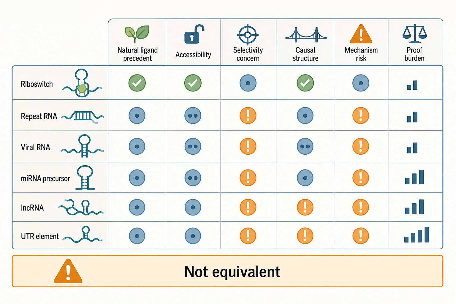
Figure 161.4. Emerging Direct RNA Target Classes and Their Failure Modes. Emerging RNA target classes are not equivalent. Riboswitches have natural ligand-binding precedent, whereas many host transcript targets require stronger proof that the mature RNA structure is causal, accessible, and selective.
Repeat-expansion RNAs provide another target class. Diseases such as myotonic dystrophy and several neurodegenerative disorders can involve expanded CUG, CCUG, GGGGCC, CGG, or other repeats. Repeat RNAs may form hairpins, G-quadruplexes, R-loops, nuclear foci, or RNP assemblies; they may sequester RNA-binding proteins; and some repeats undergo repeat-associated non-AUG translation to produce toxic peptides. A direct RNA ligand might disrupt protein sequestration, alter repeat structure, reduce foci, or change translation. The boundary case is crucial: the disease mechanism may include toxic RNA, toxic protein, DNA instability, chromatin effects, and stress responses at once. A compound that reduces foci is not automatically disease modifying unless the relevant toxic pathway is shown to change.
Viral RNAs are attractive because viruses often require conserved RNA structures for replication, translation, frameshifting, packaging, or immune evasion. Examples include internal ribosome entry sites, programmed ribosomal frameshift elements, replication signals, packaging signals, and structured untranslated regions. The druggability argument is strong when the viral RNA element is conserved, functionally constrained, structurally characterized, and exposed in infected cells. The development risk is also clear. Viral populations mutate. Host RNAs may contain similar motifs. Some viral RNA structures are shielded by proteins or membranes. A ligand must reach infected cells at the right time and concentration without triggering host toxicity.
MicroRNA precursors offer compact, structured host RNA targets. A primary microRNA or precursor microRNA is processed by Drosha or Dicer to produce a mature microRNA that guides Argonaute-mediated repression. A small molecule could bind a pri-miRNA or pre-miRNA hairpin and block processing, alter strand selection, or change abundance of a disease-linked microRNA. This strategy is conceptually different from antagomirs and locked nucleic acid anti-miRs, which bind mature microRNAs by base pairing. The selectivity challenge is substantial because hairpin motifs recur throughout the transcriptome, and changes in mature microRNA abundance can be indirect consequences of transcription, Dicer activity, cell state, or toxicity.
Long noncoding RNAs are a more heterogeneous class. Some lncRNAs act through chromatin recruitment, nuclear body architecture, transcriptional interference, RNA-RNA pairing, protein sequestration, or translation of small open reading frames. A small molecule could in principle bind a structured domain in a lncRNA and disrupt a protein interaction or conformation. The problem is that many lncRNAs lack high-confidence structure-function maps. Expression can be low and cell-type specific. Loss-of-function phenotypes may depend on the act of transcription rather than the RNA product. A direct lncRNA ligand claim therefore needs especially careful controls: genetic rescue with binding-site mutants, evidence that the mature RNA molecule is the causal entity, and orthogonal target engagement in the relevant compartment.
Structured untranslated regions in mRNAs are intermediate cases. A 5′ UTR may contain an upstream open reading frame, an internal ribosome entry element, a G-quadruplex, a thermosensor, an iron-responsive element, or another regulatory structure. A 3′ UTR may recruit RNA-binding proteins or microRNAs, localize the transcript, or control decay. A small molecule might change translation initiation, ribosome scanning, RNA stability, or localization. The danger is that translation and stability are highly sensitive to stress. A compound that globally slows translation can make a structured UTR appear regulated. A credible direct UTR-targeting program must separate local motif engagement from general changes in initiation, elongation, ribosome quality control, or mRNA decay.
Table 161.3. Emerging Direct RNA Targets. Emerging direct RNA targets vary in natural precedent, accessibility, and proof burden. Each class needs a different target-engagement strategy.
| Target class | Biological rationale | Desired drug effect | Strongest evidence type | Principal failure mode | Citation curation need |
|---|---|---|---|---|---|
| Riboswitch | Natural aptamer domains prove selective metabolite recognition by RNA. | Mimic, compete with, or distort expression-platform switching. | Bound structures, bacterial target engagement, uptake, and resistance mapping. | Poor uptake, efflux, pathway redundancy, or resistance mutations. | Riboswitch ligand-recognition reviews and antibacterial development examples. |
| Repeat-expansion RNA | Expanded repeats form disease-linked hairpins, G-quadruplexes, foci, or RNP assemblies. | Disrupt protein sequestration, repeat structure, foci, or repeat-associated translation. | Cellular occupancy plus change in toxic RNA/protein pathway and disease biomarker. | Disease may persist through toxic peptides, DNA instability, or stress arms. | Repeat-RNA targeting, RNA foci, and repeat-associated translation sources. |
| Viral RNA structure | Conserved elements can control replication, translation, frameshifting, or packaging. | Block required viral RNA function or sensitize viral replication to combination therapy. | Infected-cell engagement, conserved-site mutation, resistance map, and viral-load response. | Viral escape, host-RNA mimicry, shielding by RNPs or membranes, or indirect antiviral stress. | Viral RNA ligand reviews, UTR/IRES/frameshift examples, and resistance studies. |
| miRNA precursor | Pri-miRNA and pre-miRNA hairpins are compact processing substrates. | Block Drosha or Dicer processing, alter strand selection, or change mature miRNA abundance. | Processing-specific assay with hairpin mutation and mature-miRNA output. | Mature miRNA changes can reflect transcription, Dicer activity, toxicity, or cell state. | miRNA precursor ligand papers and microRNA-biogenesis control references. |
| lncRNA | Some lncRNAs require structured domains for protein interaction or architecture. | Disrupt causal RNA conformation, RNP assembly, or localization function. | Rescue with binding-site mutants and compartment-specific target engagement. | Phenotype may arise from DNA locus, transcription, chromatin, or neighboring genes. | lncRNA structure-function, causality standards, and ligand-targeting reviews. |
| Structured UTR | UTR motifs can regulate initiation, frameshifting, stability, localization, or decay. | Shift translation, ribosome scanning, RNA stability, or RBP/miRNA recruitment. | Local motif mutation with ribosome profiling or RNA-stability readout. | Global translation stress or decay changes can mimic UTR-specific regulation. | Structured UTR modulation, translation-control, and stress-artifact references. |
Across these target classes, several evidence questions repeat. Is the target RNA structure present in cells, or only in vitro? Is the structure populated enough for occupancy? Is the ligand active at concentrations below nonspecific RNA binding, membrane disruption, or stress response thresholds? Does a mutation that preserves general RNA abundance but disrupts the pocket reduce drug response? Does an analog that loses binding also lose cellular activity? Does target engagement precede phenotype? Does transcriptome-wide profiling show a selectivity pattern consistent with the proposed motif?
These questions make direct RNA targeting harder than simple target lists suggest. A transcript can be disease-associated without being ligandable. A ligand can bind RNA without being selective. A selective ligand can fail because the target is inaccessible. A cellular phenotype can be real but indirect. A compound can change a biomarker without improving disease. The emerging field is strongest when it treats each proposed target as a mechanistic hypothesis rather than as proof that RNA is broadly druggable.
161.5. Pharmacology, Exposure, Biomarkers, and Target Engagement
RNA-targeted small molecules must satisfy the same pharmacological requirements as other drugs: absorption, distribution, metabolism, excretion, exposure at the site of action, target engagement, pharmacodynamic response, efficacy, and safety. RNA targets add special complications. RNA molecules occupy specific compartments, appear and disappear during processing, fold into ensembles rather than single static structures, bind proteins, and may be present in only a subset of disease cells. A compound can look potent in a biochemical assay and fail because it never reaches the nuclear pre-mRNA, viral replication compartment, bacterial cytosol, neuronal tissue, or mitochondrial off-target site at the right free concentration.
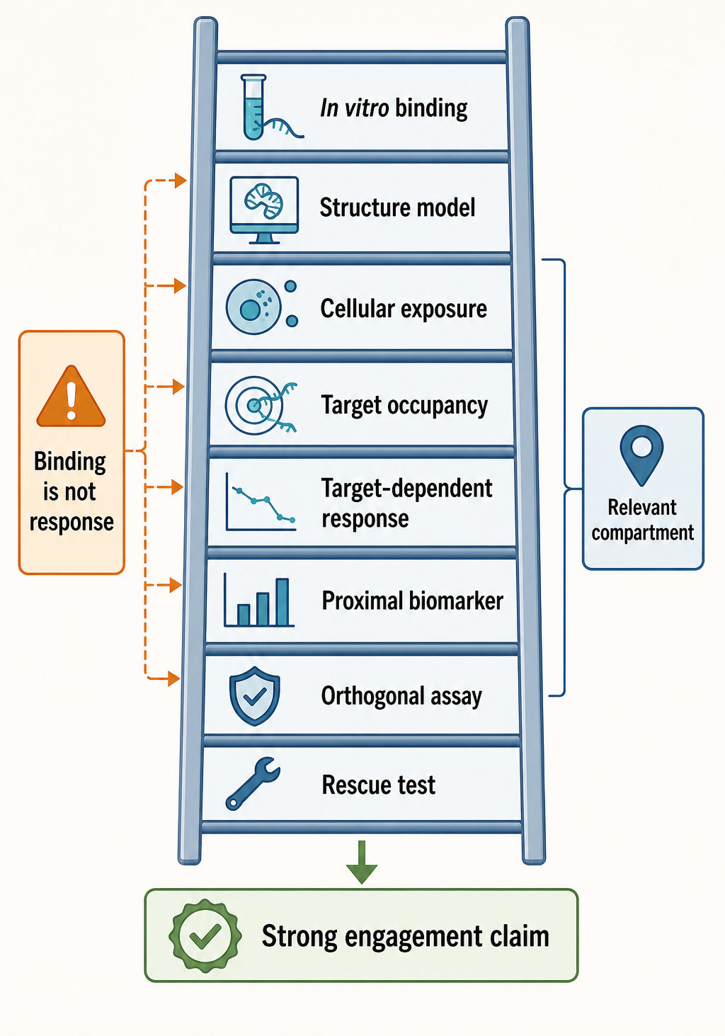
Figure 161.5. Evidence Ladder for RNA Target Engagement. RNA target engagement requires orthogonal evidence. The strongest claims connect binding, target-dependent cellular response, relevant exposure, and proximal pharmacodynamic biomarkers.
Exposure should be interpreted as free drug at the relevant compartment, not only total plasma concentration. Protein binding, lysosomal trapping, transporter activity, tissue partitioning, blood-brain barrier penetration, bacterial-envelope permeability, and intracellular pH can all alter effective concentration. Risdiplam-like systemic splicing therapy requires exposure in tissues where SMN protein matters, including motor neurons and peripheral tissues. Ribosome antibiotics require bacterial intracellular exposure while limiting host toxicity. Viral RNA ligands require exposure in infected cells and often in membrane-associated replication compartments. Repeat-expansion RNA ligands for neurological disease may require central nervous system exposure and long-term tolerability.
Biomarkers should be arranged by proximity to the target. The closest marker is direct occupancy or structural change at the RNA. Next are immediate RNA outputs such as exon inclusion, ribosome stalling at a defined site, blocked microRNA processing, altered frameshifting, or disrupted protein-RNA association. Further downstream are protein restoration, pathway correction, cell survival, bacterial killing, viral-load reduction, or clinical function. Downstream biomarkers are often more clinically meaningful, but they are less specific for target engagement. A fall in viral load could result from immune activation, polymerase inhibition, entry inhibition, or cytotoxicity rather than direct binding to a viral RNA element.
Target-engagement methods for RNA are powerful but artifact-prone. Chemical probing can reveal ligand-induced protection or reactivity changes, but changes may reflect global folding shifts, protein displacement, or altered RNA abundance. Pull-down or affinity capture can identify enriched RNAs, but sticky cationic ligands and abundant RNAs create false positives. Crosslinking can help but depends on photochemistry and proximity. Cellular thermal shift concepts are less straightforward for RNA than for proteins. Mutational resistance or loss-of-response alleles are often more convincing because they connect a sequence or structural feature to drug activity. Even then, mutations can change RNA expression or processing, so controls are required.
Transcriptome-wide profiling is necessary for selectivity but insufficient for mechanism. RNA sequencing can reveal off-target splicing, expression, or isoform changes. Ribosome profiling can detect translation changes and ribosome stalls. Structure probing can map transcriptome-wide chemical reactivity shifts. Proteomics can show downstream protein consequences. However, a large data set does not automatically identify the direct target. Cell stress, cell-cycle changes, apoptosis, innate immune activation, and differentiation state can dominate omics signatures. The best interpretation combines time course, dose response, inactive analogs, resistant target alleles, rescue experiments, and orthogonal assay types.
Table 161.4. Biomarker Proximity for RNA-Targeting Small Molecules. Proximal biomarkers are more mechanistically specific, whereas downstream outcomes are more clinically meaningful but less diagnostic of the direct target.
| Biomarker level | RNA-drug example | Interpretation | Main confounder | Validation need |
|---|---|---|---|---|
| Direct occupancy or structure change | Ligand-induced protection in target RNA or bound RNP structure. | Closest evidence for physical engagement of the intended RNA state. | Protein displacement, RNA abundance shifts, or global folding changes. | Orthogonal binding assay, inactive analog, and target-motif loss of response. |
| Local splice correction | SMN2 exon 7 percent-spliced-in increase after risdiplam-like exposure. | Proximal pharmacodynamic response for a splice-correcting drug. | Reporter or blood signal may not match disease-relevant tissue. | Endogenous transcript assay, protein linkage, exposure response, and tissue rationale. |
| Ribosome stall or frameshift change | Toeprinting, ribosome profiling, or viral frameshift reporter shift. | Translation-machine or viral-RNA functional modulation near the proposed site. | Global translation inhibition, cytotoxic stress, or reporter context artifacts. | Site mutation, resistant allele, time course, and matched active/inactive compounds. |
| Protein restoration or viral-load decline | SMN protein increase or reduced viral RNA after treatment. | Downstream biological response with stronger disease relevance than occupancy alone. | Protein turnover, immune activation, polymerase inhibition, or cell death can dominate. | Quantitative link to proximal RNA marker and mechanism-discriminating controls. |
| Clinical function or infection cure | Motor-function improvement or microbiological/virological cure. | Patient-level benefit and benefit-risk relevance. | Endpoint may not reveal whether the proposed RNA target was causal. | Prespecified clinical endpoint, exposure-response model, resistance monitoring, and safety profile. |
Pharmacodynamics also depends on target turnover. A nuclear pre-mRNA splice decision may respond quickly to drug but disappear once transcription changes. A mature lncRNA with slow turnover may require prolonged exposure before occupancy changes function. A ribosome inhibitor can act rapidly because ribosomes are abundant and essential, but bacterial killing may depend on growth state and immune context. A viral RNA structure may be present only during a replication phase. A repeat-expansion RNA focus may persist even if newly synthesized toxic RNA is reduced. Dose scheduling should match these kinetics.
Another practical problem is stoichiometry. A small molecule does not need to bind every target molecule if partial modulation is enough, but high target abundance can create a sink. Ribosomal RNA is abundant in bacteria; antibiotics overcome this through potency, uptake, and essentiality. Some repeat RNAs or highly expressed lncRNAs may require high occupancy to disrupt foci. Low-abundance pre-mRNAs may be easier stoichiometrically but harder to access and measure. Motif recurrence across the transcriptome can create distributed binding that reduces free drug and causes off-target effects.
Exposure-response modeling should distinguish concentration that changes a proximal RNA marker from concentration that changes disease biology. The two can diverge. A splicing marker in blood may not report splicing in neurons. A bacterial minimum inhibitory concentration may not predict intracellular activity in a biofilm. A viral RNA-binding signal in transfected reporter cells may not predict infected-airway pharmacology. Regulatory confidence increases when biomarkers are mechanistically proximal, measured in relevant tissue or a justified surrogate, and linked quantitatively to clinical outcomes.
For this reason, direct RNA pharmacology should avoid relying on a single signature assay. A strong package might show in vitro binding to a target motif, structural definition of the bound state, loss of activity with target mutations, cellular target engagement at unbound concentrations near therapeutic exposure, proximal RNA output correction, limited transcriptome-wide off-target modulation, exposure-response in disease tissue, and clinical or organismal benefit. Not every approved drug had all of these data at discovery, but this is the standard emerging programs should approximate before making strong mechanistic claims.
161.6. Resistance, Toxicity, Selectivity, and Regulatory Classification
Resistance and toxicity are connected by selectivity. A drug that binds a highly conserved bacterial rRNA site may be potent, but bacterial evolution can alter the pocket, protect it by methylation, or reduce intracellular drug. A drug that binds a recurring human RNA motif may face less microbial resistance but more host toxicity. A splice modifier can be exquisitely useful at one exon and harmful if it changes many other exons. The desired pharmacology is selective enough to help the patient but robust enough that the target cannot easily escape.
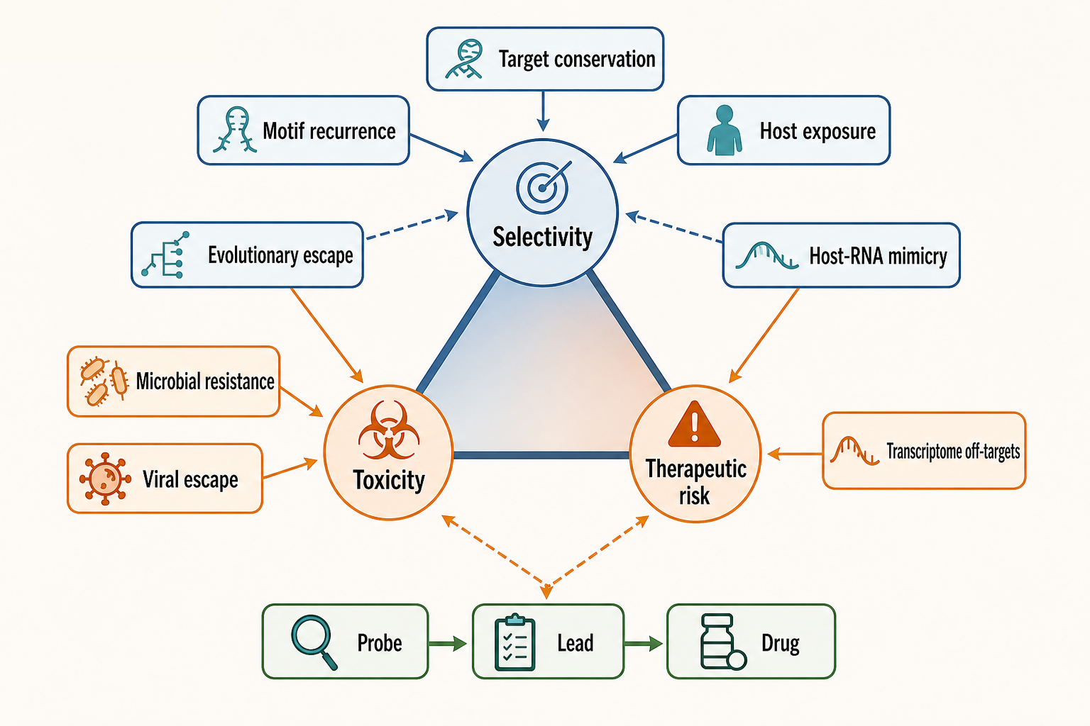
Figure 161.6. Resistance, Selectivity, and Toxicity Tradeoffs. Selectivity is a multi-scale property. Antibacterial RNA targets face microbial resistance; host RNA targets face transcriptome-wide off-target risk; viral RNA targets face both escape and host-RNA mimicry.
For antibiotics, resistance mechanisms are mature and clinically central. rRNA point mutations can reduce binding, but many bacteria carry multiple rRNA operons, so resistance level may depend on allele dosage. rRNA methyltransferases can protect functional centers. Enzymes can modify aminoglycosides or other antibiotic scaffolds. Efflux pumps and reduced permeability can lower intracellular concentrations. Biofilms and slow-growth states can create tolerance that is not the same as heritable resistance. These mechanisms mean that a beautiful RNA-binding mode is only one part of antibacterial durability.
For host RNA targets, resistance often means biological escape rather than microbial mutation. Cancer cells can alter splicing-factor expression, activate bypass pathways, mutate regulatory sequences, or change drug transport. Viruses can mutate RNA structures unless the element is highly constrained. Repeat-expansion diseases may not develop resistance in the same way, but target biology can be redundant: RNA toxicity, protein toxicity, and DNA instability may persist through different arms. A clinical program should ask how the disease system could remain pathological despite target occupancy.
Toxicity can arise from on-target exaggeration, near-target binding, or general compound properties. On-target exaggeration occurs when too much inhibition of bacterial ribosomes causes microbiome damage or when too much splice modulation disrupts essential isoforms. Near-target binding occurs when a compound binds related host RNAs, mitochondrial ribosomes, or related splice sites. General properties include hERG channel inhibition, phospholipidosis, lysosomal accumulation, genotoxic impurities, reactive metabolites, and transporter liabilities. RNA affinity alone does not predict these risks, but RNA-targeting scaffolds with cationic, planar, or highly aromatic features may require careful profiling for nonspecific nucleic acid binding and cellular accumulation.
Selectivity should be measured across molecular and organismal scales. At the molecular scale, does the ligand prefer the intended RNA pocket over related motifs? At the transcriptome scale, does it perturb a limited set of RNAs in a pattern consistent with target biology? At the cellular scale, does it avoid stress signatures at active concentrations? At the tissue scale, does it reach the disease site without accumulating in sensitive organs? At the organismal scale, does it spare the host microbiome or mitochondrial translation when that matters? A single binding-selectivity panel cannot answer all of these questions.
Regulatory classification can be confusing because “RNA drug” may refer to drug substance or mechanism. A small molecule that binds RNA is regulated as a small-molecule drug, not as an oligonucleotide, even if the target is RNA. A ribosome antibiotic is an antibacterial small molecule with resistance and stewardship considerations. A splicing modifier for a genetic disease is a small-molecule therapy with chronic exposure, reproductive safety, pediatric, and long-term follow-up concerns. A compound that changes a viral RNA structure is an antiviral small molecule and must show antiviral efficacy, resistance profile, and safety. Companion diagnostics may be needed when the target RNA sequence, repeat length, splice genotype, or biomarker defines the treatable population.
Several interpretation mistakes recur in RNA-targeting programs. Binding RNA does not make a compound selective. Selectivity in vitro does not guarantee selectivity in cells. A transcriptomic change does not prove direct target engagement. A structure predicted by software is not automatically a cellular binding pocket. An RNA target in bacteria does not imply equivalent opportunity in human transcripts. A favorable biomarker does not prove clinical benefit unless the biomarker is validated or strongly mechanistic. These cautions are not pessimism; they are the standards needed to separate durable drugs from interesting probes.
The boundary with chemical biology is also important. A probe can be valuable with micromolar potency, partial selectivity, and a clear experimental use. A drug usually needs potency, exposure, safety, manufacturability, formulation, durable pharmacodynamics, and clinical benefit. Some RNA-binding compounds are best treated as probes for discovering principles. Others may become leads after medicinal chemistry. Only a few have mature clinical validation. Chapter 162 treats discovery platforms and degrader-like designs; this chapter emphasizes the pharmacological and clinical bar.
Box 161.3. Probe, Lead, or Drug?
An RNA-binding compound can be valuable at different development stages. A probe should perturb a defined RNA or RNP question reproducibly, include an inactive or weaker analog when possible, and be used at concentrations that do not dominate cells with stress or nonspecific nucleic acid binding. A lead adds medicinal-chemistry traction: potency improves with structure-activity relationships, target engagement is measurable in cells, and off-target RNA or protein effects are bounded enough to justify optimization. A drug candidate must satisfy a higher bar: relevant tissue exposure, pharmacokinetics, manufacturability, safety margins, proximal pharmacodynamic biomarkers, and a plausible path to clinical benefit. Moving from probe to drug is therefore not a change in vocabulary alone. It requires evidence that the RNA-binding event survives cellular context, reaches the disease site, and produces a benefit-risk profile competitive with other therapeutic options.
161.7. Clinical Pipelines, Boundaries, and Failure Modes
Clinical pipelines in RNA-directed small-molecule pharmacology cluster around several strategies. Approved or established anchors include ribosome-targeting antibiotics and systemic splicing correction for SMN2. Investigational areas include additional splice modifiers, oncology spliceosome modulators, antibacterial riboswitch ligands, viral RNA structure ligands, repeat-expansion RNA binders, microRNA biogenesis inhibitors, lncRNA modulators, and structured UTR regulators. The maturity of these areas is uneven. Some have human efficacy. Some have compelling cellular target engagement. Some remain primarily discovery chemistry.
The most common failure mode is indirectness. A compound is found in a phenotypic screen that changes a reporter, RNA abundance, or cellular survival. Later experiments show that the compound affects transcription, translation, cell stress, mitochondrial function, or a protein target rather than the proposed RNA. This does not make the compound useless, but it changes the development path. The target claim must follow the evidence. A pathway-adjacent drug may still be clinically valuable; it should not be described as a direct RNA binder without proof.
Another failure mode is motif overabundance. RNA-binding ligands often recognize recurrent internal loops, bulges, or base-paired motifs. If the same motif appears in many transcripts, selectivity may depend on context rather than binding. Context can help when the intended RNA is more accessible, more abundant in disease cells, or coupled to a sensitive process. Context can hurt when off-target RNAs in essential tissues are also accessible. Transcriptome-scale binding and functional profiling are therefore not optional for host RNA targets.
A third failure mode is poor translation from in vitro structure to cellular RNA. Many RNAs fold differently during transcription, processing, translation, or RNP assembly than they do as purified fragments. Proteins can mask pockets or create new ones. Magnesium, metabolites, helicases, ribosomes, and RNA modifications can shift conformational ensembles. Chemical probing in cells can help, but probing reactivity is not a direct picture of structure. The strongest programs use cellular data to refine biochemical hypotheses rather than treating in vitro structures as final.
Clinical endpoint selection is another boundary. For bacterial ribosome antibiotics, microbiological cure, infection-site exposure, resistance, and safety drive evaluation. For splicing modifiers in genetic disease, clinical outcomes may include survival, motor milestones, respiratory support, functional scales, protein biomarkers, and genotype-defined subgroup response. For viral RNA ligands, viral-load decline must be linked to clinical benefit and resistance suppression. For repeat-expansion diseases, slow progression and tissue accessibility make trials challenging; biomarkers such as foci reduction, splice correction, toxic peptide reduction, or neurofilament changes may need validation.
Recent Consensus
The current consensus is cautious optimism. RNA is not undruggable by small molecules. The ribosome, riboswitches, and splice-modifier examples disprove that simplistic view. At the same time, RNA is not broadly druggable merely because it folds. The strongest opportunities combine a structured and accessible RNA or RNP site, a causal disease mechanism, a sensitive functional output, a feasible exposure profile, and a biomarker that reports proximal target modulation. The weakest claims rely on predicted structure, nonspecific binding, or downstream phenotypes without target-engagement controls.
Approved ribosome antibiotics remain the clearest clinical precedent for RNA-rich small-molecule pharmacology, but the consensus lesson is not that isolated RNA fragments are generally easy drug targets. The ribosome is an abundant, essential, structurally organized RNP machine with deep structural, genetic, biochemical, microbiological, and clinical evidence. Its limitations are also instructive: resistance through rRNA mutation or methylation, drug-modifying enzymes, permeability, efflux, growth-state tolerance, microbiome effects, and mitochondrial translation liabilities must be treated as core pharmacology rather than late development details.
For splice-modifying small molecules, the best-supported model is local stabilization or modulation of a defined pre-mRNA-splicing-factor state, not broad activation of splicing. Risdiplam-like SMN2 exon 7 correction is the anchor because the target exon, disease mechanism, proximal RNA biomarker, protein restoration logic, and clinical response can be linked. Consensus does not support extrapolating that success to every alternative-splicing event. Disease relevance, tissue exposure, transcriptome-wide splice selectivity, chronic safety, and clinically interpretable biomarkers remain decisive.
For emerging direct RNA ligands, the field increasingly separates chemical biology probe value from therapeutic readiness. Riboswitches have natural ligand-binding precedent; repeat-expansion RNAs, viral RNA structures, microRNA precursors, long noncoding RNAs, and structured untranslated regions each have plausible opportunities but different proof burdens. A direct RNA pharmacology claim is strongest when target engagement is shown in the relevant cellular compartment, activity is lost with target-motif disruption or resistant alleles, off-target RNA and stress signatures are bounded, and the proximal RNA response is quantitatively connected to disease biology.
Regulatory classification follows the drug substance and development context. A small molecule that binds RNA is still developed as a small-molecule drug; an antibacterial RNA-targeting compound also carries resistance and stewardship expectations; a chronic splice modifier carries long-term safety, developmental, reproductive, and pediatric considerations when relevant; and a viral RNA ligand must satisfy antiviral efficacy, resistance, and combination-therapy logic. The target being RNA changes the evidence package, not the basic requirement to show exposure, safety, efficacy, manufacturability, and a favorable benefit-risk profile.
Open Questions, Controversies, Deprecated Models, and Common Misconceptions
Open questions:
- Which RNA motifs are truly ligandable in living cells?
- Can medicinal chemistry routinely improve RNA selectivity without creating nonspecific nucleic acid binding or poor pharmacokinetics?
- How often can a small molecule distinguish one transcript from thousands of related folded motifs?
- Which target-engagement assays will become accepted for regulatory decision-making?
- Can direct RNA ligands be combined with oligonucleotides, protein-targeted drugs, or RNA degraders to improve efficacy?
- How should resistance be monitored for viral and bacterial RNA targets?
- How should long-term off-target splicing or translation effects be evaluated in pediatric or chronic indications?
Controversies:
- Boundary cases remain difficult. RNA-rich RNP modulators can be mechanistically direct without binding naked RNA, pathway-adjacent RNA drugs can be therapeutically important without being direct RNA binders, and lncRNA programs must distinguish activity through the RNA molecule from activity through the DNA locus, transcriptional process, or neighboring chromatin.
- Resistance and toxicity remain open translational tests rather than peripheral risks. Bacterial RNA targets face mutation, methylation, enzymatic drug modification, permeability, efflux, and tolerance; viral RNA targets face escape; and host RNA targets face off-target motif recurrence, tissue-specific exposure gaps, mitochondrial or microbiome liabilities, chronic splice perturbation, and general compound toxicology.
- The clinical boundary is clearest when the proposed mechanism changes patient management. Direct RNA ligands, viral RNA ligands, lncRNA ligands, repeat-expansion programs, and riboswitch antibacterials each require mechanism-specific evidence before target engagement is interpreted as therapeutic validation.
Common misconceptions:
- “A reporter change proves direct RNA binding.” Reporter responses can be indirect; direct binding needs biochemical, structural, or target-engagement evidence.
- “A transcriptome-wide expression change proves that the intended RNA was engaged.” Expression changes can reflect stress, toxicity, pathway feedback, or off-target activity without direct target engagement.
- “A chemical-probing protection pattern is automatically a binding site.” Protection can reflect structure, protein occupancy, ligand binding, modification, or indirect conformational change.
- “A favorable proximal biomarker is itself clinical benefit.” Biomarkers need a validated causal connection to patient-relevant outcomes.
- “A downstream clinical endpoint alone reveals whether the proposed RNA target was the causal pharmacological site.” Clinical benefit establishes efficacy, but mechanism still needs target engagement and causal pharmacology evidence.
Deprecated or weakened claims:
- RNA should not be described as intrinsically undruggable by small molecules; ribosome antibiotics, natural riboswitch ligand recognition, and splice-modifier pharmacology contradict that blanket claim.
- A folded RNA, a predicted pocket, or an in vitro binding curve should not be treated as establishing a therapeutic target.
- RNA-directed drug discovery should not be treated as protein drug discovery applied to a different polymer. RNA folding is co-transcriptional and context-dependent, RNA pockets can be transient or RNP-created, and functional readouts often reflect processing, translation, localization, or decay rather than simple occupancy.