Chapter 162. Direct RNA Ligand Discovery, RNA Degraders, Structure Remodeling, and Chemical Biology Platforms
Scope Note
This chapter explains how small molecules and bifunctional chemical probes are discovered, validated, and developed when the intended pharmacological object is RNA itself rather than an RNA-binding protein or a downstream pathway. The chapter covers RNA ligandability, discovery platforms, computational prediction, chemical biology evidence, cellular target engagement, ligand-induced RNA remodeling, RNA-binding protein displacement or recruitment, RIBOTAC-like degrader strategies, and the practical failure modes that separate useful probes from drug-like leads. Scientific examples are therefore presented as field-established examples requiring expert citation curation before final publication.
Executive Summary
Direct RNA ligand discovery asks whether a small molecule can recognize a biologically meaningful RNA conformation at concentrations, selectivity, residence time, and cellular exposure compatible with useful pharmacology. The central difficulty is not that RNA never binds small molecules. Natural riboswitch aptamers, ribosomal antibiotics, viral RNA elements, aminoglycoside-binding motifs, and selected aptamers show that RNA can form highly specific binding sites. The harder problem is that many cellular RNAs are flexible, heavily solvated, negatively charged, protein-associated, low-abundance, or available only transiently during transcription, processing, translation, localization, or decay. A compound that binds an RNA hairpin in vitro may fail in cells because the target conformation is rare, the RNA is masked by proteins, the compound partitions into membranes, the apparent binding comes from aggregation or electrostatics, or the measured phenotype is caused by a protein off-target.
RNA ligandability is therefore an evidence ladder rather than a yes-or-no property. The first rung is a physically plausible pocket, motif, or dynamic ensemble. The second rung is reproducible binding by orthogonal biophysical assays. The third rung is occupancy in the correct biological context. The fourth rung is a mechanistic consequence such as altered RNA structure, altered RNA-protein binding, changed splicing, changed translation, or changed RNA decay. The final rungs are selectivity, exposure-response behavior, resistance or rescue logic, and therapeutic window. This ladder matters because RNA-targeting compounds often look promising in one assay and collapse when tested against cellular abundance, transcriptome complexity, innate immune activation, efflux, metabolism, or nonspecific nucleic acid binding.
The discovery platforms are complementary. Fragment and structure-guided screens find weak but interpretable starting points around a known RNA motif. Phenotypic screens can reveal compounds that alter splicing, translation, or viral replication but require intensive deconvolution. RNA-focused chemical probing and chemoproteomic methods connect compounds to cellular RNAs by detecting structure changes, covalent or photo-crosslinked adducts, affinity enrichment, or competition with a labeled probe. AI-assisted methods can prioritize pockets, ligands, or RNA-ligand poses, but RNA conformational heterogeneity and sparse benchmark data make experimental validation indispensable.
RIBOTAC-like and proximity-induced RNA pharmacology extends direct binding into induced function. A bifunctional molecule can, in principle, bind a target RNA with one module and recruit an RNase, RNA-modifying enzyme, RBP, or endogenous decay pathway with another module. The conceptual analogy to protein degraders is useful but incomplete. RNA degraders must operate in compartments where the target RNA and effector are accessible, avoid degrading beneficial isoforms or homologous RNAs, and show that degradation is caused by proximity rather than by innate immune activation, transcriptional shutdown, or generic RNA damage. The strongest current view is cautious optimism: direct RNA ligands and RNA proximity pharmacology are scientifically real, but probe-to-lead progression depends on unusually demanding target-engagement, selectivity, and pharmacology evidence.
Concept Inventory
- RNA ligandability: the degree to which a particular RNA sequence, structure, or ensemble can bind a small molecule in a way that is selective, reproducible, biologically accessible, and capable of producing a useful functional consequence.
- RNA pocket: a local three-dimensional arrangement of bases, ribose-phosphate backbone, ions, water, and sometimes protein contacts that creates a small-molecule binding site. RNA pockets may be preorganized or ligand-stabilized.
- Motif: a recurring RNA structural or sequence element, such as an internal loop, bulge, junction, pseudoknot, G-quadruplex, riboswitch aptamer domain, repeat hairpin, or viral regulatory element.
- Target engagement: evidence that a compound physically occupies or perturbs the intended RNA target in the relevant biological context at pharmacologically meaningful exposure.
- Chemoproteomics in this chapter: chemical biology workflows that use affinity, covalent, photoaffinity, competition, mass spectrometry, sequencing, or hybrid readouts to identify compound-bound RNA, RNA-associated proteins, or compound-induced RNP remodeling.
- RBP displacement: reduction of an RNA-binding protein’s occupancy on an RNA because a ligand competes with the protein site, changes RNA structure, or changes RNP assembly.
- RBP recruitment: increased association of an RNA-binding protein or effector complex with an RNA because a ligand stabilizes a motif or bridges the RNA to an effector.
- RIBOTAC-like degrader: a bifunctional or multifunctional molecule that binds a target RNA and induces cleavage or decay by recruiting or activating a ribonuclease or RNA decay machinery.
- Proximity pharmacology: drug action in which a molecule produces an effect by bringing two biological objects close together, such as an RNA and an RNase, rather than by simply blocking an active site.
- Probe-to-lead progression: the path from a chemical probe that demonstrates a targetable mechanism to a lead series with potency, selectivity, cellular activity, pharmacokinetics, safety, and translational biomarkers.
What to Know Before Reading This Chapter
The reader should know that RNA is not a uniform polymeric target. RNA can form helices, loops, junctions, pseudoknots, tertiary folds, quadruplexes, and ribonucleoprotein particles. RNA structure is often an ensemble, meaning that several conformations coexist and interconvert. A small molecule can bind a rare member of an ensemble and shift the population toward that conformation, or a small molecule can bind only after another factor exposes the site. RNA-binding proteins, helicases, ribosomes, spliceosomes, ribonucleases, and RNA modification enzymes constantly remodel cellular RNAs, so the same sequence can present different binding surfaces at different times and compartments.
The reader should also separate binding from pharmacology. Binding is a physical interaction. Pharmacology is a dose-dependent biological effect caused by the interaction under exposure conditions that can be achieved in cells, tissues, or organisms. Many positively charged aromatic molecules bind nucleic acids, but broad binding to many RNAs or DNA is usually toxic and mechanistically uninformative. A useful direct RNA ligand normally requires a defined target RNA element, an interpretable binding mode, evidence of target occupancy, and a causal link between occupancy and a biological readout.
162.1. RNA pockets, motifs, dynamics, and ligandability criteria
RNA-targeted ligand discovery begins with a structural question: what exactly is the compound supposed to bind? In protein drug discovery, medicinal chemists often imagine a pocket as a cleft in a relatively stable folded domain. RNA can also form pockets, but RNA pockets are usually made from base edges, stacking surfaces, grooves, ribose 2′-hydroxyl groups, metal ions, structured water, and the geometry of bulges or junctions. The electrostatic environment is dominated by the polyanionic phosphate backbone. Many RNA surfaces are shallow and solvent exposed, yet some motifs form cavities, clefts, and preorganized recognition sites that are as chemically specific as protein pockets.
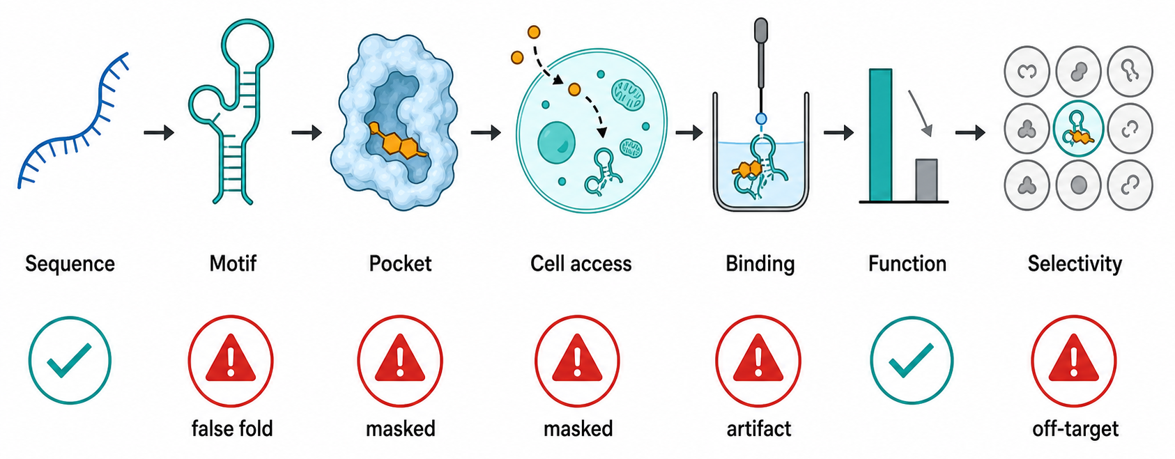
Figure 162.1. RNA ligandability as a structural and cellular evidence ladder. RNA ligandability is not established by a pocket model alone. A credible direct RNA target progresses through structural plausibility, ensemble and cellular accessibility, orthogonal binding, cellular target engagement, functional consequence, and selectivity testing.
The best natural proof that RNA can be ligandable is the riboswitch aptamer. A riboswitch aptamer domain is a structured RNA element, often in a bacterial untranslated region, that directly binds a metabolite such as a nucleotide derivative, amino acid, coenzyme, metal ion, or signaling molecule. Ligand binding changes the folding or stability of an adjacent expression platform and thereby changes transcription termination, translation initiation, splicing, or RNA decay. Riboswitches are not simply exceptions; they show that RNA can evolve pockets with molecular shape complementarity, hydrogen-bond patterns, stacking interactions, and ion-mediated contacts sufficient for biologically selective recognition. Ribosomal RNA antibiotic sites provide another proof of principle. Aminoglycosides, macrolides, oxazolidinones, tetracyclines, and other antibiotic classes act at RNA-rich functional centers of the bacterial ribosome, even when the clinically optimized drug may contact both RNA and ribosomal proteins.
The lesson from riboswitches and ribosomal antibiotics is not that every RNA is druggable. Natural ligand-binding RNAs are unusually structured or embedded in RNP machines that stabilize ligandable states. Many mRNAs and lncRNAs have local structures that are short-lived, partially occupied, or shielded by proteins. An internal loop in a purified RNA oligonucleotide may look like a pocket in nuclear magnetic resonance spectroscopy or molecular dynamics, but in cells the same sequence may be unwound by a helicase, covered by an RBP, modified at a key nucleotide, or never folded because translation or transcription kinetics favor another state.
Ligandability is therefore best defined as a compound-accessible functional ensemble. A target RNA is more ligandable when several conditions align. First, the RNA presents a recurring structural motif with a shape and chemical pattern that distinguishes it from abundant background RNAs. Second, the motif exists at meaningful fractional occupancy in the relevant cell type, stage, and compartment. Third, the motif is close to a functional decision point, such as a splice site, translation initiation region, viral frameshift element, processing site, RBP-binding site, or decay determinant. Fourth, occupancy by a small molecule can alter that decision without requiring unrealistically high compound concentrations. Fifth, the target RNA is sufficiently different from essential RNAs such as rRNA, tRNA, spliceosomal snRNAs, and abundant housekeeping mRNAs to allow a selectivity window.
Table 162.1. RNA motif classes and ligandability considerations. RNA motifs differ in pocket preorganization, cellular accessibility, selectivity risk, and functional consequences. Motif class is a starting hypothesis, not proof of druggability.
| Motif class | Binding opportunity | Functional readout | Major selectivity risk | Evidence needed |
|---|---|---|---|---|
| Riboswitch aptamer domains | Preorganized metabolite-binding pockets with defined base, ribose, ion, and water contacts | Ligand-dependent transcription termination, translation initiation, splicing, or RNA decay | Native pocket may require the full expression platform and cellular folding context | Orthogonal binding, structural or probing support, ligand-response mutations, and expression-platform readout |
| Ribosomal RNA functional centers | RNA-rich decoding, peptidyl-transferase, or exit-tunnel sites stabilized by the ribosome | Translation inhibition, altered decoding fidelity, or bacterial growth inhibition | Host cytosolic or mitochondrial ribosome toxicity and broad translation effects | Structural occupancy, resistance mutations, ribosome biochemistry, and host-selectivity profiling |
| Internal loops and bulges | Exposed base edges, stacking platforms, and local grooves in mRNAs or ncRNAs | Altered RBP binding, splicing, translation, localization, or decay | Similar short motifs across the transcriptome and nonspecific cationic binding | Biophysical binding, in-cell probing, motif mutants, inactive analogs, and transcriptome-wide counterscreens |
| Junctions, pseudoknots, and viral RNA elements | Composite tertiary grooves and mechanically sensitive folds near regulatory decisions | Changed frameshifting, IRES activity, replication, packaging, or antiviral phenotype | Conformational heterogeneity, host RNP remodeling, and viral escape variants | Cellular structure support, functional mutants or rescue variants, antiviral dose-response, and target-engagement timing |
| G-quadruplexes | Planar stacking surfaces and cation-stabilized channels | Changed translation, localization, stability, or protein binding | DNA G-quadruplex binding and broad RNA G-quadruplex cross-reactivity | G4-disrupting variants, cellular occupancy or probing, DNA/RNA counterscreens, and mechanism-linked output |
| Repeat-expansion RNAs | Repeated hairpins or internal loops that support avid or multivalent binding | Reduced RNA foci, altered RBP sequestration, splicing rescue, or toxic RNA reduction | Aggregation, unclear stoichiometry, and effects on other repeat-containing RNAs | Repeat-length dependence, RBP-release assays, inactive analogs, RNA abundance controls, and off-target profiling |
| RNP interfaces and spliceosome-stage pockets | Composite RNA-protein surfaces present only in a specific assembly state | Exon inclusion, splice-site choice, RNP stabilization, or effector recruitment | Protein-dominant mechanisms and global splicing perturbation | Stage-specific engagement, target variants, endogenous isoform panels, and RNP occupancy measurements |
RNA motifs differ in the way they create ligandable space. Internal loops and bulges expose base edges and stacking platforms. Junctions bring multiple helices together and can form composite pockets. Pseudoknots can create tertiary folds with defined grooves and ligand-sensitive mechanical properties. G-quadruplexes offer planar stacking surfaces and cation-stabilized channels, but G-quadruplex ligands often suffer from selectivity problems because similar aromatic ligands can bind DNA quadruplexes and many RNA quadruplexes. Repeat-expansion RNAs, such as CUG, CCUG, GGGGCC, or CGG repeat structures, can present repeated internal-loop or hairpin motifs; repetition increases avidity and can permit multivalent ligand designs, but it also complicates stoichiometry and can produce broad interactions with other structured repeats. Viral RNA elements, including internal ribosome entry sites, frameshift elements, untranslated-region structures, and packaging signals, are attractive because viral replication can be highly dependent on RNA structure. However, viral sequence variation and host-cell RNP remodeling create resistance and context-dependence.
RNA dynamics can help or hurt discovery. A preorganized pocket may bind a ligand with low entropic penalty and clear structure-activity relationships. A flexible motif may bind weakly but be tunable by ligands that stabilize one conformation. The latter mechanism is important because many RNA drugs do not simply occupy a static hole; they remodel a folding pathway. For example, a splicing modifier that binds a pre-mRNA-spliceosome interface can stabilize exon inclusion by creating or reinforcing contacts among RNA, small nuclear RNA, and spliceosomal proteins. The pharmacological target is then not a naked RNA hairpin, but a transient RNP conformation. Similar logic applies to viral RNA structures whose conformational equilibria control translation, frameshifting, replication, or packaging.
The strongest ligandability assessments combine sequence conservation, structure probing, structural biology, genetic perturbation, and chemical binding data. Conservation alone is not enough because RNA sequence can be conserved for coding potential, protein binding, splicing signals, or overlapping regulatory grammar rather than a small-molecule pocket. A predicted low free-energy structure is also not enough because thermodynamic models can miss pseudoknots, tertiary contacts, protein effects, RNA modifications, co-transcriptional folding, salt conditions, and cellular crowding. A useful ligandability dossier asks whether mutations that disrupt the motif change function, whether compensatory mutations restore function, whether structure probing supports the motif in cells, whether an orthogonal binding assay detects compound interaction, and whether compound-resistant or rescue variants shift the dose-response in the predicted direction.
Do not overgeneralize the word “pocket.” In RNA, a ligandable site may be a pocket, a groove, a stack, an induced-fit cleft, a hydrated ion site, or a surface that becomes druggable only when the RNA is part of a protein complex. Conversely, a visually attractive cavity in an RNA structure may be irrelevant if the conformation is absent in cells or if occupancy has no functional consequence. Chapter 53 treats RNA motifs and tertiary architecture in more detail, and Chapter 59 treats structural methods that can distinguish static models from experimentally supported conformations.
162.2. Fragment, phenotypic, and structure-guided screens
RNA ligand discovery uses several entry points because no single screen captures all forms of useful RNA pharmacology. Fragment screening starts with small, low-molecular-weight compounds that bind weakly but efficiently. A fragment hit may have a dissociation constant in the high micromolar or millimolar range, but if the fragment makes a specific contact in a defined RNA pocket, medicinal chemistry can grow, merge, or link the fragment into a stronger ligand. Fragment screening is attractive for RNA because it can reveal minimal recognition elements rather than overwhelming a target with large cationic scaffolds. The tradeoff is sensitivity: weak RNA-fragment interactions require careful biophysical assays and controls for aggregation, fluorescence interference, metal chelation, and nonspecific electrostatic binding.
Common fragment-screening readouts include nuclear magnetic resonance chemical-shift perturbation, surface plasmon resonance, isothermal titration calorimetry, differential scanning methods, mass spectrometry, X-ray or cryo-electron microscopy where crystals or complexes are available, and competition against a known ligand or labeled probe. For RNA, each readout has a specific artifact profile. Fluorescence displacement assays can mistake compound fluorescence quenching for binding. Immobilized-RNA assays can enrich compounds that bind the linker, bead, folded tag, or nonspecific nucleic acid surface. Thermal-shift-like assays can be hard to interpret because RNA may unfold through several transitions. NMR can show local perturbations but may be limited by RNA size, exchange, and sample stability. No single readout is decisive; the useful hit is the one that survives orthogonal assays and structure-activity logic.
Phenotypic screening begins from a biological output rather than from binding. A screen may ask whether compounds correct a splicing defect, suppress translation from a viral internal ribosome entry site, reduce toxic repeat RNA foci, inhibit viral replication, induce decay of an oncogenic lncRNA, or restore a reporter controlled by an RNA element. Phenotypic screens are powerful because they select for cellular permeability, exposure, and pathway relevance from the beginning. They also detect RNP-interface mechanisms that would be missed by screens against purified RNA. The cost is target ambiguity. A compound that changes a splicing reporter may bind the target pre-mRNA, a spliceosomal protein, a kinase regulating splicing factors, a transcription factor changing reporter expression, or a stress pathway that indirectly alters splice-site choice.
Good phenotypic screens therefore require deconvolution from the start. A robust design includes a primary reporter, counterscreens against unrelated reporters, transcript-level measurement of the endogenous target, dose-response curves, cytotoxicity assessment, time-course analysis, and rescue or resistance experiments. If the intended mechanism is direct RNA binding, the screen should test target RNA variants that disrupt or restore the candidate motif. It should also compare closely related transcripts or isoforms to ask whether the compound respects the proposed RNA grammar. In repeat-expansion systems, for example, a useful compound should distinguish the disease-associated repeat motif and its RNP consequences from unrelated repeat-containing RNAs, abundant structured RNAs, and general stress granule formation.
Box 162.1. Deconvoluting a Phenotypic RNA-Ligand Hit
Deconvolution checklist for a phenotypic hit
A phenotypic hit should be treated as a mechanistic lead, not as proof of direct RNA binding. First ask whether the compound changes the endogenous RNA-dependent process, not only an engineered reporter. Then test unrelated reporters, matched inactive analogs, cytotoxicity, stress markers, and time courses to remove common artifacts. If the hypothesis is direct binding to an RNA motif, mutate or swap the motif and ask whether the dose-response moves as predicted. Measure the RNA species itself: abundance, isoform choice, localization, structure, or RNP occupancy may reveal whether the phenotype follows RNA engagement. Finally, compare closely related transcripts, repeat lengths, viral variants, or splice isoforms. A credible hit survives when phenotype, target engagement, variant logic, and selectivity all point to the same RNA-dependent mechanism.
Structure-guided screening starts with a model or experimental structure of the RNA target. The structure may come from crystallography, nuclear magnetic resonance spectroscopy, cryo-electron microscopy, chemical probing constrained modeling, comparative analysis, or a ligand-bound structure of a related motif. Docking can then prioritize compounds predicted to complement a pocket. Structure-guided design is most powerful when the RNA conformation is validated under conditions close to the discovery assay and when medicinal chemistry can test predicted contacts. A single ligand-bound structure can transform a project by revealing whether potency comes from base stacking, hydrogen bonding to base edges, shape complementarity in a junction, cation-mediated contacts, or interaction with a protein-stabilized RNP surface.
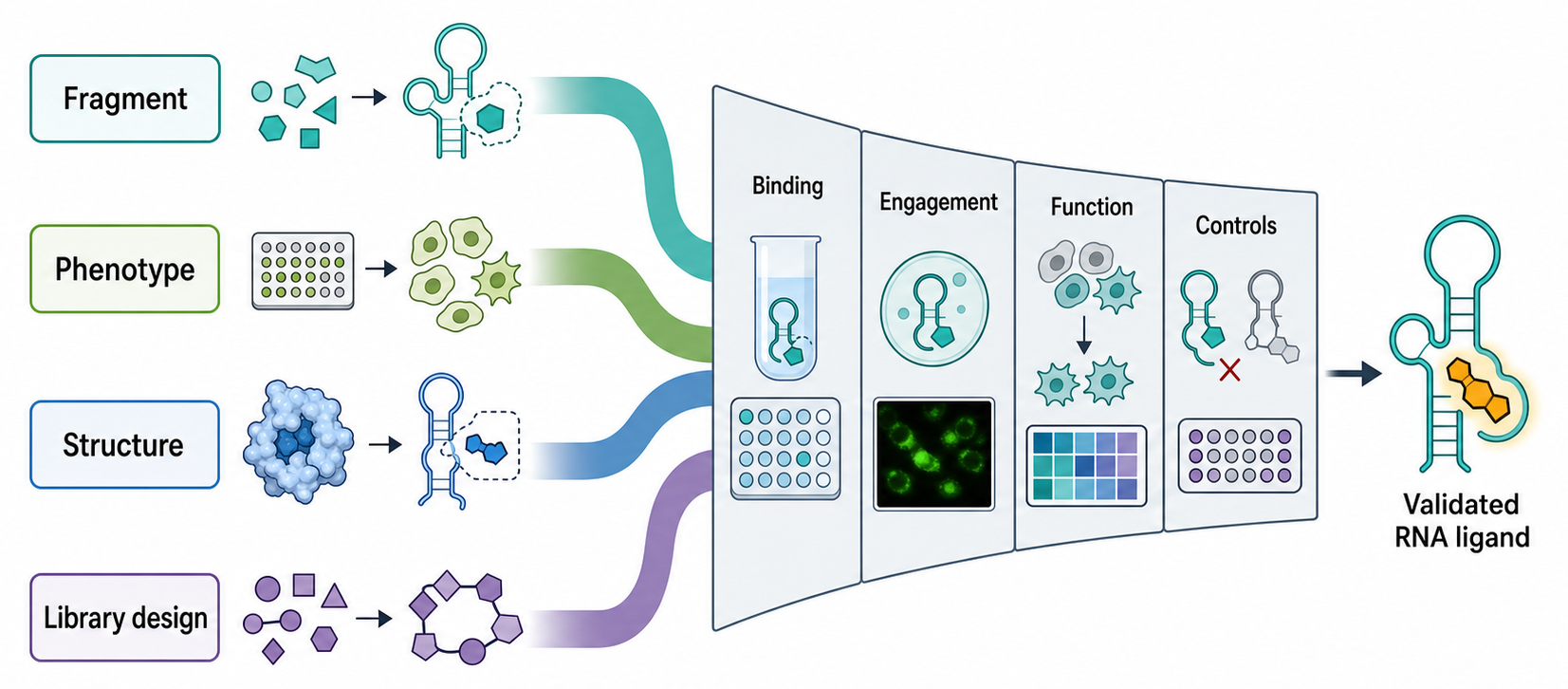
Figure 162.2. Discovery routes for RNA-targeted ligands. RNA ligand discovery can begin from weak interpretable fragments, cell-based phenotypes, or structure-guided hypotheses. The routes converge only when binding, cellular engagement, functional response, and artifact controls agree.
Structure-guided screens can mislead when the chosen RNA construct is too artificial. Short constructs may remove flanking helices, tertiary restraints, protein contacts, or competing structures. Modified nucleotides used for crystallization or stabilization may change pocket geometry. High magnesium or nonphysiological salt may stabilize conformations that are rare in cells. Docking against a single conformation may miss ligands that bind an alternative ensemble member. The best practice is to test hits against target-length variants, related motifs, mutant controls, and cell-relevant functional assays before treating a docking pose as a mechanism.
Library design is a strategic issue. General screening libraries optimized for protein pockets may underrepresent RNA-binding chemotypes, while older nucleic-acid-binding libraries may be enriched for planar polyaromatics and polycationic scaffolds that bind broadly and generate toxicity. RNA-focused libraries often seek a middle ground: enough hydrogen-bond donors and acceptors to read base edges, enough three-dimensionality to avoid generic intercalation, controlled cationic character, and shapes that can exploit grooves, loops, and junctions. Fragment libraries should preserve ligand efficiency and solubility rather than chasing potency at the cost of selectivity.
The output of screening is not a drug; it is a hypothesis. A fragment hit hypothesizes that a motif contains a chemically addressable contact. A phenotypic hit hypothesizes that a compound can alter a cellular RNA-dependent process. A structure-guided hit hypothesizes that a model has captured a biologically relevant binding mode. Progress requires convergence among these hypotheses. When binding, structure, function, and cellular occupancy agree, confidence rises. When they conflict, the conflict is often the most informative part of the project. It may reveal a protein off-target, a cell-state-specific RNA conformation, a compound artifact, or a hidden RNP mechanism.
162.3. AI-assisted ligand discovery and RNA-small-molecule prediction
AI-assisted RNA ligand discovery uses computational models to prioritize targets, pockets, compounds, poses, or chemical transformations. The phrase includes several different tasks. A model may predict whether an RNA sequence is likely to fold into a ligandable motif, whether a candidate pocket resembles known RNA-ligand sites, whether a compound is likely to bind RNA rather than protein or DNA, whether docking poses are plausible, whether a ligand will change splicing or translation, or whether a chemical series can be optimized without losing selectivity. These tasks differ in data requirements and failure modes, so they should not be collapsed into one claim that “AI predicts RNA drugs.”
The input data problem is severe. Protein-ligand machine learning benefits from large numbers of structures, affinity measurements, and medicinal chemistry series. RNA-ligand datasets are much smaller, more heterogeneous, and often biased toward ribosomal RNA, riboswitches, aminoglycoside-like scaffolds, selected aptamers, and a few intensively studied disease motifs. Negative data are sparse because failed RNA-binding campaigns are less likely to be published. Binding measurements may use different constructs, salt conditions, labels, immobilization methods, or endpoints. A model trained on these data can learn assay conditions, ligand charge, target family, or publication bias rather than transferable RNA recognition principles.
Table 162.2. AI-assisted RNA ligand discovery tasks and validation needs. AI-assisted workflows answer different questions and require different validation. A predicted pocket, predicted pose, or predicted transcript response becomes useful only when tested by experiments matched to the prediction.
| Computational task | Input data | Useful output | Main failure mode | Required validation |
|---|---|---|---|---|
| Target or motif prioritization | Transcript sequence, conservation, disease genetics, expression, perturbation data, and probing maps | Ranked RNA elements worth biochemical and cellular testing | Confusing disease correlation or high expression with ligandable causal structure | Endogenous expression checks, motif perturbation, cellular structure support, and disease-relevant functional readout |
| Structure and pocket prediction | RNA sequence, covariance, probing constraints, solved structures, and ensemble models | Candidate conformers, pockets, and accessibility hypotheses | False precision from one modeled conformation or missing protein/modification effects | In vitro and cellular probing, compensatory mutations, structural assays, and pocket-disrupting variants |
| Docking and pose ranking | Modeled or solved RNA conformers plus ligand libraries and protonation states | Contact maps and analog hypotheses for synthesis or purchase | Generic docking to charged grooves or aromatic stacks without selectivity | Structure-activity relationships, orthogonal binding assays, footprinting, and unrelated RNA/DNA counterscreens |
| Molecular dynamics and ensemble sampling | Starting structures, force fields, solvent, ions, ligands, and sampling protocol | Pocket opening, water or ion networks, induced-fit paths, and residence-time hypotheses | Force-field or sampling artifacts that overstate rare conformers | NMR, time-resolved probing, structural snapshots, and analogs that test predicted dynamic contacts |
| Generative chemistry or analog design | Hit series, predicted contacts, physicochemical constraints, and selectivity objectives | Proposed analogs with improved potency, solubility, permeability, or effector geometry | Improving predicted affinity by adding nonspecific aromatic, cationic, or hydrophobic surface | Potency-selectivity matrix, permeability and stability assays, tag-free analogs, and broad off-target profiling |
| Phenotypic-signature interpretation | Transcriptomic, splicing, proteomic, translation, morphology, or stress-response signatures | Mechanism-class hypotheses such as spliceosome effect, stress response, decay, or target-specific modulation | Training-set bias and misclassification of general stress as direct RNA engagement | Direct target-engagement assays, rescue or resistance tests, time course, and orthogonal pathway counterscreens |
| Selectivity and off-target prediction | RNA motif catalogs, transcript abundance, chemical descriptors, and prior off-target profiles | Priority off-target RNAs, DNA liabilities, RNP risks, and counterscreen panels | Sparse negative data and domain shift outside known RNA-ligand chemotypes | Transcriptome-wide engagement, RNA abundance profiling, RBP occupancy, translation profiling, and cell-panel assays |
Several computational layers can still be useful. RNA structure prediction and ensemble modeling can identify motifs that are plausibly folded and accessible. Molecular dynamics can sample pocket opening, water networks, ion positions, and ligand-induced rearrangements, although the accuracy depends on force fields and sampling. Docking can generate hypotheses about contacts, especially when the target structure is high quality and the ligand chemotype is not dominated by nonspecific electrostatics. Similarity methods can map a new RNA motif to a known ligand-binding motif. Generative chemistry can propose analogs that preserve predicted contacts while improving solubility, permeability, or metabolic stability. Transcriptome-scale informatics can prioritize targets whose expression, disease association, conservation, and functional genetics justify chemical effort.
The key pedagogical point is that AI assistance is triage, not proof. A predicted RNA pocket is a reason to run a binding or probing experiment. A predicted ligand pose is a reason to synthesize analogs that test the contact map. A predicted target transcript is a reason to measure whether the RNA exists in the relevant cell type and compartment. A predicted selectivity profile is a reason to perform transcriptome-wide engagement or functional profiling. The model’s value is judged by how efficiently it enriches experimentally validated hypotheses, not by how visually convincing a pose looks.
Box 162.2. Turning an AI Prediction into a Testable RNA-Ligand Experiment
From predicted pose to falsifiable claim
The useful output of an AI-assisted workflow is not a picture of a compound in an RNA groove; it is a claim that can fail. A pocket prediction should name the nucleotides, conformer, ions, or protein contacts that make the site accessible. A pose prediction should identify contacts that analogs can remove or strengthen. A target-prioritization model should specify the cell type, transcript isoform, and functional readout in which the RNA element matters. A selectivity model should name the most likely off-target RNAs or DNA liabilities. The first validation experiment should match the prediction: footprinting for a local contact, compensatory variants for a structural model, analog structure-activity relationships for a pose, cellular probing for accessibility, and transcriptome-wide profiling for predicted selectivity.
RNA-specific models must handle conformational multiplicity. An RNA sequence can have several secondary structures with comparable free energies. Co-transcriptional folding can trap a structure that is not the global minimum. Proteins and modifications can stabilize or destabilize motifs. A ligand can bind a minor state and shift the ensemble. If a model returns one structure and one docking pose, the apparent precision may be false. A better workflow samples multiple structures, ranks pockets across the ensemble, asks which conformers are supported by probing or comparative data, and designs experiments that distinguish conformational selection from induced fit.
AI-assisted phenotypic interpretation is also emerging. If a compound changes hundreds of transcripts, a model can classify whether the pattern resembles spliceosome inhibition, integrated stress response, transcriptional suppression, nonsense-mediated decay inhibition, ribosome stalling, innate immune activation, or target-specific modulation. This is useful because many alleged direct RNA ligands produce broad transcriptomic effects. However, such classifiers inherit the biases of their training signatures and should not replace direct target-engagement experiments.
There are several practical boundaries. First, high predicted affinity is not meaningful if the model ignores compound aggregation, protonation, tautomerism, RNA counterions, or cellular concentration. Second, a ligand that docks to many RNA grooves may be a promiscuous nucleic-acid binder rather than a lead. Third, deep learning models can hallucinate confidence outside their domain of applicability. Fourth, model benchmarks can be inflated by near-duplicate targets, ligand analog leakage, or retrospective enrichment against easy decoys. Fifth, a model optimized for binding may not optimize the desired functional outcome, such as exon inclusion, translation repression, or RNase recruitment.
The appropriate consensus is constructive skepticism. AI-assisted approaches can improve target selection and reduce experimental search space, especially when paired with chemical probing, structural biology, and medicinal chemistry feedback. They cannot yet replace rigorous biochemical and cellular validation for direct RNA target engagement. Chapter 143 treats RNA foundation models and sequence-to-function prediction more broadly, while Chapter 63 treats probing-constrained modeling that can supply experimental constraints for ligand-discovery models.
162.4. Chemical probing, chemoproteomics, pull-downs, and target engagement
Target engagement is the central evidentiary problem in direct RNA pharmacology. A compound may bind a purified RNA, alter a reporter, or produce a disease-relevant phenotype, but none of those observations alone prove that the compound occupies the intended RNA in cells. Target engagement asks whether the compound physically contacts, stabilizes, remodels, or protects the target RNA at the concentration and time scale that produce the phenotype. In RNA drug discovery, engagement evidence is difficult because RNA is dynamic, often low abundance, and embedded in RNPs; small molecules may interact with many RNAs weakly; and the target may be a transient pre-mRNA or viral replication intermediate.
Chemical probing provides one route. Reagents such as dimethyl sulfate, SHAPE electrophiles, or other structure-sensitive chemicals modify nucleotides according to accessibility, flexibility, or base-pairing environment. If a ligand binds or remodels an RNA, the modification pattern can change. In vitro probing can map the local footprint of a compound. Cellular probing can test whether similar structural changes occur in living cells. Mutational profiling and sequencing convert chemical modification into transcript-level maps. The evidence is strongest when ligand-dependent changes occur at the predicted motif, are dose dependent, are lost in motif-disrupting mutants, and correlate with a functional readout.
Probing has limits. A change in chemical reactivity does not always mean direct binding at that nucleotide. The compound may alter protein binding, transcription rate, RNA processing, translation, or RNA abundance. Some reagents cannot access compartments equally. RNA degradation or altered isoform ratios can masquerade as structure change if analysis does not control for abundance. Ligands can interfere with probing chemistry or reverse transcription. Therefore chemical probing should be paired with orthogonal evidence such as binding assays, pulldowns, competition, mutation, structural models, and functional rescue.
Chemoproteomic and chemotranscriptomic approaches use chemical handles to connect compounds with cellular molecules. A ligand can be modified with an affinity tag, photo-crosslinker, clickable alkyne, electrophile, or isotopic label. After treatment, the tagged compound or its covalent adducts are captured, identified, and quantified by sequencing or mass spectrometry. For RNA targets, one can enrich bound RNAs and identify them by sequencing, or identify proteins whose RNA association changes upon ligand treatment. Competitive experiments, in which excess free compound reduces capture by a tagged analog, are essential because beads, tags, linkers, and photoreactive groups create nonspecific enrichment.
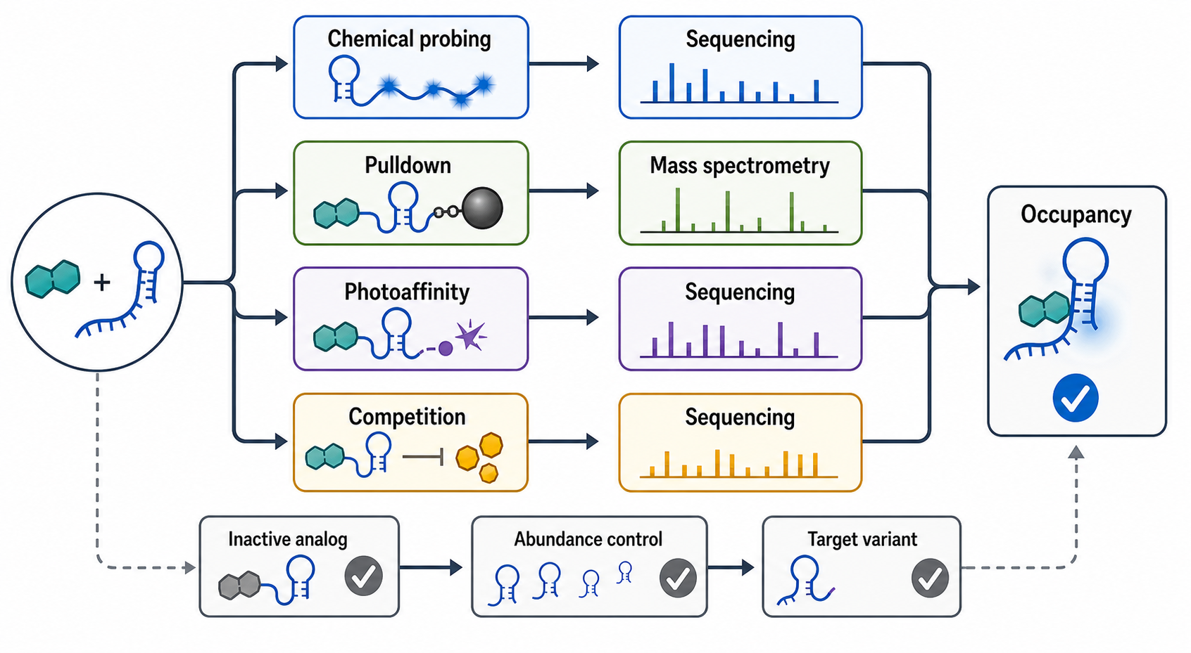
Figure 162.3. Chemical biology evidence for cellular RNA target engagement. RNA target engagement can be inferred from convergent chemical-probing, enrichment, competition, and functional data. Each readout has artifacts, so inactive analogs, competition, abundance controls, and target variants are part of the evidence rather than optional extras.
Pulldown assays are intuitive but easily overinterpreted. An immobilized compound can enrich an RNA because of direct binding, indirect binding through a protein, association with a large RNP, nonspecific charge, hydrophobic bead interactions, or differential RNA abundance. Conversely, a true cellular target may be missed if the tag disrupts binding, the linker blocks the pocket, the target RNA is low abundance, the binding is reversible and lost during washing, or the relevant conformation depends on live-cell conditions. Strong pulldown evidence includes inactive analog controls, competed capture by active free compound, target-mutant controls, RNase or protease sensitivity tests when mechanistically appropriate, and quantitative comparison with transcript abundance.
Occupancy assays ask a more pharmacological question: what fraction of target RNA is bound at a given compound concentration in cells? For proteins, occupancy can sometimes be inferred from thermal stabilization, activity blockade, or covalent labeling. For RNA, occupancy may be inferred from probe competition, protected chemical-probing sites, ligand-induced structural signatures, allele-specific response, or downstream functional saturation. The challenge is that functional response may be nonlinear. A small amount of occupancy at a regulatory bottleneck can produce a large phenotype, whereas high occupancy at an abundant nonfunctional motif may do little.
Target engagement also requires temporal logic. If a compound changes splicing, the compound must engage the pre-mRNA before or during spliceosome assembly. If a compound changes translation initiation, engagement must occur before ribosome recruitment or scanning. If a compound induces decay, engagement must precede the decay event and not merely bind the remaining RNA fragments. Time-course experiments can distinguish immediate structural engagement from delayed transcriptional feedback. Washout and pulse-chase experiments can measure whether the compound’s functional effect tracks compound exposure or persists because the RNA population has been irreversibly remodeled or degraded.
RNA-centric proteomics connects direct RNA ligands to RNP remodeling. A ligand may displace an RBP, recruit an RBP, expose a decay element, block a helicase, or stabilize a spliceosomal intermediate. Methods derived from RNA pulldown, interactome capture, CLIP-like occupancy profiling, and quantitative mass spectrometry can measure changes in protein association. These experiments must distinguish genuine target-specific remodeling from global stress responses that change many RNPs. For example, translation inhibition, heat shock, oxidative stress, or innate immune activation can reorganize stress granules and RNA-binding proteins broadly, creating secondary effects that resemble target engagement unless controls are rigorous.
An evidence hierarchy is useful. Low confidence: one assay shows in vitro binding or a cellular phenotype. Moderate confidence: orthogonal in vitro binding, structure-activity relationship, and target-dependent reporter response agree. High confidence: cellular target-engagement evidence maps to the intended motif, target mutations shift binding and function, inactive analogs fail, transcriptome-wide data show limited off-target activity, and the timing of engagement precedes the biological response. Very high confidence: structural or biochemical evidence defines the binding mode, resistance or rescue variants validate causality, and pharmacokinetic exposure supports the effect in disease-relevant cells or models.
Table 162.3. Target-engagement evidence hierarchy for RNA ligands. RNA target engagement is strongest when orthogonal binding, cellular occupancy, target variants, inactive analogs, timing, and selectivity profiling converge on the same mechanism.
| Confidence level | Evidence package | What it proves | What remains unresolved |
|---|---|---|---|
| Low | One in vitro binding assay, one reporter response, or one cellular phenotype | The compound and RNA-linked readout form a testable hypothesis | Cellular occupancy, directness, selectivity, assay interference, and causal mechanism |
| Moderate | Orthogonal in vitro binding, early structure-activity relationship, and target-dependent reporter or purified-system response | The interaction is reproducible and can affect a simplified RNA-dependent system | Endogenous target engagement, exposure-response coherence, off-targets, and disease-cell relevance |
| High | Motif-local cellular probing or occupancy, inactive analog controls, target-disrupting variants, dose and time dependence, and limited transcriptome-wide disruption | The compound engages the intended RNA state in cells and the engagement is linked to function | Exact atomic contacts, tissue exposure, resistance behavior, and long-term safety margin |
| Very high | Defined binding or remodeling mode, endogenous rescue or resistance variants, pharmacokinetic/pharmacodynamic exposure support, disease-relevant model response, and broad selectivity profiling | The RNA mechanism is causally supported and plausible for lead progression | Patient, tissue, genotype, infection-stage, or chronic-dosing variability |
Chapter 131 covers high-throughput RNA structure probing, Chapter 133 covers RNA-protein interaction mapping, and Chapter 138 covers RNA-centric proteomics. The present chapter uses those methods as components of target engagement for chemical pharmacology.
162.5. RNA structure remodeling and RBP displacement or recruitment
Many direct RNA ligands act by changing the behavior of an RNP rather than by blocking an enzyme-like active site. RNA structure remodeling means that ligand binding shifts the RNA from one conformational state to another or changes the kinetics of folding and unfolding. RBP displacement means that a ligand reduces the association of an RNA-binding protein with the RNA. RBP recruitment means that a ligand increases the association of a protein or complex with the RNA. These outcomes can arise from direct competition, allostery within the RNA, stabilization of an RNP interface, or creation of a new composite surface.
A simple displacement mechanism occurs when a ligand and an RBP compete for overlapping RNA features. If an RBP recognizes an internal loop and a small molecule binds that same loop with sufficient affinity and residence time, the compound can reduce RBP occupancy. Repeat-expansion RNAs illustrate this logic. Expanded repeats can sequester RBPs into nuclear foci or abnormal RNPs; a ligand that binds the repeat structure may release or prevent recruitment of the RBP and thereby restore splicing or other RNA-processing programs. This mechanism is attractive because the repeated motif can supply many binding sites, but it is also risky because multivalent binding can drive aggregation, alter RNA foci without correcting disease mechanisms, or affect other repeat-containing transcripts.
Allosteric remodeling occurs when ligand binding at one site changes a distal regulatory feature. Riboswitches are the natural paradigm: ligand binding to an aptamer domain changes the expression platform. In therapeutic discovery, allostery may occur in viral RNA elements, untranslated regions, or pre-mRNAs where a local ligand stabilizes a helix, exposes a splice site, hides a start codon context, or changes a frameshift stimulatory structure. The important evidence is not only that structure changes, but that the changed structure is the causal route to the biological output. Compensatory mutations and ligand-response mutations are especially powerful because they can test whether the predicted structural communication is necessary.
Splicing-modifier mechanisms show that recruitment and stabilization can be as important as displacement. Some small molecules promote inclusion of an exon by stabilizing interactions among a pre-mRNA sequence, U1 small nuclear ribonucleoprotein or other spliceosomal components, and nearby protein factors. In such cases the compound may contact both RNA and protein surfaces or stabilize a composite RNP interface. Calling this a “direct RNA ligand” is partly correct but incomplete; the pharmacological target is the assembly state. This distinction matters for selectivity. A compound may be selective not because the RNA sequence alone is unique, but because only a particular pre-mRNA-spliceosome intermediate presents the right geometry.
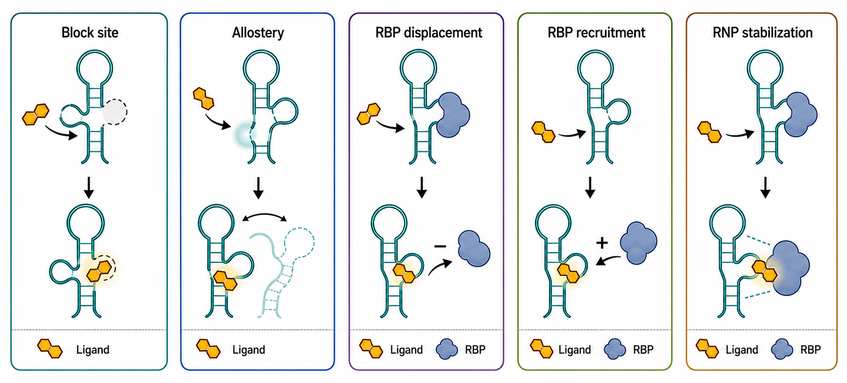
Figure 162.4. Ligand-induced RNA and RNP remodeling mechanisms. Direct RNA ligands can change function by blocking an RNA site, shifting an RNA ensemble, displacing an RBP, recruiting an RBP, or stabilizing a transient RNP assembly. These mechanisms require different assays and controls.
Translation changes provide another set of examples. A ligand that binds a viral internal ribosome entry site, a frameshift element, an upstream open reading frame region, or a structured 5′ untranslated region can alter ribosome recruitment, scanning, start-codon choice, elongation pausing, or recoding. Evidence requires separating direct RNA effects from general translation inhibition. Polysome profiling, ribosome profiling, reporter variants, endogenous protein measurement, and transcript abundance controls can distinguish translation-specific regulation from RNA decay or transcriptional effects. For coding RNAs, one must also ask whether a compound binds the mRNA structure or an RNA-rich ribosomal site that affects many mRNAs.
Ligand-induced RNA decay can arise without an engineered degrader. Stabilizing a structure may expose a decay element, block translation and make an mRNA more decay-prone, recruit a surveillance factor, or trigger nuclear retention and exosome targeting. Conversely, a ligand may stabilize an RNA by preventing endonuclease access or RBP displacement. Interpreting abundance changes therefore requires measuring transcription, processing, translation, decay rate, and RNA localization where feasible. A reduced RNA level is not automatically evidence of direct degradation by the ligand; it may be a downstream consequence of stress, feedback, or cell-state selection.
RBP recruitment can be desired or undesired. Desired recruitment might bring a splicing factor, stabilizing protein, decay factor, editing enzyme, or RNase to a target RNA. Undesired recruitment may generate toxic RNP assemblies, stress granules, or innate immune complexes. Cellular concentration matters. Recruiting a limiting RBP to one abundant target may deplete the RBP from other RNAs. Recruiting an RNase without local control may create collateral degradation. The pharmacology of recruitment is therefore stoichiometric as well as structural.
The assays for remodeling should match the proposed mechanism. If the mechanism is displacement, use direct RBP occupancy measurements such as CLIP-like assays, RNA pulldown, electrophoretic mobility shift, or quantitative RNP proteomics, plus functional rescue. If the mechanism is recruitment, show increased effector association and dependence on the effector. If the mechanism is structure remodeling, use chemical probing, structural biology, compensatory mutations, and time-resolved functional readouts. If the mechanism is translation change, measure ribosome occupancy and protein output. If the mechanism is splicing change, measure endogenous isoforms and spliceosome-stage dependence. Each assay has artifacts, but convergent evidence can define a credible causal chain.
The main misconception is that RNA binding automatically means functional remodeling. Many ligands bind without changing the relevant ensemble enough to matter. Others remodel RNA but only at concentrations that also perturb membranes, ribosomes, mitochondria, or stress pathways. A functional direct RNA ligand should satisfy three linked statements: the compound binds the RNA target in the relevant context, binding changes a defined molecular state, and the molecular state change explains the phenotype.
162.6. RIBOTAC-like degraders and proximity-induced mechanisms
RIBOTAC-like molecules extend RNA ligand discovery from occupancy to induced degradation or effector recruitment. The conceptual design has at least two modules. One module binds a target RNA motif. Another module recruits, activates, or positions a ribonuclease, decay factor, RNA-modifying enzyme, or other effector close enough to act on the target RNA. A linker or scaffold connects the modules. The intended pharmacological event is proximity-induced action: the compound does not need to occlude a functional site if it can bring an effector to the RNA and cause cleavage, decay, editing, or remodeling.
The analogy to proteolysis-targeting chimeras is useful but imperfect. Protein degraders recruit an E3 ubiquitin ligase to a target protein and rely on ubiquitin-proteasome degradation. RNA degraders must choose among many possible effector pathways: RNase L-like cleavage, RNase H-like mechanisms when an oligonucleotide is involved, endogenous endonucleases, exosome-related decay, nonsense-mediated decay, nuclear surveillance, cytoplasmic deadenylation and decapping, or designed ribonuclease recruitment. A small-molecule RIBOTAC-like degrader is typically imagined as fully small molecule or small molecule plus effector-binding ligand, whereas antisense gapmers are nucleic-acid drugs that recruit RNase H through Watson-Crick pairing. The distinction matters because the exposure, tissue distribution, immune sensing, and selectivity rules are different.
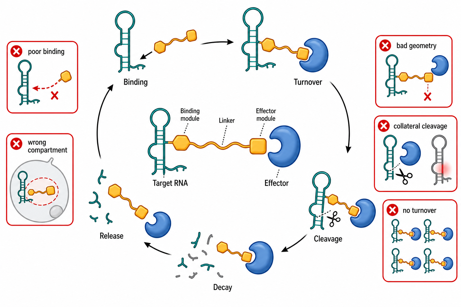
Figure 162.5. RIBOTAC-like proximity-induced RNA degradation. A RIBOTAC-like molecule is designed to bind a target RNA and recruit an RNA-degrading or RNA-remodeling effector. Productive degradation depends on cellular target engagement, effector availability, linker geometry, cleavage, product release, and selectivity.
A useful RNA degrader must solve four physical problems. First, the target-binding module must recognize the RNA in cells. Second, the effector-recruiting module must bind an effector that is present in the same compartment and not saturated or inaccessible. Third, the linker must allow productive geometry for cleavage or remodeling. Fourth, the resulting ternary or encounter complex must be selective enough to avoid broad RNA damage. Potency can fail at any step. A target ligand with good biochemical affinity may not bind the cellular RNP. An RNase ligand may recruit an enzyme but orient it poorly. A linker that is too short or too rigid may prevent productive cleavage. A linker that is too long or hydrophobic may create nonspecific binding or poor pharmacokinetics.
Proximity-induced cleavage has catalytic appeal. In principle, one degrader molecule could bind, recruit an RNase, induce cleavage, release, and act again. In practice, catalytic turnover depends on binding kinetics, product release, effector availability, and whether cleavage fragments remain associated. High affinity is not always beneficial; a molecule that binds too tightly to the target or effector may become stoichiometric. This is a familiar principle in proximity pharmacology: productive ternary complex formation and turnover can matter more than binary affinity. For RNA, one must also account for target abundance and synthesis rate. A rapidly transcribed RNA may require sustained degradation, whereas a low-copy toxic RNA may respond to partial occupancy.
RNase recruitment raises safety boundaries. RNases are powerful enzymes, and many are part of antiviral or stress pathways. Recruiting or activating an RNase can produce innate immune signaling, global RNA decay, apoptosis, or inflammation if the degrader is not locally controlled. The target RNA may share motifs with other transcripts. The effector may cleave near the recruited site but not exactly at one nucleotide, creating heterogeneous fragments that themselves have biological activity. In viral infection, broad RNA degradation may appear antiviral but be host-toxic. In cancer, reducing an oncogenic ncRNA may be beneficial only if normal tissues tolerate the same target or effector engagement.
RIBOTAC-like designs also need a rigorous dependency test. If a molecule reduces target RNA abundance, one must show that degradation depends on both the target-binding module and the effector-recruiting module. Inactive analogs that lack target binding, effector binding, or productive linker geometry are essential. Knockdown, knockout, inhibition, or compartmental depletion of the proposed effector should reduce degrader activity. Cleavage sites or decay intermediates should match the proposed effector where possible. The effect should be dose and time dependent, and target RNA reduction should precede downstream phenotypes. Transcriptome-wide RNA abundance and structure data should quantify collateral effects.
Box 162.3. When RNA Loss Counts as Degrader Mechanism
Evidence ladder for an RNA degrader claim
An RNA abundance decrease is only the starting observation. A degrader claim needs module logic: a binding-defective target ligand, an effector-binding-defective analog, and an unproductive linker control should lose activity. The proposed effector should be required by knockdown, knockout, inhibition, relocalization, or rescue. Target RNA loss should occur after cellular engagement and before downstream phenotypes such as splicing rescue, toxicity, or antiviral response. Decay-rate measurements, cleavage-site mapping, or decay-intermediate detection should support the proposed pathway. Controls should rule out transcriptional shutdown, reporter silencing, processing defects, export blockade, cytotoxicity, innate immune activation, and selection against target-expressing cells. Transcriptome-wide profiling then asks whether the degrader is local and selective or simply damaging RNA metabolism.
Programmable small-molecule conjugates occupy a spectrum between classical small molecules and oligonucleotide therapeutics. A conjugate may combine an RNA-binding small molecule with a ribonuclease-recruiting ligand, a reactive warhead, a photocatalyst, a localization tag, an RBP ligand, or a delivery-enhancing group. The more complex the conjugate, the harder it becomes to optimize drug-like properties. Molecular weight, polar surface area, ionization state, solubility, permeability, efflux, metabolism, plasma protein binding, tissue distribution, and lysosomal trapping can dominate biology. A beautiful ternary mechanism in vitro may fail because the molecule never reaches the compartment where the RNA lives.
Table 162.4. RIBOTAC-like degrader design variables and failure modes. RNA degrader design is constrained by both induced-proximity geometry and RNA pharmacology. Activity requires target binding, effector recruitment, productive cleavage or decay, turnover, and tolerable collateral effects.
| Design variable | Desired property | Common failure | Control experiment |
|---|---|---|---|
| Target-binding module | Selectively engages the intended cellular RNA motif or RNP state | Binds the purified construct but not the folded, modified, protein-bound, or compartmentalized RNA | Binding-defective analogs, cellular probing or occupancy, target-motif mutants, and inactive transcript controls |
| Effector-recruiting module | Recruits an RNase, decay factor, or remodeling effector present in the same compartment | Effector is absent, saturated, mislocalized, or globally activated | Effector knockdown, knockout, inhibition, or relocalization tests plus stress and innate-immune controls |
| Linker or scaffold geometry | Positions target and effector for productive cleavage, remodeling, release, and turnover | Unproductive ternary complex, steric clash, stoichiometric trapping, or hydrophobic nonspecific binding | Linker-length and rigidity series, ternary-complex assays, cleavage time courses, and product-release measurements |
| Compartment and exposure | Reaches the nucleus, cytoplasm, granule, viral replication site, or tissue where the RNA and effector coexist | Efflux, serum binding, lysosomal trapping, wrong tissue distribution, or poor free intracellular concentration | Free-drug and fractionation measurements, localization controls, compartment-specific readouts, and PK/PD comparison |
| Cleavage or decay chemistry | Reduces target RNA through the proposed effector-dependent pathway before downstream phenotype | Apparent RNA loss from transcriptional inhibition, processing changes, cytotoxicity, or stress-mediated decay | Nascent transcription assays, decay-rate measurements, cleavage-site mapping, and early time-course ordering |
| Selectivity and collateral effects | Degrades or remodels the target while sparing unrelated RNAs and essential RNPs | Shared motif cleavage, broad RNA damage, RBP depletion, or innate immune activation | RNA-seq, structure or engagement profiling, RBP occupancy, translation profiling, and immune-marker panels |
| Drug-like properties | Preserves mechanism after tag removal while maintaining solubility, permeability, stability, and tolerability | High molecular weight, high polarity, metabolism, plasma protein binding, or probe tag dependence dominates biology | Tag-free analogs, serum and microsomal stability, permeability and efflux assays, cell-panel toxicity, and PK/PD linkage |
The strongest near-term opportunities are likely targets where a modest reduction in a pathogenic RNA has a large therapeutic effect, the target presents a repeated or highly distinctive motif, the relevant cell type is accessible, and biomarkers can measure both RNA degradation and functional rescue. Repeat-expansion RNAs, viral RNAs, disease-associated splice isoforms, and selected oncogenic or toxic ncRNAs are plausible categories. The weakest opportunities are targets whose RNA motif is shared across many essential transcripts, targets that require complete knockdown in inaccessible tissues, and targets where the disease mechanism is mostly protein-driven despite an RNA-binding readout.
The field should avoid treating “degrader” as a potency label. A compound that lowers RNA abundance is not necessarily a degrader; it may inhibit transcription, alter processing, block export, suppress cell growth, activate stress, or select against cells expressing the target. A RIBOTAC-like mechanism requires evidence of induced proximity and effector-dependent RNA loss. Chapter 150 discusses RNase H-recruiting antisense gapmers, and Chapter 158 discusses pharmacokinetics, pharmacodynamics, immunogenicity, toxicology, and regulatory science for RNA drugs.
162.7. Probe-to-lead translation and failure-mode analysis
Probe-to-lead progression is where many direct RNA ligand projects fail. A chemical probe is a tool compound that tests a biological hypothesis. A lead is a chemical series with a plausible path toward therapeutic exposure, selectivity, safety, manufacturability, and clinical biomarkers. The gap is large for RNA targets because early probes often rely on high charge, planar aromatic surfaces, affinity tags, photo-crosslinkers, or physicochemical properties that are poorly compatible with drug-like behavior. A useful probe may be too promiscuous, insoluble, unstable, impermeable, effluxed, metabolized, immunostimulatory, or toxic to become a lead without major redesign.
The first failure mode is nonspecific nucleic-acid binding. RNA is abundant, negatively charged, and structurally repetitive at the level of helices and grooves. Cationic and aromatic compounds can bind many RNAs and DNA. Such compounds may produce attractive biochemical binding curves and broad cellular phenotypes but little target selectivity. Counterscreens against unrelated RNAs, DNA, rRNA-rich lysates, tRNA, heparin, and cell extracts can expose nonspecific binding. Structure-activity relationships should show that small chemical changes alter target activity in ways consistent with a defined binding mode rather than generic charge or hydrophobicity.
The second failure mode is assay interference. RNA assays often use fluorescence, reverse transcription, sequencing, polymerases, ribosomes, nucleases, or beads. Compounds can fluoresce, quench dyes, inhibit reverse transcriptase, inhibit polymerases, bind beads, aggregate proteins, chelate magnesium, change pH, intercalate into reporter constructs, or cause cell stress. Orthogonal assays are not optional. If a compound is active only in one reporter or only with one label, the safest assumption is artifact until proven otherwise. Chapter 5 provides general evidence standards, but RNA pharmacology requires especially aggressive artifact control because nucleic acid-binding compounds are chemically promiscuous by default.
The third failure mode is target misassignment. Phenotypic screens are prone to discovering protein targets, pathway effects, or general stress responses. A compound that corrects a splicing reporter may inhibit a kinase, alter transcription elongation, change chromatin, or perturb spliceosome assembly globally. A compound that inhibits viral replication may target a host kinase, membrane trafficking, polymerase activity, or translation rather than a viral RNA structure. Target engagement experiments must therefore be built into lead progression, not appended after medicinal chemistry has optimized an unknown mechanism.
The fourth failure mode is cellular exposure mismatch. A compound can bind RNA in vitro at 1 micromolar but require 50 micromolar in cells because of poor permeability, efflux, serum binding, lysosomal trapping, metabolism, or competition with abundant off-target RNAs. Conversely, a compound can appear potent in cells because it accumulates in a compartment or triggers stress at low nominal concentration. Measuring free intracellular concentration is difficult but important. Exposure-response should be compared with engagement-response and phenotype-response. If phenotype occurs far below detectable engagement or only at cytotoxic concentrations, the proposed mechanism needs revision.
The fifth failure mode is selectivity collapse during optimization. Medicinal chemistry often increases affinity by adding aromatic surface, cationic groups, or hydrophobic contacts. Those changes may improve target binding while increasing binding to many RNAs, DNA, membranes, or proteins. Optimization must therefore track selectivity as actively as potency. Transcriptome-wide engagement, RNA abundance profiling, translation profiling, RBP occupancy, and cell-panel assays can reveal whether a series is becoming broadly disruptive. Early selectivity filters are more useful than late toxicity surprises.
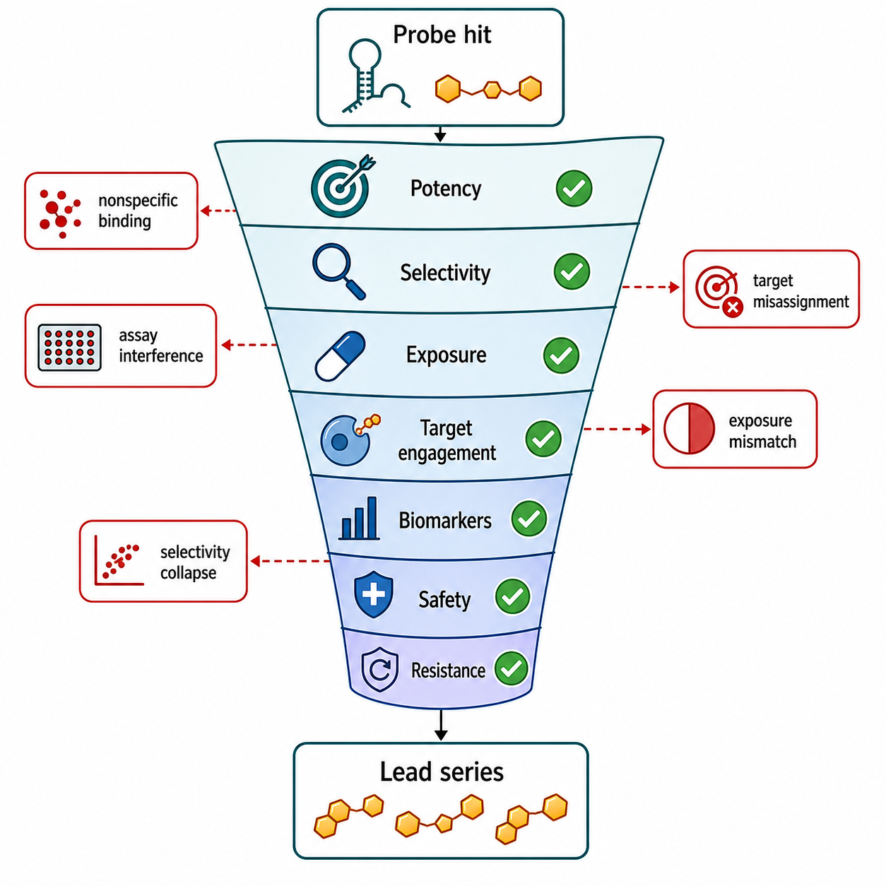
Figure 162.6. Probe-to-lead decision funnel for direct RNA pharmacology. A direct RNA probe becomes a credible lead only when medicinal chemistry preserves mechanism while improving selectivity, exposure, safety, and translational biomarkers. Many RNA hits fail because nonspecific binding, assay interference, or target misassignment is discovered late.
Clinical translation adds target and disease constraints. The target RNA must be causally linked to disease, expressed in accessible tissues, and modulated in a way that predicts benefit. Biomarkers should measure target engagement, RNA structural or abundance change, downstream pathway correction, and safety. For infectious disease, resistance monitoring must include RNA sequence variation that disrupts ligand binding while preserving viral fitness. For genetic disease, patient stratification may depend on repeat length, splice variant, tissue expression, or modifier genes. For cancer, tumor selectivity may be undermined if the RNA target is also important in proliferating normal tissues.
Regulatory classification can be complex. A direct RNA small molecule may be regulated like a conventional small molecule, but its safety package must address nucleic-acid-related liabilities, immune activation, genotoxicity where DNA binding is plausible, reproductive toxicity when germline or developmental RNAs are affected, and off-target transcriptome effects where mechanistically relevant. A RIBOTAC-like conjugate may raise additional questions about effector recruitment, degradation products, long-lived tissue accumulation, and class-specific immunotoxicity. Companion diagnostics may be needed when the target is a patient-specific RNA variant, repeat expansion, splice isoform, or viral genotype.
The most useful development logic is a disciplined funnel. Start with a defined RNA element and disease hypothesis. Demonstrate binding by orthogonal assays. Map the binding or remodeling site. Establish cellular target engagement at pharmacologically reasonable exposure. Show target-dependent functional response using mutants, rescue, or resistance. Quantify selectivity across transcriptome and proteome. Optimize chemistry while preserving mechanism. Establish pharmacokinetic and pharmacodynamic relationships. Only then treat the series as a therapeutic lead rather than a chemical-biology probe.
Recent Consensus
The current working consensus is that direct RNA targeting is real but technically demanding. RNA can form ligandable motifs, and several natural or clinical precedents show that small molecules can modulate RNA-rich biological systems. The field has moved beyond the obsolete idea that RNA is generally undruggable. At the same time, the field has also moved beyond simplistic optimism. Most cellular RNAs are not automatically accessible drug targets, and apparent RNA binding must be separated from nonspecific nucleic-acid affinity, protein off-targets, assay artifacts, and stress responses.
The consensus evidence ladder emphasizes target engagement. Strong projects combine structural or probing evidence, biochemical binding, cellular occupancy, functional genetics, selectivity profiling, and exposure-response relationships. RIBOTAC-like proximity pharmacology is an important frontier, but degrader claims require effector-dependence and induced-proximity evidence, not merely reduced RNA abundance. AI-assisted methods are valuable for prioritization but remain limited by sparse RNA-ligand training data and conformational complexity.
Open Questions, Controversies, Deprecated Models, and Common Misconceptions
Open questions:
- How often do disease-relevant RNAs present stable ligandable pockets in cells?
- Which RNA motifs provide enough selectivity over abundant background RNAs?
- Can small-molecule conjugates recruit RNases with acceptable tissue-level safety?
- How should cellular RNA target engagement be quantified in a way comparable across laboratories?
- Which computational benchmarks genuinely predict prospective RNA-ligand success?
- How often are phenotypic RNA-targeting hits actually protein-targeting hits?
Common misconceptions:
- “In vitro binding is the same as cellular engagement.” Cellular engagement depends on permeability, localization, competition, RNP context, abundance, and target accessibility.
- “RNA abundance reduction is the same as degrader mechanism.” RNA loss can arise from transcriptional repression, toxicity, decay, processing defects, or indirect stress unless degrader recruitment is shown.
- “A predicted structure is the cellular target state.” RNA target states in cells depend on proteins, ligands, modifications, translation, compartment, and kinetics.
- “Broad transcriptomic change proves broad RNA binding.” It may reflect a narrow upstream perturbation.
- “A selective phenotype proves selective target engagement.” Pathway bottlenecks can amplify weak or off-target interactions.
- “A compound touching RNA in a ribonucleoprotein complex is best understood as a naked-RNA ligand.” RNP context can make the pharmacological target a composite RNA-protein surface rather than isolated RNA.