Chapter 121. Cross-Disease RNA Mechanisms: Causality, Convergence, Biomarkers, and Therapeutic Validation
Scope Note
This chapter compares RNA mechanisms across disease classes. It does not replace the chapters that teach individual rare disorders, cancers, neurological syndromes, immune pathways, infections, or extracellular-RNA biology. Instead, it asks which reasoning tools transfer between those fields: how a disease-associated RNA observation becomes a causal claim, which molecular failures recur in otherwise unrelated disorders, how evidence from genetics, perturbation and rescue, biochemistry and structure, longitudinal observation, and clinical intervention should be integrated, and when a biomarker or therapeutic target claim outruns the data. Mendelian disorders supply unusually legible causal anchors, whereas cancer, neurodegeneration, infection, inflammation, and metabolic disease reveal the difficulty of resolving convergent mechanisms in heterogeneous tissues and changing cell states.
Executive Summary
RNA disease mechanisms converge on a limited set of molecular operations even when their initiating causes differ. Sequence variants and somatic mutations can alter splice-site choice, RNA ends, modification, localization, translation, surveillance, or ribonucleoprotein assembly. Repeat-expanded transcripts can create multivalent RNA surfaces and unconventional translation products. Tumors, infected tissues, inflamed organs, and degenerating nervous systems can all accumulate double-stranded or mislocalized RNA, remodel RNA-binding proteins, change decay and translation, or produce extracellular RNA. Convergence does not mean equivalence: the initiating lesion, vulnerable cell type, magnitude, timing, reversibility, and evidence standard determine whether the same-looking RNA phenotype has the same causal meaning.
The hardest interpretive problem is causality. A disease-associated RNA feature may be a driver, mediator, modifier, compensatory response, marker of cell composition, consequence of tissue injury, technical artifact, or correlate of another lesion. No single evidence type resolves every category. Human genetics can anchor direction and lifelong exposure but can be confounded by linkage, pleiotropy, and developmental effects. Perturbation and rescue test manipulability but may introduce dose, delivery, or model artifacts. Biochemistry and structure can establish physical plausibility without proving that the mechanism operates at physiological occupancy in the affected tissue. Longitudinal data provide temporal ordering but do not by themselves remove confounding or reverse causation. Clinical intervention can be powerful evidence when target engagement changes a disease endpoint, yet failure may reflect delivery, exposure, timing, or endpoint choice rather than an incorrect target. The chapter therefore uses triangulation: independent evidence classes should converge on the same causal chain while their distinct failure modes are tested.
The ownership boundary is deliberate. Chapter 38 and Chapter 41 own detailed rare processing disorders and their molecular machinery; Chapter 53 owns cancer-specific transcriptomic mechanisms; Chapter 95, Chapter 99, and Chapter 103 own neurological localization, repeat-expansion, and RNA-binding-protein pathology; Chapter 107 owns extracellular-RNA biogenesis and communication; and the immunity and host-pathogen chapters own receptor pathways and infection-specific mechanisms. This chapter owns the comparison: what rare variants teach about causality, which processes recur across classes, how a biomarker crosses the thresholds from analytical validity to clinical utility, and how a putative target survives genetic, mechanistic, pharmacological, and clinical validation.
Concept Inventory
- RNA-processing defect: a disease-relevant error in the conversion of a primary RNA transcript into a mature or functional ribonucleoprotein. The defect may affect capping, splicing, cleavage and polyadenylation, editing, modification, nuclear export, localization, translation, surveillance, decay, or assembly with proteins. A processing defect can be caused by a variant in a cis element on the RNA, by a mutation in a trans-acting protein or RNA factor, or by a cellular state that changes factor concentration, localization, or activity.
- Transcriptomic signature: a reproducible pattern of RNA abundance, isoform usage, editing, modification, fusion transcript expression, or RNA localization associated with a disease state or clinical outcome. The term signature does not itself imply mechanism. A signature can be diagnostic, prognostic, predictive of therapy response, or merely descriptive.
- Toxic RNA: an RNA molecule whose sequence, structure, abundance, localization, or bound-protein composition directly contributes to pathology. Repeat-expanded RNAs in neurological disease are common examples because expanded CUG, CGG, GGGGCC, CAG, or related repeat RNAs can form abnormal structures, accumulate in foci, recruit RNA-binding proteins, and perturb RNA metabolism.
- Repeat-RNA phase transition: a concentration-, sequence-, and valency-dependent change in which repeat-containing RNA molecules demix or crosslink into an RNA-rich assembly. A liquid-like phase-separated focus and a solid-like gel are not interchangeable material states. Demonstrating either state explains an assembly route, but it does not by itself prove that the assembly causes disease.
- RNA granule: a non-membrane ribonucleoprotein assembly whose formation depends on multivalent interactions among RNAs and proteins. Stress granules, neuronal transport granules, processing bodies, nuclear speckles, and disease-associated condensates are related but not interchangeable. Condensate involvement in disease is plausible when altered material properties, composition, persistence, localization, or clearance can be connected to cell dysfunction, but condensate presence alone is not proof of causality.
- RNA biomarker: an RNA measurement used to infer a biological or clinical state. Biomarkers include expression signatures, splice isoforms, fusion transcripts, viral RNAs, extracellular RNAs, circulating tumor RNAs, immune-response transcripts, and RNA modifications. Biomarker validation requires analytical validity, clinical validity, clinical utility, and attention to sampling, stability, normalization, and confounding by cell mixture.
- Causal anchor: an observation that strongly fixes one link in a disease mechanism, such as a segregating variant with an experimentally confirmed RNA consequence or a selective intervention with verified target engagement. An anchor strengthens a chain but does not validate every downstream step.
- Convergent mechanism: an RNA-level process reached from different initiating lesions or disease contexts, such as cryptic splicing, double-stranded-RNA accumulation, altered RNA decay, RBP misassembly, or translation stress. Convergence is a mechanistic hypothesis that requires context-specific testing, not proof that diseases are interchangeable.
- Triangulation: causal inference from evidence classes with different assumptions and biases. Agreement among human genetics, perturbation and rescue, physical mechanism, patient time courses, and intervention is stronger than repetition of one assay type.
- Reverse causation: a situation in which disease state changes an RNA measurement even though the RNA change is interpreted as causing the disease. Longitudinal sampling and upstream perturbation help address reverse causation but do not automatically eliminate it.
What to Know Before Reading This Chapter
The chapter assumes familiarity with the central steps of gene expression: transcription makes a precursor RNA, processing converts the precursor into a mature RNA or RNP, export and localization place the RNA in a cellular compartment, and translation or noncoding function connects RNA to phenotype. The reader should also distinguish a gene from a transcript, an exon from an intron, a splice isoform from a protein isoform, and a biomarker from a causal mechanism.
Two recurring examples carry much of the chapter. First, a rare splice-altering variant can turn a normally innocuous intronic sequence into a pseudoexon, creating a frameshifted mRNA or an unstable transcript. Second, a repeat expansion can create a gain-of-function RNA that binds proteins and perturbs RNA granules. These examples show why RNA disease biology often sits between genetics, molecular cell biology, and clinical evidence.
The most important evidence rule is simple: association is not enough. RNA abundance changes can be caused by altered cell composition, ischemia, immune infiltration, drug exposure, sample handling, degradation, sequencing depth, or annotation choice. Splicing and editing changes can be secondary to stress or dedifferentiation. Noncoding RNA claims can be inflated by correlation without perturbation. For every example, ask six questions: What changed? In which cell, compartment, and disease stage? How was it measured? What plausible causal path connects it to phenotype? Which intervention distinguishes cause from consequence? Which neighboring chapter owns the disease-specific detail needed to judge the example?
121.1. Mendelian and rare diseases as causal RNA-mechanism anchors
Mendelian disease is disease in which variants at one genetic locus have a large effect on risk or phenotype. Rare disease is broader; it includes Mendelian disorders, chromosomal disorders, mitochondrial disorders, developmental syndromes, and individually uncommon disease entities whose molecular cause may be heterogeneous. RNA-processing defects occur when a disease-causing variant disrupts the RNA-level steps between DNA sequence and mature functional product. The same variant can be described genetically as a point mutation, but mechanistically as a splice defect, RNA stability defect, RNA modification defect, RNA localization defect, or translation defect.
The first RNA-processing lesson in rare disease is that coding sequence is not the only sequence that matters. A variant at a canonical splice donor or acceptor can block intron removal. A deep intronic variant can create a cryptic splice site that inserts a pseudoexon into the mature mRNA. A synonymous coding variant can alter an exonic splicing enhancer, reduce binding by a serine/arginine-rich splicing factor, and change exon inclusion without changing the encoded codon in the reference transcript. Variants in untranslated regions can alter RNA-binding protein sites, microRNA sites, polyadenylation signals, upstream open reading frames, or RNA structures that affect translation and stability. For clinical interpretation, a DNA variant is therefore incomplete evidence until the relevant RNA consequence has been tested or predicted with adequate confidence.
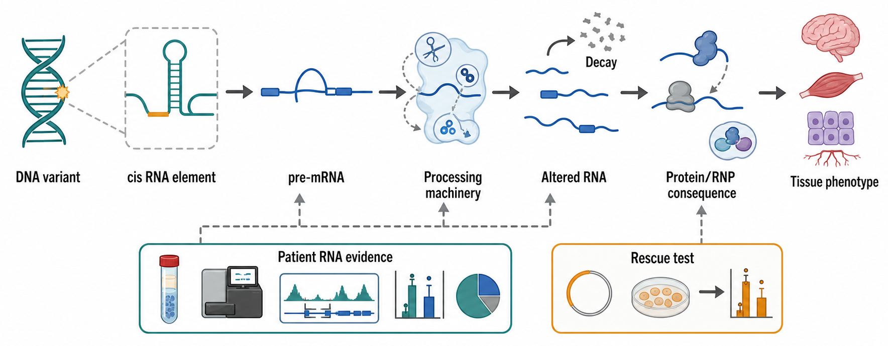
Figure 121.1. From Variant to RNA-Processing Disease. A disease-associated DNA variant can act through RNA by disrupting splice-site recognition, RNA stability, untranslated-region regulation, modification, export, or translation. The visual separates variant annotation from mechanism and highlights where patient RNA evidence and rescue experiments can test causality.
A practical causal chain for a splice-altering rare disease variant has four steps. First, the variant changes a cis-regulatory RNA element: a splice site, branch point, polypyrimidine tract, exonic enhancer, intronic silencer, or cryptic exon boundary. Second, the spliceosome or associated regulatory proteins choose a different splice pattern. Third, the altered RNA either encodes an altered protein, introduces a premature termination codon that triggers nonsense-mediated decay, changes an untranslated region, or produces a nonproductive RNA. Fourth, tissue dysfunction follows if the affected gene is dosage-sensitive, developmentally required, cell-type restricted, or part of a pathway with limited compensation. Each step can fail as an explanation if the altered isoform is rare, if the tissue studied is not the disease tissue, if the transcript is naturally variable, or if the variant is in linkage disequilibrium with another causal lesion.
Box 121.1. Interpreting a Splice-Altering Variant
A splice prediction is a hypothesis, not a diagnosis. A strong RNA-disease interpretation asks five questions in order. Does the variant affect a recognizable splice donor, acceptor, branch point, polypyrimidine tract, enhancer, silencer, or cryptic exon boundary? Is the abnormal RNA detected in a tissue or model that expresses the relevant transcript at the disease-relevant stage? Does the RNA outcome plausibly alter function through frameshift, premature termination and nonsense-mediated decay, exon loss, altered domain composition, untranslated-region change, or dosage loss? Is the event allele specific, segregating with disease, recurrent in unrelated patients, or supported by a calibrated functional assay? Finally, does correction of the splice event restore RNA, protein, or cellular phenotype? A negative blood RNA result is weak if the gene is not expressed in blood, and a positive minigene result is incomplete if endogenous chromatin, cell type, or decay conditions differ from the patient tissue.
Rare disease also exposes the importance of trans-acting RNA-processing factors. A protein that recognizes splice sites, modifies transfer RNAs, processes ribosomal RNAs, edits organellar RNAs, trims small RNAs, or remodels ribonucleoproteins may affect many RNA substrates. Disease specificity then arises not because only one RNA is affected, but because some cell types have narrow tolerance for a processing imbalance. A neuron may be sensitive to small changes in local mRNA translation, a hematopoietic progenitor to ribosome biogenesis defects, a skeletal muscle cell to repeat-RNA accumulation, and a developing embryo to dosage-sensitive splicing of transcription factors. The mechanism is often networked rather than one RNA to one symptom.
Table 121.1. RNA-Processing Defect Classes in Rare Disease. Classify rare-disease mechanisms by affected RNA step, example consequence, useful assay, and key caveat.
| RNA step | Lesion type | Possible disease consequence | Evidence assay | Interpretation caveat |
|---|---|---|---|---|
| Splice-site recognition | Canonical donor, acceptor, branch-point, or enhancer variant | Exon skipping, intron retention, frameshift, premature termination, or nonsense-mediated decay | Patient RNA-seq, targeted RT-PCR, minigene or endogenous correction assay | The assayed tissue must express the relevant isoform and distinguish pathogenic change from normal splice variation. |
| Cryptic exon control | Deep intronic variant creates a splice site or weakens local repression | Pseudoexon inclusion, unstable transcript, altered protein dosage, or nonproductive RNA | Junction reads, long-read isoform sequencing, allele-specific RNA evidence | Nonsense-mediated decay can erase the abnormal transcript before sampling. |
| 3′ end formation | Cleavage, polyadenylation, or UTR-regulatory defect | Altered 3′ UTR, changed stability, disrupted RBP or microRNA regulation, or altered translation | 3′ end sequencing, long-read RNA sequencing, UTR reporter assay | Alternative polyadenylation is tissue- and state-dependent, so a shifted end is not automatically pathogenic. |
| RNA modification or editing | Defect in a modifying enzyme, editing enzyme, or substrate-recognition element | Impaired decoding, folding, stability, translation, immune discrimination, or RNP assembly | Modification profiling, direct RNA evidence where appropriate, substrate class analysis, factor rescue | Broad trans-acting defects produce many secondary RNA changes; primary substrate evidence is needed. |
| RNA decay and surveillance | Premature termination, NMD sensitivity, decay-factor imbalance, or abnormal RNA persistence | Loss of gene dosage or accumulation of aberrant transcripts | Allele-specific expression, expression outlier analysis, decay-pathway perturbation, rescue assay | Steady-state abundance alone cannot separate transcription, processing, and decay effects. |
| RNP assembly, export, or localization | RBP mutation, localization element defect, or impaired RNP remodeling | Mislocalized RNA, defective local translation, granule imbalance, or tissue-selective vulnerability | RNA FISH, fractionation, CLIP-family mapping, patient-derived cell models | Visible mislocalization or granules require a functional link to disease phenotype. |
Evidence for a trans-acting processing disorder usually requires a different argument from evidence for a cis splice variant. A cis variant can often be connected to one transcript by allele-specific RNA sequencing, minigene assays, or correction of the local sequence. A trans-acting defect may produce hundreds of RNA changes. The central question becomes which RNA changes are primary and which are downstream of cell stress. Stronger evidence comes from biochemical knowledge of the factor, reproducible substrate classes, patient-genotype correlation, rescue by restoring the factor, and convergence on disease-relevant pathways. For example, a defect in an RNA modification enzyme should be evaluated by asking which RNA species lose the modification, whether the affected RNA species are expressed in the vulnerable tissue, and whether the loss changes translation, decoding, folding, stability, or immune sensing in a way that explains the phenotype.
RNA processing can also be the reason a DNA test looks incomplete. Some pathogenic events are better detected in RNA than DNA: aberrant exon skipping, cryptic exon inclusion, allele-specific expression, fusion transcripts, tissue-specific isoforms, and expression outliers. Long-read RNA sequencing can connect distant exons and reveal full-length isoforms, while short-read RNA sequencing can quantify exon junctions and expression. However, clinical RNA testing has limits. The disease tissue may be inaccessible, blood may not express the relevant gene, nonsense-mediated decay can erase the abnormal transcript, and sample handling can alter RNA integrity. A negative RNA test therefore does not exclude a pathogenic variant unless the assay covers the right tissue, developmental stage, transcript model, and degradation conditions.
Therapeutically, RNA-processing defects are unusually actionable when the disease mechanism can be reduced to a mis-spliced exon or an unstable transcript. Splice-switching antisense oligonucleotides can block a splice site or regulatory element, redirect exon inclusion, suppress a poison exon, or restore a productive reading frame. Small molecules can alter splicing factor activity or stabilize a desired RNP state. These approaches require a precise target transcript, a therapeutic window, and evidence that changing the RNA improves a disease-relevant cellular or clinical endpoint. They also require caution: changing one splice event may alter other transcripts, and increasing a partially functional transcript may not restore normal protein localization, stoichiometry, or developmental timing.
Do not overgeneralize the term RNA-processing disease. Many disorders show RNA changes because sick cells change transcription, immune activation, metabolism, or viability. A disorder should be called RNA-processing driven only when evidence links a specific processing abnormality to disease pathogenesis through genetics, molecular mechanism, and perturbation. Detailed mechanisms of individual processing disorders belong to Chapter 38 and Chapter 41; the transferable lesson here is that a strong rare-disease anchor joins the initiating variant, the measured RNA consequence, the vulnerable cell, and the phenotype without skipping a link.
121.2. Convergent RNA dysregulation across cancer and proliferative disease
Cancer is a genetic disease, but it is also a transcriptome disease. A tumor cell changes what it transcribes, how it splices transcripts, which RNA ends it uses, which RNAs it edits or modifies, which noncoding RNAs it expresses, and which fusion transcripts it produces from rearranged or misregulated genomes. The cancer transcriptome is not merely a downstream readout. In some cases, RNA-level changes create oncogenic proteins, disable tumor suppressor pathways, rewire immune recognition, alter cell identity, or create drug vulnerabilities. In other cases, RNA changes are passengers that report proliferation, hypoxia, inflammation, lineage, or treatment exposure.
The simplest cancer RNA mechanism is a fusion transcript. A chromosomal rearrangement can place exons from two genes into one transcript. If the fusion preserves an open reading frame and joins functional protein domains, the mature mRNA can encode a chimeric oncogenic protein. If the fusion places a coding region under a strong promoter or removes regulatory regions, expression can change without producing a novel protein. Fusion detection is an RNA problem because the clinically relevant product is often the expressed junction rather than the DNA breakpoint alone. RNA sequencing can reveal whether a predicted rearrangement is transcribed, which exons are joined, and whether the junction could encode a functional product. The limitation is that low-level readthrough, template switching during library construction, mapping artifacts, and passenger rearrangements can mimic or complicate fusion calls.
Alternative splicing in cancer has several mechanistic routes. Mutations in spliceosome components or splicing regulators can globally alter exon recognition. Oncogenic signaling can change phosphorylation and localization of splicing factors. Dedifferentiation can reactivate fetal or progenitor isoforms. Tumor hypoxia and stress can alter RNA-binding protein activity. The consequences include expression of pro-survival isoforms, loss of tumor-suppressive exons, intron retention, altered untranslated regions, and neoantigen formation from abnormal junctions. The same splicing event can be clinically attractive if it is recurrent, tumor-selective, required for tumor fitness, and accessible to an antisense or small-molecule intervention. It is less attractive if it is heterogeneous, secondary to cell composition, or redundant with many other transcriptomic changes.
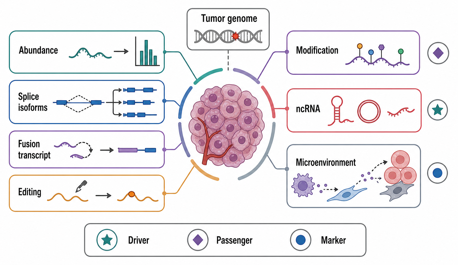
Figure 121.2. Layers of the Cancer Transcriptome. Cancer transcriptomes contain many kinds of RNA alteration. Some are drivers, some are biomarkers, and many are passengers of lineage, proliferation, stress, immune infiltration, or tumor purity. Interpretation depends on mechanism, recurrence, context, and perturbation evidence.
RNA editing and RNA modifications add another layer. Adenosine-to-inosine editing by ADAR enzymes can recode codons, alter splice sites, change RNA structure, or reduce innate immune sensing of double-stranded RNA. RNA modifications such as N6-methyladenosine can affect RNA stability, splicing, export, translation, and decay through reader, writer, and eraser proteins. In cancer, altered editing or modification can be interpreted in three ways: it may promote tumor growth, reflect the cellular state of the tumor, or represent an adaptive response to stress and immune pressure. Because editing and modification detection is technically demanding, claims require attention to mapping bias, modification stoichiometry, antibody specificity for enrichment assays, base-calling error, and whether the measured event occurs on the RNA species that carries the proposed function. The transferable rule is to establish the molecular species and event stoichiometry before assigning an effector pathway or disease role.
Noncoding RNAs in cancer include microRNAs, long noncoding RNAs, enhancer RNAs, circular RNAs, repeat-derived RNAs, and small fragments from transfer RNAs or other stable RNAs. A microRNA can repress tumor suppressor mRNAs or oncogenic mRNAs depending on its targets and cellular context. A long noncoding RNA can recruit chromatin regulators, scaffold proteins, alter nuclear organization, modulate splicing, sponge regulatory factors, or act as a marker of lineage. A circular RNA can be stable and abundant enough to act as a biomarker, but most proposed circular RNA mechanisms require careful stoichiometric validation. The key caution is that noncoding RNA expression is often highly cell-type specific. A tumor-associated noncoding RNA may mark the abundance of stromal cells, immune cells, necrosis, or a developmental lineage rather than directly regulate malignant behavior.
Box 121.2. Testing Whether a Cancer Noncoding RNA Is Causal
Overexpression is not mechanism. A noncoding RNA that is higher in tumors may be a driver, but it may also mark cell lineage, proliferation, hypoxia, stromal admixture, immune infiltration, copy-number change, enhancer activity, or tumor purity. Causality is stronger when perturbing the RNA changes a malignant phenotype at physiological expression levels and when rescue restores the phenotype with an RNA molecule that cannot be explained by editing the DNA locus. For long noncoding RNAs and enhancer RNAs, experiments should distinguish transcript-dependent effects from promoter, enhancer, chromatin, or transcription-through effects at the same genomic region. For circular RNAs, the back-splice junction must be confirmed and the proposed binding, translation, or biomarker function must be plausible at measured molecule numbers. For microRNAs, target repression should be shown for disease-relevant targets, not inferred from seed matches alone.
Table 121.2. Cancer RNA Alterations: Driver, Marker, or Passenger? Help readers distinguish mechanistic cancer RNA alterations from state markers and artifacts.
| RNA alteration | Possible driver evidence | Possible biomarker use | Common confounder | Validation requirement |
|---|---|---|---|---|
| Fusion transcript | Recurrent expressed junction with functional domains, dependency, or therapy response | Tumor classification, treatment selection, minimal residual disease tracking | Readthrough transcription, template switching, mapping artifacts, passenger rearrangements | Orthogonal junction confirmation, frame assessment, expression evidence, and clinical context. |
| Splice isoform | Tumor-selective isoform required for growth, immune escape, invasion, or drug resistance | Subtype, prognosis, splice-factor state, or target-engagement marker | Dedifferentiation, hypoxia, stress, annotation version, cell mixture | Isoform-specific quantification plus perturbation and rescue of the proposed splice event. |
| ADAR editing event | Edited site changes coding sequence, splicing, structure, or immune sensing with tumor phenotype | Interferon state, immune evasion state, or pathway activity marker | Mapping bias, unfiltered DNA variants, sequencing error, double-stranded RNA abundance | Matched DNA control, site-level quantification, and perturbation of editing enzyme or edited site. |
| RNA modification | Mark stoichiometry and reader engagement change stability, splicing, export, translation, or decay | Pathway activity, differentiation state, or treatment-response signal | Antibody specificity, base-calling error, low stoichiometry, tumor purity | Orthogonal modification mapping, transcript-context measurement, and writer, eraser, or reader perturbation. |
| Long noncoding RNA | Transcript or locus perturbation alters malignant phenotype with a defined mechanism | Lineage, subtype, stromal, immune, or disease-stage marker | Cell-type specificity, nearby transcription effects, correlation with chromatin state | Separate transcript-dependent from locus-dependent effects and test rescue or dose response. |
| Circular RNA | Stable back-splice product has sufficient abundance and a tested RBP, miRNA, translation, or biomarker role | Stable tumor or liquid-biopsy marker | Back-splice artifacts, host-gene expression, low molecule count | Junction-specific assays, independent quantification, and stoichiometric plausibility. |
| Expression signature | Locked score predicts response or reflects a dependency beyond tumor composition | Diagnosis, prognosis, immune state, therapy response, or residual disease | Tumor purity, stage, treatment access, batch effects, platform drift | Independent external validation, calibration, decision thresholds, and comparison with standard care. |
Cancer transcriptomics has transformed classification. Expression signatures can separate tumor lineages, molecular subtypes, immune-infiltrated tumors, proliferative states, and therapy-resistant states. Single-cell RNA sequencing can separate malignant cells from stromal and immune compartments and can reveal rare resistant populations. Spatial transcriptomics can map tumor-immune neighborhoods. Long-read sequencing can resolve isoforms and fusion structures that short reads fragment. These methods are powerful, but they also create interpretive hazards. A differential expression result may reflect changed cell proportions. A cluster marker may be a dissociation artifact. A tumor-specific isoform may depend on annotation version. A prognostic signature may encode stage, grade, purity, or treatment access rather than an RNA mechanism.
Clinical cancer RNA interpretation is strongest when the RNA measurement answers a defined question. A diagnostic assay may ask whether a tumor expresses a known fusion transcript. A treatment-selection assay may ask whether a target pathway is active or whether an immune checkpoint program is induced. A minimal residual disease assay may ask whether a tumor-specific transcript remains detectable after therapy. A mechanistic study may ask whether a splice isoform is required for invasion or drug resistance. These questions use overlapping technologies but different evidence standards. A discovery RNA-seq result can generate hypotheses for all of them, but the path to a clinical report, a drug target, or a mechanistic model diverges after discovery.
Therapeutic targeting in cancer must separate transcriptomic association from dependency. A tumor-enriched RNA is not necessarily a good target. Stronger evidence includes genetic or pharmacologic perturbation, rescue with an RNA-insensitive construct, dose-response relationships, conservation across models, patient-derived model validation, and evidence that target modulation affects tumor growth, immune escape, metastasis, or therapy response at exposures achievable in patients. RNA-directed therapies can target oncogenic transcripts, splice junctions, fusion transcripts, noncoding RNAs, or RNA-processing factors. RNA-processing factors are challenging because many are essential in normal cells. Selectivity may come from tumor-specific dependencies, synthetic lethality, altered dosage, or a splice event uniquely required by a cancer subtype. Chapter 53 owns cancer-specific examples; this chapter uses them to show why genetic dependency, therapeutic index, and biomarker enrichment must be tested as separate propositions.
121.3. Neurological, repeat-expansion, and proteostasis-linked RNA mechanisms
Neurological disease is a central arena for RNA pathology because neurons are long-lived, highly polarized, locally translating cells that depend on RNA transport, RNA granules, splicing programs, and precise protein homeostasis. A mature neuron can store and translate mRNAs far from the nucleus, regulate synaptic proteins through local RNA-binding proteins, and sustain stress-response granules under demanding metabolic conditions. These features make neurons vulnerable to repeat-expanded RNAs, RNA-binding protein aggregation, altered splicing, and defects in granule dynamics.
Repeat-expansion disorders illustrate toxic RNA with unusual clarity. A simple sequence repeat can expand beyond a pathogenic threshold in a gene or noncoding region. The expanded repeat may change DNA stability, transcription, RNA structure, protein coding, or translation. At the RNA level, repeated CUG, CCUG, CGG, GGGGCC, CAG, or related motifs can form hairpins, G-quadruplexes, or other repetitive structures. These structures can recruit RNA-binding proteins, create multivalent RNA-RNA contacts, and accumulate in nuclear or cytoplasmic foci. The downstream consequences can include redistribution of RNA-binding proteins, altered splicing and RNA transport, changed nuclear retention, and interaction with stress-granule or nucleolar pathways. Repeat-associated non-AUG translation can also produce repetitive peptides from expanded repeat RNAs even when a conventional start codon is absent, adding protein toxicity to RNA toxicity.
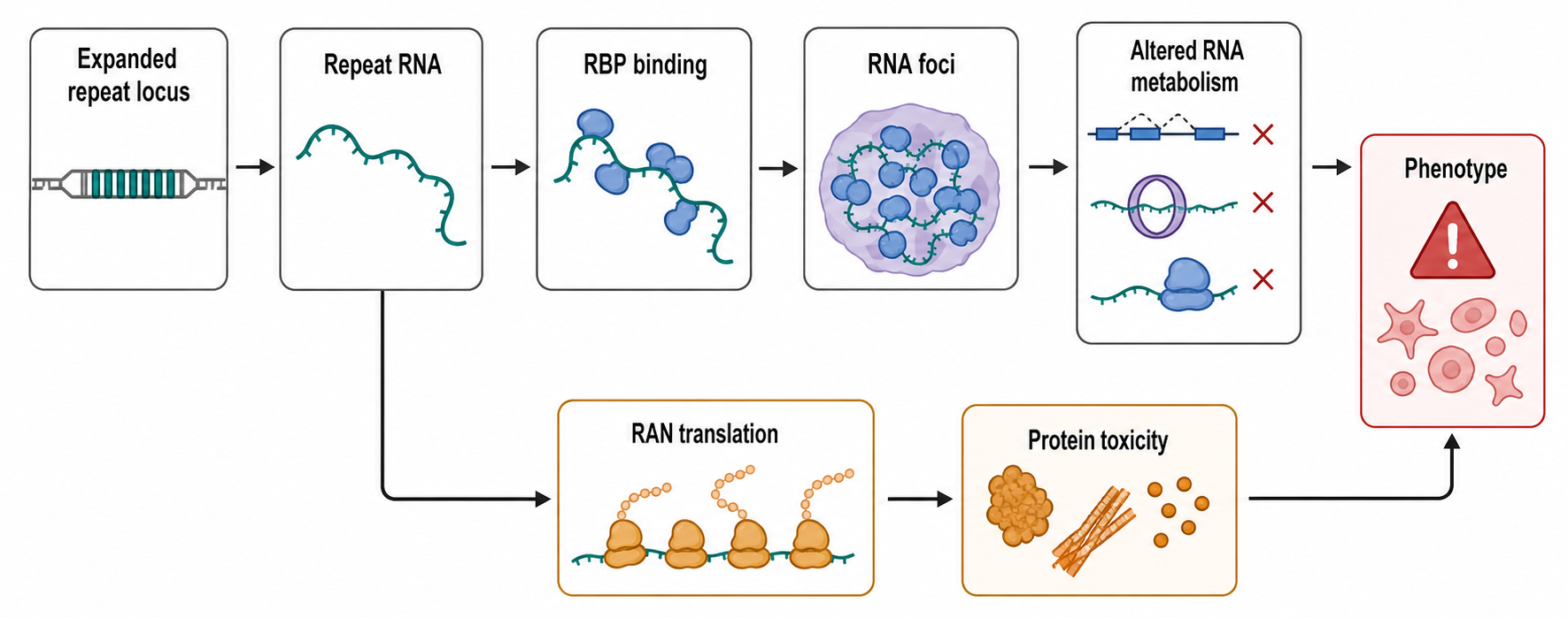
Figure 121.3. Toxic Repeat RNA and RBP Misassembly. Repeat-expanded RNAs can create multivalent RNA-RNA and RNA-protein interaction surfaces. Purified solid-like RNA gels, dynamic cellular foci, and RNP condensates are distinct observations; each requires material-state and functional evidence before it is connected to toxicity. The same disease locus may also involve altered transcription, repeat instability, sense and antisense RNAs, RAN translation, protein toxicity, and loss of gene function.
A causal pathway for toxic repeat RNA can be described step by step. First, the repeat expands in the genome and is transcribed into an RNA containing many copies of the repeated motif. Second, the repeat-containing RNA folds into abnormal structures or multivalent interaction surfaces. Third, RNA-binding proteins bind the repeat RNA with enough affinity and valency to become redistributed, sequestered, or misassembled. Fourth, normal RNA targets of those proteins are misprocessed, mislocalized, mistranslated, or destabilized. Fifth, affected neurons or muscle cells accumulate functional deficits that outpace compensation. Jazurek and colleagues review methods for identifying proteins that bind specific repeat RNAs and emphasize that repeat-binding experiments require careful discrimination between direct binding, indirect co-purification, and disease-relevant occupancy.
Jain and Vale supplied a complementary physical mechanism for the second step. Purified CAG- and CUG-repeat RNAs formed RNA-rich assemblies only after repeat number crossed a sequence-dependent threshold, about 30 triplets under the reported in vitro conditions. Multivalent intermolecular base pairing drove a sol-gel transition: the purified assemblies were RNA-enriched and showed little fluorescence recovery after photobleaching, consistent with a solid-like gel. In human U-2OS reporter cells, long CAG-repeat RNAs formed nuclear foci that fused and exchanged RNA more readily, indicating a more liquid-like and ATP-remodeled state. GGGGCC-repeat RNA also assembled in a repeat-number-dependent manner, but its G-quadruplex-compatible foci were less dynamic and were sensitive to flanking sequence and cellular context. Antisense oligonucleotides and agents that disrupted base pairing or ionic interactions dissolved reporter or patient-cell foci. These experiments establish that sequence-specific RNA-RNA interactions can generate a thresholded assembly, but the authors explicitly did not establish that foci formation itself causes cellular toxicity.
The term RNA-binding protein aggregation covers several related but distinct phenomena. Some RNA-binding proteins form physiological granules through low-complexity domains, RNA-binding domains, and reversible multivalent interactions. Others mislocalize, become post-translationally modified, enter persistent assemblies, or mature into less dynamic aggregates. Disease-associated proteins may bind RNA normally but aggregate when mutated, overexpressed, mislocalized from the nucleus to the cytoplasm, or exposed to chronic stress. RNA can promote assembly by increasing local concentration and multivalency, but RNA can also buffer aggregation by keeping proteins soluble. The same RNA-protein interaction can therefore be protective, neutral, or harmful depending on concentration, modification state, and cellular context.
Han and colleagues frame RNA condensates as disease-relevant assemblies whose material state and composition can influence pathology, but the review also supports a cautious interpretation: a visible granule is not automatically a disease driver. A granule claim needs evidence that the assembly changes a disease-relevant function, not only that a protein or RNA appears in a punctum. Important evidence includes live-cell dynamics, perturbation of assembly interfaces, separation of RNA-binding from aggregation effects, rescue of downstream RNA-processing defects, and correlation with patient-relevant phenotypes. Phase separation describes demixing, whereas gelation describes the development of a crosslinked, mechanically arrested network; neither word should be inferred from a fixed-cell image alone. Granule persistence, impaired clearance, altered composition, or conversion into less dynamic states can plausibly contribute to neurodegeneration, but each disease context must be tested.
Box 121.3. Reading Repeat-RNA Foci Without Overclaiming
A punctum is an observation, not yet a mechanism. Repeat-RNA foci show that an expanded RNA accumulates, localizes, or assembles abnormally in cells. A fixed image cannot distinguish a dynamic liquid-like focus from a crosslinked gel or an aggregate. Material-state evidence therefore requires live-cell exchange or fusion measurements, concentration and repeat-number dependence, and perturbations of the proposed RNA-RNA or RNA-protein interface. The foci become stronger evidence for toxicity when they contain the repeat RNA by strand-specific detection, recruit specific RNA-binding proteins by orthogonal assays, and coincide with mis-splicing, altered transport, translation defects, stress-granule changes, or neuronal dysfunction. Colocalization alone is weaker because a protein can appear in a focus through indirect binding, fixation artifacts, high local concentration, or a protective buffering response. C9ALS/FTD adds a clinical caution: repeat-RNA foci and dipeptide-repeat inclusions can be abundant outside the regions with greatest neurodegeneration. Stronger tests lower the relevant sense or antisense RNA, disrupt the implicated interaction, or restore the affected RNA-processing pathway and then ask whether cellular and disease phenotypes improve. Absence of visible foci also does not exclude RNA toxicity, because small dynamic assemblies or diffuse repeat-RNA interactions may be pathogenic but hard to image.
Repeat expansion disease also challenges simple genotype-to-phenotype reasoning. Repeat length, repeat interruptions, somatic instability, tissue-specific expansion, bidirectional transcription, methylation, RNA structure, RAN translation, and protein aggregation can all influence disease severity. A CAG repeat in one locus may primarily encode a polyglutamine protein, whereas another repeat context may generate toxic RNA, antisense transcripts, or RAN products. Franklin and colleagues discuss a proposed reverse-transcription mechanism for expandable repeats, illustrating that repeat instability itself remains an area where models can compete and where DNA, RNA, and repair pathways may intersect. The practical point is that repeat disease cannot be reduced to “long repeat equals one mechanism.”
The C9orf72 expansion in amyotrophic lateral sclerosis and frontotemporal dementia (C9ALS/FTD) makes the mixed-mechanism problem concrete. The GGGGCC expansion can be somatically mosaic, so repeat length in blood can differ greatly from repeat length in brain; the conventional 30-repeat pathogenic cutoff is operational, and the clinical meaning of approximately 30-100 repeats remains uncertain. Penetrance, age at onset, and ALS-versus-FTD presentation also vary with ancestry, family, somatic repeat behavior, and other genetic or environmental modifiers. At the molecular level, the locus produces sense and antisense repeat RNAs, five detectable dipeptide-repeat classes through repeat-associated translation, reduced C9orf72 protein expression, and DNA-level chromatin effects. Nuclear repeat-RNA foci and dipeptide-repeat inclusions are specific pathological markers, but their regional abundance does not consistently track the most affected tissue or clinical severity. TDP-43 nuclear loss and cytoplasmic pathology provide a major point of convergence with most non-SOD1, non-FUS ALS, while C9orf72 loss can impair membrane trafficking, autophagy-lysosome function, and immune homeostasis. The locus therefore cannot be interpreted as a simple sense-RNA gain-of-function disease.
Table 121.3. Repeat-Expansion Mechanism Layers. Summarize how one repeat-expansion locus can produce multiple mechanistic outputs.
| Mechanism layer | RNA involvement | Example evidence | Boundary caveat |
|---|---|---|---|
| Toxic RNA foci | Repeat-expanded RNA accumulates in nuclear or cytoplasmic foci | RNA FISH, repeat-length association, colocalization with RNA-processing factors | Foci are evidence of abnormal RNA state, not proof of toxicity by themselves. |
| Sequence- and valency-dependent RNA phase transition | Repeat motifs create multivalent RNA-RNA contacts that can yield RNA-rich phases or crosslinked gels | Repeat-number and concentration series, fluorescence recovery after photobleaching, fusion behavior, ionic or antisense perturbation, endogenous-locus confirmation | Purified solid-like gels and dynamic cellular foci are distinct; reporter abundance, flanking sequence, ionic conditions, and cellular remodeling alter thresholds, and assembly does not establish toxicity. |
| RBP sequestration or misassembly | Structured or multivalent repeat RNA recruits RNA-binding proteins away from normal targets | Repeat RNA pull-down, CLIP-family evidence, biochemical binding, downstream mis-splicing or mislocalization | Direct binding, indirect co-purification, and disease-relevant occupancy must be separated. |
| RAN translation | Repeat-containing RNA supports repeat-associated non-AUG translation | RAN peptide detection, reporter assays, reduction after lowering repeat RNA | RAN peptides can coexist with RNA toxicity, loss of function, or conventional protein toxicity. |
| Conventional protein toxicity | Repeat in a coding context alters the encoded protein or protein aggregation | Mutant protein detection, aggregation phenotypes, protein-directed rescue | Some repeat diseases are primarily protein driven, so RNA-centered evidence must be locus-specific. |
| DNA instability or epigenetic silencing | Expanded repeat changes transcription, methylation, or allele expression before RNA output is measured | Repeat sizing, methylation assays, allele-specific expression, family or tissue instability data | RNA changes may be downstream of DNA-level instability rather than the initiating mechanism. |
| Antisense or bidirectional transcription | Sense and antisense repeat RNAs can form distinct structures, bind proteins, or produce peptides | Strand-specific RNA-seq, strand-specific FISH, antisense transcript perturbation | Sense and antisense contributions can be difficult to assign without strand-resolved assays. |
| Granule and condensate remodeling | Repeat RNAs and RBPs alter assembly dynamics, persistence, localization, or clearance | Live-cell dynamics, assembly-domain perturbation, rescue of RNA-processing defects | Physiological granules are not pathological unless altered material state or composition changes function. |
| Clinical molecular-coverage test | A therapy lowers one repeat RNA or repeat-derived product while other sense, antisense, protein, loss-of-function, or downstream mechanisms remain active | Exposure, regional target engagement, species-specific biomarkers, and clinical endpoints | Lower CSF polyGP or polyGA after sense-repeat lowering establishes partial pathway engagement, not correction of the full C9ALS/FTD mechanism or clinical efficacy. |
Therapeutic strategies for neurological RNA disease include lowering the toxic RNA, blocking its interaction with proteins, correcting splicing, altering repeat-associated translation, improving granule dynamics, or reducing toxic protein products. Antisense oligonucleotides can degrade or sterically block target RNAs; small molecules can bind repeat structures or modulate downstream pathways; gene therapy can supplement missing functions; and splice-switching approaches can correct specific mis-splicing events. The C9ALS/FTD experience shows why the molecular entity must be specified. Early clinical programs that lowered sense repeat-containing C9orf72 transcripts reduced cerebrospinal-fluid polyGP or polyGA biomarkers but did not show clinical benefit. Those biomarker changes document engagement of part of the repeat-derived pathway, not correction of antisense RNA, all dipeptide repeats, C9orf72 loss, TDP-43 dysfunction, or downstream degeneration. Potential lowering of non-expanded C9orf72 transcripts further complicates interpretation. The results argue that sense-RNA lowering alone was insufficient in those trials; they do not establish that every repeat-RNA intervention is futile.
Verma and colleagues illustrate the earlier, preclinical end of this evidence ladder. A shape-similarity search of roughly 250,000 National Cancer Institute compounds yielded three candidates, B1, B4, and B11. Fluorescence binding, isothermal titration calorimetry, nuclear magnetic resonance, circular dichroism, thermal denaturation, and gel-shift assays supported preferential interaction with expanded CGG-repeat RNA relative to selected RNA and DNA controls. In COS7 reporter cells expressing a 99-CGG FMR1 5′-untranslated-region construct, the compounds reduced FMRpolyG inclusions and corrected SMN2 and BCL2L1/Bcl-x minigene splicing toward the control pattern without reducing the downstream reporter or altering a selected control splicing event. The work connects a defined RNA-binding hypothesis to two cellular phenotypes, but it remains a micromolar-dose reporter and minigene study. Direct displacement of the proposed sequestered proteins was inferred rather than measured, transcriptome-wide selectivity was not established, and efficacy, pharmacokinetics, brain exposure, and safety were not tested in animals or patients.
Neurological disease biomarker work often measures RNA in blood, cerebrospinal fluid, induced pluripotent stem cell-derived neurons, postmortem tissue, or animal models. Each source answers a different question. Blood can reveal immune or systemic signatures but may not report neuronal RNA metabolism. Cerebrospinal fluid is closer to the central nervous system but is still a mixed extracellular compartment. Postmortem brain can capture disease tissue but is affected by agonal state, cell loss, RNA degradation, and treatment history. Patient-derived neurons can model genetic background but may resemble developmental rather than aged neurons. Causality in neurological RNA disease therefore benefits from convergent evidence across patients, cells, animal models, biochemical assays, and targeted correction.
A recurring neurological caveat is that RNA mechanisms can be stage-specific. During early disease, a toxic RNA may disturb splicing or transport before overt neuronal loss. During late disease, RNA profiles may be dominated by gliosis, inflammation, neuronal depletion, or medication. A therapy that lowers a toxic RNA may therefore show molecular target engagement before clinical stabilization, and failure to reverse late degeneration does not necessarily disprove the early RNA mechanism. Conversely, an RNA abnormality detected only in terminal tissue may be a consequence rather than a cause. Longitudinal patient samples, natural-history cohorts, and model systems that capture early cellular phenotypes are especially valuable for separating initiation from progression.
121.4. Shared RNA mechanisms in infection, immunity, metabolism, and inflammation
RNA is central to infection because many pathogens use RNA genomes or RNA intermediates, and host cells use RNA features to discriminate self from nonself. Viral RNA can be detected by endosomal Toll-like receptors, cytosolic RIG-I-like receptors, protein kinase R, oligoadenylate synthetase/RNase L pathways, and other antiviral systems. The same sensors can also respond to self RNA when localization, modification, processing, or clearance fails. Disease can therefore arise from too little RNA sensing, allowing infection, or too much RNA sensing, causing autoinflammation, autoimmunity, tissue damage, or chronic interferon signaling. The dedicated immunity chapters own receptor-specific signaling; the comparison here is the recurring imbalance between ligand production, compartmentalization, modification, clearance, sensing threshold, and feedback.
The basic self-nonself logic is molecular rather than philosophical. A viral RNA may carry a 5′ triphosphate, double-stranded structure, unusual length, missing cap modification, replication intermediates, or compartmental mislocalization. A host RNA may become immunostimulatory if mitochondrial double-stranded RNA escapes normal processing, if endogenous retroelement transcripts accumulate, if defective RNA decay exposes double-stranded structures, or if therapeutic RNA lacks appropriate chemical and end modifications. RNA modifications can reduce or reshape immune sensing, but the effect depends on the receptor, RNA species, dose, delivery vehicle, and cell type. This point connects directly to RNA vaccine and therapeutic chapters, where immunogenicity can be useful for vaccination and harmful for replacement or editing therapies.
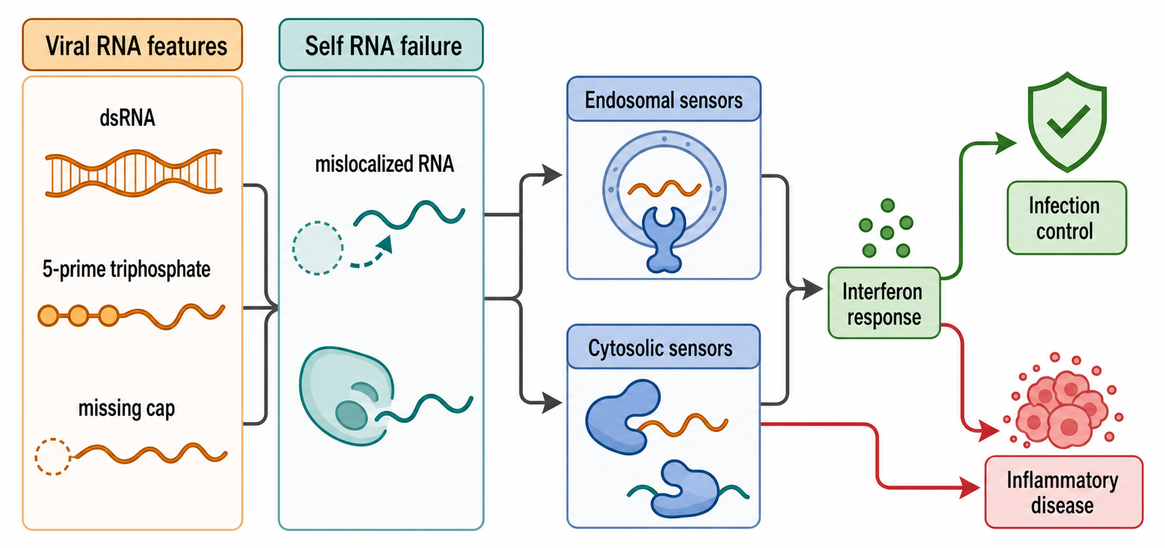
Figure 121.4. Self and Nonself RNA in Infection and Inflammation. Antiviral receptors detect molecular features such as double-stranded RNA, 5′ triphosphate ends, missing cap modifications, replication intermediates, or abnormal localization. Similar signaling can become pathogenic when self RNA accumulates or escapes normal processing.
Infection changes host RNA biology beyond direct sensing. Interferon signaling induces hundreds of interferon-stimulated genes, many of which encode RNA-binding proteins, nucleases, helicases, editing enzymes, and translation regulators. Viral infection can trigger host shutoff, stress granules, altered splicing, RNA decay, and translation arrest. Viruses counteract these pathways by capping or mimicking host RNA ends, hiding replication intermediates, modifying RNA structures, encoding RNA-binding antagonists, blocking interferon induction, or remodeling cellular membranes. An RNA change measured during infection may therefore represent viral replication, host defense, viral immune evasion, tissue injury, or changing cell composition.
Immune disease can involve RNA in several ways. Autoantibodies can target RNPs. Nucleic-acid sensing pathways can be activated by self RNA or RNA-DNA hybrids. RNA decay defects can increase immunostimulatory RNA. Noncoding RNAs can regulate immune-cell differentiation, cytokine expression, and inflammatory memory. Splicing and editing can alter immune receptors, cytokines, antigen presentation, and cell-state transitions. Because immune activation itself strongly changes RNA expression, causality is difficult. A transcript that rises in autoimmune tissue may be a driver of inflammation, a marker of infiltrating immune cells, a protective feedback response, or a consequence of therapy.
Metabolic disease and inflammation connect to RNA through nutrient sensing, stress signaling, mitochondrial function, and tissue remodeling. Obesity, diabetes, fatty liver disease, and atherosclerosis involve chronic changes in immune-cell composition, endothelial activation, adipocyte state, hepatocyte metabolism, and mitochondrial stress. RNA-level changes include altered splicing, microRNA expression, long noncoding RNA expression, RNA modifications, translation, and extracellular RNA release. The evidence challenge is that metabolic disease is systemic and slow. RNA perturbations in cultured cells may not capture organism-level endocrine feedback, tissue cross-talk, diet, microbiome effects, or medication exposure. Consequently, a specific RNA becomes a credible cross-disease mediator only when tissue-resolved perturbation, organism-level physiology, temporal ordering, and independent human evidence agree.
Inflammatory RNA programs are also temporally layered. Immediate innate immune responses may depend on preexisting sensor proteins and rapid post-transcriptional control of cytokine mRNAs. Later responses involve transcriptional induction, alternative splicing, RNA decay, and changes in immune-cell differentiation. Resolution of inflammation requires RNA programs that turn signals off as well as programs that activate defense. A snapshot RNA profile can therefore miss the direction of causality. A high cytokine mRNA level may indicate ongoing receptor signaling, failed decay, increased numbers of cytokine-producing cells, or delayed sampling after a transient stimulus. Time-course experiments, perturbation of RNA decay factors, and cell-type-resolved measurements are often needed to connect an RNA change to inflammatory pathology.
Inflammation also changes extracellular RNA. Damaged cells, activated immune cells, platelets, extracellular vesicles, and pathogens can release RNA into plasma or tissue fluids. Extracellular RNA can serve as a biomarker and may in some contexts act as a signaling or procoagulant molecule. However, extracellular RNA measurements are vulnerable to hemolysis, platelet activation during sampling, nuclease activity, vesicle isolation bias, carrier-protein association, and small-RNA normalization artifacts. A disease-associated extracellular RNA should not be interpreted as an active intercellular signal unless uptake, target engagement, dose, and functional consequence are shown in a relevant system.
Therapeutic targeting of RNA pathways in infection and immunity includes antiviral nucleoside analogs, RNA polymerase inhibitors, RNA vaccines, RNAi or antisense targeting of viral transcripts, modulation of innate immune sensors, and correction of immunostimulatory self-RNA accumulation. The therapeutic boundary is narrow. Suppressing antiviral sensing can reduce harmful inflammation but may increase infection risk. Increasing RNA sensing can improve vaccine adjuvanticity or antitumor immunity but may worsen autoimmunity. Degrading viral RNA can be highly specific if conserved accessible sites exist, but viral mutation, delivery to infected tissue, and timing can limit effect. The final clinical claim must be disease-specific, tissue-specific, and exposure-specific.
121.5. Cross-disease RNA biomarkers: state, specificity, and causal limits
An RNA biomarker is a measured RNA feature used to infer diagnosis, prognosis, treatment response, disease activity, residual disease, tissue origin, or biological mechanism. RNA biomarkers are attractive because RNA is dynamic, cell-type informative, and close to phenotype. The same dynamism creates risk: RNA changes with stress, sampling time, medication, cell composition, processing delay, and degradation. A clinically useful RNA biomarker must therefore be more than statistically significant. It must be measurable reproducibly, add information beyond existing tests, and support a clinical decision.
Liquid biopsy refers to disease measurement from blood or another body fluid rather than a tissue biopsy. RNA liquid biopsy can measure cell-free RNA, extracellular vesicle RNA, platelet-associated RNA, circulating tumor RNA, viral RNA, immune-cell RNA, or whole-blood expression signatures. The biological source matters. Cell-free RNA can derive from dying cells, secreted vesicles, ribonucleoprotein particles, blood cells, placenta, tumor, or injured tissue. Extracellular vesicle RNA can be enriched for some molecules but isolation methods differ. Platelet RNA can reflect platelet activation and tumor interaction but is sensitive to handling. Whole-blood RNA can be robust for immune states but is dominated by leukocyte composition.
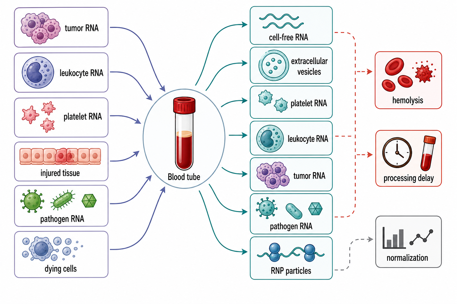
Figure 121.5. Liquid-Biopsy RNA Sources and Confounders. Circulating RNA can originate from tumor cells, immune cells, platelets, injured tissue, placenta, pathogens, extracellular vesicles, ribonucleoprotein particles, or dying cells. Sampling and processing can change the signal before sequencing begins.
Expression signatures are among the most widely used RNA biomarker forms. A signature may consist of one transcript, a ratio, a multigene score, a pathway score, or a machine-learning classifier. Expression signatures can classify tumor subtype, estimate immune activation, detect infection, stratify prognosis, or predict therapy response. The evidence basis has three layers. Analytical validity asks whether the assay measures the RNAs accurately and reproducibly. Clinical validity asks whether the signature is associated with the clinical state in independent cohorts. Clinical utility asks whether using the signature improves decisions or outcomes. Many published signatures pass discovery statistics but fail independent validation, portability across platforms, or clinical-utility testing.
Splicing signatures can be more disease-specific than total expression when a disease mechanism changes isoform choice. A splice junction, exon inclusion level, intron-retention event, or full-length isoform can report a splice-factor mutation, cryptic exon activation, developmental state, or therapeutic response. Splicing signatures require careful annotation and quantification. Short reads can quantify junctions but may not connect distant exons. Long reads can resolve isoforms but may have lower throughput or different error profiles. A disease-associated splice event may be invisible in a reference annotation, may differ by tissue, or may be confounded by RNA degradation. Clinical use therefore benefits from targeted assays after discovery.
Fusion transcripts are diagnostically useful when they define a tumor subtype, guide therapy, or confirm a pathogen or rearrangement. RNA-based fusion tests can identify expressed fusions and exact transcript junctions. They are especially valuable when DNA breakpoints are large, intronic, repetitive, or hard to interpret. Yet fusion testing has artifacts: template switching, readthrough transcription, paralogous mapping, low expression, and contamination. Diagnostic reporting should distinguish a known actionable fusion from a novel expressed junction of uncertain significance.
Extracellular RNA diagnostics require preanalytical discipline. Blood collection tube, processing time, centrifugation, storage temperature, freeze-thaw cycles, hemolysis, platelet contamination, extraction chemistry, spike-ins, library preparation, and normalization can dominate biological signal. Small RNAs are particularly vulnerable because abundant red blood cell microRNAs and platelet RNAs can distort disease associations. A robust extracellular RNA biomarker program uses standardized protocols, negative controls, orthogonal assays, independent cohorts, and explicit reporting of sample handling. Chapter 107 owns extracellular-RNA biogenesis and transfer; this section owns the clinical inference boundary between a reproducible analyte and a causal signal.
Table 121.4. RNA Biomarker Validation Hazards. Separate analytical validity, clinical validity, and clinical utility while listing common RNA-specific failure modes.
| Validation dimension | Question answered | RNA-specific hazard | Mitigation |
|---|---|---|---|
| Sample collection | Was the specimen collected without changing the RNA signal? | Hemolysis, platelet activation, processing delay, nuclease exposure, freeze-thaw damage | Standardized tubes, timing, centrifugation, storage, hemolysis checks, and complete preanalytical metadata. |
| RNA extraction and library preparation | Does the workflow recover the intended RNA class? | Size bias, vesicle-isolation bias, degradation, inhibitors, spike-in failure, low input | Prespecified extraction chemistry, quality metrics, controls, spike-ins, and orthogonal confirmation. |
| Normalization | Are samples comparable after technical and biological scaling? | Cell-mixture shifts, abundant blood-cell small RNAs, global RNA changes, unstable reference genes | Composition-aware normalization, stable references, negative controls, and sensitivity analyses. |
| Model training | Did the classifier learn disease biology rather than cohort noise? | Batch effects, hospital workflow, ancestry, medication, comorbidity, disease severity imbalance | Locked feature set, site-aware splitting, preregistered modeling choices, and held-out testing. |
| External validation | Does the RNA signature work outside the discovery cohort? | Platform drift, annotation changes, specimen differences, population shift | Independent cohorts, calibrated performance estimates, and prespecified endpoints. |
| Clinical decision threshold | Does the result support a defined clinical action? | Retuned cutoffs, gray-zone samples, unreported false positives or false negatives | Predefined thresholds, reporting categories, decision-curve analysis, and comparison with current care. |
| Clinical utility or surrogate use | Does using the RNA measurement improve decisions or patient outcomes? | Confusing association, prognosis, or target engagement with benefit | Prospective utility testing and surrogate-endpoint use only when outcome linkage is established. |
Diagnostic RNA interpretation should also avoid overfitting. A classifier trained on one hospital cohort may learn batch effects, ancestry, comorbidities, medication use, disease severity, or sampling workflow. A single-cell signature may fail in bulk data if the cell type is rare. A tumor expression signature may encode tumor purity instead of tumor biology. A viral RNA test may be analytically sensitive but clinically ambiguous if it detects residual noninfectious RNA. Good RNA diagnostics therefore use prespecified endpoints, locked models, external validation, calibration, decision thresholds, and comparison with current standard care.
RNA biomarkers can still be mechanistically useful even before clinical adoption. A biomarker can identify a patient subgroup for mechanistic study, select a disease stage for intervention, track target engagement in a trial, or reveal unexpected tissue involvement. The key is to label the use correctly. A pharmacodynamic marker that changes after treatment is not automatically a surrogate endpoint. A prognostic RNA signature does not prove that the measured RNAs cause poor outcome. A diagnostic RNA panel is not necessarily a therapeutic target list.
The strongest RNA diagnostic studies treat the assay as a complete workflow rather than a list of genes. The workflow begins with the clinical question and specimen type, then fixes collection conditions, RNA extraction, quality metrics, library or amplification method, normalization, classifier or threshold, and reporting categories. It then tests the locked workflow in independent samples. This discipline matters because RNA assays are easy to retune after seeing the data. Without a locked workflow, a biomarker can appear accurate because the analysis has absorbed cohort-specific noise. With a locked workflow, failure is informative: it may reveal that the original signal was a batch effect, that the disease is biologically heterogeneous, or that the specimen is not close enough to the affected tissue.
121.6. From RNA mechanism to target validation across disease classes
The unifying problem in RNA disease biology is deciding what kind of claim an RNA observation supports. An RNA can be a mechanism, a marker, a mediator, a modifier, a passenger, or an artifact. A mechanism claim says that changing the RNA or its processing changes disease pathogenesis through a defined molecular route. A marker claim says that the RNA measurement reports disease state but may not cause it. A mediator claim says the RNA lies between an upstream lesion and downstream phenotype. A modifier claim says the RNA changes severity or penetrance without being the primary cause. A passenger claim says the RNA changes because disease changes the cell. An artifact claim says the RNA pattern comes from measurement or analysis rather than biology.
The ownership and handoff matrix consolidates the boundary used throughout the chapter: disease-specific chapters own molecular detail, while this chapter owns the transferable causal and translational questions.
Table 121.5. Ownership and Handoff Matrix for Cross-Disease RNA Mechanisms. Distinguish this chapter’s comparative synthesis from disease-specific primary owners and show which inference problem transfers across fields.
| Disease context | Primary chapter owner | Cross-disease mechanism used here | Evidence anchor | Non-transferable boundary |
|---|---|---|---|---|
| Rare cis-processing disorders | Chapter 38 | Variant-to-RNA-to-phenotype chain | Segregation, patient RNA, endogenous correction, rescue | Tissue-specific expression and developmental timing differ by gene and disorder. |
| Rare trans-processing and RNP disorders | Chapter 41 | Substrate-class convergence and vulnerable-cell selectivity | Factor biochemistry, substrate mapping, allelic series, factor rescue | A broad transcriptomic response does not identify the causal substrate set. |
| Cancer and proliferative disease | Chapter 53 | Driver, dependency, state marker, and passenger separation | Recurrent lesion, isoform-specific perturbation, patient-derived model, predictive biomarker | Tumor lineage, purity, heterogeneity, and therapeutic window dominate transferability. |
| Neurological localization and repeat disease | Chapter 95, Chapter 99, Chapter 103 | Toxic sense and antisense RNA, repeat-RNA phase state, RBP occupancy, RAN translation, protein and loss-of-function mechanisms | Genetics, structure and material-state measurement, endogenous occupancy, neuronal perturbation, rescue, molecular target engagement, clinical outcome | Repeat motif, locus, neuronal subtype, somatic instability, molecular coverage of the intervention, and reversibility are disease-specific. |
| Infection and immunity | Chapter 108-Chapter 111 | Ligand production, localization, modification, clearance, sensing threshold, and feedback | Pathogen load, receptor genetics, temporal profiling, sensor perturbation | Receptor and ligand repertoires vary across cells, pathogens, and organisms. |
| Extracellular RNA and biomarkers | Chapter 107 | Analyte source, preanalytics, analytical validity, clinical validity, utility | Locked workflow, reference materials, external validation, decision study | Extracellular detection does not establish secretion, uptake, or signaling function. |
| Therapeutic translation | Chapter 154, Chapter 157, Chapter 163 | Causal target, modality access, target engagement, selectivity, timing, endpoint | Human genetics, pharmacology, target engagement, intervention | Chemistry, biodistribution, safety, population, and endpoint constraints are modality-specific. |
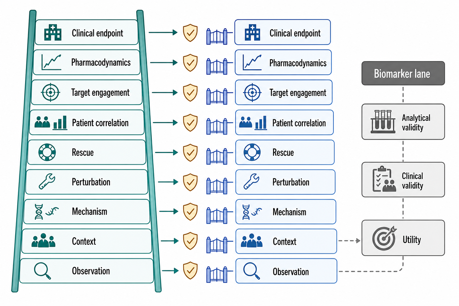
Figure 121.6. Evidence Ladder for RNA Disease Causality and Target Validation. RNA disease claims become stronger when reproducible observation is followed by tissue context, plausible molecular mechanism, perturbation, rescue, patient relevance, target engagement, and clinical benefit. Human genetics raises the prior probability of target success but does not guarantee direction, modality access, safety, or efficacy. Biomarker claims follow a related but distinct validation path, and partial molecular engagement must not be shown as a surrogate for benefit.
A useful validation ladder begins with observation but does not end there. First, the RNA feature must be measured reproducibly with appropriate controls. Second, the RNA feature must be connected to the relevant cell type, tissue, developmental stage, and disease state. Third, the molecular mechanism must be physically plausible: the RNA must be abundant enough, localized correctly, structured or modified as proposed, and able to bind the claimed partners. Fourth, perturbation must move the phenotype in the predicted direction. Fifth, rescue must show specificity: restoring the RNA, isoform, binding event, or downstream product should correct the phenotype more convincingly than a general stress reduction. Sixth, patient evidence should connect the mechanism to severity, progression, treatment response, or genotype. Seventh, therapeutic modulation should show target engagement, pharmacology, and benefit at tolerable exposure.
Human genetics is often the first independent evidence class. A high-penetrance variant that segregates with disease can identify an initiating lesion; allelic series can relate molecular severity to phenotype; naturally occurring loss-of-function variants can approximate lifelong target inhibition; and somatic mutations can expose acquired dependencies in cancer. Genetics is not automatically decisive. A variant may be linked to the causal allele rather than causal itself, affect several transcripts or cell types, act during development, or produce phenotypes through protein and RNA routes simultaneously. Population associations can also be distorted by ancestry, selection, ascertainment, and horizontal pleiotropy. For an RNA mechanism, the useful question is not merely whether a locus is associated with disease, but whether the allele changes the proposed RNA species in the relevant tissue and whether that molecular effect lies in the direction predicted by the disease model.
Historical drug-development data quantify why human genetics is valuable without making it determinative. Nelson and colleagues linked drug target-indication pairs to genome-wide association and Mendelian evidence and found direct genetic support for 2.0% of mechanisms whose latest stage was phase I versus 8.2% of approved mechanisms among well-studied indications. Their model estimated an approximately twofold higher probability of progression from phase I to approval for genetically supported pairs. This is population-level enrichment, not a guarantee for an individual target. The analysis depended on historical pipeline records, disease ontologies, and uncertain variant-to-gene mappings; support also varied greatly among therapeutic areas. Genetics must still specify the causal gene, molecular direction, relevant tissue, disease stage, and compatibility between a lifelong allele and the magnitude and timing of a drug.
Perturbation and rescue form a second evidence class. Deleting a transcript, editing its splice site, lowering it with an antisense oligonucleotide, blocking an RNA-protein interface, or changing a processing factor asks whether the system is manipulable. A rescue asks whether restoring the proposed causal entity reverses the phenotype: for example, whether an RNA-insensitive cDNA restores function after transcript knockdown, whether correction of a cryptic exon restores protein and cellular behavior, or whether reintroducing a binding-competent but catalytically inactive factor separates scaffolding from enzyme activity. Perturbations can still mislead through supraphysiological dose, incomplete cell specificity, innate immune activation, off-target sequence complementarity, clonal adaptation, or general toxicity. The most informative designs use multiple perturbation chemistries, graded doses, orthogonal readouts, and rescues that distinguish transcript, DNA-locus, protein, and neighboring-gene effects.
Biochemistry and structure provide a third class by testing whether the proposed molecular steps are physically possible. Binding affinity, stoichiometry, kinetic rate, modification fraction, RNA copy number, subcellular concentration, accessibility in an RNP, and structural state can rule mechanisms in or out. A repeat RNA that binds an RBP in vitro at micromolar concentration is not necessarily occupied in a neuron; a small molecule that recognizes a naked RNA hairpin may fail when the cellular RNA is remodeled or protein coated; and an enrichment peak does not report what fraction of transcripts carry a modification. Reconstitution, crosslinking, structural biology, quantitative imaging, and occupancy measurements are strongest when they connect the purified mechanism to endogenous molecules and disease-relevant concentrations. Physical plausibility is necessary for many mechanistic claims, but an elegant structure alone does not establish that the interaction controls disease.
Longitudinal patient observation and clinical intervention provide the fourth and fifth evidence classes. Serial samples can determine whether an RNA signature precedes clinical deterioration, tracks a flare, follows tissue injury, or changes after treatment. Time ordering narrows causal explanations but does not remove time-varying confounding: medication, infection, cell composition, circadian phase, and disease severity can move together. An intervention supplies a stronger test when it selectively changes the proposed mechanism, demonstrates exposure and target engagement, and improves a clinically meaningful outcome. A negative trial remains ambiguous if the molecule never reached the cell, target engagement was absent, the dose was intolerable, treatment began after irreversible injury, the endpoint was insensitive, or enrolled patients lacked the target-dependent mechanism. A positive molecular response without clinical benefit may show that the RNA is a pharmacodynamic marker, that the mechanism is insufficient, or that downstream damage has become autonomous.
C9ALS/FTD provides a particularly informative intervention boundary because target engagement and clinical outcome diverged. Lower CSF polyGP or polyGA after a sense-repeat-directed antisense oligonucleotide shows that the drug reached the central compartment and changed at least part of the repeat-derived pathway. The absence of clinical benefit then narrows—but does not uniquely identify—the failure: sense repeat RNA may be insufficient as a target, antisense RNA or C9orf72 loss may remain active, treatment may have started after self-sustaining TDP-43 pathology, the magnitude or distribution of engagement may have been inadequate, or simultaneous lowering of normal C9orf72 may have offset benefit. A biomarker is most informative when its molecular coverage is explicit; one repeat-derived protein cannot stand in for every pathogenic entity at a complex locus.
Triangulation does not require every project to collect every evidence type, but it requires explicit accounting. A rare-disease splice claim may begin with genetics, patient RNA, and rescue. A cancer dependency may begin with recurrent somatic alteration, perturbation across models, and a predictive biomarker. A neurodegenerative mechanism may require structure, occupancy, longitudinal progression, and intervention in disease-relevant cells. A biomarker claim may require no mechanistic rescue at all, but it cannot skip analytical reproducibility, independent clinical validation, calibration, and a defined decision context. Evidence classes should be weighted by what they can actually establish, not arranged as a ceremonial checklist.
The evidence ladder differs by RNA class. For a splice defect, strong evidence includes variant segregation, abnormal junction detection in disease-relevant tissue, minigene or endogenous transcript validation, restoration by a splice-switching oligonucleotide, and improvement in protein or cellular phenotype. For a toxic repeat RNA, strong evidence includes repeat-length association, RNA foci or abnormal RNA-protein binding, downstream mis-splicing or translation defects, reduction of toxicity after lowering the repeat RNA, and model-system concordance. For a repeat-RNA phase-transition mechanism, strong evidence additionally requires repeat- and concentration-dependent assembly, material-state measurements in living cells, perturbations that distinguish RNA-RNA from RNA-protein interactions, endogenous-locus confirmation, and a functional link from assembly to toxicity; foci dissolution alone tests assembly, not disease rescue. For a broader RNA condensate mechanism, strong evidence includes altered assembly properties, disease-linked perturbation of assembly interfaces, functional rescue, and distinction between physiological granules and persistent pathological assemblies. For a biomarker, strong evidence is not mechanistic rescue but analytical and clinical validation.
Therapeutic target validation has special constraints for RNA. A target RNA must be reachable by the modality. Nuclear RNAs may be accessible to some antisense oligonucleotides, while cytosolic mRNAs may be more amenable to RNA interference in certain tissues. Brain targets face delivery barriers. Tumor targets face heterogeneity and toxicity in normal proliferating cells. Immune targets face narrow safety margins. Repeat RNAs may have similar sequences elsewhere in the transcriptome. RNA structures targeted by small molecules may be transient, protein-covered, or context-dependent. A target that works in cultured cells can fail because tissue exposure, intracellular uptake, endosomal escape, RNP occupancy, or compensatory pathways differ in patients.
Target-validation failures can therefore be classified before development begins. A causal failure occurs when changing the RNA does not alter disease because it was a passenger or marker. A mechanistic failure occurs when the intervention changes a measured RNA feature but not the downstream effector thought to matter. A modality failure occurs when the target is sound but chemistry, biodistribution, cellular uptake, endosomal escape, nuclear access, or RNP accessibility prevents sufficient modulation. A selectivity failure occurs when homologous transcripts, physiological isoforms, or the same pathway in healthy tissue creates toxicity. A population failure occurs when only a molecularly defined subgroup depends on the target but the trial population is broader. A timing or reversibility failure occurs when target dependence existed early but tissue damage is autonomous by treatment. An endpoint failure occurs when the assay is noisy, the follow-up is too short, or a pharmacodynamic marker is mistaken for a validated surrogate. These failure modes demand different remedies; treating all of them as evidence against the biological target discards information, while treating all as delivery problems protects weak targets from falsification.
Clinical evidence also has layers. A molecular endpoint can show target engagement, such as reduced toxic RNA, corrected exon inclusion, or decreased fusion transcript. A pharmacodynamic endpoint can show pathway movement. A clinical endpoint can show improved survival, function, symptoms, imaging, or disease progression. A surrogate endpoint is acceptable only when the field has evidence that changing the surrogate predicts clinical benefit. Rare diseases and neurological disorders often require creative endpoints, natural-history studies, and patient-specific logic, but the need for careful causal reasoning does not disappear.
Target validation should also consider reversibility. Some RNA lesions act continuously, so reducing the pathogenic RNA or correcting an isoform can plausibly improve function after disease onset. Other RNA lesions act during development, causing anatomical or circuit changes that may not fully reverse in adulthood. Some RNA mechanisms produce secondary damage, such as inflammation, proteostasis stress, or cell loss, that becomes partly independent of the original RNA trigger. A therapeutic program should therefore state whether the intended effect is prevention, slowing, symptomatic improvement, reversal, or target engagement for patient stratification. This distinction is especially important when molecular correction is easier to demonstrate than clinical recovery.
Common misconceptions recur across RNA disease biology. Do not assume every disease-associated noncoding RNA is functional. Do not assume every altered splice isoform is translated or pathogenic. Do not assume an RNA-binding protein found in a granule is sequestered from all normal targets. Do not assume that a repeat RNA mechanism excludes protein toxicity or DNA instability. Do not assume a biomarker signature reveals the therapeutic target. Do not assume that correcting a molecular marker will improve disease unless the marker lies on the causal path. These cautions are not skepticism for its own sake; they are the difference between a useful RNA disease mechanism and an attractive but misleading association.
Recent Consensus
The current consensus is strongest for five broad statements. First, RNA-processing defects are bona fide causes of human disease when genetic variants or trans-acting factor defects reproducibly alter RNA maturation in disease-relevant cells. Second, cancer transcriptomes provide clinically useful classification and therapeutic information, but transcriptomic alteration must be separated from tumor purity, lineage, stress, and passenger effects. Third, repeat-expanded RNAs can undergo sequence- and valency-dependent assembly, recruit RNA-binding proteins, and coexist with repeat-associated translation, protein toxicity, loss of gene function, and DNA-level effects; the contribution of each layer must be established per locus. Fourth, RNA biomarkers are powerful but require validation standards that differ from mechanistic disease claims. Fifth, human genetic support enriches for successful target-indication pairs at a portfolio level but does not replace direction-of-effect, tissue, modality, safety, or clinical-efficacy evidence for an individual target.
The field also agrees on a negative principle: similar RNA measurements do not imply shared etiology. An interferon-response signature can arise from infection, self-nucleic-acid sensing, tissue injury, or treatment; intron retention can arise from splice-factor mutation, differentiation, stress, or RNA degradation; and extracellular RNA can reflect secretion, cell death, blood-cell contamination, or altered clearance. Cross-disease synthesis is therefore useful only when it preserves initiating cause, tissue context, temporal order, and assay limitations.
Open Questions, Controversies, Deprecated Models, and Common Misconceptions
Open questions:
- Which disease-associated RNA modifications are causal rather than adaptive?
- How often do long noncoding RNAs in disease act through sequence-specific mechanisms rather than through transcription, chromatin context, or cell identity?
- Which RNA condensates are protective stress responses and which are pathological material states?
- How should clinical laboratories integrate short-read RNA-seq, long-read RNA-seq, targeted panels, and DNA sequencing for rare disease?
- Which extracellular RNA measurements are stable enough for routine clinical diagnostics?
- How much target engagement is enough for an RNA-directed therapy when the clinical endpoint develops slowly?
Deprecated or weakened claims:
- Deprecated or overextended models should be handled explicitly. The phrase “junk RNA” is obsolete as a blanket dismissal, but the opposite error is to call every transcribed sequence functional. The phrase “oncogenic lncRNA” should not be used for a tumor-correlated transcript without perturbation and mechanism. The phrase “phase separation causes disease” should be replaced by a specific claim about assembly composition, dynamics, persistence, localization, or downstream RNA metabolism. The phrase “liquid biopsy RNA detects cancer” should specify analyte, specimen, stage, sensitivity, specificity, and comparison test.
Common misconceptions:
- “The same RNA signature in two diseases proves a shared cause.” Similar signatures can arise from common cell-state responses, cell-mixture changes, treatment, or technical processing; shared causality requires independent mechanistic evidence.
- “A rescue experiment proves the complete mechanism.” Rescue supports specificity under the tested conditions, but an overexpressed construct, general stress relief, or downstream bypass can rescue without validating every upstream molecular step.
- “A biomarker that predicts outcome is a therapeutic target.” Prediction establishes clinical validity only in a defined setting; target status additionally requires causal, modality, exposure, selectivity, and therapeutic-window evidence.
- “Failure of an RNA-directed clinical trial disproves the target.” Trial failure may instead reflect insufficient exposure, absent target engagement, wrong population, late intervention, toxicity, or an insensitive endpoint; distinguishing these possibilities requires pharmacology and biomarker data.
- “Human genetic support guarantees that pharmacological modulation will work.” Genetic effects may be lifelong, developmental, allele-specific, or pleiotropic and may not mimic the magnitude, tissue distribution, or timing of a medicine.