Chapter 67. Canonical Translation Initiation Machinery Across Biological Systems
Scope Note
Translation initiation is the set of molecular events that recruits a ribosome, places initiator transfer RNA in the P site, selects a start codon, and joins the large ribosomal subunit. This chapter owns the canonical RNA signals, initiation factors, ribosomal states, and cross-domain comparisons required to explain those steps in bacteria, archaea, eukaryotic cytosol, mitochondria, chloroplasts, and viral RNAs that recruit host canonical machinery. Regulated initiation, internal ribosome entry sites (IRESs), ribosome shunting, reinitiation, upstream-open-reading-frame control, stress signaling, and other noncanonical routes are treated in Chapter 70.
Executive Summary
Translation initiation converts an mRNA into a ribosome-loaded coding template by solving three problems: recruiting a small ribosomal subunit or complete ribosome to an RNA, positioning an initiator tRNA in the ribosomal P site, and selecting a start codon in the correct reading frame. Bacteria often use base pairing between a purine-rich Shine-Dalgarno sequence in the mRNA and the anti-Shine-Dalgarno sequence at the 3′ end of 16S rRNA, but bacterial initiation is not reducible to that motif. Start-codon identity, spacing, mRNA structure, leaderless transcripts, standby sites, initiation factors IF1, IF2, and IF3, and RNA-binding regulators all contribute to initiation efficiency. Chloroplasts retain bacterial-like logic but have lineage-specific RNA maturation and nucleus-encoded regulatory proteins.
Archaeal initiation is a mosaic. Archaeal ribosomes and information-processing proteins resemble eukaryotic systems in many respects, yet many archaeal mRNAs use bacterial-like Shine-Dalgarno pairing, while others are leaderless and initiate near the 5′ end. Archaeal initiation factors include homologs of eukaryotic factors involved in initiator-tRNA delivery and start-site selection, but the diversity of archaeal transcript leaders prevents a single rule from explaining all archaeal mRNAs.
In eukaryotic cytosol, the dominant pathway for many cellular mRNAs is cap-dependent scanning. The 5′ cap recruits eIF4F, the 43S preinitiation complex loads near the 5′ end, helicases help the small subunit traverse the leader, and AUG recognition triggers GTP-dependent factor rearrangement and 60S joining. The Kozak context tunes start recognition, but the conserved mechanistic core is recruitment, scanning, codon inspection, and subunit joining.
Organelles retain initiation features inherited from bacterial ancestors while using lineage-specific ribosomes, RNA architectures, and imported factors. Viral transcripts that carry or acquire host-like caps can recruit the same eukaryotic machinery, making them useful contexts for separating canonical factor use from virus-specific regulation. Detailed noncanonical and stress-responsive viral strategies belong to Chapter 70. Core initiation defects cause disease when mutations disrupt ribosome recruitment, initiator-tRNA delivery, start-site fidelity, subunit joining, or organellar protein synthesis.
Concept Inventory
- Translation initiation: assembly of an initiation-competent ribosome with initiator tRNA at a selected start codon before peptide-bond formation begins.
- Start codon: the mRNA triplet decoded first in a coding sequence. AUG is most common, but GUG, UUG, CUG, and other codons can function in specific organisms or contexts.
- Initiator tRNA: a specialized tRNA used for the first amino acid. Bacteria use N-formylmethionyl initiator tRNA in most cytosolic translation, whereas eukaryotic cytosol uses methionyl initiator tRNA without formylation.
- Ribosome-binding site: an RNA region that promotes ribosome recruitment or positioning. In bacteria this often includes a Shine-Dalgarno motif; in eukaryotes the phrase is less precise because recruitment commonly occurs at the cap and scanning selects the start site.
- Shine-Dalgarno sequence: a purine-rich mRNA element upstream of a bacterial or organellar start codon that can base-pair with the anti-Shine-Dalgarno sequence in small-subunit rRNA.
- Anti-Shine-Dalgarno sequence: the complementary sequence near the 3′ end of bacterial 16S rRNA, and related rRNAs, that pairs with Shine-Dalgarno motifs.
- 5′ untranslated region: the mRNA leader between the 5′ end or cap and the coding sequence; its length, structure, and start-codon context affect canonical ribosome recruitment and scanning.
- 43S preinitiation complex: a eukaryotic small-subunit complex containing the 40S ribosomal subunit, initiator Met-tRNAi, eIF2-GTP, eIF1, eIF1A, eIF3, and associated factors before start-codon recognition.
- Scanning: movement of the eukaryotic small ribosomal subunit along the mRNA leader until it recognizes a start codon in a favorable context.
- Kozak context: the nucleotide environment around a eukaryotic start codon that affects start recognition, especially positions upstream and downstream of AUG.
- Initiation-factor analogy: a comparison based on shared molecular task, such as initiator-tRNA delivery or subunit joining; functional analogy does not by itself establish one-to-one homology or identical regulation.
What to Know Before Reading This Chapter
The ribosome has a small subunit that decodes mRNA and a large subunit that catalyzes peptide-bond formation. The small subunit contains the P site, where the initiator tRNA pairs with the start codon before elongator tRNAs enter the A site. Translation initiation is therefore a start-site selection problem before it is a peptide-synthesis problem.
Readers should distinguish an mRNA leader from a promoter. A promoter is a DNA element that controls transcription initiation, whereas an mRNA leader is an RNA segment that affects translation, decay, localization, and surveillance after transcription. Readers should also distinguish ribosome recruitment from start-codon selection. A strong recruitment element can still lead to poor translation if the start codon is inaccessible or in weak context, and a strong start codon cannot be used efficiently if ribosomes seldom reach it.
This chapter uses mRNA, viral RNA, bacterial operons, mitochondrial transcripts, and chloroplast transcripts as running examples. Chapter 66 explains codon usage and initiator-tRNA identity, while Chapter 68 follows the ribosome after initiation into elongation.
67.1. Bacterial initiation and Shine-Dalgarno logic
Bacterial translation initiation usually begins with a 30S small ribosomal subunit, an initiator transfer RNA charged with N-formylmethionine, and initiation factors IF1, IF2, and IF3. The mRNA contributes a start codon and often a ribosome-binding site upstream of the start codon. The textbook model is Shine-Dalgarno initiation: a purine-rich mRNA sequence, frequently resembling AGGAGG, base-pairs with the anti-Shine-Dalgarno sequence at the 3′ end of 16S ribosomal RNA. This RNA-RNA interaction positions the start codon near the P site so that the initiator tRNA can pair with it. IF2, a GTPase, promotes initiator-tRNA placement and joining of the 50S subunit; IF3 helps prevent premature large-subunit joining and disfavors incorrect initiation complexes; IF1 occupies the A site and helps organize the decoding center. Recent review coverage of bacterial and chloroplast initiation is provided by Webster (2025).
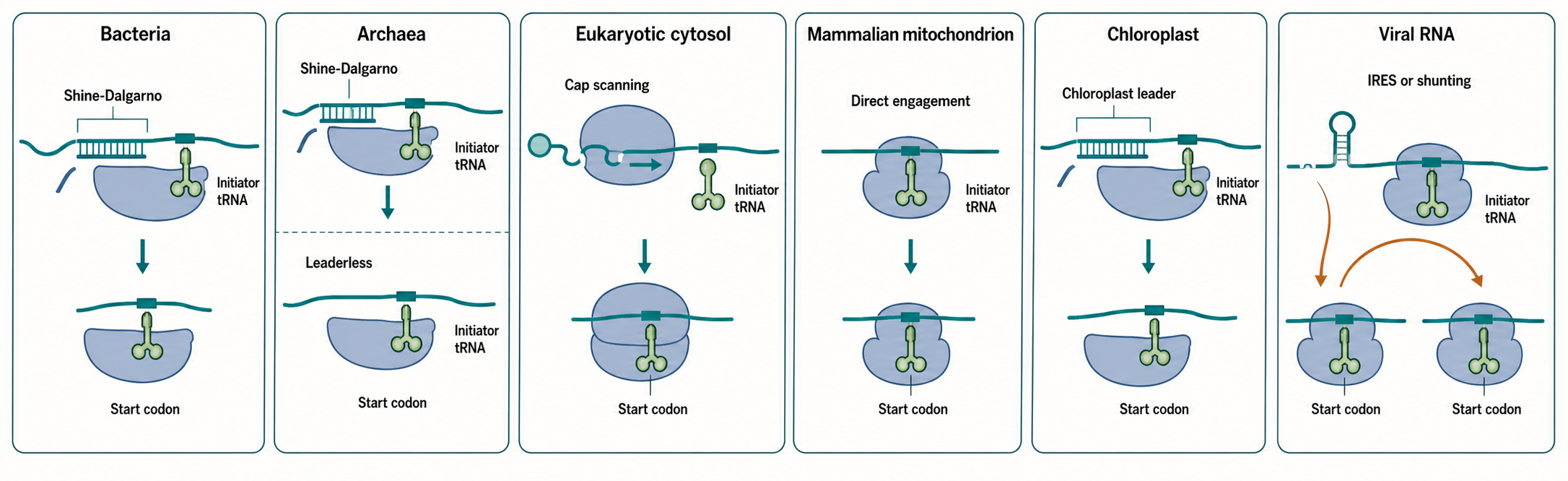
Figure 67.1. Comparative Initiation Logic Across Systems. Six translation contexts—bacteria, archaea, eukaryotic cytosol, mammalian mitochondria, chloroplasts, and capped viral messenger RNAs—are aligned by four shared tasks: ribosome recruitment, initiator-tRNA delivery, start-site selection, and large-subunit joining. Bacterial and chloroplast systems commonly use Shine-Dalgarno pairing; archaeal transcripts span leadered and leaderless modes; eukaryotic cytosol uses cap-dependent 43S loading and scanning; mitochondria use organelle-specialized factors on short-leadered or leaderless transcripts; and capped viral RNAs can recruit the host canonical eukaryotic pathway. The figure explicitly labels regulated and noncanonical initiation as a handoff to Chapter 70.
Table 67.1. Initiation Factors and Functional Analogies Across Systems. Each translation system solves the shared problems of ribosome recruitment, initiator-tRNA delivery, start-site selection, and large-subunit joining with a distinct but often evolutionarily related set of factors.
| System | Recruitment signal | Initiator tRNA delivery | Start-site fidelity | Subunit joining | Common boundary cases |
|---|---|---|---|---|---|
| Bacteria | Shine-Dalgarno / 16S rRNA base pairing | IF2 (GTPase) with fMet-tRNA | IF3, IF1; start codon identity | IF2 GTP hydrolysis on 50S joining | Leaderless mRNAs; structured leaders; polycistronic translational coupling |
| Archaea | Shine-Dalgarno (leadered) or 5′ end (leaderless) | aIF2 (GTPase) with Met-tRNA | aIF1, aIF1A | aIF5B | Coexisting leadered and leaderless transcript classes in same genome |
| Eukaryotic cytosol | 5′ cap + eIF4F (eIF4E, eIF4G, eIF4A) | eIF2-GTP-Met-tRNAi ternary complex | eIF1, eIF1A, eIF3; Kozak context | eIF5B | Structured leaders and lineage-specific context preferences |
| Mammalian mitochondria | Short or absent 5′ UTR; direct ribosome engagement | mtIF2 (GTPase) with fMet-tRNA | mtIF3 | mtIF2 GTP hydrolysis | Leaderless or near-leaderless mRNAs; membrane-coupled synthesis |
| Chloroplasts | Shine-Dalgarno + nucleus-encoded RNA-binding proteins | Bacterial-like factor | Nucleus-encoded factors; RNA editing may create start codon | Bacterial-like | PPR-dependent leader stabilization; RNA editing that creates or corrects start codons |
| Capped viral messenger RNA | Viral or host-derived cap compatible with eIF4F | Host eIF2-GTP-Met-tRNAi | Host scanning and start-recognition factors | Host eIF5B | Canonical dependence must be tested; virus-specific remodeling belongs to Chapter 70 |
The important point is that Shine-Dalgarno pairing is a positioning mechanism, not a universal bacterial law. Many bacterial genes have obvious Shine-Dalgarno motifs, but some have weak motifs, atypical spacing, structured leaders, or leaderless transcripts. Optimal spacing between the Shine-Dalgarno element and the start codon is usually on the order of several nucleotides; too short or too long a spacer can misalign the start codon relative to the P site. Start codon identity also matters. AUG is generally strongest, GUG and UUG can initiate in many bacteria, and rarer codons require favorable surrounding context or specialized circumstances. The initiator tRNA must be selected over elongator tRNAs, so initiation depends on both codon-anticodon pairing and factor-mediated discrimination.
RNA structure is part of the canonical recruitment substrate. If the Shine-Dalgarno sequence or start codon is buried in a stem-loop, the 30S subunit cannot productively engage even when the sequence motif is present. Polycistronic mRNAs likewise present multiple start regions, each of which must expose an appropriately spaced recruitment signal and start codon. These baseline accessibility constraints explain why sequence motifs alone cannot predict initiation. Temperature-, ligand-, protein-, or small-RNA-dependent switching of accessibility is regulated initiation and is treated in Chapter 70.
Experimental evidence for bacterial initiation mechanisms comes from mutational analysis of ribosome-binding sites, toeprinting assays that map ribosome position on mRNA, ribosome profiling that detects initiating or translating ribosomes genome-wide, biochemical reconstitution with purified subunits and factors, and structural studies of initiation complexes. Each method has limitations. Reporter assays can separate a leader from its native operon and RNA context. Ribosome profiling gives a population snapshot shaped by drug treatment, nuclease digestion, and library bias. Toeprinting can show a stalled complex in vitro but may not capture competition among cellular RNAs. A robust initiation claim is strongest when mutations in the RNA element, compensatory changes, ribosome binding, and protein output agree.
Do not overgeneralize Shine-Dalgarno logic. Some bacteria, including lineages with reduced genomes or unusual transcript architecture, use many leaderless or weakly leadered mRNAs. Some coding regions contain internal Shine-Dalgarno-like sequences that affect elongation or pausing rather than initiation. Conversely, a sequence resembling a Shine-Dalgarno motif is not necessarily functional if it is inaccessible, incorrectly spaced, or outside an initiation context. Chapter 68 discusses downstream elongation effects of mRNA sequence, and Chapter 25 provides the folding background needed to evaluate occlusion and accessibility.
67.2. Archaeal initiation mechanisms
Archaeal translation initiation cannot be described simply as bacterial or eukaryotic. Archaeal ribosomes are prokaryotic in cellular organization because translation occurs in the cytoplasm without a nucleus, but archaeal information-processing systems share many homologous factors and structural features with eukaryotes. Archaeal mRNAs may be leadered, with a 5′ untranslated region before the start codon, or leaderless, with the start codon very close to the 5′ end. Many leadered archaeal transcripts contain Shine-Dalgarno-like motifs that can pair with small-subunit rRNA, whereas leaderless transcripts appear to recruit ribosomes by mechanisms that depend on the 5′ end and start codon accessibility.
The archaeal initiation factor set includes proteins related to eukaryotic initiation factors, including aIF1, aIF1A, aIF2, and aIF5B. aIF2 is a heterotrimeric GTPase that delivers initiator methionyl-tRNA to the small ribosomal subunit in a manner conceptually related to eukaryotic eIF2. aIF1 and aIF1A contribute to start-site selection and an open preinitiation state, while aIF5B promotes subunit joining after start-codon recognition. These factor relationships are one reason archaeal systems are important for understanding the evolution of eukaryotic initiation. At the same time, archaeal mRNA leaders and ribosome-binding signals preserve bacterial-like solutions in many genes.
Leaderless initiation is especially important in archaea. A leaderless mRNA begins at or very near the start codon, leaving little or no upstream region for a Shine-Dalgarno interaction. In such transcripts, the ribosome must identify the 5′-proximal start codon without scanning a long leader. This can favor a mechanism in which an initiation-competent ribosomal subunit, initiator tRNA, and factors assemble directly at the exposed 5′ start region. Leaderless initiation also occurs in bacteria and eukaryotic organelles, but its prevalence in archaeal transcriptomes makes it a central archaeal theme.
Archaeal initiation is regulated by RNA structure, transcript processing, and small RNAs, but the coverage in the current local bibliography is incomplete. Gomes-Filho et al. (2018) provides a review anchor for archaeal small RNAs, but direct support for many mechanistic archaeal initiation claims requires additional archaeal translation-specific references. Final bibliography item: add recent reviews and primary structural or biochemical papers on archaeal translation initiation factors, leaderless mRNA initiation, and archaeal Shine-Dalgarno usage.
The evidence standards are the same as in other systems but the available datasets can be more uneven. Comparative genomics can identify Shine-Dalgarno motifs and leader lengths, but motif frequency does not prove factor use. Transcript-end mapping can classify leaderless mRNAs, but start-site artifacts or condition-specific transcription start sites can alter estimates. In vitro initiation experiments clarify factor requirements but may use model mRNAs that do not represent genomic diversity. Therefore, archaeal initiation should be taught as a set of coexisting modes whose prevalence differs by lineage, growth condition, and transcript class.
The broader handoff is evolutionary. Chapter 10 discusses the ribosome and adaptor logic in deep time, while Chapter 21 covers archaeal and eukaryotic transcription systems. Translation initiation in archaea illustrates how cellular systems can combine homologous protein machinery with RNA signals that look superficially bacterial.
67.3. Eukaryotic cap-dependent scanning
Most eukaryotic nuclear-encoded mRNAs use cap-dependent scanning. The 5′ cap is a modified nucleotide structure added to RNA polymerase II transcripts; the common cap 0 structure contains 7-methylguanosine linked by an unusual 5′-to-5′ triphosphate bridge to the first transcribed nucleotide, and many metazoan mRNAs contain additional ribose methylations. Pelletier et al. (2021) review the eukaryotic cap structure and its multiple roles. In translation initiation, the cap helps recruit eIF4E, the cap-binding component of the eIF4F complex. eIF4G acts as a scaffold that binds eIF4E, the RNA helicase eIF4A, poly(A)-binding protein, and other factors. This assembly connects the 5′ end, the mRNA body, and sometimes the 3′ poly(A) tail into a translation-promoting messenger ribonucleoprotein.
The small ribosomal subunit enters through the 43S preinitiation complex. In a simplified sequence, eIF2 bound to GTP carries initiator Met-tRNAi to the 40S subunit, while eIF1, eIF1A, and eIF3 help maintain an open scanning-competent complex. The mRNA-bound eIF4F complex and associated factors recruit the 43S complex near the 5′ end, generating a 48S complex on mRNA. The 40S subunit then scans in the 5′-to-3′ direction through the 5′ untranslated region. Scanning is not passive sliding over a featureless string. RNA secondary structures, RNA-binding proteins, cap-proximal obstacles, modified nucleotides, and helicases influence how quickly and accurately the complex moves. Wang et al. (2022) provides primary evidence on rapid 40S scanning and regulation by mRNA structure.
Start-codon recognition occurs when the initiator tRNA anticodon pairs with an AUG codon in a favorable nucleotide context. The Kozak context refers to sequence preferences around the start codon; in vertebrates, a purine at position -3 and a G at +4 often strengthen recognition. Context is quantitative rather than an absolute rule, and the canonical machinery couples codon-anticodon pairing to conformational closure of the small subunit. Regulated bypass, reinitiation, and alternative start-site selection are handed to Chapter 70.
After start recognition, the scanning complex changes conformation. eIF1 release, eIF2 GTP hydrolysis, phosphate release, and rearrangement of initiation factors help convert an open scanning state into a closed start-codon-locked state. eIF5B, related to bacterial IF2, promotes joining of the 60S large ribosomal subunit. The result is an 80S initiation complex with initiator tRNA in the P site, ready for elongation. This sequence establishes the canonical mechanistic baseline needed to interpret any departure from cap-dependent scanning.
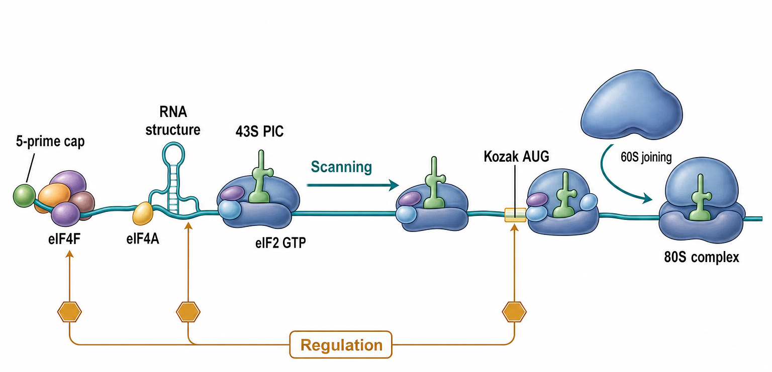
Figure 67.2. Eukaryotic Cap-Dependent Scanning. Initiation of many cytosolic eukaryotic mRNAs proceeds through five coupled stages: the 7-methylguanosine cap recruits eIF4E as part of eIF4F; eIF4G scaffolds eIF4E and eIF4A and helps recruit the 43S preinitiation complex near the 5′ end; the 40S subunit scans through the leader; start-codon recognition in a favorable context triggers eIF1 release and eIF2 GTP hydrolysis; and eIF5B promotes 60S joining, yielding an 80S initiation complex with initiator tRNA in the P site. The visual ends at the elongation-ready canonical complex.
The factor names differ across domains, so comparisons must distinguish homology from analogous molecular work. Bacterial IF1 and eukaryotic or archaeal IF1A-family activities help organize the small subunit, bacterial IF2 and eukaryotic or archaeal IF5B-family GTPases promote subunit joining, and bacterial IF3 and eukaryotic factors contribute to fidelity through differently partitioned mechanisms. These correspondences clarify conserved tasks without implying identical complexes or regulatory wiring. Chapter 25 provides the mRNP background, Chapter 66 explains codon context and initiator-tRNA identity, and Chapter 70 owns regulated and noncanonical pathways.
67.4. Organellar initiation systems
Mitochondria and chloroplasts inherited translation systems from bacterial ancestors but reshaped them under organellar genome reduction, nuclear control, and specialized RNA processing. Organellar initiation cannot be inferred by copying the rules of Escherichia coli or the eukaryotic cytosol. Each organelle combines bacterial-like ribosomal ancestry with organelle-specific mRNA leaders, RNA modifications, imported proteins, and genome architecture.
Mammalian mitochondrial mRNAs are typically processed from polycistronic precursor transcripts and often have very short or absent 5′ untranslated regions. Many are leaderless or nearly leaderless, and mitochondrial ribosomes are specialized for membrane-associated synthesis of oxidative phosphorylation proteins. Mitochondrial initiation uses organelle-specific initiation factors, commonly named mtIF2 and mtIF3 in animals, that are related to bacterial factors but adapted to mitochondrial ribosomes and mRNAs. The start codon is selected in a context where the mRNA may have little upstream sequence and where translation is coupled to inner-membrane protein biogenesis.
Plant and algal chloroplasts often retain more bacterial-like features. Chloroplast mRNAs can use Shine-Dalgarno-like interactions, but many chloroplast transcripts require nucleus-encoded RNA-binding proteins that define RNA ends, stabilize transcripts, edit nucleotides, or remodel leaders. Webster (2025) explicitly treats bacterial and chloroplast initiation together, making it the strongest local reference anchor for this subsection. Small et al. (2023) reviews plant organellar RNA maturation, which is essential context because initiation depends on the mature RNA species actually presented to ribosomes.
Organellar RNA editing can change codons, create start codons, restore conserved amino acids, or alter RNA structure. In plant mitochondria and chloroplasts, pentatricopeptide repeat proteins and related factors bind specific RNA sequences and shape maturation, stability, and translation. The initiation consequence is indirect but important: a translation start site may be unusable until processing exposes an appropriate leader, editing creates the correct codon, or an RNA-binding protein stabilizes a conformation that permits ribosome binding.
Evidence for organellar initiation includes genetics of organelle-targeted initiation factors, ribosome profiling adapted to mitochondria or chloroplasts, organellar polysome analyses, in organello translation assays, comparative transcript mapping, and structural studies of organellar ribosomes. Method-specific caveats are substantial. Mitochondrial ribosome profiling requires separating mitochondrial translation from cytosolic footprints and dealing with compact transcripts. Chloroplast translation measurements can be confounded by photosynthetic state, development, and transcript stability. A mutation that lowers protein abundance may affect initiation, elongation, RNA maturation, or protein stability; distinguishing these requires orthogonal assays.
The organellar section connects directly to Chapter 17, which treats organellar RNA genes and transcript processing. The present chapter focuses on the initiation step, while later chapters on translation quality control and disease should address how organellar initiation defects contribute to respiratory-chain disorders, photosynthetic defects, and developmental phenotypes.
67.5. Canonical machinery recruitment in viral translation contexts
Viruses do not possess autonomous ribosomes, so every viral messenger RNA must recruit a cellular translation system. This section uses viral transcripts only to clarify when host canonical machinery remains the operative initiation engine. Viral regulation, host shutoff, IRES classes, shunting, and stress-tolerant initiation are owned by Chapter 70, while virus-family genome-expression programs are developed in Chapter 115-Chapter 117.
Many DNA-virus and retroviral transcripts are synthesized and processed like host messenger RNAs. Their 5′ caps bind eIF4E, eIF4G scaffolds the cap-bound messenger ribonucleoprotein, the 43S preinitiation complex is recruited, and the 40S subunit scans to a start codon before 60S joining. Some cytoplasmic RNA viruses encode capping enzymes, and others acquire capped leaders from host RNAs. These origins differ, but once a functional cap and compatible leader are presented, the downstream canonical factor sequence can closely resemble cellular cap-dependent initiation.
The experimental question is therefore not simply whether a viral RNA is translated, but which host components it requires. Cap-competition experiments, initiation-factor depletion, toeprinting, ribosome profiling, initiation-complex reconstitution, and structural studies can test dependence on eIF4E, eIF4G, eIF4A, eIF2, eIF3, and scanning. Reporter output alone cannot distinguish altered recruitment from RNA abundance or stability. A claim of canonical recruitment is strongest when intact viral RNA, factor dependence, ribosome position, and protein output converge.
Viral contexts also enforce a useful boundary: acquisition of a cap does not prove that every subsequent step is canonical, and detection of host factors on an RNA does not prove that their ordinary roles are retained. This chapter stops once the canonical machinery and its required steps are established. Any virus-specific bypass, factor cleavage, internal recruitment, or stress-response remodeling is a handoff to Chapter 70.
67.6. Comparative initiation-factor dependencies and disease-linked core defects
All translation systems must recruit a ribosomal small subunit or complete ribosome, deliver an initiator tRNA, verify a start codon in the P site, and join the large subunit. The proteins that perform these tasks differ across bacteria, archaea, eukaryotic cytosol, and organelles. Comparative reasoning should therefore map molecular tasks first and names second: similar names can conceal lineage-specific functions, while differently named factors can perform related steps.
Bacterial IF2 and archaeal or eukaryotic IF5B-family GTPases illustrate a conserved subunit-joining task with different pathway placement. Bacterial IF1 and eukaryotic or archaeal IF1A occupy related regions of the small subunit, whereas start-site fidelity is partitioned differently among bacterial IF3 and the larger eukaryotic factor network. Initiator-tRNA delivery likewise uses bacterial IF2 but archaeal and eukaryotic a/eIF2 ternary complexes. These comparisons are mechanistic correspondences, not claims that the full complexes are interchangeable.
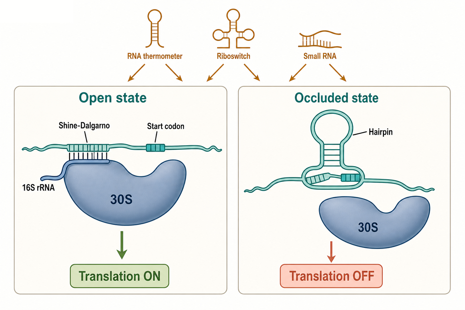
Figure 67.3. Bacterial Start-Region Accessibility. Bacterial translation depends not only on the sequence of a Shine-Dalgarno motif but on whether that motif and the adjacent start codon are accessible to the 30S subunit. In the open state, both elements are available for pairing with 16S rRNA and initiator tRNA; in the occluded state, a stem-loop sequesters them and blocks 30S engagement. The figure teaches accessibility as a prerequisite for canonical recruitment without developing the regulatory inputs that shift the ensemble.
Disease-linked core defects are most interpretable when assigned to a discrete initiation step. Variants that impair cap recognition or scaffold assembly reduce ribosome recruitment; defects in eIF2-family activity alter initiator-tRNA delivery; defects in fidelity factors change start-site selection; defects in IF5B-family or organellar factors impede subunit joining. Mitochondrial initiation defects can reduce synthesis of oxidative-phosphorylation subunits, with strong effects in energy-demanding tissues. The phenotype depends on residual activity, tissue context, transcript architecture, and compensation by other translation components.
Evidence for a core defect should connect genotype or perturbation to a biochemical state rather than infer initiation from final protein abundance alone. Reconstituted complexes and structural studies identify the blocked step; toeprinting and ribosome profiling localize ribosomes; pulse labeling tests new protein synthesis; and complementation can establish causality. Protein abundance also reflects RNA production and decay, elongation, protein folding, and proteolysis, so it is not by itself a specific initiation readout.
Regulated factor availability, mTOR signaling, integrated stress responses, uORF-dependent selectivity, and therapeutic manipulation of initiation are important but distinct. Chapter 70 owns regulatory and stress-dependent mechanisms, while Chapter 153 owns therapeutic messenger-RNA design.
Recent Consensus
Current consensus treats translation initiation as a conserved set of molecular tasks implemented by domain-specific machinery. Bacterial systems commonly use 30S recruitment and Shine-Dalgarno-guided positioning; archaea combine bacterial-like leader architectures with factors related to eukaryotic machinery; eukaryotic cap-dependent initiation proceeds through eIF4F recruitment, 43S loading, scanning, start recognition, and 60S joining; and organelles modify bacterial-derived mechanisms. Viral RNAs that present host-compatible caps and leaders can recruit this canonical machinery, whereas virus-specific bypass and regulatory routes require separate analysis in Chapter 70.
The strongest recent evidence comes from structural biology of initiation complexes, genome-scale ribosome profiling and leader-function assays, biochemical reconstitution, and targeted mutational studies. The field increasingly recognizes that translation initiation must be interpreted together with RNA maturation, localization, modification, and decay.
Open Questions, Controversies, Deprecated Models, and Common Misconceptions
Open questions:
- Archaeal initiation diversity is under-sampled relative to bacterial and eukaryotic systems; leaderless mechanisms and factor requirements need broader comparative evidence.
- Predicting initiation efficiency from sequence remains imperfect because RNA structure, RNA-binding proteins, transcript processing, and cellular state are difficult to model together.
- Mitochondrial and chloroplast initiation systems differ across lineages enough that animal, plant, algal, and protist organelles should not be collapsed into one generic organellar pathway.
- Functional correspondences among initiation factors do not always map cleanly onto one-to-one evolutionary homology, especially where ancestral tasks have been repartitioned among several eukaryotic factors.
- For many viral transcripts, the point at which host-like canonical recruitment gives way to virus-specific remodeling remains incompletely resolved.
Common misconceptions:
- “A Shine-Dalgarno-like motif proves bacterial initiation, and absence of a strong motif proves an mRNA is untranslated.” Initiation depends on accessibility, spacing, start codon context, leader structure, initiation factors, and organism-specific mechanisms.
- “Factors with analogous initiation functions are interchangeable across bacteria, archaea, eukaryotes, and organelles.” Related tasks can be executed by nonidentical complexes with different factor dependencies and pathway order.
- “A reduction in protein abundance proves an initiation defect.” RNA abundance, elongation, protein folding, and degradation can produce the same endpoint; initiation requires step-specific evidence.