Chapter 160. Antiviral Ribonucleoside Analogs and RNA Polymerase-Pathway Drugs
Scope Note
This chapter explains how antiviral ribonucleoside and ribonucleotide analogs are designed, activated, incorporated, and selected to disrupt viral RNA synthesis. The emphasis is on RNA-dependent RNA polymerase (RdRP) pathways in RNA viruses and on closely related RNA-templated or RNA-linked antiviral drug logic, including prodrug chemistry, intracellular phosphorylation, chain termination, delayed termination, polymerase stalling, lethal mutagenesis, resistance, proofreading, host polymerase toxicity, mitochondrial risk, and combination strategies. The chapter uses remdesivir, molnupiravir, ribavirin, favipiravir, and sofosbuvir-like drugs as recurring examples. Chapter-local references now contain automated DOI/PMID coverage anchors; final claim-level citation keys still require expert review.
Executive Summary
Antiviral ribonucleoside analogs are small molecules that imitate natural ribonucleosides closely enough to enter host metabolic pathways and viral polymerase active sites, but differ enough to damage viral RNA synthesis or viral genetic stability. A nucleoside analog usually begins as an uncharged base-sugar molecule or as a masked nucleotide prodrug. The pharmacologically active species is commonly a nucleoside triphosphate analog that competes with adenosine triphosphate, guanosine triphosphate, cytidine triphosphate, or uridine triphosphate during viral RNA synthesis. The route from swallowed, infused, or inhaled drug to polymerase substrate is not incidental chemistry; it is a central determinant of potency, tissue selectivity, resistance risk, and toxicity.
Viral RdRPs are attractive targets because many RNA viruses depend on them, because humans do not encode cytosolic RdRPs for ordinary mRNA replication, and because RdRP active sites must repeatedly bind nucleoside triphosphates during every replication cycle. Selectivity is nevertheless incomplete. Host kinases, nucleotidases, transporters, mitochondrial polymerases, host RNA polymerases, and innate immune sensors can all encounter analogs or their metabolites. Antiviral drug design therefore balances three forms of selectivity: selective activation in infected or permissive tissues, selective use by viral polymerases, and selective damage to viral replication before host damage becomes clinically unacceptable.
Polymerase-pathway drugs act through more than one biochemical pattern. Some analogs terminate RNA synthesis immediately after incorporation because the altered sugar lacks a required 3′ hydroxyl or because the next nucleotide cannot be accommodated. Some allow one or several further additions before the altered primer terminus or altered RNA duplex geometry blocks translocation, stalls the polymerase, or promotes template-dependent pausing. Some are mutagenic base analogs that pair ambiguously, increasing the mutation frequency of viral genomes until enough progeny are nonviable. Other analogs perturb nucleotide pools, interfere with capping, or modulate host antiviral responses. One named drug may combine more than one mechanism in a virus-, cell-, and concentration-dependent way.
The most important misconception is that all nucleoside analog antivirals are simple chain terminators. Remdesivir is better understood as a delayed-termination and polymerase-stalling drug in many coronavirus polymerase contexts; molnupiravir is better understood as a mutagenic ribonucleoside prodrug; ribavirin has multiple context-dependent mechanisms; favipiravir can act as a mutagenic and polymerase-inhibitory purine analog; and sofosbuvir-like drugs illustrate how careful prodrug design can deliver nucleotide analogs to hepatocytes and terminate hepatitis C virus RNA synthesis with clinically useful selectivity. The clinical value of each mechanism depends on viral proofreading, resistance pathways, host metabolism, tissue exposure, timing of therapy, and the therapeutic window.
Concept Inventory
- Nucleoside analog: a molecule resembling a natural nucleoside, usually a nucleobase attached to a sugar-like moiety, that can be transported into cells and enzymatically converted to nucleotide metabolites.
- Nucleotide analog: a molecule resembling a natural nucleotide, usually with one or more phosphate or phosphate-like groups. Many antiviral nucleotide analogs are administered as prodrugs that mask the monophosphate.
- Triphosphate active metabolite: the intracellular form that competes with natural nucleoside triphosphates at a polymerase active site.
- Prodrug: a compound administered in an inactive or less active form that is chemically or enzymatically converted to the active drug or active metabolite.
- Chain termination: cessation of polymerase elongation after analog incorporation because the RNA primer cannot be extended efficiently.
- Lethal mutagenesis: antiviral strategy in which a mutagenic analog increases viral genome error frequency beyond the population’s tolerance, producing noninfectious or less fit progeny.
- Proofreading: correction of misincorporated or abnormal nucleotides. Some viruses, especially coronaviruses, encode exonuclease-mediated proofreading.
- Therapeutic window: the exposure range in which antiviral benefit is achieved without unacceptable host toxicity.
What to Know Before Reading This Chapter
Readers should be comfortable with the polarity of RNA synthesis: polymerases extend an RNA chain by adding nucleoside triphosphates to the 3′ hydroxyl of the nascent strand while reading a template. The nucleotide addition cycle includes substrate binding, base pairing, metal-dependent catalysis, pyrophosphate release, and translocation of the polymerase relative to the template and product. A drug can interfere with any of these steps.
Readers should also distinguish nucleobases, nucleosides, nucleotides, and nucleotide triphosphates. A nucleobase is the heterocyclic base. A nucleoside is base plus sugar. A nucleotide adds phosphate. A ribonucleoside triphosphate is the immediate substrate for RNA polymerization. Many antiviral compounds are named as parent nucleosides, but the active inhibitor is often a triphosphate formed inside cells.
The running examples in this chapter serve different teaching roles. Remdesivir illustrates an adenosine-like prodrug whose triphosphate can be incorporated by viral RdRPs and then stall polymerase elongation. Molnupiravir illustrates a mutagenic ribonucleoside prodrug whose active form can produce ambiguous base-pairing during viral RNA replication. Ribavirin illustrates why a single drug can have multiple mechanisms and why mechanism assignments require careful system-specific evidence. Favipiravir illustrates a purine analog whose activity depends on ribosylation and phosphorylation to an active ribofuranosyl triphosphate. Sofosbuvir-like drugs illustrate nucleotide prodrug design, hepatocyte activation, and chain termination against hepatitis C virus polymerase.
160.1. Nucleoside and Nucleotide Analog Activation
An antiviral ribonucleoside analog is not automatically an RNA polymerase inhibitor when it enters the body. The parent compound must reach susceptible cells, cross membranes or use transporters, avoid premature degradation or clearance, and become a nucleotide metabolite that competes with natural ribonucleoside triphosphates. This activation path is why two analogs that look similar on paper can differ sharply in antiviral spectrum, route of administration, tissue exposure, and toxicity.
The simplest activation scheme has three phosphorylation steps. A nucleoside analog enters a cell and a host kinase adds the first phosphate, generating the monophosphate. A second kinase produces the diphosphate. A nucleoside diphosphate kinase or related enzyme produces the triphosphate. The triphosphate analog then competes with the natural nucleotide in a viral polymerase active site. In practice, the first phosphorylation is often the bottleneck because kinases are selective for sugar geometry, base identity, cellular compartment, and concentration. If the first kinase does not recognize an analog, high extracellular drug concentration may still yield little active triphosphate.
Activation is inseparable from deactivation. Nucleoside analogs and their phosphorylated metabolites can be deaminated, cleaved, dephosphorylated, exported, or trapped in compartments that do not support viral replication. A cell line that activates an analog efficiently can overestimate antiviral potency for a tissue in which the relevant kinase is weakly expressed. Conversely, a tissue with efficient activation can be a site of both antiviral activity and host toxicity. This is a recurring problem in extrapolating from permissive cell culture to patients.
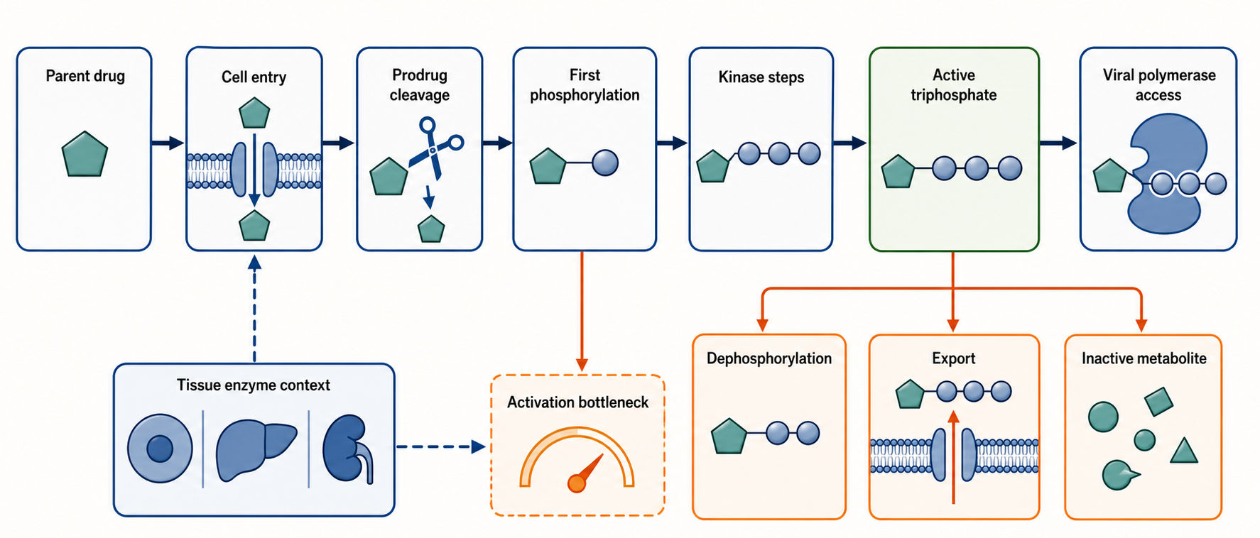
Figure 160.1. Activation path from parent drug to active triphosphate. A ribonucleoside analog becomes a viral polymerase substrate only after cell entry and metabolic activation. The first phosphorylation step can be rate-limiting for nucleoside analogs, whereas nucleotide prodrugs are designed to bypass or reshape that bottleneck. Competing deactivation and tissue-specific enzyme expression determine active triphosphate exposure.
The natural nucleotide pools also matter. Viral polymerases do not encounter an analog in isolation; they encounter a competition between analog triphosphate and natural ribonucleoside triphosphate. A weakly incorporated analog can become effective if the competing natural nucleotide is scarce, if the analog triphosphate accumulates to high concentration, or if the viral polymerase has poor discrimination at the relevant active-site step. Conversely, high natural nucleotide concentration can protect viral RNA synthesis from an analog that relies on direct competition. This competition is one reason pharmacodynamic interpretation should consider intracellular metabolite concentrations rather than only plasma parent-drug levels.
The term “nucleoside analog activation” should therefore be read as a multi-step pharmacological process, not as a single enzymatic conversion. Uptake transporters influence whether the parent nucleoside enters a cell. Esterases, amidases, phosphoramidases, and kinases influence whether a prodrug releases the correct nucleotide intermediate. Nucleotidases and phosphatases oppose accumulation. The viral replication compartment determines whether the active triphosphate is physically available to the polymerase. For positive-strand RNA viruses that remodel membranes into replication organelles, the local concentration near replication complexes may not equal the average cytosolic concentration.
A concrete example is remdesivir. The administered compound is a prodrug of an adenosine analog. It is designed to bypass inefficient first phosphorylation and deliver a monophosphate-like intermediate that host enzymes can convert to a triphosphate. The triphosphate resembles adenosine triphosphate enough for incorporation by several viral RdRPs, but its modified sugar and cyano-substituted architecture alter downstream polymerase movement. The same example shows why naming a drug as a nucleoside analog can hide the medicinal chemistry: remdesivir is not simply an adenosine look-alike; it is a delivery system for an intracellular nucleotide analog.
Molnupiravir illustrates a different activation logic. It is a prodrug of a ribonucleoside analog that is converted to the corresponding triphosphate. Rather than acting mainly as a steric block after incorporation, the active analog can adopt base-pairing states that allow it to pair in more than one way during viral RNA synthesis. The activation requirement remains essential: without formation of the triphosphate, the analog cannot be efficiently used by viral polymerases. Its mechanism therefore connects metabolic activation to mutagenesis rather than to simple termination.
Activation evidence comes from several experimental layers. Biochemical studies test whether purified polymerases incorporate a triphosphate analog. Cell-based assays test whether parent compounds or prodrugs inhibit viral replication in living cells. Metabolite profiling measures intracellular mono-, di-, and triphosphate forms. Resistance selection asks whether polymerase mutations reduce susceptibility. Toxicology assays ask whether host polymerases, mitochondrial processes, or nucleotide-pool metabolism are harmed at relevant exposures. No single layer proves the whole mechanism. For example, a polymerase assay can show incorporation, but not whether the active metabolite accumulates in lung epithelium, liver, or immune cells at safe dosing.
Box 160.1. Reading an Activation Claim
Activation claim checklist. When a paper says that an antiviral ribonucleoside analog is activated, ask what molecule is being tracked. Parent-drug concentration, monophosphate release, and triphosphate accumulation are different measurements. Then ask where the measurement was made. A permissive transformed cell line can reveal a possible pathway but may not represent airway epithelium, hepatocytes, kidney, immune cells, or infected tissue. Next, ask whether natural nucleotide pools were measured, because polymerase inhibition depends on competition between analog triphosphate and natural NTPs. Finally, ask whether deactivation was considered. Deamination, dephosphorylation, export, and compartmental sequestration can prevent a chemically plausible analog from reaching viral replication complexes. Strong activation evidence connects time-resolved metabolite profiling, relevant cell types, viral inhibition, and polymerase use of the same triphosphate species.
Table 160.1. Activation bottlenecks and evidence types. Activation claims require evidence at each step from cell entry to active triphosphate accumulation. A polymerase-active triphosphate does not prove that the parent drug generates that metabolite in infected tissue.
| Step | Possible bottleneck | Example evidence | Interpretation caveat |
|---|---|---|---|
| Cell entry and tissue delivery | Parent nucleoside or prodrug does not reach infected cells, uses weak transport routes, or is cleared before target-tissue exposure. | Parent-drug pharmacokinetics, tissue or cell exposure measurements, transporter assays, and infected-cell antiviral assays. | Plasma exposure does not prove active metabolite exposure in the infected tissue or replication compartment. |
| Prodrug unmasking or first phosphorylation | Chemical masks are cleaved too early or too slowly, or the first kinase poorly recognizes the analog. | Enzyme assays, time-resolved metabolite profiling, and comparison of parent nucleoside with monophosphate-prodrug forms. | Efficient activation in a permissive cell line can overstate activation in airway, liver, kidney, immune, or gastrointestinal tissues. |
| Di- and triphosphate formation | Downstream kinases are limiting, dephosphorylation competes with activation, or natural nucleotide pools dilute analog competition. | LC-MS measurement of mono-, di-, and triphosphate species with natural NTP pool measurements across time. | Total intracellular triphosphate concentration may not equal local concentration near viral replication organelles. |
| Viral polymerase use | Viral RdRP rejects the analog, incorporates it inefficiently, or extends past the incorporated analog. | Purified polymerase kinetics, primer-extension assays, structural trapping, and resistance substitutions near active-site or translocation motifs. | Incorporation in vitro does not by itself prove that the parent drug inhibits viral replication by that mechanism in cells. |
| Competing deactivation and host exposure | Deamination, phosphatase activity, export, mitochondrial access, or host polymerase use narrows the therapeutic window. | Metabolite decay studies, mitochondrial function assays, host polymerase assays, cytotoxicity, and safety monitoring. | A safety signal or absence of signal is exposure-, tissue-, duration-, and patient-context dependent. |
Do not overgeneralize activation from one virus, one cell line, or one dosing route. An analog can be potent in vitro but weak in vivo because the active triphosphate is not formed in the infected tissue. Another analog can appear broadly active in cell culture but fail clinically because achievable tissue exposure is below the concentration needed for polymerase competition. Activation can also vary with age, inflammation, liver function, kidney function, genetic variation in metabolic enzymes, and coadministered drugs. These factors make nucleoside analog pharmacology a bridge between RNA enzymology and clinical pharmacology rather than a purely virological topic.
160.2. Prodrug Design and Viral RNA Synthesis Targeting
Prodrug design is the medicinal-chemistry answer to a biological obstacle: nucleotide analogs are often too polar to enter cells efficiently, and nucleoside analogs may not be phosphorylated efficiently enough after entry. A prodrug temporarily masks a charge, improves absorption or tissue delivery, or bypasses a slow metabolic step. Once inside the target tissue, host enzymes reveal the monophosphate or nucleoside that can be converted into the active triphosphate. In antiviral ribonucleoside pharmacology, prodrug design often decides whether an elegant polymerase inhibitor becomes a useful medicine.
The first major problem is membrane permeability. Natural nucleotides carry negatively charged phosphate groups that restrict passive diffusion across lipid bilayers. Cells use specialized transporters and metabolic pathways to handle nucleotide synthesis internally rather than importing large amounts of phosphorylated nucleotide from outside. A nucleoside analog without phosphate can often cross membranes more readily or use nucleoside transporters, but it then depends on kinases. A monophosphate analog can bypass the first kinase, but it usually needs chemical masking to cross membranes. Prodrug groups solve this problem by hiding charged phosphate oxygen atoms until intracellular enzymes remove the masks.
The second problem is tissue targeting. A prodrug can be tuned so that the enzymes required for unmasking are abundant in the desired tissue. Sofosbuvir-like hepatitis C virus drugs provide a central example. Their ProTide-like design masks a nucleotide monophosphate with groups that improve hepatocyte delivery and intracellular release. Because hepatitis C virus replicates primarily in hepatocytes, delivering high active-metabolite exposure to liver cells can produce a favorable antiviral window. The same design principle would not automatically work for respiratory viruses, neurotropic viruses, or systemic hemorrhagic fever viruses because the target tissue and activation enzymes differ.
The third problem is chemical stability. A prodrug must survive formulation, storage, gastrointestinal or plasma exposure, and first-pass metabolism long enough to reach the relevant cells. It must then be labile enough inside cells to release the correct intermediate. If the prodrug is cleaved too early, the charged intermediate may be trapped outside target cells or cleared. If it is too stable, active triphosphate levels may remain too low. Prodrug design is therefore a kinetic design problem as much as a structural one.
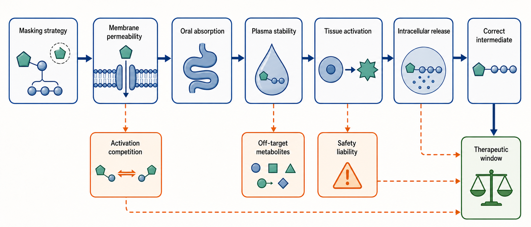
Figure 160.2. Prodrug design decision tree. Prodrug design balances permeability, stability, tissue targeting, and intracellular unmasking. A useful nucleotide prodrug must survive long enough to reach target cells, release the correct intermediate efficiently, and avoid off-target metabolites that narrow the therapeutic window.
Targeting viral RNA synthesis requires a second level of selectivity after activation. Viral RdRPs share a palm-domain catalytic architecture and use two-metal-ion chemistry to add nucleotides, but they differ in active-site geometry, initiation mode, accessory proteins, template-product duplex handling, and proofreading. A triphosphate analog that is accepted by one viral polymerase may be rejected by another. A drug that inhibits a purified polymerase can also fail if viral replication complexes exclude the analog, if the analog is excised, or if the virus encodes compensating functions.
The basic catalytic cycle gives a useful map for drug action. First, the polymerase binds a template base and an incoming nucleoside triphosphate. Second, base pairing and active-site closure align the alpha phosphate for attack by the 3′ hydroxyl of the primer. Third, phosphodiester-bond formation extends the RNA and releases pyrophosphate. Fourth, the polymerase translocates so that the next template base is positioned. An analog can reduce binding, mislead base selection, distort catalysis, alter the product RNA, block translocation, or make subsequent extension inefficient. Prodrug design delivers the analog; polymerase targeting defines what the analog does after delivery.
Remdesivir again illustrates the connection. Its prodrug design supports intracellular formation of an adenosine analog triphosphate. Viral polymerases can incorporate that triphosphate opposite uridine in the template. After incorporation, the altered sugar substituent affects later stages of elongation, especially when the polymerase has moved several positions beyond the incorporated analog. The antiviral mechanism therefore depends on both the upstream delivery of the monophosphate and the downstream mechanics of polymerase translocation.
By contrast, molnupiravir prodrug design delivers a mutagenic ribonucleoside analog. The active triphosphate can be used during viral RNA synthesis, but the consequence is not primarily physical blockade. The analog can behave as one base-pairing partner during one round of RNA synthesis and as another during a later round. In a positive-strand RNA virus, such ambiguous pairing can affect both negative-strand and positive-strand synthesis, increasing transition mutations in viral genomes. The prodrug’s success depends on achieving enough active metabolite in infected cells during the time window when the virus is replicating rapidly.
Ribavirin and favipiravir show why polymerase-pathway targeting is broader than direct active-site blockade. Ribavirin can be converted to phosphorylated forms, can affect nucleotide metabolism, can be incorporated by some viral polymerases, and can influence mutagenesis or immune context depending on the system. Favipiravir requires conversion to a ribofuranosyl triphosphate form and is used by some viral polymerases as a purine-like substrate. For both drugs, mechanism claims must specify virus, cell type, concentration, metabolic state, and assay. A blanket statement such as “ribavirin is a chain terminator” or “favipiravir causes error catastrophe” is too imprecise.
Table 160.2. Prodrug and polymerase-targeting contrasts. Similar naming can hide different pharmacology. Antiviral ribonucleoside and nucleotide analogs differ in activation strategy, active metabolite, polymerase mechanism, and safety constraints.
| Drug class/example | Activation strategy | Active species | Principal polymerase-pathway effect | Major caveat |
|---|---|---|---|---|
| Remdesivir-like adenosine nucleotide prodrug | Masked monophosphate-like prodrug bypasses inefficient first phosphorylation, then host enzymes generate the triphosphate. | Adenosine analog triphosphate. | RdRP incorporation followed by delayed stalling or translocation interference in susceptible polymerase contexts. | Clinical interpretation depends on timing, tissue exposure, viral polymerase context, and proofreading or resistance. |
| Molnupiravir-like mutagenic ribonucleoside prodrug | Oral prodrug releases a ribonucleoside analog that is phosphorylated to the triphosphate. | Mutagenic ribonucleoside triphosphate. | Ambiguous base pairing increases transition mutations and can reduce infectious progeny. | Host genotoxicity, reproductive risk, and early-replication treatment window must be evaluated directly. |
| Ribavirin-like pleiotropic nucleoside analog | Nucleoside uptake and phosphorylation produce metabolites that can affect nucleotide metabolism and viral RNA pathways. | Phosphorylated ribavirin metabolites, including mono- and triphosphate forms. | Context-dependent mixture of nucleotide-pool depletion, polymerase effects, mutagenesis, capping effects, and immune modulation. | Dominant mechanism varies by virus, cell type, concentration, and regimen; hemolytic anemia and teratogenicity are key boundaries. |
| Favipiravir-like purine analog | Intracellular ribosylation and phosphorylation generate an active ribofuranosyl triphosphate. | Favipiravir ribofuranosyl triphosphate. | Viral RdRP substrate that can promote mutagenesis, elongation inhibition, or chain-termination-like effects depending on system. | Broad preclinical activity does not by itself establish clinical efficacy or mechanism for a specific indication. |
| Sofosbuvir-like HCV nucleotide prodrug | ProTide-like masking improves hepatocyte delivery and bypasses first phosphorylation. | Uridine analog triphosphate. | NS5B incorporation followed by functional chain termination in hepatitis C virus RNA synthesis. | Hepatocyte-targeted HCV success does not automatically generalize to nonhepatic or acute respiratory RNA viruses. |
Evidence for prodrug targeting is strongest when chemical, biochemical, cellular, and clinical data align. Chemical studies define the activation route. Intracellular metabolite measurements show that the active triphosphate accumulates. Purified polymerase assays show incorporation or inhibition. Structural studies show where the analog sits in the active site or product RNA. Viral sequencing shows the expected mutation spectrum or resistance pattern. Clinical samples show viral-load reduction at exposures consistent with the mechanism. Weak evidence arises when only one layer is present, such as cell culture inhibition without active-metabolite measurement or biochemical inhibition at concentrations never achieved in patients.
Clinical and regulatory boundaries also enter at the prodrug stage. A prodrug group is not an inert wrapper. It can create metabolites with their own toxicity, interact with transporters, alter distribution, or complicate drug-drug interactions. Oral outpatient antivirals require different properties from intravenous hospital drugs. A drug intended for early infection must be safe enough for use before severe disease develops and before all patients can be certain to benefit. A drug intended for life-threatening infection may tolerate different risk. These boundaries shape which polymerase inhibitors move forward.
160.3. Chain Termination, Delayed Termination, and Polymerase Stalling
Chain termination is the most intuitive mechanism for a nucleoside analog: the analog is incorporated into nascent RNA, and the polymerase can no longer extend the chain. This intuition comes partly from DNA and reverse-transcriptase inhibitors, where absence or masking of the sugar 3′ hydroxyl can directly prevent formation of the next phosphodiester bond. RNA virus polymerases, however, encounter a wider range of analog effects. Some analogs terminate immediately, some terminate after several further additions, some slow elongation without absolute termination, and some create template-dependent stalls that depend on sequence context.
Immediate chain termination occurs when the incorporated analog leaves no chemically competent primer terminus or forces the active site into a geometry incompatible with the next addition. The primer’s 3′ end is the nucleophile in RNA synthesis. If that end lacks the correct hydroxyl or cannot align with the incoming nucleotide’s alpha phosphate, elongation stops. The mechanism is straightforward in principle, but even immediate termination can be incomplete if the polymerase occasionally extends the analog-terminated primer or if repair and excision systems remove the analog.
Delayed chain termination is more subtle. The analog is incorporated, the polymerase adds one or more additional nucleotides, and only later does elongation stall. Delayed termination can occur because the altered nucleotide affects the shape of the RNA product duplex after it has moved away from the catalytic site. The polymerase must translocate the template-product duplex through a constrained channel. An analog substituent that is tolerated at the active site may clash with a conserved residue or structural element during translocation several nucleotides later. Alternatively, the altered base or sugar may distort duplex geometry enough to destabilize active-site alignment at a downstream position.
Remdesivir is the central example for this chapter. In coronavirus polymerase systems, the active triphosphate can be incorporated in place of adenosine. The polymerase may then add several more nucleotides before stalling. Structural and biochemical models explain the stall as a consequence of the incorporated analog encountering a downstream constraint during translocation. This is a richer mechanism than the phrase “chain terminator” suggests. The drug does not simply remove the 3′ hydroxyl; instead, it allows partial extension and then interferes with polymerase movement or geometry.
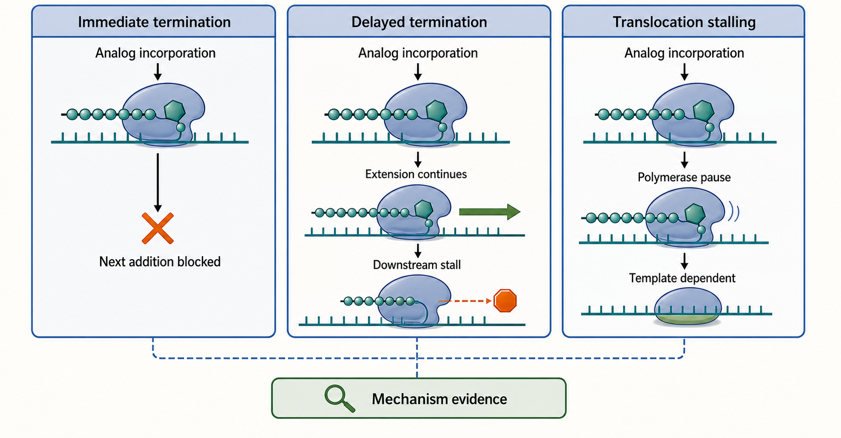
Figure 160.3. Immediate termination, delayed termination, and translocation stalling. Ribonucleotide analogs can stop viral RNA synthesis at different points in the nucleotide addition cycle. Some block the next addition immediately, some allow extension before a downstream stall, and some slow translocation in a template-dependent manner.
Template-dependent stalling adds another layer. Polymerases do not synthesize all sequences with equal kinetics. Local template sequence, RNA structure, nascent-product stability, and accessory proteins can influence pausing. If an analog creates a marginal defect, a difficult template context may convert slowing into effective termination, whereas an easy context may permit readthrough. This means that inhibition measured on a short synthetic template may not fully predict inhibition during full viral genome replication. For long RNA virus genomes, even modest increases in pausing or premature termination can reduce the number of complete genomes produced.
Polymerase translocation is often the hidden step in analog action. After catalysis, the polymerase must move so that the newly added nucleotide shifts from the active insertion site to a post-translocation position, and the next template base becomes available. Translocation involves coordinated movement of the RNA duplex, template strand, product strand, and polymerase motifs. An analog can be accepted during catalysis but disrupt this mechanical step. The biological consequence is a population of stalled replication complexes. Stalling can reduce full-length genome synthesis, alter subgenomic RNA production, expose abnormal RNA structures to innate sensors, or change recombination patterns.
Not every stall is equivalent to permanent termination. A polymerase may pause and then resume, backtrack, dissociate, transfer template, or be rescued by viral or host factors. In biochemical assays, a strong band at a particular RNA length may represent a pause, a dead-end product, or a mixture. Time-course experiments, chase experiments, template variation, and structural trapping help distinguish these possibilities. In infected cells, the same distinction is harder because viral RNA products are degraded, copied, packaged, or sensed by host pathways. Therefore, mechanistic labels should be tied to specific evidence.
Box 160.2. When a Stall Is Not a Stop
Evidence ladder for a stopped RNA product. A single primer-extension band shows that one RNA length accumulated under one assay condition. It does not by itself prove an irreversible terminator. A stronger interpretation asks whether the polymerase can resume during a chase, whether changing the template sequence shifts the stall, whether viral cofactors alter readthrough, and whether structural or kinetic data identify the blocked step. Immediate termination should stop at or directly after analog incorporation. Delayed termination should allow defined additional nucleotides before a reproducible downstream block. A pause should be reversible or strongly time-dependent. Translocation stalling should be supported by evidence that catalysis occurred but movement to the next register failed. In infected cells, degradation and selection obscure these states, so biochemical mechanism should be paired with replication-product and resistance evidence.
Sofosbuvir-like drugs illustrate a different termination architecture. Sofosbuvir is a nucleotide prodrug used against hepatitis C virus. Its active triphosphate is incorporated by the viral NS5B RdRP and acts functionally as a chain terminator because the modified sugar prevents efficient extension. Hepatocyte-targeted prodrug delivery and viral polymerase selectivity contribute to clinical usefulness. The example is important because it shows how a cleaner termination mechanism can be embedded in a broader system of prodrug activation, viral genotype variation, combination therapy, and resistance management.
Table 160.3. Mechanism labels and required evidence. Immediate termination, delayed termination, polymerase pausing, and translocation stalling require different experimental support. A single gel band or cell-culture inhibition result is rarely sufficient to assign mechanism.
| Mechanism label | Minimal evidence | Stronger evidence | Common overinterpretation |
|---|---|---|---|
| Immediate chain termination | Primer-extension products stop at or immediately after analog incorporation. | Time-course and chase experiments show a dead-end product, with structural or kinetic support and repair or excision assessed. | Any shortened RNA product proves an obligate, irreversible terminator. |
| Delayed chain termination | Polymerase incorporates the analog, adds additional nucleotides, and then stalls at a reproducible downstream position. | Sequence-context tests, structural models of downstream clash, resistance mapping, and infected-cell replication products support the same stall. | A delayed pause is permanent termination in every viral polymerase or template context. |
| Template-dependent pausing or elongation slowing | Specific templates show slower extension or enriched intermediate products after analog incorporation. | Chase assays, template variants, viral cofactors, and cellular replication assays distinguish pause, readthrough, dissociation, and degradation. | A strong gel band or sequencing drop-off alone identifies the exact biochemical step. |
| Translocation stalling | Catalysis occurs, but the incorporated analog disrupts movement of the template-product duplex or next-template positioning. | Pre-steady-state kinetics, trapped structural states, single-molecule or time-resolved assays, and resistance changes near translocation elements align. | Active-site binding or incorporation automatically demonstrates a translocation block. |
| Mutagenic substrate or lethal-mutagenesis mechanism | Drug exposure raises viral mutation frequency while infectious titer falls. | Base-pairing chemistry, polymerase incorporation, high-accuracy sequencing, infectivity loss, and rescue or resistance data support mutation-driven loss of viability. | Increased mutation frequency alone proves formal error catastrophe or excludes other antiviral effects. |
Polymerase stalling evidence should be interpreted with a hierarchy of assays. Purified polymerase extension assays can map where termination or pausing occurs, but they may omit viral cofactors. Structural studies can show steric clashes or altered positioning, but they often capture selected states. Cell-based replication assays show antiviral activity but may not identify the exact polymerase step. Viral RNA sequencing can show reduced genome completion or altered mutation spectra but may reflect downstream selection. A robust chain-termination model usually combines several evidence types.
Resistance mutations provide functional tests of termination mechanisms. A mutation near the polymerase active site, template channel, or translocation pathway can reduce analog incorporation, improve extension after incorporation, or alter excision sensitivity. However, resistance is constrained because polymerases must preserve natural nucleotide use. High-level resistance mutations often reduce viral fitness unless compensated. This creates opportunities for combination therapy: a second drug can exploit the fitness cost or block a different step so that escape from one mechanism does not restore replication.
Chain termination is not a binary property of a molecule. It is a property of a molecule in a particular polymerase, sequence context, metabolic state, and assay. The same analog may be an efficient terminator for one viral polymerase, a weak pause-inducer for another, and a mostly mutagenic substrate in a third. Reader-facing terminology should therefore specify “immediate chain termination,” “delayed chain termination,” “polymerase stalling,” or “elongation slowing” when the evidence supports that distinction.
160.4. Lethal Mutagenesis and Error Catastrophe
Lethal mutagenesis is an antiviral strategy that attacks the genetic integrity of a viral population rather than stopping every polymerase molecule. RNA viruses replicate with mutation rates high enough to support rapid adaptation, immune escape, and drug resistance, but low enough to preserve functional genomes. A mutagenic ribonucleoside analog can push the mutation burden upward until many progeny genomes encode defective proteins, broken regulatory signals, or nonviable combinations of mutations. The result is reduced infectious virus even if RNA synthesis continues.
The term “error catastrophe” comes from theoretical models in which a replicating population loses genetic information when mutation rates exceed an error threshold. In antiviral pharmacology, the phrase is often used loosely. It is safer to distinguish lethal mutagenesis, which can be demonstrated by increased mutation frequency and loss of infectivity, from full theoretical error catastrophe, which requires stronger population-genetic evidence. A drug can be clinically useful by increasing deleterious mutations without proving a formal error-threshold transition.
Mutagenic ribonucleosides work because base pairing is a chemical process, not an immutable code. Natural bases have preferred hydrogen-bonding patterns, but tautomeric states, protonation, and analog modifications can change pairing. A mutagenic analog may pair like cytidine in one context and like uridine in another, or like adenine in one step and guanine in another. When a viral polymerase copies a genome containing that analog, the next round of synthesis can convert ambiguous pairing into transition mutations. Repeated cycles amplify the genetic damage.
Molnupiravir provides the key example. It is converted in cells to a ribonucleoside triphosphate analog that can be incorporated into viral RNA. The incorporated analog can support continued RNA synthesis, which is important: a pure terminator would not be copied into progeny genomes. During subsequent copying, ambiguous base-pairing behavior can increase transition mutations. The antiviral effect depends on viral replication cycles producing genomes that are copied again, translated, packaged, and selected. Deep sequencing can detect shifts in mutation spectra, while infectivity assays test whether those mutations reduce viable virus.
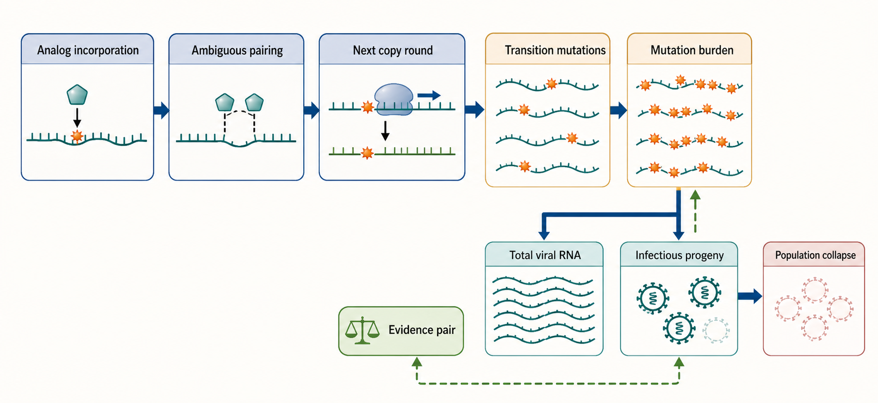
Figure 160.4. Lethal mutagenesis across replication cycles. A mutagenic ribonucleoside can be incorporated into viral RNA, copied ambiguously in a later round, and increase transition mutations across the viral population. Lethal mutagenesis is supported when mutation burden rises and infectious progeny decline.
Favipiravir can also be understood through mutagenesis and polymerase inhibition, depending on the virus and assay. The drug is converted to an active ribofuranosyl triphosphate. Viral polymerases can incorporate it as a purine analog, and its incorporation can increase mutation frequencies or impair elongation. The relative contribution of mutagenesis versus direct inhibition may vary across influenza virus, flaviviruses, arenaviruses, bunyaviruses, and other RNA virus systems. This variability is a caution against treating “mutagenic analog” as a complete mechanism.
Ribavirin is a long-standing example of mechanism complexity. It has been associated with inosine monophosphate dehydrogenase inhibition and guanosine nucleotide pool depletion, altered capping, immunomodulatory effects, direct polymerase incorporation, and mutagenesis. Which mechanism dominates depends on virus, host cell, concentration, and combination partner. In hepatitis C treatment history, ribavirin’s value was especially evident in combination regimens even when it was not a potent direct-acting polymerase inhibitor by modern standards. In other viral systems, its mutagenic or nucleotide-pool effects may be more prominent.
There are several experimental signatures of lethal mutagenesis. Viral RNA or progeny virus should show an increased mutation frequency at drug exposures that reduce infectivity. The mutation spectrum should fit the analog’s base-pairing chemistry. Infectious titer should fall more than total viral RNA if many genomes are produced but defective. Removing the drug should not immediately restore infectivity from already damaged genomes, although surviving genomes can repopulate. Resistance mutations may improve polymerase discrimination against the analog or alter activation and pool balance indirectly.
There are also traps. Sequencing errors can masquerade as viral mutagenesis unless high-fidelity library preparation, controls, and error-correction strategies are used. Population bottlenecks can make mutation spectra noisy. Cytotoxicity can reduce viral replication and secondarily change mutation patterns. Host innate immune activation can suppress virus independently of mutagenesis. A mutagenesis claim is strongest when biochemical base-pairing evidence, polymerase incorporation evidence, sequencing, infectivity loss, and rescue or resistance data agree.
Lethal mutagenesis has a distinctive safety concern: if a drug is designed to promote ambiguous base pairing, could it mutagenize host DNA or RNA? The answer depends on activation, substrate selectivity, DNA versus RNA polymerase use, ribonucleotide reductase pathways, repair, exposure, and reproductive or developmental context. A mutagenic RNA antiviral does not automatically become a host genotoxin, but the concern is mechanistically reasonable and must be tested. Regulatory evaluation therefore pays attention to genotoxicity assays, reproductive toxicity, duration of treatment, patient population, and contraindications.
The population genetics of RNA viruses creates both opportunity and risk. High mutation rates make viruses vulnerable to additional mutational pressure. Large population sizes and rapid replication also allow selection of variants with reduced analog sensitivity. Some resistance pathways may increase polymerase fidelity, which can reduce mutagenic susceptibility but also reduce adaptability or fitness. Other pathways may change activation, nucleotide pools, or proofreading. Combination therapy can be designed to make escape from mutagenesis costly by pairing a mutagenic analog with a terminator, protease inhibitor, entry inhibitor, or immune-modulating intervention.
Do not overgeneralize the phrase “error catastrophe” as if it were directly observed whenever a mutagenic drug reduces viral titer. A careful chapter, paper, or regulatory review should state the measured evidence: increased transition frequency, reduced specific infectivity, loss of viable progeny, altered consensus sequence, reduced RNA accumulation, or clinical viral-load decline. The term lethal mutagenesis is often the more precise mechanism-level label.
160.5. Viral Proofreading, Resistance, Host Polymerase Toxicity, and Mitochondrial Risk
Viral polymerases must balance speed, fidelity, and evolvability. If fidelity is too low, genomes accumulate lethal defects. If fidelity is too high, adaptation slows. Nucleoside analogs exploit this balance by either being accepted as abnormal substrates or by increasing errors. Viruses respond through polymerase discrimination, excision, proofreading, altered replication-complex context, or compensatory mutations. Host cells impose a second constraint: the same analogs must not damage essential host polymerases, mitochondrial nucleic acid metabolism, or nucleotide homeostasis beyond acceptable limits.
Proofreading is especially important for coronaviruses. Coronaviruses have unusually large RNA genomes for RNA viruses and encode a proofreading exonuclease activity within the replication-transcription complex. This activity can remove some misincorporated nucleotides or analogs from nascent RNA, reducing susceptibility to certain mutagens or terminators. A drug that works well against a nonproofreading RdRP virus may be less effective against a coronavirus unless the analog evades excision, stalls after moving beyond the immediate proofreading window, overwhelms repair, or acts through a mechanism not easily corrected.
Remdesivir’s delayed-stalling behavior can be viewed partly in this light. If an analog caused immediate obvious mispairing at the primer terminus, a proofreading exonuclease might remove it efficiently. If the analog is incorporated and only later interferes with polymerase translocation, the replication complex may face a different repair problem. This does not mean proofreading is irrelevant; rather, the timing and structural location of the analog-induced defect influence whether proofreading can rescue synthesis.
Resistance to nucleoside analogs can arise at several levels. Polymerase active-site mutations may reduce analog incorporation while preserving natural nucleotide incorporation. Mutations in translocation-associated motifs may allow extension past a delayed terminator. Mutations in proofreading enzymes may increase or decrease susceptibility depending on whether proofreading removes the analog or maintains genome viability under mutagenic stress. Mutations in viral accessory proteins may change replication-complex architecture. In cell culture, resistance can be selected under drug pressure, but clinical resistance depends on viral population size, treatment duration, immune pressure, adherence, and combination therapy.
Resistance has a cost because nucleotide selection is central to viral replication. A polymerase that rejects an analog too strongly may also reject or mishandle natural nucleotides, lowering fitness. A fidelity-increasing mutation may reduce adaptability. A proofreading change may make the virus more sensitive to ordinary replication errors. Fitness costs are clinically useful when they reduce transmission or when a second drug exploits the weakened state. However, compensatory mutations can restore fitness, so resistance interpretation requires whole-genome viral sequencing and phenotypic testing rather than single-mutation storytelling.
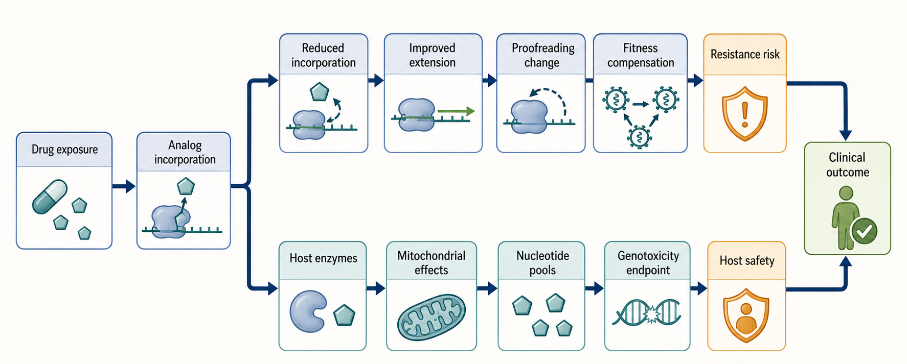
Figure 160.5. Resistance and host-toxicity checkpoints. Viral resistance can reduce analog incorporation, improve extension, alter proofreading, or compensate for fitness costs. Host toxicity can arise when analog metabolites affect mitochondrial or nuclear enzymes, nucleotide pools, or genotoxicity endpoints.
Host polymerase toxicity is the mirror image of viral polymerase targeting. Human nuclear RNA polymerases, mitochondrial RNA polymerase, mitochondrial DNA polymerase gamma, and other nucleotide-using enzymes all handle natural nucleotides. An analog that is sufficiently similar to be used by a viral RdRP may also be used or bound by host enzymes. The degree of risk depends on intracellular concentration, compartment access, enzyme selectivity, repair capacity, tissue turnover, and treatment duration. Short courses for acute viral infection can tolerate some exposures that would be unacceptable for chronic prophylaxis or long-term therapy.
Mitochondria deserve special attention. Mitochondria have their own DNA replication, transcription, RNA processing, translation, and nucleotide metabolism. Mitochondrial toxicity can appear as impaired oxidative phosphorylation, lactic acidosis, myopathy, neuropathy, liver injury, pancreatitis, or other tissue-specific syndromes depending on the drug class and exposure. Ribonucleoside analogs may affect mitochondrial RNA polymerase or nucleotide pools, while deoxynucleoside analogs are classically associated with mitochondrial DNA polymerase concerns. The boundary is not absolute because ribonucleotide metabolism and deoxyribonucleotide metabolism are connected.
Mitochondrial risk assessment uses multiple assays. Biochemical assays test incorporation or inhibition by mitochondrial polymerases. Cultured-cell assays measure mitochondrial DNA copy number, mitochondrial RNA expression, oxygen consumption, membrane potential, and lactate. Animal studies and clinical monitoring look for tissue injury. A negative result in one assay is not complete reassurance because mitochondrial toxicity can be tissue-specific, delayed, or dependent on metabolic stress. Conversely, a high-concentration cell-culture signal may not translate to clinical toxicity if exposures are not achieved in patients.
Host mutagenicity is a separate but related concern for mutagenic ribonucleosides. A drug that increases viral RNA mutations should be tested for host DNA mutagenesis, chromosomal damage, reproductive toxicity, and developmental risk. Mechanistic selectivity can arise because the active ribonucleotide is used mainly by viral RdRPs in infected cytoplasm, because treatment is short, because DNA polymerases reject the analog, or because metabolism does not efficiently generate deoxynucleotide forms. These are testable claims, not assumptions. Clinical restrictions may reflect residual uncertainty even when benefit outweighs risk for selected populations.
Box 160.3. Host Safety Is Mechanism Specific
Three safety questions should stay separate. First, can the active ribonucleotide analog enter host RNA pathways or mitochondrial nucleotide metabolism at clinically relevant exposure? This is tested with host polymerase assays, metabolite profiling, mitochondrial function assays, and tissue-sensitive toxicology. Second, can the compound or a metabolite reach DNA replication chemistry? Genotoxicity and reproductive-risk assays address this question, especially for mutagenic ribonucleosides. Third, does the intended treatment context change the acceptable risk? A short course for high-risk acute infection, chronic therapy, prophylaxis, pregnancy, pediatric use, and immunocompromised prolonged infection are not equivalent. The scientific caution is two-sided: do not dismiss host risk because the target is viral RNA, and do not assume unacceptable genotoxicity merely because viral lethal mutagenesis is the intended antiviral mechanism.
Innate immune effects complicate toxicity and efficacy. Viral RNA synthesis products and analog-containing RNA may be sensed by endosomal Toll-like receptors, RIG-I-like receptors, protein kinase R, oligoadenylate synthetase pathways, or other RNA surveillance systems. A drug can reduce viral replication and thereby lower inflammatory RNA burden, but it can also alter RNA species in ways that affect sensing. Ribavirin’s immunological effects have been discussed in multiple systems. For modern analogs, innate immune consequences should be evaluated in relevant primary cells and infection models rather than inferred only from chemical class.
Clinical resistance and toxicity boundaries vary with treatment context. A life-threatening viral hemorrhagic fever, severe hospitalized respiratory infection, chronic hepatitis C infection, and outpatient early respiratory infection have different acceptable risks, durations, endpoints, and combination strategies. Pediatric use, pregnancy, renal impairment, hepatic impairment, immunocompromise, and transplant settings require special attention because activation, clearance, viral kinetics, and safety margins differ. Regulatory labels often encode these distinctions in dosing restrictions, contraindications, monitoring requirements, and population-specific evidence statements.
160.6. Remdesivir, Molnupiravir, Ribavirin, Favipiravir, and Sofosbuvir-Like Drugs
The named drugs in this section are not interchangeable examples of one mechanism. They define a vocabulary of antiviral ribonucleoside pharmacology. Remdesivir emphasizes nucleotide prodrug activation and delayed RdRP stalling. Molnupiravir emphasizes mutagenic ribonucleoside activation and viral error loading. Ribavirin emphasizes pleiotropic mechanisms and combination-dependent clinical value. Favipiravir emphasizes purine-analog activation and virus-dependent mutagenic or inhibitory effects. Sofosbuvir-like drugs emphasize hepatocyte-targeted nucleotide prodrug design and effective direct-acting therapy against hepatitis C virus. Comparing them teaches the design space better than memorizing each drug alone.
Remdesivir is administered as a prodrug of an adenosine analog. The parent prodrug is converted intracellularly to a triphosphate metabolite that competes with adenosine triphosphate. In susceptible viral polymerases, the analog can be incorporated into viral RNA. In coronaviruses, biochemical and structural models support delayed stalling after additional nucleotides are added. The drug’s clinical interpretation depends on timing: an RdRP inhibitor is expected to work best when active viral replication is a major driver of disease, and less well when late pathology is dominated by host inflammation, tissue damage, thrombosis, or secondary complications.
Molnupiravir is an orally available prodrug of a mutagenic ribonucleoside analog. Its active triphosphate can be incorporated during viral RNA synthesis and can promote transition mutations during subsequent copying. The therapeutic logic is early outpatient suppression of viral replication by driving viral genomes toward nonviability. The safety logic is more delicate because mutagenic intent raises host genotoxicity and reproductive concerns that must be addressed by assays, treatment restrictions, and risk-benefit analysis. Molnupiravir therefore illustrates both the power and the regulatory sensitivity of lethal-mutagenesis strategies.
Ribavirin is chemically older and mechanistically broader. It is a guanosine-like triazole carboxamide nucleoside analog whose phosphorylated metabolites can affect viral and host pathways. It can deplete guanosine nucleotide pools through inosine monophosphate dehydrogenase inhibition, alter viral RNA synthesis, contribute to mutagenesis, affect capping for some viruses, and modulate immune responses. In some settings, ribavirin alone has limited potency or unacceptable toxicity, while in combinations it has contributed to improved outcomes. Hemolytic anemia and teratogenic risk are key clinical concerns. The drug is a warning against reducing mechanism to a single cartoon.
Favipiravir is a pyrazinecarboxamide-derived antiviral that is converted inside cells to an active ribofuranosyl triphosphate. It can act as a purine analog for viral RdRPs and has been studied across multiple RNA viruses. Its mechanism can include mutagenesis, chain-termination-like effects, or elongation inhibition depending on viral polymerase and experimental system. Clinical translation has varied by indication, geography, trial design, and endpoint. Favipiravir is useful pedagogically because it shows how broad-spectrum activity in preclinical systems does not automatically guarantee a clear clinical role.
Sofosbuvir-like drugs represent a major success of nucleotide prodrug logic. Sofosbuvir is designed to deliver a uridine nucleotide analog to hepatocytes, where hepatitis C virus replicates. Its active triphosphate is incorporated by NS5B polymerase and causes chain termination. In combination with other direct-acting antivirals, sofosbuvir-containing regimens transformed hepatitis C therapy by enabling high cure rates with finite oral treatment courses. The example also shows why combination context matters: polymerase inhibition, viral genotype coverage, resistance barrier, pharmacokinetics, and companion drugs all determine clinical performance.
Table 160.4. Named antiviral teaching anchors. Representative antiviral ribonucleoside and nucleotide analogs teach different principles. Mechanism, activation, resistance, clinical timing, and toxicity must be specified rather than inferred from chemical class.
| Example | Natural nucleotide mimic | Active metabolite | Primary teaching mechanism | Major safety or interpretation boundary |
|---|---|---|---|---|
| Remdesivir | Adenosine-like. | Adenosine analog triphosphate. | Nucleotide prodrug activation followed by RdRP incorporation and delayed polymerase stalling in susceptible viruses. | Intravenous use, tissue exposure, disease stage, viral proofreading, resistance, and liver or renal-context safety shape interpretation. |
| Molnupiravir | Cytidine-like mutagenic ribonucleoside analog. | Mutagenic ribonucleoside triphosphate. | Ambiguous base pairing drives transition-heavy viral error loading and loss of infectious progeny. | Genotoxicity and reproductive-risk evaluation, treatment duration, patient selection, and early outpatient timing are central. |
| Ribavirin | Guanosine-like triazole carboxamide nucleoside analog. | Phosphorylated ribavirin metabolites. | Pleiotropic mechanism spanning IMPDH-linked GTP depletion, capping or polymerase effects, mutagenesis, and immune modulation. | Mechanism is virus- and regimen-specific; hemolytic anemia and teratogenicity limit use. |
| Favipiravir | Purine-like base analog after metabolic conversion. | Favipiravir ribofuranosyl triphosphate. | RdRP substrate that can promote viral mutagenesis or direct polymerase inhibition depending on virus and assay. | Clinical translation varies by indication, endpoint, geography, and trial design; reproductive and metabolic cautions require source review. |
| Sofosbuvir-like drugs | Uridine-like nucleotide analogs. | Uridine analog triphosphate. | Hepatocyte-targeted nucleotide prodrug delivery and NS5B-mediated chain termination in hepatitis C virus. | HCV liver-targeted success depends on genotype, resistance barrier, companion drugs, pharmacokinetics, and drug interactions. |
These examples also differ in route and timing. Intravenous therapy can be appropriate for hospitalized severe disease but may miss the early replication window for many acute infections. Oral therapy can reach patients earlier but must meet higher safety and drug-interaction expectations for broad outpatient use. Chronic viral infections require sustained tolerability and resistance management. Acute high-consequence infections may require rapid deployment under uncertainty. A drug’s mechanism cannot be separated from the clinical scenario in which the mechanism is asked to matter.
Another comparison is breadth. A broad-spectrum RdRP analog is attractive for emerging viruses because the polymerase target is conserved across many RNA viruses. But broad-spectrum potential is limited by activation, tissue distribution, viral proofreading, polymerase selectivity, and safety. A narrow but highly optimized drug, such as a hepatitis C virus nucleotide analog in a combination regimen, may have greater clinical impact than a broader compound with weaker exposure or safety. Antiviral preparedness needs both: platform knowledge for rapid deployment and disease-specific optimization for durable clinical use.
Resistance barriers differ across the examples. Sofosbuvir has a clinically useful resistance profile partly because key resistance substitutions can impair viral fitness. Remdesivir resistance can involve polymerase changes that alter susceptibility, but the clinical relevance depends on treatment context and viral replication duration. Mutagenic drugs may select for fidelity changes or other pathways. Ribavirin resistance is harder to summarize because its mechanisms are multiple. Favipiravir susceptibility can vary with polymerase and activation. Resistance claims should therefore identify the drug, virus, mutation, phenotypic assay, and fitness cost.
Toxicity profiles also differ. Ribavirin’s hemolytic anemia and reproductive toxicity concerns are central to its clinical use. Molnupiravir’s mutagenicity-related restrictions are tied to mechanism and patient population. Remdesivir safety discussions include liver enzyme elevations, renal considerations linked partly to formulation and patient context, and infusion setting. Sofosbuvir-like regimens require attention to drug-drug interactions and disease-specific contraindications, even though their tolerability transformed hepatitis C treatment. Favipiravir has its own reproductive and metabolic cautions. Mechanism-based teaching should not imply class-uniform toxicity.
Do not overstate any of these drugs as generic “RNA virus cures.” Viral disease outcome depends on replication timing, tissue injury, immune response, access to care, route of administration, adherence, variant susceptibility, and combination partners. Polymerase-pathway drugs are most powerful when given early enough, at exposures sufficient for active metabolites, against viruses whose polymerases or replication pathways remain susceptible, and in patients whose safety profile supports treatment.
160.7. Future Analogs and Combination Strategies
Future antiviral ribonucleoside analog design will be shaped by lessons from both success and disappointment. A useful new analog must do more than inhibit a viral polymerase in a purified assay. It must be activated in relevant tissues, reach infected cells at the right time, avoid rapid clearance, evade or exploit viral proofreading, maintain selectivity over host polymerases, resist common resistance pathways, fit feasible manufacturing and formulation constraints, and combine rationally with other therapies. The field is moving from single-mechanism cartoons toward integrated design across chemistry, enzymology, virology, pharmacology, and clinical deployment.
One future direction is better tissue-targeted prodrug design. Respiratory viruses, neurotropic viruses, hepatotropic viruses, enteric viruses, and systemic viruses require different exposure patterns. An orally available drug for early respiratory infection must produce active triphosphate in airway-relevant cells quickly and safely. A drug for viral encephalitis faces blood-brain barrier and neurotoxicity constraints. A drug for chronic liver infection can exploit hepatocyte targeting but must tolerate longer dosing. Prodrug chemistry, transporter knowledge, and tissue-specific metabolite measurements will be central.
Another direction is proofreading-aware design. For coronaviruses and other viruses with repair or excision functions, analogs can be designed to avoid immediate recognition, stall after translocation, overwhelm proofreading, inhibit the proofreading enzyme indirectly, or pair with drugs that disable replication-complex repair. Proofreading inhibition itself is attractive but risky because viral exonucleases may share catalytic logic with host enzymes or have complex roles in replication. A combination that pairs an RdRP analog with a proofreading-pathway inhibitor could increase potency but would require strong selectivity evidence.
Combination therapy is already central in chronic viral infections and is likely to remain important for acute infections when resistance, incomplete efficacy, or high viral burden are concerns. A chain terminator can be paired with a mutagenic analog, but such combinations need careful evaluation because one drug that stops elongation too efficiently might reduce the number of replication cycles needed for the mutagenic drug to exert its effect. A polymerase inhibitor can pair with a protease inhibitor, entry inhibitor, capsid inhibitor, immune modulator, monoclonal antibody, or host-targeted antiviral. The best pairings block independent steps, have nonoverlapping toxicities, and suppress resistance more than either drug alone.
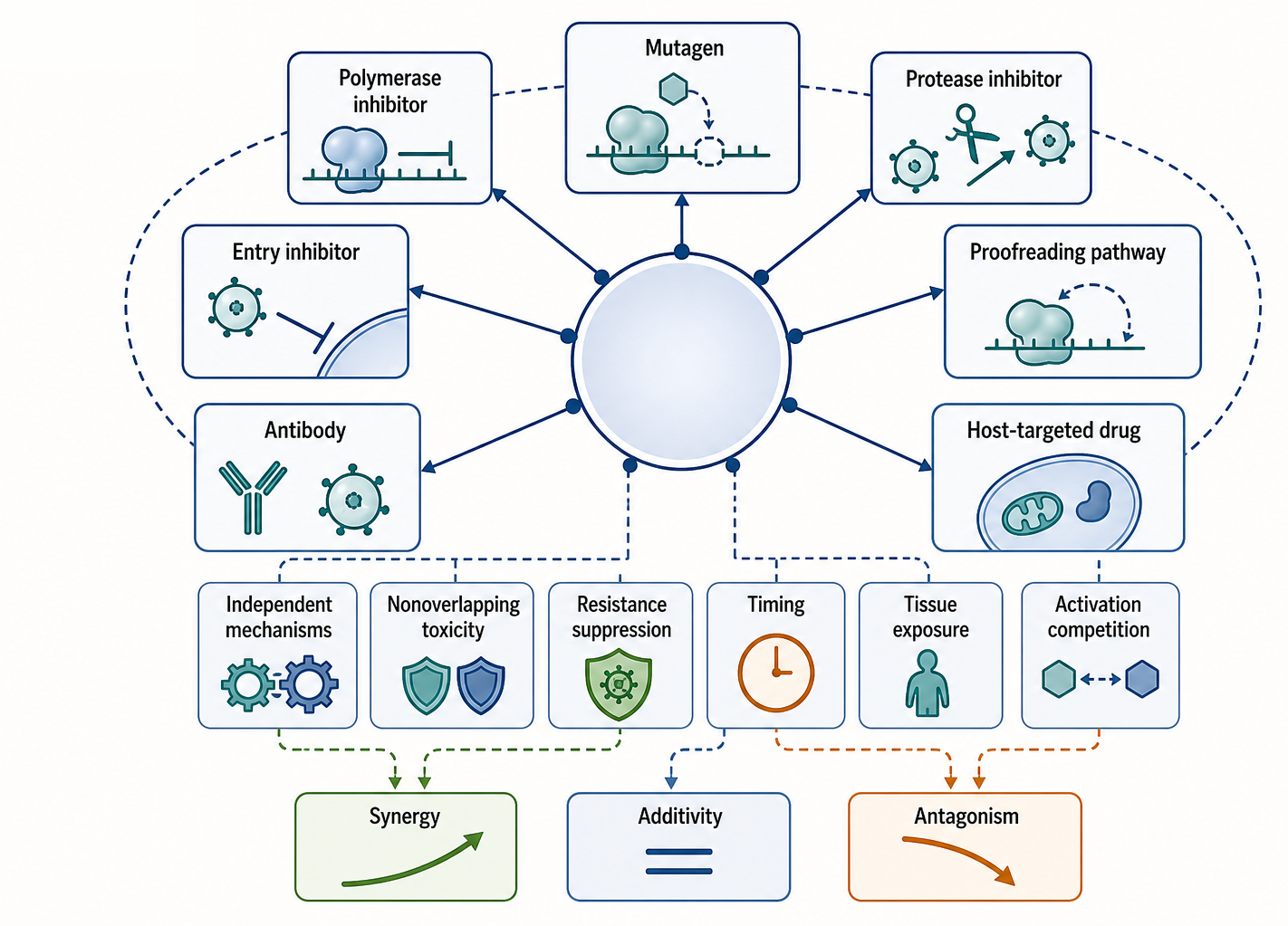
Figure 160.6. Future combination strategy matrix. Combination strategies should pair independent antiviral mechanisms, nonoverlapping toxicities, and resistance-suppressing fitness costs. Activation competition, timing, and tissue exposure can convert expected synergy into additivity or antagonism.
Combination design should be mechanism-based rather than simply additive. If two drugs compete for the same activation kinase, one may reduce the other’s active metabolite. If two drugs stress mitochondrial nucleotide metabolism, toxicity can become limiting. If one drug lowers viral replication too rapidly, it may reduce activation of an immune partner or alter biomarker interpretation. If one drug selects for high-fidelity polymerase variants, it may change susceptibility to a mutagenic partner. Dose-response matrices, time-of-addition studies, resistance selection, and animal models can reveal synergy, additivity, antagonism, or toxicity.
Future analog discovery will also benefit from better polymerase structural dynamics. Static structures are valuable, but analog action often depends on transient active-site closure, translocation, backtracking, pausing, and proofreading handoff. Cryo-electron microscopy, time-resolved structural approaches, single-molecule assays, molecular dynamics, and high-throughput biochemical profiling can map these states. The goal is not only to see where an analog binds, but to understand how the replication complex moves after incorporation.
Sequencing technologies will refine lethal-mutagenesis evaluation. High-accuracy viral population sequencing can distinguish true analog-induced mutations from sequencing noise, bottlenecks, and selection artifacts. Linked-read or long-read approaches can measure mutation combinations on the same genome, which matters because viability depends on haplotypes, not only per-site mutation rates. Time-resolved sequencing can show whether mutations accumulate before infectivity falls. These methods will make “error catastrophe” claims more precise and may identify resistance before clinical failure.
Host safety science must develop alongside antiviral potency. Mitochondrial assays, genotoxicity assays, stem-cell and developmental models, tissue organoids, and human genetic diversity panels can identify risks earlier. For mutagenic analogs, reproductive and developmental boundaries should be addressed explicitly before broad deployment. For nucleotide prodrugs, metabolite liabilities and off-target enzyme interactions should be mapped. For broad-spectrum preparedness drugs, safety expectations may be especially high because they could be stockpiled or deployed quickly during outbreaks.
Clinical trial strategy is part of mechanism. A polymerase inhibitor for an acute respiratory virus may fail in a hospitalized late-disease trial yet work in early outpatient disease. A mutagenic analog may need viral sequencing endpoints, not only symptom endpoints, to confirm mechanism. A combination may require resistance endpoints even if short-term viral load improves. Immunocompromised patients with prolonged viral replication can reveal resistance and persistent infection dynamics not seen in ordinary acute infection. Trial design should match the biological window of the polymerase pathway.
The most likely future is not one universal nucleoside analog for all RNA viruses. A more realistic goal is a portfolio: optimized disease-specific drugs, broad-spectrum candidates with known activation and safety profiles, rapid-screening systems for emerging polymerases, combination rules grounded in mechanism, and surveillance for resistance. The same chemical class can serve outbreak response, chronic infection treatment, post-exposure prophylaxis, and combination cure strategies, but each use has different constraints.
Consensus is strong on several points. Active triphosphate formation is central for most ribonucleoside analog RdRP inhibitors. Viral polymerase selectivity is necessary but insufficient without favorable host metabolism and tissue exposure. Chain termination, delayed termination, stalling, and mutagenesis are distinct mechanisms that should not be conflated. Viral proofreading and resistance can substantially alter susceptibility. Host toxicity, especially mitochondrial and genotoxicity risk, must be evaluated mechanistically rather than assumed from class labels.
Open questions remain. Which chemical features best allow analogs to evade coronavirus proofreading while preserving host selectivity? How can respiratory-tissue activation be measured predictively in humans? Which mutation-spectrum endpoints best predict clinical benefit for mutagenic analogs? Can proofreading-pathway inhibitors be made selective enough for combination therapy? How should broad-spectrum candidates be prioritized before an outbreak identifies the target virus? Which organoid, animal, or human challenge systems best forecast tissue-specific activation and safety?
Several overgeneralizations should be retired. Nucleoside analogs are not all chain terminators. Broad-spectrum cell-culture activity is not the same as clinical broad-spectrum utility. Mutagenic antivirals do not automatically cause host genetic damage, but the risk cannot be dismissed without data. A polymerase resistance mutation is not clinically meaningful until susceptibility, fitness, transmission, and treatment context are measured. A prodrug group is not merely a delivery label; it is a major determinant of where, when, and how much active inhibitor is produced.
Recent Consensus
The current mechanistic consensus is that antiviral ribonucleoside and nucleotide analogs should be described by activation pathway, active metabolite, polymerase interaction, viral context, and host safety profile. The older shorthand of “nucleoside analog equals chain terminator” is inadequate for modern RNA virus pharmacology. RdRP inhibitors can terminate, delay termination, stall translocation, induce mutagenesis, alter nucleotide pools, or combine several effects.
There is also consensus that local tissue pharmacology matters. Plasma exposure alone is not enough to infer antiviral activity. Intracellular active triphosphate levels in relevant infected cells are often the better mechanistic exposure metric, although they can be difficult to measure in patients. Resistance, proofreading, mitochondrial toxicity, and genotoxicity concerns should be addressed as mechanism-linked issues rather than afterthoughts.
Open Questions, Controversies, Deprecated Models, and Common Misconceptions
Open questions:
- How to predict tissue-specific activation from preclinical systems?
- How to design analogs that remain active against proofreading viruses?
- How to interpret low-frequency mutagenesis signals in clinical samples?
- How to combine mutagenic and terminating drugs without antagonism?
- The best regulatory framework for broad-spectrum antivirals intended for future outbreaks when definitive efficacy data may be hard to obtain before emergency use
Deprecated or weakened claims:
- Deprecated or oversimplified models include the idea that all antiviral nucleoside analogs act by immediate chain termination, that cell-culture potency is a stable proxy for clinical potency, and that a mutagenic mechanism automatically implies unacceptable host genotoxicity. Each statement contains a recognizable concern, but each becomes misleading when stripped of activation, exposure, enzyme selectivity, duration, and evidence context.