Chapter 76. Developmental and Cell-Type Programs of Local Translation
Scope Note
This chapter compares how localized messenger RNAs are stored, activated, translated, and cleared in developmental and cell-type-specific programs. It owns maternal RNA activation and embryonic patterning, migration and polarity, regeneration, activity-dependent local translation, comparative neuronal and glial cases, and evidence standards that distinguish spatial protein synthesis from RNA enrichment, transported protein, or contamination. General cis-signal, RBP, motor, anchoring, and remodeling grammar belongs in Chapter 74; membrane and organelle targeting belongs in Chapter 75; and full neural/glial systems, disease, and circuit integration belongs in Chapter 103.
Executive Summary
Local translation is protein synthesis from an RNA template at a restricted subcellular site. In a neuron, the site may be a dendritic spine, a presynaptic terminal, an axonal growth cone, or an injured axon segment. In a glial cell, the site may be an astrocyte process near a synapse or an oligodendrocyte process near a myelin sheath. In an embryo, the site may be a region of an oocyte or early embryo where a maternal mRNA is stored and later activated. In a migrating or polarized cell, the site may be a leading edge, basal or apical domain, protrusion, adhesion, or cleavage-plane-associated region. The common logic is that cells use RNA localization and translational control to place newly synthesized proteins near the structures that need them. Local translation is useful because distance, timing, and specificity matter. Neurons can be centimeters or longer in vertebrates, so waiting for every protein to be synthesized in the soma and transported as protein can be too slow or imprecise for synaptic plasticity, axon guidance, mitochondrial maintenance, or injury responses. Oocytes and early embryos often begin development before widespread zygotic transcription, so maternal RNAs must be stored, masked, positioned, and activated in a spatially and temporally ordered way. Migrating cells and polarized epithelia continually remodel their edges, adhesions, membranes, and cytoskeleton; local translation can deliver actin regulators, signaling proteins, receptors, or scaffold proteins close to the relevant cellular domain.
Mechanistically, local translation depends on a messenger ribonucleoprotein particle, or mRNP, changing from a localized or stored state to a translation-competent state. The general transport and remodeling machinery is introduced only as a prerequisite bridge to Chapter 74. This chapter asks how developmental timing, cell identity, injury, polarity, and activity determine when repression is relieved, poly(A)-tail or initiation state changes, ribosomes engage, locally synthesized protein acts, and the transcript returns to storage or decay.
The strongest evidence for local translation combines several measurements. RNA localization shows that the template is present. Ribosome association or nascent-peptide detection shows that translation occurs. Spatial isolation or imaging shows where translation occurs. Perturbation shows which RNA element, RNA-binding protein, motor, or signaling pathway is necessary. Rescue shows whether local synthesis, rather than global protein abundance, explains the phenotype. No single method is sufficient. Puromycin labeling can diffuse or report stress; ribosome profiling can lose spatial information during lysis; metabolic labeling has limited spatial resolution; reporter tags can perturb RNA localization or translation; and purified neurite, synapse, or glial-process fractions can carry somatic, nuclear, mitochondrial, or ambient RNA contamination.
The local bibliography provides useful anchors for neuronal and glial local translation and for maternal translation dynamics, including Das et al. (2021), Gala et al. (2023), Biever et al. (2020), Dai et al. (2025), and Xiong et al. (2022). It remains weaker for classic localized maternal determinants, embryonic-axis patterning, and direct migration/polarity mechanisms. Those topics are marked for verified source expansion rather than supported with unrelated citations.
Concept Inventory
- Local translation: spatially restricted protein synthesis from an mRNA template. It requires evidence for translation at the site, not merely RNA enrichment or protein enrichment.
- Synaptic translation: local translation at or near synapses, including dendritic spines, dendritic shafts, presynaptic terminals, and perisynaptic glial processes.
- Axonal translation: protein synthesis from mRNAs within axons. It supports growth cones, injury responses, mitochondrial function, cytoskeletal remodeling, and axon survival.
- Glial local translation: local protein synthesis in astrocytic or oligodendrocytic processes. The term should not be restricted to neurons, because glial cells also extend specialized processes far from the nucleus.
- Maternal mRNA: an mRNA deposited into an oocyte by the mother before fertilization or early embryogenesis. Maternal mRNAs can be stored in a repressed state and activated by cytoplasmic polyadenylation, localization, or developmental signals.
- Translational masking: repression that keeps a stored or transported mRNA from being translated prematurely.
- Cytoplasmic polyadenylation: extension of a cytoplasmic poly(A) tail after nuclear export. In oocytes, embryos, and neurons it can activate translation by improving poly(A)-binding protein recruitment and initiation-factor coupling.
- Growth cone: motile structure at the tip of a developing or regenerating axon that senses guidance cues and remodels cytoskeleton locally.
- Synapse-to-nucleus communication: signaling from synaptic or neuritic compartments to the nucleus, often through calcium waves, kinase cascades, transcription factors, locally synthesized proteins, or retrograde transport.
- Contamination artifact: false evidence for local translation caused by mixing of compartments, adherent soma, broken membranes, ambient RNA, diffusion of labels, reporter overexpression, or stress-induced redistribution.
What to Know Before Reading This Chapter
The reader should know that translation requires ribosomes, mRNAs, charged tRNAs, initiation factors, elongation factors, termination factors, and energy. The reader should also know that eukaryotic mRNAs usually have a 5′ cap, a coding sequence, untranslated regions, and a poly(A) tail. These features are not only generic expression elements. In spatially complex cells, untranslated regions and associated RNA-binding proteins often determine where the mRNA travels, whether it is stored, and when ribosomes can initiate.
The chapter uses three recurring examples. The first is a dendritic mRNA whose translation is stimulated after synaptic activity. This example teaches how activity-dependent signaling can convert a stored transcript into a local protein source. The second is an axonal mRNA in a growth cone or injured axon. This example teaches why long-distance cell architecture makes local synthesis biologically useful. The third is a maternal mRNA in an oocyte or early embryo. This example teaches how translation can be delayed until the proper developmental time and place.
Several distinctions matter throughout the chapter. Local RNA enrichment is not local translation. Local protein enrichment is not local translation, because the protein might have been made elsewhere and transported. Ribosome association is not always productive translation, because stalled ribosomes, scanning complexes, or contaminating ribosomes can be captured. A developmental or neuronal phenotype after perturbing an RNA-binding protein is not automatically a local-translation phenotype, because the same protein may regulate splicing, export, stability, localization, stress granules, and global translation.
Core Mechanisms and Molecular Players
76.1. Comparative local-translation programs in neurons, glia, and polarized cells
Neurons made local translation unavoidable as a biological problem. A neuron can have a compact cell body, an elaborate dendritic tree, and an axon that extends far from the nucleus. Dendrites receive synaptic input, axons send output, growth cones interpret guidance signals, and presynaptic terminals release neurotransmitter. These compartments need proteins on different time scales. Some proteins can be synthesized in the soma and transported as proteins, but other responses require a local template and local ribosomes.
Dendritic local translation is closely tied to synaptic plasticity, the ability of synapses to change strength, shape, composition, or signaling state after activity. A dendritic mRNA can be transported in a repressed mRNP, held near a synapse, and activated by local calcium entry, kinase signaling, receptor stimulation, or neurotrophins. The causal chain is: synaptic activity creates a local signal; the signal modifies RNA-binding proteins or initiation factors; repression is relieved or initiation is enhanced; ribosomes synthesize protein near the synapse; the new protein changes receptor trafficking, actin remodeling, scaffold composition, or signaling feedback. This mechanism allows one branch or spine to respond without forcing the whole neuron to synthesize the same protein everywhere.
Axonal local translation solves related but distinct problems. During development, a growth cone must turn toward or away from guidance cues. A local cue can stimulate asymmetric translation of cytoskeletal regulators, membrane proteins, or signaling factors, biasing growth-cone movement. In mature or injured axons, local translation contributes to mitochondrial maintenance, stress responses, retrograde signaling, and regeneration. Axonal translation is especially sensitive to interpretation errors because axonal samples are often close to cell bodies, glia, extracellular debris, and severed fragments. A claim that an axon translates a specific mRNA is strongest when the experiment physically isolates axons, verifies absence of somatic and nuclear markers, detects the mRNA in the axon, detects translation in the axon, and shows a local phenotype after selectively perturbing axonal translation.
Synaptic local translation includes both postsynaptic and presynaptic settings. Postsynaptic dendritic spines have been the best studied because they are experimentally accessible and strongly linked to activity-dependent plasticity. Presynaptic translation is harder to study in many systems because terminals are small and close to other cellular material, but evidence supports local protein synthesis in selected presynaptic compartments. Biever et al. (2020) reported that monosomes can actively translate synaptic mRNAs in neuronal processes. That study is important pedagogically because it challenges the assumption that active translation at synapses must always appear as large polysomes. A single ribosome on a transcript can still make protein, especially for compartment-limited synthesis, although monosome-associated footprints must be interpreted with controls for nonproductive ribosome binding.
Glia extend the concept beyond neuron-centered models. Astrocytes send fine processes around synapses and blood vessels; oligodendrocytes extend processes that form myelin; microglia survey tissue with motile branches. Gala et al. (2023) reviewed glial mRNA localization in synaptic plasticity, emphasizing that glial processes contain localized RNAs and can influence synapse function. Glial local translation could supply proteins near synapses, myelin segments, endfeet, or injury sites without relying entirely on transport from the glial soma. The field is less mature than neuronal local translation, partly because glial processes are thin, intertwined with neuronal processes, and hard to purify without contamination. The correct conclusion is not that glial local translation is rare, but that many glial claims need stricter spatial and cell-type-specific evidence.
The comparative conclusion is that neural and glial compartments use the same broad principle—local templates answer spatially restricted demand—but differ in geometry, stimuli, transcript sets, and contamination risks. This chapter retains those contrasts as local-translation programs. Detailed neuron-glia systems integration, disease mechanisms, and circuit consequences belong in Chapter 103, where local translation can be evaluated alongside splicing, editing, transport, decay, and broader RNA metabolism.
76.2. Maternal RNA storage, activation, and embryonic patterning
Maternal mRNAs are transcripts produced during oogenesis and deposited into the egg before fertilization or early embryogenesis. They are essential because early embryos often undergo rapid divisions before robust zygotic transcription begins. During this period, the embryo uses maternal RNAs and proteins to control cell cycle progression, axis formation, germ-layer patterning, germ-cell specification, and the transition to zygotic gene expression. Maternal RNA regulation is therefore a temporal and spatial translation problem: which stored transcript is translated, where it is translated, and when it is destroyed.
Storage begins with translational masking. A maternal mRNA may be exported from the nucleus and packaged with RNA-binding proteins that prevent efficient initiation. Repression can involve short poly(A) tails, inhibitory proteins bound to 3′ UTR elements, closed-loop disruption, sequestration in RNP granules, or competition with activating factors. This is not a dead state. Stored mRNAs are poised molecules. They can be transported, anchored, remodeled, polyadenylated, translated, or degraded when fertilization, maturation, localization, or developmental timing cues arrive.
Cytoplasmic polyadenylation is a central activation mechanism in many oocytes and early embryos. The general causal sequence is: a cis-element in the 3′ UTR recruits a cytoplasmic polyadenylation regulatory complex; developmental signaling changes the activity or phosphorylation state of the complex; a cytoplasmic poly(A) polymerase extends the poly(A) tail; poly(A)-binding protein binds the longer tail; initiation-factor interactions improve; ribosome recruitment increases. The result is translational activation without new transcription. This logic connects maternal mRNA regulation to Chapter 29 on polyadenylation and Chapter 70 on translational control by mRNA features.
Embryonic patterning adds spatial control. A localized maternal mRNA can create a local source of protein that defines an embryonic region. Classic examples include anterior, posterior, vegetal, and germ-plasm-associated mRNAs in model organisms. The exact molecules and mechanisms differ among flies, frogs, fish, nematodes, mammals, and other animals. The general principle is that a maternal transcript can be transported to a region, anchored, held repressed during transit, and translated after localization. The protein product can then act as a morphogen, determinant, translation regulator, cytoskeletal regulator, or signaling factor. The local bibliography for this chapter does not include the classic primary literature for these examples, so detailed claims about particular maternal mRNAs should be treated as final bibliography items for later citation enrichment rather than citation-backed statements here.
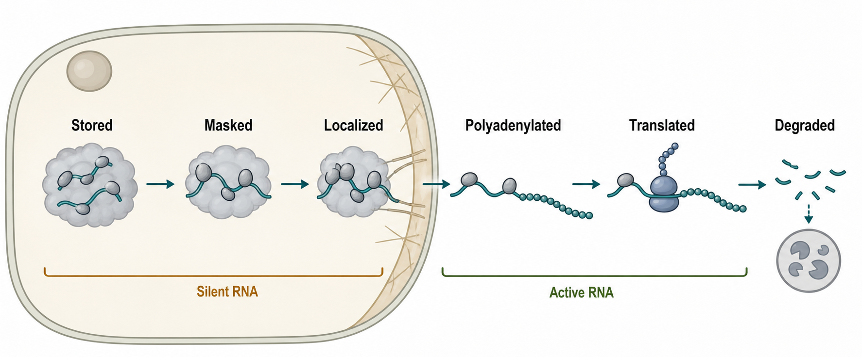
Figure 76.2. Maternal mRNA States During Early Development. A maternal mRNA moves through stored, masked, localized, cytoplasmically polyadenylated, translated, and degraded states during oocyte maturation and early embryogenesis. The figure should show that spatial localization and translational activation are separable steps.
Table 76.1. Biological Contexts of Local Translation. Local translation occurs in neuronal, glial, developmental, migratory, and polarized-cell contexts with different RNA states and outputs; evidence must connect the relevant compartment to nascent protein while controlling for transport and contamination.
| Context | Representative site | Likely RNA state | Biological output | Strongest evidence type | Major artifact risk |
|---|---|---|---|---|---|
| Dendritic spine | Postsynaptic spine or nearby dendritic shaft. | Repressed mRNP activated by calcium, kinase, receptor, or neurotrophin signaling. | Receptor trafficking, actin and scaffold remodeling, synaptic plasticity. | Endogenous RNA imaging plus local nascent-chain or ribosome evidence and spine-scale perturbation. | Stored RNA, transported mature protein, or perisynaptic glial signal mistaken for synthesis. |
| Axonal growth cone | Distal tip of developing axon. | Transported repressed mRNP activated by local guidance cue. | Asymmetric cytoskeletal remodeling, turning, membrane addition, cue response. | Microfluidic axon isolation with RNA imaging, local reporter or nascent-chain readout, and cue-dependent perturbation. | Soma or dendrite carryover, culture injury, glial material, reporter mislocalization. |
| Injured axon | Axotomy site or distal regenerating segment. | Injury-responsive mRNP, often unmasked or recruited after damage. | Wound sealing, retrograde injury signaling, mitochondrial support, regeneration. | Isolated axons with compartment markers, local nascent-protein labeling, inhibitor or rescue design. | Injury-induced global stress translation, severed-cell debris, glial contamination. |
| Astrocyte process | Perisynaptic fine process or vascular endfoot. | Localized glial mRNA; direct translation evidence is emerging and context-dependent. | Synaptic support, uptake or release machinery, plasticity modulation. | Cell-type-resolved RNA localization paired with glial ribosome or nascent-chain markers. | Neuronal RNA or protein contamination, extracellular-vesicle transfer, ambient RNA. |
| Oligodendrocyte process | Myelin-forming process or internode-adjacent region. | Process-localized mRNP linked to myelin or support functions; translation extent is context-dependent. | Myelin maintenance, local membrane remodeling, axon support. | Process imaging with oligodendrocyte markers and a translation-sensitive readout. | Axonal material, compact myelin preparation artifacts, mixed glial fractions. |
| Oocyte cortex | Animal, vegetal, or cortical region of the oocyte. | Stored maternal mRNP, often masked until poly(A)-tail extension or repressor release. | Local determinant or regulator production after maturation or fertilization. | RNA localization plus poly(A)-tail, ribosome, or nascent-protein timing. | RNA localization or protein gradient mistaken for synthesis site. |
| Embryonic patterning region | Anterior, posterior, vegetal, or germ-plasm-associated region. | Localized maternal transcript translated after positioning; local citations remain thin. | Axis, germline, or cell-fate patterning. | Timed imaging, translational activation assay, and localization-defective rescue. | Protein diffusion or stabilization, bulk embryo averaging, under-cited classic examples. |
| Migrating-cell leading edge | Protrusion or adhesion-rich cell front. | Protrusion-enriched mRNP activated by local signaling; local citations remain thin. | Actin dynamics, adhesion turnover, directional persistence. | Protrusion imaging or isolation plus localization-element perturbation. | Abundant mRNA carryover, global translation effects, fragile protrusion contamination. |
| Polarized epithelial domain | Apical or basolateral cortex. | Domain-enriched transcript whose translation must be separated from transport or stabilization. | Domain identity, membrane remodeling, cytoskeletal organization. | Domain-resolved RNA and nascent-synthesis readouts with polarity markers. | ER association, selective decay, cell geometry, or transported protein mistaken for local synthesis. |
Maternal RNA interpretation requires special artifact control. First, RNA localization in an oocyte does not prove local translation, because many maternal RNAs are deliberately stored in silent states. Second, maternal protein gradients do not prove local translation, because proteins can diffuse, be transported, or be locally stabilized. Third, early embryo phenotypes after perturbing an RBP may reflect altered oocyte development, RNA stability, cytoplasmic organization, fertilization competence, or zygotic genome activation rather than direct local translation. Fourth, bulk embryo measurements can average distinct blastomeres, cortical regions, germ plasm, yolk-associated material, and somatic compartments.
The strongest maternal RNA evidence combines localization imaging, poly(A)-tail measurement, ribosome or nascent-protein evidence, timed perturbation, and rescue. For example, a strong study would show that a specific 3′ UTR element localizes a maternal mRNA, that the mRNA remains translationally repressed before localization, that cytoplasmic polyadenylation or repressor release activates translation at the correct developmental stage, and that a localization-defective but protein-coding-competent RNA fails to rescue patterning while a localized construct succeeds. Without such separation, it is easy to confuse local translation with RNA localization, local protein accumulation, or general developmental competence.
The references.md file lists several embryo-related automated hits, but most are not direct anchors for maternal mRNA storage and localized translation. Sahin et al. (2023) concerns embryonic arrest; Wang et al. (2023) concerns maternal and embryonic signals in uterine epithelial receptivity; Wu et al. (2025) concerns a sperm-derived circRNA and zygotic genome activation. These may be useful for adjacent developmental RNA topics, but they do not replace verified references on canonical maternal mRNA localization, cytoplasmic polyadenylation, and patterning. This chapter therefore marks maternal RNA bibliography as reference_support_limited.
76.3. Local translation in migration, regeneration, and cell polarity
Migrating cells are polarized cells. They have a front that protrudes, senses signals, and forms adhesions; a rear that contracts and detaches; and internal trafficking routes that deliver membranes, receptors, cytoskeletal regulators, and signaling proteins. Local translation can reinforce this polarity by producing proteins near the leading edge or protrusion instead of distributing newly synthesized protein throughout the cytoplasm. The main object is not one universal “migration mRNA.” It is a set of transcripts whose local synthesis can tune actin assembly, adhesion turnover, membrane remodeling, or signal transduction.
The causal chain in a migrating cell resembles the neuronal chain on a shorter length scale. An mRNA contains localization or regulatory elements. RNA-binding proteins package the transcript and repress translation during transport or storage. Cytoskeletal motors, actin-associated factors, or membrane trafficking bring the mRNP near a protrusion or adhesion. Local signaling activates translation. Newly synthesized protein changes protrusion persistence, adhesion strength, actin branching, contractility, or directional sensing. Because migration is mechanically dynamic, the relevant time scale can be minutes rather than hours.
Cell polarity is broader than migration. Epithelial cells have apical and basolateral domains; stem cells and early embryos can divide asymmetrically; immune cells polarize toward an antigen-presenting cell or target; neurons maintain axonal and dendritic identity; oligodendrocytes polarize toward myelin segments. Local translation can help maintain these domains, but the mechanism must be shown for each system. A transcript enriched near an apical domain might be there because of directed transport, local anchoring, local stabilization, ER association, selective decay elsewhere, or geometry of cytoplasm. Local translation is one possible output of localization, not an automatic consequence.
Regenerating axons provide a particularly clear reason for local translation. After injury, distal axon segments need proteins for wound sealing, cytoskeletal remodeling, stress signaling, retrograde injury signaling, mitochondrial support, and growth. Some responses occur far from the soma, so local translation can provide proteins before new transcriptional responses arrive. A locally synthesized protein can also enter retrograde transport complexes and help communicate injury state to the nucleus. Fernandopulle et al. (2021) discuss disease and injury contexts where RNA transport and local translation contribute to neuronal maintenance and pathology.
The local bibliography for migration is weak. Song et al. (2025) is a broad review of cell migration in diabetic wound healing and may help with general migration biology, but it is not a strong citation for RNA localization or local translation mechanisms. Camand et al. (2012) concerns N-cadherin effects on glial cell migration, but the listed metadata does not make it a direct local-translation reference. For this reason, mechanistic claims in this section are presented as synthesis from the established local-translation framework and should receive dedicated migration-local-translation citations in a later bibliography pass.
Experimental design in migration and polarity studies must avoid three common confusions. First, protrusions are fragile and easy to contaminate with cell-body material during isolation. Second, mRNAs encoding cytoskeletal or adhesion proteins are often abundant, so their presence at a leading edge may reflect abundance rather than selective localization. Third, inhibiting translation globally can impair migration by reducing energy, stress tolerance, or protein homeostasis, not specifically by blocking local synthesis. The most informative experiments perturb a localization element or local activation mechanism while preserving overall mRNA and protein expression.
76.4. Activity-dependent regulation and synapse-to-nucleus communication
Activity-dependent regulation means that neuronal activity changes RNA localization, translation, stability, or RNP composition. At an excitatory synapse, receptor activation can allow calcium entry, activate kinases and phosphatases, remodel the actin cytoskeleton, change membrane trafficking, and alter local translation. Inhibitory synapses, neuromodulatory inputs, glial signals, and neurotrophins can also influence local translation. The key point is that translation responds to local signaling, not only to global nutrient or stress pathways.
At a dendritic synapse, a simplified causal sequence is useful. First, synaptic activity creates a local biochemical signal. Second, that signal modifies proteins bound to mRNAs or translation factors nearby. Third, selected mRNAs become more or less available to ribosomes. Fourth, newly synthesized proteins alter the synapse or surrounding dendrite. Fifth, feedback from translation products, decay pathways, or transport changes resets the local state. This sequence can support long-lasting synaptic changes because translation converts a transient signal into new molecular material.
Local translation also contributes to synapse-to-nucleus communication. A synapse can signal to the nucleus through calcium waves, kinase cascades, transcription-factor movement, endosome-based retrograde signaling, locally synthesized proteins, or changes in soma-directed transport. Wild et al. (2019) reported synapse-to-nucleus communication through NFAT mediated by L-type calcium channel calcium spike propagation to the soma. That paper is not specifically a local-translation paper, but it is relevant because it shows that activity signals can travel from synaptic regions to nuclear transcription systems. Local translation can intersect with such pathways when a locally synthesized protein stabilizes a retrograde signal, modulates calcium handling, changes transport complexes, or produces a factor that later enters the nucleus.
Synapse-to-nucleus logic is important because local translation is not only a local endpoint. A dendrite or axon may use local synthesis to decide which signals are sent back to the soma. In an injured axon, locally translated signaling proteins can become part of retrograde injury-signaling complexes. In a stimulated dendrite, local translation can alter the strength or duration of transcriptional responses. In glia, local translation near synapses may tune glial release, uptake, metabolism, or structural support, thereby changing neuronal activity that eventually affects nuclear programs in neurons or glia.
The boundary case is that activity-dependent translation is not always spatially local. Neuronal activity can alter global initiation, stress pathways, mTOR signaling, integrated stress response pathways, and transcription. A rise in translation after stimulation does not prove that the translation occurred at synapses. Conversely, a local synaptic translation event may be too small to dominate whole-cell measurements. Strong experiments therefore match the scale of the question: spine or synapse imaging for local events, isolated neurites for compartment events, and whole-cell or nuclear assays for downstream transcriptional responses.
Another caution concerns ribosome state. Biever et al. (2020) supports active translation by monosomes in neuronal processes, but ribosome footprints or monosome association must be interpreted with care. A monosome can be a productive translating ribosome, a stalled ribosome, a scanning or initiating complex, or an artifact of sample preparation depending on the assay. Combining ribosome association with nascent-chain detection, inhibitor sensitivity, codon phasing, and local perturbation helps separate productive translation from ribosome occupancy.
This section owns the local-translation program and the distinction between activity-triggered synthesis and retrograde signaling. Chapter 103 integrates these events with neuronal and glial RNA processing, editing, transport, disease, and circuit-level physiology.
76.5. Evidence standards for developmental and cell-type local translation
A local translation claim asks a demanding question: where in the cell was a protein molecule synthesized from a particular mRNA? Methods usually answer pieces of that question rather than the entire question. RNA imaging shows where the template is. Ribosome profiling shows where ribosomes protected fragments, but usually after homogenization. Nascent-chain reporters show where new peptide appears, but reporters can change the RNA or protein. Puromycin-based methods label newly synthesized peptides, but the label can diffuse and stress the cell. Metabolic amino-acid labeling measures new protein synthesis, but spatial assignment can be limited. Fractionation enriches compartments, but contamination can dominate low-abundance signals.
The first evidence axis is template and synthesis identity. RNA imaging or compartment enrichment establishes that a candidate template is present, whereas nascent-chain reporters, metabolic labeling, puromycylation, or ribosome-state assays address synthesis with different degrees of transcript specificity. A strong conclusion links the local RNA to its nascent product and shows translation dependence. Instrument-specific tagging and imaging mechanics are treated in Chapter 74 and Chapter 135; this chapter focuses on whether the chosen combination resolves the biological program.
The second axis is spatial and cell-type attribution. Neuronal, glial, embryonic, protrusion, and regenerating compartments have different contamination structures. Synaptic material can mix pre- and postsynaptic membranes with glial processes; axon preparations can include soma, dendrite, debris, or injury-induced responses; embryo regions can average distinct blastomeres or cortical domains. Compartment markers, microscopy, negative markers, spike-ins, and cell-type-resolved ribosome or nascent-chain signals should match the scale of the claim.
The third axis is causality. A local-translation program is strengthened when a localization or activation element, cell-state signal, or translation regulator is perturbed without simply eliminating total RNA or global protein synthesis. Rescue should distinguish correct local synthesis from restored whole-cell abundance. The phenotype should also be spatially appropriate: turning, regeneration, polarity, patterning, or synaptic remodeling should be connected to synthesis in the relevant compartment.
Table 76.3. Distinguishing Local Protein Sources. Protein detected in a subcellular region can arise from local translation, transport, stabilization, secretion, uptake, or sample contamination; source-specific perturbations are needed to distinguish these alternatives.
| Possible source of local protein | Expected evidence | Decisive perturbation |
|---|---|---|
| Local translation | Local mRNA, ribosome or nascent-chain signal, translation-sensitive protein appearance, and local phenotype coincide. | Disrupt the localization element or local initiation at the site, then rescue with a construct restoring local synthesis rather than only total protein. |
| Transported mature protein | Protein appears locally without matching mRNA or nascent-chain signal and depends on soma or cell-body transport. | Block soma-to-site transport or mark old versus newly made protein; local translation inhibition should not remove already delivered protein. |
| Local stabilization | Protein half-life is longer at the site without evidence for local synthesis. | Alter local anchoring, degradation, or turnover while leaving mRNA translation unchanged. |
| Local secretion or uptake | Protein tracks extracellular, donor-cell, receptor, or secretory-pathway markers. | Block secretion or uptake, or remove donor-cell production, while testing recipient local translation separately. |
| Extracellular vesicle transfer | Vesicle markers carry RNA, ribosomal proteins, or mature protein from donor cells to the local region. | Inhibit vesicle release or uptake and use donor-specific cargo labels to distinguish transfer from recipient synthesis. |
| Contamination during preparation | Signal covaries with nuclear, somatic, glial, mitochondrial, ER, or ambient RNA markers. | Improve purification, reject contaminated fractions, and validate the signal by in situ imaging with negative markers. |
Evidence should be graded rather than declared binary. Template localization alone supports availability; ribosome occupancy supports possible engagement; nascent-chain evidence supports synthesis; local perturbation supports causality; rescue and a compartment-specific phenotype connect synthesis to function. The evidence-ladder figure and local-protein-source table summarize these levels without repeating general imaging or transport tutorials.
Experimental Foundations and Evidence
The strongest foundation for this chapter comes from convergent methods rather than a single technique. Reviews by Das et al. (2021) and Fernandopulle et al. (2021) support the broad consensus that mRNA transport and local translation are central to neuronal and disease biology. Biever et al. (2020) provides direct evidence that monosomes can actively translate synaptic mRNAs in neuronal processes. Gala et al. (2023) extends the field into glial processes and synaptic plasticity. Wild et al. (2019) anchors synapse-to-nucleus communication as an activity-dependent signaling problem that local translation can intersect with, although it should not be cited as direct proof of local translation.
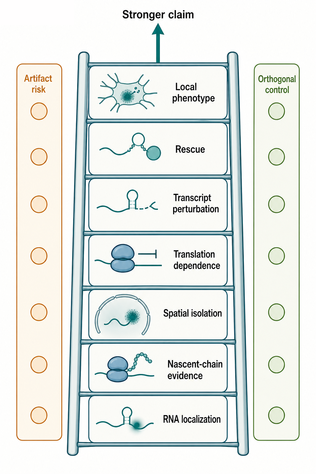
Figure 76.3. Evidence Ladder for Local Translation. Evidence for local translation becomes stronger as studies combine template localization, ribosome or nascent-chain evidence, spatial isolation, translation dependence, transcript-specific perturbation, rescue, and local phenotype. Each rung should list common artifacts and the best complementary control.
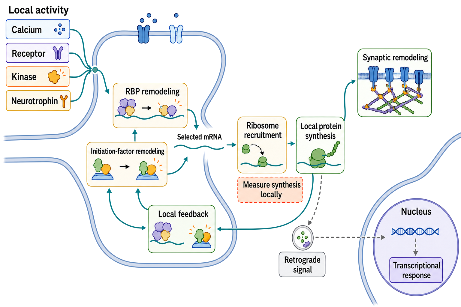
Figure 76.5. Activity-Dependent Local Translation from Synaptic Signal to Local and Nuclear Response. Calcium entry, receptor activation, kinase signaling, and neurotrophin input can remodel RNA-binding proteins and initiation factors at a synapse, making selected local mRNAs available for ribosome recruitment and protein synthesis. Newly synthesized proteins can remodel the synapse, reset the local RNA state through feedback, or contribute to a retrograde signal that changes nuclear transcription; these routes intersect but are not equivalent.
Evidence remains uneven across contexts. Neuronal dendrites and axons have comparatively rich evidence. Glial local translation is increasingly supported but methodologically difficult. Maternal RNA storage and activation are central to developmental biology, but the chapter-specific bibliography currently lacks the classic references needed for detailed citation-backed treatment. Migration and polarity have strong conceptual links to local translation, but the local reference list provides only weak direct support.
Biological Contexts Across Systems
Neurons use local translation for plasticity, axon guidance, presynaptic and postsynaptic remodeling, injury responses, and long-distance maintenance. Glia likely use local translation to support synapses, myelin, vascular contacts, and injury responses, but the compartmental evidence must distinguish glial from neuronal material. Oocytes and embryos use stored maternal RNAs to control development before zygotic transcription fully takes over. Migrating and polarized cells use local translation to reinforce asymmetry, but the field must separate local synthesis from local protein transport and local stabilization.
Across all systems, local translation should be treated as a state program: a transcript can be localized, repressed, activated, translated, returned to storage, or degraded in response to developmental stage and cell context. General transport and anchoring machinery is treated in Chapter 74.
Technology, Computational, Clinical, and Engineering Links
Technology links include neurite microfluidics, live RNA imaging, endogenous tagging, spatial proteomics, ribosome profiling, proximity labeling, and spatial transcriptomics. Computational links include prediction of localization motifs, classification of compartment-enriched transcripts, and correction of contamination. Clinical links include neurodevelopmental disorders, neurodegeneration, axon injury, demyelinating disease, infertility, embryonic arrest, and wound repair. Engineering links include synthetic 3′ UTRs, localization elements, optogenetic control of translation, and reporter systems that test local protein synthesis.
The clinical boundary is important. A disease-associated RBP or translation regulator does not prove a local-translation mechanism. This chapter supplies the spatial evidence standard; Chapter 103 owns integration with neural and glial disease mechanisms, while developmental and regenerative disease contexts should likewise be evaluated against their whole-system chapters.
Recent Consensus
The current consensus is that local translation is a widespread strategy in spatially complex cells and embryos, not a rare neuronal curiosity. Dendrites, axons, glial processes, oocytes, early embryos, protrusions, and regenerating compartments can all use localized mRNAs as protein templates. The strongest consensus exists for neuronal mRNA transport and local translation, supported by reviews and direct studies such as Das et al. (2021), Fernandopulle et al. (2021), and Biever et al. (2020).
A second consensus is that local translation is inseparable from RNA localization, RNP remodeling, translation repression, activation signals, and RNA decay. A transcript is not simply delivered to a site and translated automatically. It is stored, inspected, remodeled, and often coupled to signaling pathways.
A third consensus is methodological: local translation claims require orthogonal evidence. RNA position, ribosome occupancy, nascent protein synthesis, perturbation, rescue, and contamination controls answer different parts of the claim.
Open Questions, Controversies, Deprecated Models, and Common Misconceptions
Open questions:
- How many localized transcripts are translated under basal conditions versus stored for rare stimuli? This distinction matters especially in synapses, glial processes, and oocytes. A localized RNA may be a ready template rather than an actively translated template.
- How general glial local translation is? Glial processes are biologically suited for local translation, and evidence is growing, but the field needs more cell-type-resolved, compartment-resolved translation assays.
- How much local translation contributes to disease compared with global RNA metabolism defects? Many disease-linked RBPs regulate multiple steps in RNA metabolism, so local translation may be one output among several.
Common misconceptions:
- “RNA enrichment at a synapse proves local translation.” The transcript may be stored, stalled, decaying, or merely nearby.
- “Local protein enrichment proves local synthesis.” The protein may have been synthesized elsewhere and transported or locally stabilized.
- “A purified axon, synapse, or glial-process fraction is automatically clean.” Contamination from soma, glia, nuclei, mitochondria, ER, ambient RNA, and broken processes must be measured.
- “Maternal mRNA localization always implies immediate translation.” Many maternal RNAs are localized while translationally masked and become active only after developmental cues.