Chapter 72. Native mRNA Grammar: Cap, UTRs, CDS, Poly(A), Structure, and RBP Code
Scope Note
Messenger RNA is often introduced as a linear carrier of protein-coding information, but a mature endogenous mRNA is better understood as an integrated regulatory molecule. The 5′ cap, 5′ untranslated region (5′ UTR), coding sequence (CDS), 3′ untranslated region (3′ UTR), poly(A) tail, RNA structure, covalent modifications, and RNA-binding protein (RBP) occupancy jointly determine where an mRNA goes, when ribosomes initiate, how translation proceeds, how long the transcript persists, and which surveillance pathways inspect it. This chapter owns the native whole-transcript grammar that connects those features and is the general mechanistic owner of CPEB-directed cytoplasmic polyadenylation and translational activation. Chapter 29 owns nuclear cleavage, initial polyadenylation, and tailing-enzyme comparison; Chapter 66 owns decoding and tRNA systems; Chapter 36 owns causal codon-optimality-mediated decay; and Section 153.3 in Chapter 153 owns therapeutic codon engineering.
Executive Summary
An mRNA molecule integrates sequence, chemistry, structure, and protein occupancy across its entire length. The 5′ cap recruits cap-binding proteins, protects the 5′ end from exonucleases, and helps license translation initiation. The poly(A) tail and its poly(A)-binding proteins stabilize many cytoplasmic mRNAs and communicate with initiation factors, although the classical closed-loop picture is an oversimplified snapshot rather than a universal physical state. The 5′ UTR acts as an initiation control region whose length, structure, upstream open reading frames, start-codon context, internal ribosome entry elements, and protein-binding sites influence how scanning preinitiation complexes reach a start codon. The CDS encodes protein sequence while also encoding codon usage, codon-pair context, local RNA structure, elongation kinetics, co-translational folding opportunities, and decay signals. The 3′ UTR carries recognition sites for RBPs and microRNAs, localization elements, stability determinants, and alternative polyadenylation outputs that can change mRNA behavior without changing the protein sequence. The 3′ end can also be remodeled after an mRNA has entered the cytoplasm. In cytoplasmic polyadenylation, cis elements in a 3′ UTR recruit cytoplasmic polyadenylation element-binding proteins (CPEBs) and associated factors that switch selected mRNAs between a short-tailed, translationally repressed state and a longer-tailed, translation-competent state. The best-defined example is the staged activation of dormant maternal mRNAs during Xenopus oocyte maturation. In that system, signaling-dependent CPEB phosphorylation and mRNP remodeling shift the balance away from PARN or other deadenylases and toward a noncanonical poly(A) polymerase such as TENT2/GLD2. Tail extension promotes poly(A)-binding protein recruitment and productive cap-associated initiation. The component list, kinase input, and relationship between tail length and translation vary across organisms and cell states; the mechanism is therefore a regulated mRNP transition, not a universal rule that longer tails always translate better.
The mature mRNA is also a ribonucleoprotein particle, or mRNP. Bound proteins and RNA modifications provide identity marks that record aspects of the transcript’s history and condition: nuclear processing, export, localization, translational engagement, stress response, and decay competence. N6-methyladenosine (m6A), cap-adjacent N6,2′-O-dimethyladenosine (m6Am), pseudouridine, 5-methylcytidine, inosine, and other marks can affect RNA structure, reader-protein recruitment, translation, localization, or stability, but modification studies require careful controls because antibody bias, stoichiometry uncertainty, and indirect perturbations can produce misleading claims. Predictive models can combine sequence, structure, isoform choice, UTR features, codon patterns, RBP motifs, modifications, and cell-state measurements to estimate translation, localization, and half-life. Such models remain context-dependent because native mRNA fate emerges from coupled molecular processes rather than a single universal code.
Concept Inventory
- 5′ cap: A modified nucleotide structure attached to the 5′ end of many eukaryotic and viral mRNAs. The canonical cap0 contains 7-methylguanosine linked by a 5′ to 5′ triphosphate bridge; cap1 and cap2 include 2′-O-methylation on the first and second transcribed nucleotides. Cap structures affect stability, translation, processing, export, immune recognition, and decapping.
- 5′ UTR: The transcribed region between the cap or transcription start site and the main coding sequence start codon. The 5′ UTR can regulate translation initiation through scanning distance, secondary structure, upstream open reading frames, upstream start codons, binding motifs, and specialized initiation mechanisms.
- Coding sequence: The region translated into a polypeptide. The CDS also contains regulatory information through codon choice, codon-pair patterns, RNA structure, ribosome pause sites, nascent peptide effects, and embedded protein- or RNA-binding motifs.
- 3′ UTR: The region between the stop codon and the poly(A) tail or transcript end. The 3′ UTR commonly contains binding sites for RBPs and miRNAs, localization elements, stability signals, and alternative polyadenylation outputs.
- Poly(A) tail: A tract of adenosines added to most nuclear-encoded eukaryotic mRNAs after cleavage of the pre-mRNA 3′ end. Tail length and associated proteins influence stability and translation, with strongest effects in early development and selected regulatory contexts.
- Cytoplasmic polyadenylation: Regulated extension of an existing mRNA poly(A) tail in the cytoplasm, usually after nuclear cleavage and initial polyadenylation have already occurred. In the best-defined developmental systems, this remodeling activates selected stored mRNAs for translation.
- Cytoplasmic polyadenylation element (CPE): A U-rich 3′-UTR cis element whose activity depends on sequence, number, spacing, position relative to the polyadenylation signal, developmental stage, and the CPEB-family factors present. A CPE-like motif alone does not establish regulation.
- CPEB: A member of the cytoplasmic polyadenylation element-binding protein family. CPEBs are RNA-binding proteins that connect selected 3′-UTR elements to translational repression, poly(A)-tail remodeling, localization, or activation; paralogs are not functionally interchangeable.
- TENT2/GLD2: A noncanonical terminal nucleotidyltransferase that can act as a cytoplasmic poly(A) polymerase when recruited to RNA by specificity factors. GLD2-family composition and target selection differ among animal lineages and cell types.
- PARN-polymerase balance: The regulated competition or sequential action of poly(A)-specific ribonuclease (PARN), other deadenylases, and cytoplasmic poly(A) polymerases on a selected mRNP. It is a mechanistic balance, not a universal stoichiometric switch.
- RBP code: A shorthand for the combinatorial occupancy of RNA-binding proteins across an mRNA. The term is useful only if treated as probabilistic and context-dependent; protein binding does not automatically imply regulation.
- CDS codon pattern: The arrangement of synonymous codons and codon pairs within a native coding sequence. Codon patterns covary with GC content, RNA structure, RBP or miRNA motifs, modification motifs, and nascent-peptide context. Chapter 66 explains decoding and tRNA supply; Chapter 36 explains causal codon-dependent decay.
- MicroRNA response element: A site, usually in a 3′ UTR, that can be recognized by an Argonaute-loaded miRNA through seed pairing and additional pairing or contextual features. Site presence predicts potential regulation, not guaranteed repression.
- MRNP identity mark: A chemical modification, bound protein, exon junction mark, cap state, tail state, or structural feature that helps define the molecular history and future behavior of an mRNA.
- Predictive mRNA model: A statistical, mechanistic, or machine-learning model that estimates translation, stability, localization, isoform behavior, or protein output from native mRNA features and cell-state measurements.
What to Know Before Reading This Chapter
The reader should already know that RNA is a ribose-phosphate polymer with 5′ to 3′ polarity and that eukaryotic protein-coding transcripts are processed before most translation occurs. A mature cytoplasmic mRNA usually contains a 5′ cap, untranslated regions, a coding sequence, and a 3′ poly(A) tail, but each feature has exceptions. Histone mRNAs in many animals end in a stem-loop rather than a poly(A) tail, many viral RNAs use unusual 5′ or 3′ strategies, and organellar mRNAs follow different processing rules.
Two distinctions are especially important. First, a sequence motif is not the same as a functional regulatory element. A motif becomes a regulatory element only in a specific molecular context with a factor that binds it, a condition in which binding matters, and evidence that altering the motif changes mRNA behavior. Second, mRNA fate is not decided at one step. Transcription, processing, export, localization, translation, and decay are coupled through shared proteins, physical competition, and feedback from ribosomes and decay enzymes.
Throughout this chapter, the running examples are a conventional mammalian mRNA, alternative 3′ UTR isoforms produced by alternative polyadenylation, a neuronal mRNA transported to a subcellular compartment, and an endogenous coding sequence whose codon pattern covaries with structure and RBP occupancy. These examples show why the effect of one feature depends on the rest of the transcript and on the cell state in which the mRNP is assembled and read.
72.1. Cap, UTR, CDS, and poly(A)-tail integration
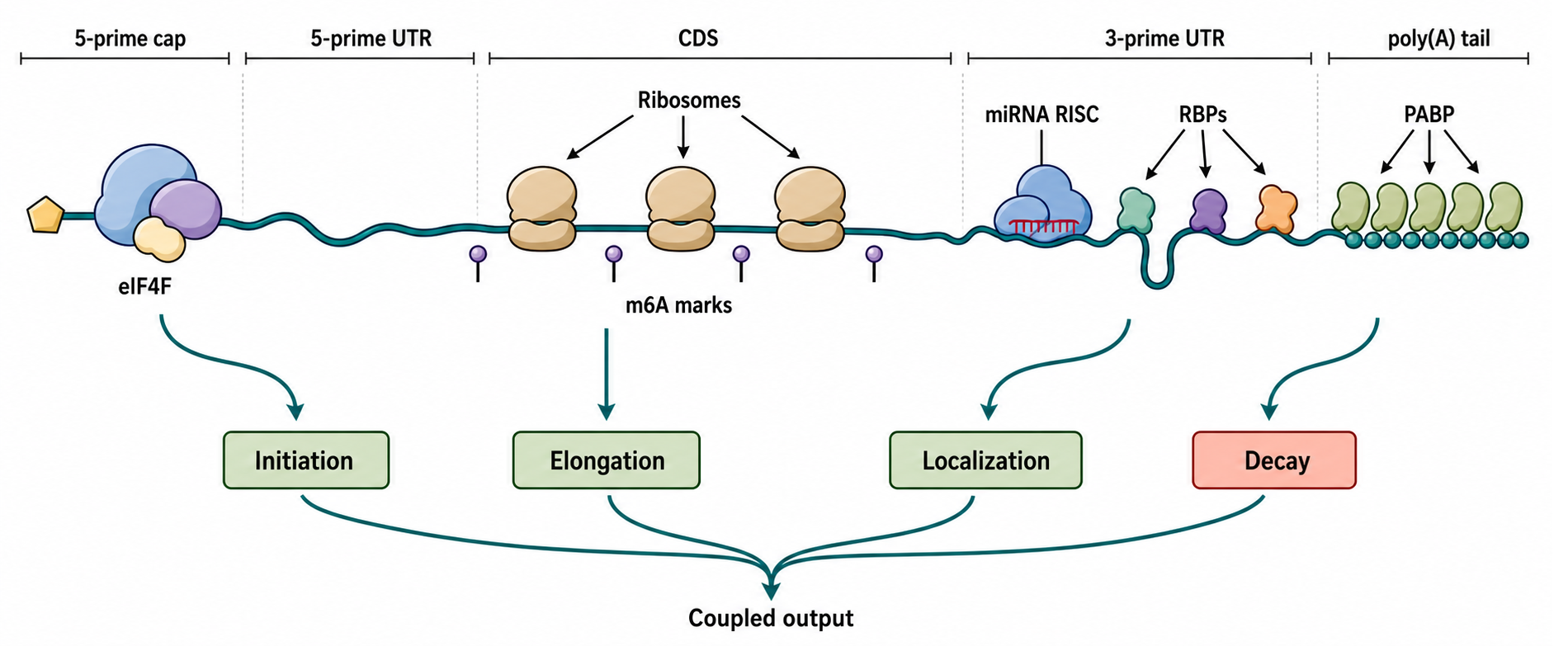
Figure 72.1. Integrated mRNA Architecture Map. A mature eukaryotic mRNA integrates regulatory information across its entire length through coupled architectural features. This schematic maps the major elements of a representative mRNA—5′ cap, 5′ UTR, coding sequence (CDS), 3′ UTR, and poly(A) tail—alongside their molecular readers: cap-binding proteins and the eIF4F complex at the 5′ end, translating ribosomes over the CDS, Argonaute–miRNA complexes and RBPs in the 3′ UTR, and poly(A)-binding proteins on the tail, with m6A and other covalent modifications indicated at representative positions. Arrows connect each architectural feature to its major regulatory outputs—translation initiation, elongation, localization, and decay—emphasizing that these features act as a coupled system in which strengthening or weakening one element can reinforce or expose the effects of another.
An mRNA begins to acquire regulatory architecture before it is fully synthesized. In many nuclear protein-coding genes, the 5′ cap is added co-transcriptionally soon after RNA polymerase II has produced a short nascent RNA. Splicing, exon junction complex deposition, 3′ cleavage, polyadenylation, export-factor loading, and surveillance all contribute to the protein composition of the mature messenger ribonucleoprotein particle. By the time a transcript reaches the cytoplasm, the mRNA is not bare RNA but a dynamic mRNP whose sequence and protein occupancy affect translation and decay.
Table 72.1. Architectural Features of Eukaryotic mRNA: Readers, Outputs, Methods, and Caveats. Summary of the major mRNA architectural features discussed in this chapter, listing the primary molecular readers that recognize each feature, the common regulatory output, representative experimental methods used to study it, and a key interpretive caveat.
| Feature | Main molecular readers | Common output | Representative evidence methods | Main caveat |
|---|---|---|---|---|
| 5′ cap | eIF4E, cap-binding complex, DCP1/DCP2 | Translation initiation, 5′-end protection, nuclear export | Cap-analog competition, decapping assays, ribosome profiling | Cap0 vs. cap1 vs. cap2 differ in immune sensing and eIF4E affinity; cap state must be specified |
| 5′ UTR structure | eIF4A/eIF4B, eIF4G, 40S subunit | Altered scanning rate, initiation efficiency | SHAPE probing, reporter libraries, hairpin-mutagenesis reporters | Structure is dynamic; cap-proximal position matters more than stability alone |
| uORF | 43S preinitiation complex, eIF2 | Reduced main-ORF translation; reinitiation under stress | Ribosome profiling, AUG-mutant reporters, eIF2α phosphorylation assays | Effect depends on uORF length, intercistronic distance, and reinitiation competence |
| CDS codon pattern | Ribosomes, RBPs, structure-sensitive factors, modification readers | Context-dependent translation, localization, and decay | Endogenous synonymous editing, matched recoding, structure probing, RBP mapping, RNA half-life assays | Synonymous changes also alter GC content, dinucleotides, RNA structure, modification motifs, and RBP or miRNA sites; decoding and causal decay require the dedicated analyses in Chapter 66 and Chapter 36 |
| CDS RNA structure | Ribosomes (helicase activity), RBPs | Elongation kinetics, co-translational folding windows, frameshifting | DMS-MaPseq, in-cell SHAPE, ribosome profiling at structured regions | Ribosomes unfold most structures; in-cell conformations differ from minimum free-energy predictions |
| 3′ UTR miRNA site | Argonaute–miRNA complex, GW182/TNRC6 | Translational repression, deadenylation, mRNA decay | Argonaute CLIP-seq, seed-site mutagenesis, miRNA perturbation with protein-output measurement | Site presence does not guarantee repression; site context, miRNA abundance, and RBP competition all modulate outcome |
| 3′ UTR RBP site | AU-rich element binding proteins, Pumilio, CPEB, LARP4 | mRNA stability, localization, translational activation or repression | eCLIP, iCLIP, motif-mutation reporters, RBP knockdown or overexpression | Binding does not prove function; overlapping sites create competitive or cooperative dynamics |
| Poly(A) tail | PABP, LARP1, LARP4, CCR4-Not | mRNA stability, translation initiation synergy with cap | Direct RNA sequencing, PAT assay, deadenylation kinetics, TAIL-seq | Tail-length effects are strongest in early development; in somatic cells, basal tail length is less predictive |
| m6A or m6Am | YTHDF1/2/3, YTHDC1/2, eIF3 | Translation efficiency, mRNA decay, splicing, nuclear export | m6A-seq, MAZTER-seq, DART-seq, nanopore direct RNA sequencing | Antibody-based maps lack stoichiometry information; many sites may be low-occupancy or condition-specific |
| RBP occupancy pattern | Multiple RBPs competing or cooperating across transcript | Combinatorial control of stability, localization, and translation | eCLIP, time-resolved RBP profiling, quantitative proteomics | Occupancy is probabilistic and condition-dependent; binding does not imply regulation without functional evidence |
The cap and poly(A) tail define the two physical ends of a typical mature eukaryotic mRNA. The cap protects the 5′ end and recruits cap-binding proteins. In the nucleus, the cap-binding complex contributes to processing and export. In the cytoplasm, eIF4E recognizes many capped mRNAs and, with scaffold protein eIF4G and helicase eIF4A, helps recruit the small ribosomal subunit through the eIF4F complex. The poly(A) tail is bound by poly(A)-binding proteins (PABPs), which protect the tail and interact with translation and decay factors. Reviews of LARP1, LARP4, and PABP emphasize that tail-associated proteins are active regulators of stability and translation rather than passive tail decorations (Mattijssen et al. 2021).
Cap-tail communication is often taught as a closed-loop model in which eIF4G bridges cap-bound eIF4E to PABP on the poly(A) tail. This model captures an important biochemical principle: factors at one mRNA end can influence processes at the other end. It should not be overgeneralized into a claim that every translating mRNA forms a stable circular molecule. Modern evidence supports dynamic, factor-dependent, transcript-specific contacts rather than a single rigid topology. The mechanistic point is integration. A strong cap, accessible 5′ UTR, favorable start-codon context, productive coding region, stabilizing 3′ UTR, and appropriate poly(A)-RNP state can reinforce each other; a weak feature can be buffered or exposed depending on the rest of the transcript.
The coding sequence participates in this integration even when the protein product is not the focus. The encoded nascent polypeptide can affect ribosome behavior, translocon engagement, localization, and mRNA quality control. Hopfler and Hegde review cases in which the nascent chain helps determine mRNA fate by altering ribosome-associated factors, membrane targeting, or surveillance exposure (Hopfler and Hegde 2023). Thus the CDS is not merely an interchangeable protein-coding cassette surrounded by regulatory UTRs. The CDS carries dual information: amino acid sequence and RNA-level regulatory features.
Alternative transcript isoforms make this integration concrete in native cells. A change in transcription start site can alter cap-proximal structure and upstream open reading frames; alternative splicing can change the CDS, exon-junction marks, and 3′ UTR; and alternative cleavage and polyadenylation can change RBP, miRNA, and localization sites together with the tail-associated mRNP. Isoforms encoding the same protein can therefore differ in initiation, localization, and decay because their surrounding RNA grammar differs. This is an endogenous systems problem rather than a collection of independent sequence modules.
The evidence for architectural integration comes from several experimental classes. Reporter libraries test designed combinations of UTRs, CDSs, caps, and tails. Ribosome profiling measures ribosome occupancy and can reveal initiation or elongation changes. RNA half-life assays measure decay after transcriptional shutoff, metabolic labeling, or time-resolved sequencing. Protein output assays measure the combined result of translation and stability. Structure probing and RBP mapping show physical features and bound factors. Each method has limitations: reporter context can miss endogenous chromatin and processing history; ribosome density is not identical to protein synthesis rate; RNA half-life estimates can be distorted by cell growth or transcriptional perturbation; and binding maps do not prove functional regulation.
The boundary cases are instructive. Bacterial mRNAs do not normally have a 7-methylguanosine cap or long poly(A) tail with the same stabilizing role, and bacterial polyadenylation can promote decay. Many viral RNAs imitate, steal, modify, or bypass host mRNA end features. Some eukaryotic mRNAs use internal ribosome entry, cap-independent initiation enhancers, or special 3′ structures. These exceptions do not disprove integrated architecture; they show that integration uses different molecular parts in different biological systems.
72.2. 5-prime UTR grammar and initiation control
The 5′ UTR is the region that a scanning eukaryotic preinitiation complex usually examines before selecting a start codon. In the canonical scanning model, a 43S preinitiation complex containing the 40S ribosomal subunit, initiator methionyl-tRNA, and initiation factors is recruited near the cap. The complex moves along the 5′ UTR in the 5′ to 3′ direction until it recognizes a start codon in a favorable sequence and structural context. This model is a central grammar for many eukaryotic mRNAs, but real transcripts use variations that include leaky scanning, reinitiation after upstream open reading frames, ribosome shunting, internal entry, and stress-dependent changes in initiation factors.
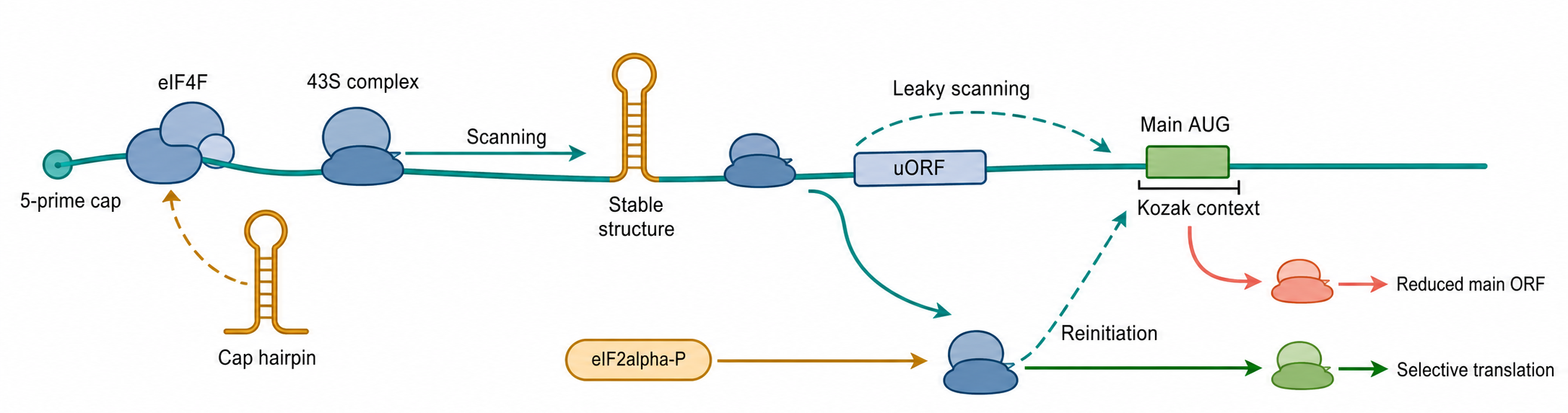
Figure 72.2. 5′ UTR Initiation Grammar. The 5′ UTR determines how and whether the ribosome reaches the main start codon. This diagram traces the canonical scanning pathway: a 43S preinitiation complex is recruited near the cap with help from eIF4F, then moves 5′ to 3′ along the UTR until it encounters an AUG in favorable sequence context, while a cap-proximal hairpin can inhibit initial complex assembly and a stable downstream structure slows scanning. Upstream open reading frames (uORFs) are shown capturing scanning ribosomes to reduce main-ORF translation, with a stress-state branch illustrating how eIF2α phosphorylation can permit selective reinitiation; leaky scanning and Kozak context are indicated to convey that start-codon recognition is probabilistic and factor-dependent rather than a binary switch.
A useful way to read 5′ UTR grammar is to ask what a ribosome sees before the main start codon. A short, weakly structured 5′ UTR with no upstream AUG codons is often permissive for cap-dependent initiation. A long UTR with stable structures near the cap can interfere with recruitment or early scanning. A stable hairpin downstream of the cap can slow scanning or require helicase activity. Upstream open reading frames (uORFs) can reduce main ORF translation by capturing scanning ribosomes; under some stress conditions, uORFs allow selective translation of downstream coding sequences by reinitiation logic. Start-codon context changes initiation probability because the ribosome recognizes AUG in a local nucleotide environment rather than as a naked triplet.
The phrase “5′ UTR structure” requires care. RNA secondary structure is a set of base-pairing interactions, not a single static fold. A 5′ UTR can contain local stem-loops, long-range contacts, protein-stabilized structures, or alternative conformations that differ between cell states. Leppek and colleagues review functional 5′ UTR structures and emphasize both biological significance and the difficulty of proving that a predicted structure is the causal regulatory feature (Leppek et al. 2018). Primary evidence from 40S scanning studies shows that mRNA structure can regulate scanning kinetics and initiation, but the effect depends on position, stability, helicase recruitment, and factor availability (Wang et al. 2022).
Cap-proximal structure is a particularly sensitive boundary case. A hairpin close to the cap can inhibit assembly of the preinitiation complex before scanning begins, while a comparable structure farther downstream may primarily affect scanning progression. Uppala and colleagues showed that cap-proximal RNA secondary structure inhibits preinitiation complex formation on HAC1 mRNA, illustrating why the same structural stability can have different effects depending on position (Uppala et al. 2022). Grayeski and colleagues found that global 5′ UTR RNA structure can regulate translation of a SERPINA1 mRNA, supporting the idea that distributed structure, not only one motif, can govern translation output (Grayeski et al. 2022).
Internal ribosome entry sites (IRESs) illustrate an alternative grammar. An IRES is an RNA element that can recruit ribosomes internally, often with a subset of canonical initiation factors or with specialized trans-acting factors. Viral IRESs are the clearest examples, and type IV picornavirus IRES elements have been reviewed as structured RNAs with defined functional domains (Li et al. 2024). Cellular IRES claims require stricter artifact control because bicistronic reporters can produce misleading signals through cryptic promoters, cryptic splicing, readthrough, or RNA instability. A robust IRES claim needs controls showing that the second cistron is translated from the same RNA and that RNA structure or associated factors are responsible.
The 5′ UTR also carries RBP motifs. Iron-responsive elements, terminal oligopyrimidine tracts, and transcript-specific structures show how proteins can connect nutrient state, stress pathways, or growth signals to initiation. LARP1-mediated regulation of 5′ terminal oligopyrimidine mRNAs connects cap-proximal sequence, mTOR signaling, and translation of ribosome-biogenesis mRNAs, although detailed coverage belongs in chapters on growth signaling and translation control. The key principle is that 5′ UTR grammar is not merely a count of AUGs and hairpin energies. The relevant grammar includes cap context, scanning path, start-codon competition, structure position, protein binding, cell state, and the kinetic availability of initiation factors.
72.3. CDS codon patterns and structure within native mRNA grammar
The coding sequence specifies a polypeptide through triplet codons, but the CDS is also part of the native RNA grammar. Synonymous codon patterns covary with GC content, dinucleotide composition, local RNA structure, modification motifs, embedded RBP or miRNA sites, and the positions at which nascent-peptide signals emerge from the ribosome. The integrative question is therefore not whether one codon is universally optimal. It is how a native CDS feature changes the interpretation of cap, UTR, tail, structure, and occupancy features in a specified cell state.
Codon-dependent decoding and decay provide one bridge into this grammar, but their primary mechanisms belong elsewhere. Chapter 66 owns codon demand, wobble, tRNA abundance, charging, and modification as a decoding system. Chapter 36 owns the causal chain from codon identity through ribosome state and factor recruitment to mRNA half-life. This chapter retains the architectural consequence: a CDS codon pattern can alter translation state and thereby change how the same 3′ UTR, poly(A) state, or RBP occupancy is expressed as protein output, localization, or decay.
Nascent-peptide context is another integrative route. A pause near a domain boundary can influence cotranslational folding, whereas emergence of a signal peptide or transmembrane segment can redirect the ribosome–mRNA complex to the endoplasmic reticulum. Hopfler and Hegde emphasize that encoded nascent polypeptides can feed back on mRNA fate through ribosome-associated factors, targeting pathways, and surveillance exposure (Hopfler and Hegde 2023). A CDS effect can therefore originate in RNA sequence, decoding state, encoded peptide, or an interaction among them.
CDS RNA structure adds another layer. Base pairing in coding regions can influence ribosome initiation if it extends into the 5′ region, elongation if it presents structures to the ribosome, and stability if it changes RBP or nuclease access. Ribosomes are powerful helicases and unfold many structures during elongation, but structure can still matter through kinetics, cotranslational folding windows, frameshift stimulation, recoding signals, or long-range interactions. RNA 3D structure prediction reviews emphasize that RNA folding cannot be inferred perfectly from sequence, especially in cells where proteins, ribosomes, modifications, and co-transcriptional folding histories reshape ensembles (Wang X et al. 2023).
Programmed ribosomal frameshifting is a boundary case where CDS architecture intentionally disrupts ordinary decoding. A slippery sequence and downstream RNA structure can promote a shift in reading frame, allowing one RNA to produce multiple proteins. Many viruses use this strategy. In ordinary host mRNAs, similar features may trigger quality control rather than productive recoding. Distinguishing regulatory pausing from harmful collision requires evidence from ribosome profiling, reporter mutagenesis, protein products, and quality-control markers.
For experimental interpretation, synonymous recoding is powerful but not clean by default. Changing codons also changes GC content, dinucleotide frequencies, RNA structure, RBP motifs, miRNA sites inside CDSs, and potential modification motifs. Native-grammar experiments should use multiple matched recodings, endogenous editing where feasible, direct structure or occupancy measurements, and separate readouts for RNA abundance, half-life, localization, and protein output. If the claim is specifically that codon identity caused decay, the mechanistic standards in Chapter 36 apply.
72.4. 3-prime UTR elements and RBP/miRNA regulation
The 3′ UTR is a major regulatory platform because it can change mRNA fate without changing protein sequence. A 3′ UTR may contain AU-rich elements, GU-rich elements, cytoplasmic polyadenylation elements, Pumilio response elements, miRNA response elements, localization motifs, stability elements, and binding sites for many other RBPs. Alternative polyadenylation can produce isoforms with different 3′ UTR lengths. A shorter isoform may remove repressive miRNA or RBP sites, whereas a longer isoform may add localization or stability information. Broad reviews of 3′ UTR function and alternative polyadenylation emphasize that these isoform changes are regulatory changes, not just annotation differences (Mayr 2019; Tian and Manley 2017; Gruber and Zavolan 2019). In cancer, 3′ UTR heterogeneity has been reviewed as one route by which gene regulation changes without protein-coding mutation (Chan et al. 2023).
MicroRNAs are small RNAs loaded into Argonaute-containing silencing complexes. A miRNA typically recognizes target mRNAs through base pairing between its seed region and complementary sites, often in the 3′ UTR. Once bound, the complex can repress translation and promote deadenylation and decay. The strongest simple rule is that conserved seed-matched sites in accessible 3′ UTR contexts are more likely to be functional than random matches. However, miRNA regulation is quantitative, combinatorial, and context-dependent. Site number, site spacing, local structure, nearby RBP occupancy, miRNA abundance, Argonaute availability, and the basal half-life of the mRNA all affect measurable repression (Bartel 2018).
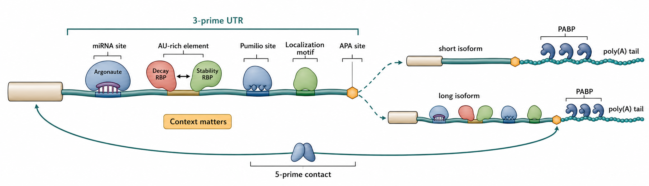
Figure 72.4. 3′ UTR Regulatory Platform. The 3′ UTR functions as a multi-input regulatory platform where sequence elements and bound factors jointly determine mRNA fate without altering the encoded protein sequence. This diagram depicts a 3′ UTR carrying a miRNA response element recognized by an Argonaute-loaded miRNA, an AU-rich element competed for by stability-promoting and decay-promoting RBPs, a Pumilio response element, a localization motif, and an alternative polyadenylation site that generates isoforms with different 3′ UTR lengths and regulatory-site compositions, with PABP on the poly(A) tail communicating with 5′-end factors. The figure emphasizes that 3′ UTR length creates regulatory possibilities but does not determine outcome alone; functional consequence depends on which factors are expressed, which sites are occupied, and whether the mRNA is in a translating, localized, or decay-competent state.
The evidence basis for miRNA regulation includes reporter assays, Argonaute CLIP mapping, miRNA perturbation, transcriptome-wide mRNA and protein measurements, evolutionary conservation, and rescue experiments. Each evidence class has traps. A reporter can exaggerate a site if it removes the element from its endogenous UTR context. CLIP peaks show physical proximity to Argonaute but do not prove functional repression. Overexpression of a miRNA can create nonphysiological targeting. Loss of a miRNA can produce indirect network effects. Disease association reviews, including cancer-focused miRNA reviews, are useful for context but should not be treated as proof that every reported miRNA-mRNA pair is a direct causal interaction (Smolarz et al. 2022; Ebrahimi et al. 2020).
RBPs can cooperate or compete with miRNAs. An RBP may open a structured region and expose a miRNA site, mask a site from Argonaute, recruit deadenylases, stabilize the transcript, or localize the mRNA to a compartment where the miRNA machinery differs. Broad RBP reviews make the same caution at transcriptome scale: binding sites and protein occupancy are evidence of molecular contact, but functional regulation must be shown with perturbation and output measurements (Hentze et al. 2018). The RBP code is therefore not a simple additive list of motifs. Binding order, protein concentration, post-translational modification, RNA structure, and translation state matter. An AU-rich element bound by a decay-promoting protein in one cell type can behave differently if a stabilizing factor occupies overlapping sequence in another.
The 3′ UTR also communicates with translation. PABP on the poly(A) tail can interact with initiation factors; deadenylation often precedes decapping and 5′ to 3′ decay; and translational repression can alter exposure to decay enzymes. Localization elements in 3′ UTRs can recruit transport granule proteins that repress translation during transport and permit translation near a destination. Neuronal dendritic mRNAs and early embryonic mRNAs provide classic examples, while detailed localization mechanisms are treated in Chapter 74.
Do not overgeneralize 3′ UTR length. Longer UTRs are not automatically more repressed, and shorter UTRs are not automatically oncogenic or highly translated. Length changes alter the set of possible regulatory sites, but functional outcome depends on which sites are present, which factors are expressed, and whether the isoform is translated, localized, or degraded. The best-supported claims combine isoform-resolved end mapping, factor-binding evidence, perturbation, and protein-output measurement.
Box 72.1. Artifact Controls for mRNA Architecture Claims
- IRES claims: rule out cryptic promoters, cryptic splice sites, read-through translation, and RNA instability using monocistronic controls, promoter-less constructs, and RNA-level verification that both cistrons derive from the same molecule.
- miRNA targeting claims: use endogenous miRNA perturbation (not only overexpression), seed-site mutant rescue in the endogenous 3′ UTR context, and protein-output measurement rather than mRNA abundance alone.
- RBP regulation claims: require a binding result plus a functional perturbation result; motif presence or CLIP enrichment alone does not establish regulation.
- RNA modification claims: require site-level mapping with a method that minimizes antibody bias, stoichiometry or occupancy information when feasible, and rescue with modification-defective RNA or writer/eraser mutants rather than indirect genetic perturbations.
- CDS codon-pattern claims: use multiple matched synonymous designs and measure GC content, dinucleotide composition, RNA structure, RBP or miRNA occupancy, and modification motifs alongside the biological output. A decoding claim requires Chapter 66 evidence, and a causal codon-decay claim requires the kinetic and pathway tests in Chapter 36.
72.5. Cytoplasmic polyadenylation, CPEB systems, and translational activation
Cytoplasmic polyadenylation is the regulated addition of adenosines to the 3′ end of an mRNA that has already been cleaved and polyadenylated in the nucleus. This definition separates two related but distinct processes. Nuclear 3′-end formation chooses a cleavage site and builds the initial tail as part of pre-mRNA maturation; its primary owner is Chapter 29. Cytoplasmic polyadenylation instead remodels the tail of a mature mRNA to change its translation, stability, or developmental timing. The canonical pathway is especially important when transcription is absent or temporarily uninformative, as in a fully grown oocyte whose future protein-production program must be executed from maternal mRNAs already stored in the cytoplasm.
The cis-regulatory grammar begins in the 3′ untranslated region. A classical cytoplasmic polyadenylation element (CPE) is U-rich and lies in functional relation to the polyadenylation signal, often AAUAAA, that was used during nuclear processing. The element is not a single context-free word. CPE sequence, number, spacing, distance from the polyadenylation signal, neighboring Pumilio-binding sites or other RBP elements, and developmental stage can alter whether an mRNA is repressed, activated early, activated late, or deadenylated. Large reporter-library experiments in frog oocytes and embryos identified UUUUA as a compact active element and showed that its effect depends on the polyadenylation signal and surrounding sequence (Xiang et al. 2024). C-rich elements and poly(rC)-binding proteins can support cytoplasmic polyadenylation in early embryos, providing a noncanonical route whose activity differs from the maturation-stage CPE pathway (Vishnu et al. 2011). Thus a motif scan predicts candidates; it does not identify an active program without stage-specific binding, tail, and translation evidence.
CPE-binding proteins provide trans specificity. Vertebrates encode four CPEB-family proteins. CPEB1 is the founding and best-characterized member, whereas CPEB2, CPEB3, and CPEB4 have overlapping but distinct RNA-recognition properties, regulatory inputs, and biological functions (Ivshina et al. 2014; Huang et al. 2023). CPEBs typically contain conserved RNA-binding domains and less-conserved regulatory regions that engage cofactors and post-translational control. CPEB occupancy can recruit a multiprotein mRNP containing cleavage and polyadenylation specificity factor (CPSF), scaffold protein symplekin, a cytoplasmic poly(A) polymerase, deadenylases, poly(A)-binding proteins, and cap-associated repressors. Not every target contains every component at once. The useful abstraction is a selected mRNP whose enzymatic and initiation states can be remodeled, not one invariant complex for all CPEB paralogs and tissues.
In a repressed Xenopus oocyte mRNP, CPEB binds the CPE while proteins such as Maskin can connect the 3′-UTR complex to cap-bound eIF4E. Maskin competes with eIF4G for eIF4E and thereby blocks productive initiation-complex assembly. Neuroguidin can perform an analogous cap-blocking role in other CPEB complexes. At the 3′ end, PARN or CCR4-NOT-associated deadenylation can oppose tail extension. A GLD2-family polymerase may already be associated with the mRNP, but net tail length remains short when deadenylation or restricted polymerase activity dominates. This coexistence explains why simply detecting TENT2/GLD2 in a complex does not prove that its target is actively polyadenylated. Enzyme perturbation must be connected to a target-specific change in tail length and then to translation or stability.
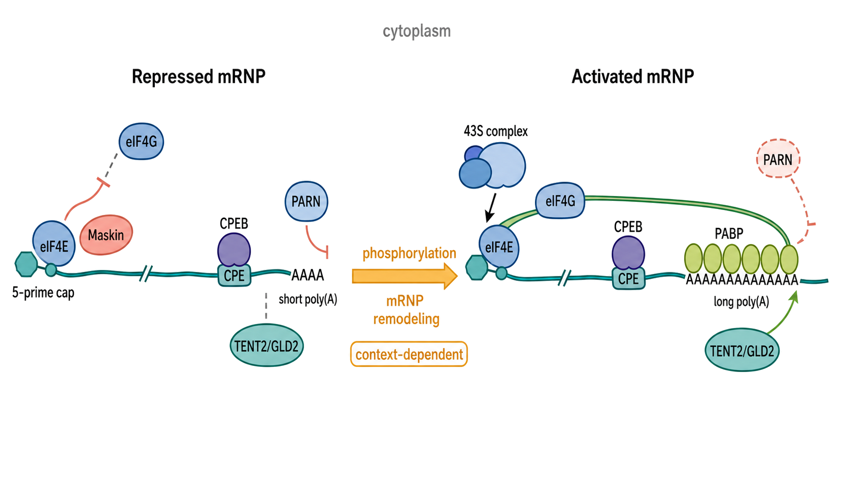
Figure 72.5. CPEB mRNP State Transition from Repression to Translational Activation. A two-state mechanism diagram should place the same capped, CPE-containing mRNA in a repressed state on the left and an activated state on the right. In the repressed state, CPEB binds a U-rich CPE near the polyadenylation signal; CPSF and symplekin provide a scaffold; PARN or another deadenylase keeps the poly(A) tail short; TENT2/GLD2 is present but does not dominate net tail metabolism; and Maskin or neuroguidin binds eIF4E and excludes eIF4G. A central signaling arrow should show CPEB and cofactor phosphorylation followed by mRNP remodeling, with an explicit note that the kinase and factor composition are context-dependent. In the activated state, deadenylase activity is reduced or displaced, TENT2/GLD2 extends the tail, PABP occupancy increases, PABP-bound eIF4G engages eIF4E, and eIF3/43S recruitment initiates translation. A bottom annotation should distinguish this cytoplasmic remodeling from nuclear cleavage and initial polyadenylation in Chapter 29 and should state that longer tails are not universally more translated in somatic cells.
Activation is a causal sequence rather than an unexplained correlation between a long tail and abundant protein. In the classical oocyte model, a maturation signal activates a kinase pathway that phosphorylates CPEB1 and other mRNP components. Phosphorylation changes CPEB interactions and promotes assembly or activity of a polyadenylation-competent complex. PARN activity is reduced or PARN leaves the mRNP in models where it is present, while a noncanonical poly(A) polymerase gains net access to the 3′ hydroxyl end. TENT2, historically called GLD2, is a catalytic subunit whose activity and target choice depend on recruitment by RNA-binding partners; xGLD2 and symplekin are required for CPEB-mediated polyadenylation in the reconstituted Xenopus framework (Barnard et al. 2004). Related terminal nucleotidyltransferases, including GLD4-family enzymes in some animals, can contribute through distinct complexes, so “the cytoplasmic poly(A) polymerase” is not a universal single-protein designation.
Tail extension then changes the protein-binding capacity of the RNA end. Additional poly(A)-binding protein (PABP) molecules occupy the elongated tail. PABP can engage eIF4G, increase productive eIF4G-eIF4E association, and help displace cap-bound repressors such as Maskin. eIF4G then connects cap recognition to eIF3 and the small ribosomal subunit, increasing the probability of translation initiation. Experiments with cyclin B1 reporters, cap-affinity purification, Maskin perturbation, polyadenylation inhibition, and PABP/eIF4G interference support this cap-to-tail handoff in Xenopus oocytes (Cao and Richter 2002). Chapter 70 owns the broader initiation-factor logic. The present section owns the upstream 3′-UTR and tail-remodeling mechanism that makes a selected mRNA competent for that initiation machinery.
Maternal mos and cyclin B mRNAs illustrate why the pathway is a developmental timer. The RNAs are synthesized and processed during oogenesis, stored with short tails and repressive proteins, and translated in a controlled sequence after progesterone triggers meiotic maturation. Mos accumulation activates a kinase cascade, whereas cyclin synthesis helps drive cell-cycle transitions; premature or mistimed translation would disrupt the order of meiosis. Different CPE arrangements and associated factors help specify early and late waves of polyadenylation. This example also establishes the evidence ladder: a 3′-UTR mutation changes polyadenylation timing; CPEB and cofactors physically associate with the RNA; kinase or factor perturbation changes tail extension; and translation and meiotic progression change in the predicted direction. The developmental consequences and integration with maternal-mRNA storage and clearance are synthesized in Chapter 102.
Tail-length coupling changes across development. Poly(A)-tail length profiling by sequencing (PAL-seq) found a strong relationship between tail length and translational efficiency in early zebrafish and frog embryos, followed by an embryonic switch after which this coupling weakens and tail length more strongly reflects stability and decay state (Subtelny et al. 2014). mTAIL-seq likewise revealed dynamic, Wispy-dependent tail regulation and strong tail-length–translation coupling during Drosophila egg activation (Lim et al. 2016). The 2024 frog and fish reporter-library study further showed that oocyte maturation occurs against widespread deadenylation and that embryos use stage-specific combinations of polyadenylation and deadenylation elements (Xiang et al. 2024). These observations correct the common assumption that a longer poly(A) tail universally predicts greater translation in somatic cells. Coupling is strongest when limiting PABP and stored maternal mRNAs make tail extension a decisive gate; after zygotic genome activation and developmental remodeling, other features can dominate.
Neurons reuse parts of the developmental logic in a spatially restricted setting. Selected mRNAs travel into dendrites in repressed mRNPs and can be activated near stimulated synapses. CPEB-associated dendritic complexes can contain GLD2, PARN, neuroguidin, and symplekin; synaptic stimulation has been linked to CPEB phosphorylation, PARN loss from the complex, tail extension, local protein synthesis, and changes in long-term potentiation (Udagawa et al. 2012). CPEB-family functions also extend to axons, learning, memory, and neurological phenotypes, although paralog-specific mechanisms remain incompletely resolved (Huang et al. 2023). A neuronal CPE or CPEB dependence does not prove that the exact oocyte complex has been redeployed. Cell compartment, stimulus, paralog, cofactor availability, and target RNA must be established independently.
Somatic boundary cases are equally important. TENT2/GLD2 and CPEBs are expressed outside germ cells and neurons, and cytoplasmic tail extension can regulate particular mRNAs or noncoding RNAs. Yet most steady-state somatic mRNAs do not obey a simple tail-length–translation rule, and deadenylation often marks progression toward decay rather than a reversible developmental switch. Other noncanonical poly(A) polymerases can stabilize germline transcripts, while in other contexts terminal nucleotidyltransferases add short mixed or non-A tails with different consequences. Cytoplasmic polyadenylation must therefore be demonstrated as a target-specific enzymatic reaction, not inferred from the presence of CPEB, a long tail, or increased protein abundance alone.
Table 72.2. Measuring Cytoplasmic Polyadenylation: Observables, Strengths, and Artifacts. Poly(A)-tail assays differ in whether they measure length distributions, transcript-specific tails, extension activity, or coupled translation; priming, ligation, amplification, and isoform ambiguity require orthogonal controls.
| Method | Primary observable | Best use | Major artifact or ambiguity | Orthogonal evidence needed |
|---|---|---|---|---|
| Poly(A) test (PAT) or extension PAT | PCR product-size distribution for one transcript | Developmental time courses, endogenous targets, and cis-element reporter mutants | Oligo(dT) priming, ligation, PCR, and gel-resolution biases; size does not identify the responsible enzyme | CPE/CPEB perturbation, catalytic-polymerase or deadenylase perturbation, and translation measurement |
| Ligation-mediated PAT (LM-PAT) | Transcript-specific tail distribution after adaptor ligation | Better anchoring of transcript 3′ ends and comparison of selected RNAs | Ligation efficiency and PCR amplification can distort abundance and tail distributions | End-mapped RNA, input controls, and an independent tail assay |
| PAL-seq | Transcriptome-wide tail-length distributions | Comparing tails with translational efficiency across stages or tissues | Library capture thresholds, short-tail recovery, and aggregation across isoforms | Isoform-resolved 3′-end mapping and ribosome/protein-output data |
| TAIL-seq or mTAIL-seq | Transcriptome-wide poly(A) length and terminal non-A residues | Small developmental samples and dynamic tail remodeling | Adapter and capture biases; low abundance and very short tails can be underrepresented | Spike-ins, replicate libraries, and targeted PAT validation |
| Nanopore direct RNA sequencing | Single-molecule isoform and poly(A)-tail estimate | Linking tail state to native transcript isoform | 3′-capture dependence, poly(A)-selection bias, signal calibration, truncated reads, and limited depth | Synthetic tail standards, cap or intact-RNA selection controls, and targeted validation |
| Ribosome or polysome profiling paired with a tail assay | Translation state associated with a measured tail distribution | Testing tail-length–translation coupling | Ribosome occupancy is not identical to protein production; population measurements need not link tail and translation on the same molecule | Pulse labeling or quantitative protein output, developmental staging, and perturbation of the tail pathway |
The principal measurements answer different questions. A poly(A) test (PAT), ligation-mediated PAT, or related transcript-specific PCR assay estimates the tail-length distribution of one mRNA and is well suited to time courses and cis-element mutants, but PCR size, priming, and ligation biases can distort the distribution. TAIL-seq and PAL-seq measure tail lengths transcriptome-wide and can be paired with ribosome profiling or protein synthesis assays, but each library chemistry has capture thresholds and may underrepresent very short tails. Nanopore direct RNA sequencing can link a tail estimate to an individual native isoform, although 3′-end capture, poly(A) selection, pore-signal calibration, truncation, and read-depth biases must be controlled. A measured tail shift is not itself proof of new synthesis: altered deadenylation, selective decay of short-tailed molecules, isoform switching, or differential capture can produce a similar population-level result.
Mechanism therefore requires orthogonal evidence. CPE or polyadenylation-signal mutants test cis dependence; CPEB CLIP or immunoprecipitation tests physical association; phosphosite mutants and timed kinase perturbations test signal dependence; catalytically inactive TENT2/GLD2 and deadenylase perturbations test enzymatic balance; PAT or sequencing measures the tail; polysome profiling, ribosome profiling, pulse labeling, or protein reporters measure translation; and rescue experiments connect the molecular lesion to the biological phenotype. Reporter RNA injected into an oocyte is powerful because stage and sequence can be controlled, but injection can bypass nuclear history and create nonphysiological RNA concentration. Endogenous validation is needed before extending reporter logic to a natural transcript.
Box 72.2. Evidence Ladder for Causal Cytoplasmic Polyadenylation
- Define the RNA substrate: map the endogenous 3′ end and isoform so a change in alternative cleavage or transcript abundance is not mistaken for a tail change.
- Test the cis element: mutate the candidate CPE or related 3′-UTR element while preserving the polyadenylation signal and other known motifs; test compensatory or context variants rather than one destructive mutation alone.
- Establish factor occupancy: show stage- or stimulus-appropriate binding of the relevant CPEB paralog and cofactors by CLIP, immunoprecipitation, or reconstituted binding, recognizing that binding alone does not establish regulation.
- Resolve enzyme balance: perturb TENT2/GLD2 or the relevant noncanonical polymerase and PARN or another deadenylase, include catalytic-dead and rescue conditions, and measure target-specific tail distributions.
- Connect signaling to remodeling: test a timed kinase or phosphosite perturbation and show that mRNP composition or enzyme activity changes before tail extension.
- Connect the tail to translation: pair PAT, PAL-seq, TAIL-seq, or nanopore measurements with polysome/ribosome profiling, pulse labeling, or protein output; distinguish greater initiation from altered mRNA stability.
- Validate biological consequence: restore the molecular pathway or target protein and rescue the oocyte, embryonic, neuronal, or somatic phenotype without relying solely on a reporter RNA.
The ownership boundary can now be stated precisely. Chapter 29 owns nuclear cleavage-site choice, canonical cleavage/polyadenylation machinery, and initial tail synthesis. Chapter 70 owns regulated recruitment of ribosomes and initiation-factor alternatives. Chapter 102 owns how maternal storage, activation, and clearance are coordinated across embryos, germ cells, and differentiating tissues. This chapter owns the general molecular bridge among a 3′-UTR cis element, CPEB-family factor, opposing tail enzymes, PABP/eIF4G recruitment, and translational activation. Cross-references provide biological depth without substituting for the complete causal mechanism given here.
72.6. RNA modifications and RNP identity marks
An mRNA’s identity is not fully specified by A, C, G, and U sequence. Chemical modifications and bound proteins can mark transcript history and influence future interactions. N6-methyladenosine (m6A) is the most extensively studied internal mRNA modification in many eukaryotic systems. It is installed by writer complexes, interpreted by reader proteins, and removed by eraser enzymes in some contexts. Other marks relevant to mRNAs include m6Am near the cap, pseudouridine, 5-methylcytidine, inosine generated by adenosine-to-inosine editing, and 2′-O-methylation in cap-adjacent positions. Structural reviews of m6A and m6Am methyltransferases clarify that modification specificity depends on enzyme architecture, cofactors, RNA context, and associated proteins (Oerum et al. 2021; Flamand et al. 2023).
The term “epitranscriptomic mark” should be used cautiously. A modification can act as a direct recognition mark if a reader binds the modified nucleotide and changes mRNA fate. A modification can act structurally if it changes base pairing, stacking, or local RNA conformation. A modification can act indirectly if perturbing a writer enzyme changes many RNAs or cellular states. In some cases, a modification may be a byproduct of processing rather than a regulatory signal. Reviews of detection techniques emphasize that mapping methods differ in resolution, stoichiometry measurement, sequence bias, and ability to distinguish similar chemical marks (Ofusa et al. 2022).
m6A provides a concrete example. m6A can recruit YTH-domain reader proteins, alter RNA structure by weakening A-U pairing, affect translation or decay, and participate in stress granule behavior. But m6A peaks from antibody-based enrichment are not exact modification sites unless validated by higher-resolution methods, and a peak does not reveal what fraction of molecules is modified. Genetic deletion of a writer enzyme can change development, stress response, splicing, or RNA abundance indirectly. A strong mechanistic claim usually requires site-level mapping, stoichiometry or occupancy information when possible, writer or reader perturbation, rescue with modification-defective mutants, and measurement of the relevant mRNA fate.
RNP identity marks include proteins deposited during nuclear processing. An exon junction complex can mark spliced mRNAs and later influence nonsense-mediated decay if translation terminates upstream of an exon junction in an inappropriate context. Cap-binding complexes mark 5′ processing history. PABPs and LARP proteins mark 3′ end state. Stress granule proteins such as G3BP can reorganize mRNPs under stress, and recent work links G3BP-driven granules, RNA-RNA interactions, and DDX3X-dependent resolution to mRNA translatability (Trussina et al. 2025). Post-translational modifications of RBPs can alter granule assembly and degradation, adding another layer of identity control (Jeon et al. 2022).
Modification state must be interpreted together with native processing and occupancy. The same mapped modification can have different consequences in alternative isoforms, different subcellular compartments, or cell states that express different reader proteins. A site-level change should therefore be connected to the transcript isoform, modification fraction, RBP occupancy, translation state, localization, and decay output rather than treated as an autonomous fate switch. Chapter 46 and Chapter 52 provide the evidence framework and cross-mark biology; this chapter owns their integration with whole-mRNA grammar.
72.7. Integration of translation, localization, and decay in native mRNAs
Translation, localization, and decay are coupled because ribosomes, transport factors, and decay enzymes read overlapping features of the same mRNP. A translating ribosome can protect parts of an mRNA from nucleases, expose poor codon decoding to quality-control factors, displace RBPs, or recruit membrane-targeting machinery through the nascent peptide. A localized mRNA can be translationally repressed during transport and activated at a destination. A decaying mRNA can remain associated with ribosomes long enough for translation-coupled surveillance to inspect termination, elongation, or collisions.
Translation-coupled quality control illustrates the integration principle because the consequence of a ribosome state depends on transcript architecture. A premature stop can be interpreted relative to exon-junction marks, 3′ UTR length, and PABP proximity; a pause can be interpreted differently depending on structure, nascent peptide, and local ribosome traffic. The detailed surveillance mechanisms belong in Chapter 35, Chapter 36, and Chapter 71. This chapter retains the architectural rule that the same local event can have different outcomes in different whole-transcript contexts.
Nonsense-mediated decay illustrates architectural interpretation. A premature termination codon is not recognized merely because it is a stop codon. The surveillance machinery evaluates stop-codon position relative to downstream exon junction complexes, 3′ UTR length, PABP proximity, and other features. A stop codon that is normal in one transcript architecture can be premature in another. This is why alternative splicing, alternative polyadenylation, and translation initiation choice can create or eliminate decay-sensitive isoforms.
Localization adds spatial coupling. Many mRNAs are transported in repressed mRNPs to compartments such as neuronal dendrites, axons, oocytes, embryos, or the endoplasmic reticulum. A localization element often resides in the 3′ UTR, but localization can also be influenced by coding-region translation, nascent peptides, RNA structure, and RBPs. Single-molecule perspectives show that mRNA transport is not a single conveyor-belt process; transcripts can diffuse, dock, undergo active transport, pause in granules, and switch translation states (Basyuk et al. 2021). Reviews of RNA trafficking emphasize the need to combine imaging, sequencing, perturbation, and predictive methods to infer localization mechanisms (Wang J et al. 2023).
Decay pathways integrate nuclear and cytoplasmic events. Nuclear mRNA decay can remove improperly processed or retained transcripts before translation, while cytoplasmic decay responds to deadenylation, decapping, endonucleolytic cleavage, miRNA recruitment, RBP binding, and translation status. Rambout and Maquat review nuclear mRNA decay networks that regulate gene expression and quality control (Rambout and Maquat 2024). The cytoplasmic branch commonly begins with deadenylation, proceeds to decapping, and then allows 5′ to 3′ exonucleolysis, although endonucleolytic and 3′ to 5′ routes can dominate in particular contexts.
Stress changes coupling. During heat shock, nutrient limitation, viral infection, or innate immune activation, translation initiation can be globally reduced, selected mRNAs can continue translating, and many mRNPs can enter stress granules or processing bodies. These assemblies are not uniform trash bins or storage containers. Their composition changes with stress type and cell state, and visible granule localization does not prove that a specific mRNA is repressed, protected, or degraded. Functional interpretation requires time-resolved measurements of RNA abundance, translation, localization, and protein binding.
72.8. Predictive models of mRNA behavior
Predictive models of native mRNA behavior try to estimate outputs such as protein production, ribosome loading, half-life, localization, or isoform-specific fate from measurable features. A simple model might use 5′ UTR length, minimum free energy near the start codon, start-context scores, CDS composition, GC content, 3′ UTR length, miRNA-site counts, and poly(A)-tail length. More advanced models can use sequence embeddings, predicted structures, RBP occupancy maps, modification calls, cell-type expression profiles, ribosome profiling, direct RNA sequencing, or high-throughput reporter measurements. The scientific objective here is to explain or predict endogenous transcript behavior; therapeutic sequence generation belongs in Chapter 153.
A predictive model has four parts: input features, assumptions, training data, and output definition. The input features determine what the model can possibly learn. A model that lacks cell-type RBP abundance cannot fully predict RBP-dependent regulation. A model trained on reporter libraries may learn useful sequence grammar but miss endogenous processing, chromatin, localization, and immune context. A model trained on one organism’s codon optimality may not transfer to another organism. The output definition also matters. Protein abundance is not the same as translation rate because protein degradation, folding, secretion, and measurement technology contribute to abundance.
Direct RNA sequencing and time-resolved RBP profiling provide richer inputs for these models. Nanopore direct RNA sequencing can read native RNA molecules and reveal transcript isoforms, poly(A) tail estimates, and modification-associated signal changes, although modification detection still requires careful validation and calibration. A 2025 human transcriptome study used nanopore direct RNA sequencing to examine complexity and crosstalk among mRNA modifications and regulatory features, illustrating the move toward molecule-resolved architecture (Kim et al. 2025). Time-resolved profiling of RBPs across the mRNA life cycle adds dynamic occupancy information that static motif scans cannot provide (Choi et al. 2024).
The most important limitation is that prediction is not mechanism by itself. A model may correctly rank endogenous transcript isoforms without revealing why their behaviors differ. Conversely, a mechanistic feature can be real but contribute little to prediction in a particular dataset because another feature is dominant or correlated. Model interpretation therefore requires perturbation. If a model predicts that a 5′ UTR hairpin controls translation, mutating the hairpin while preserving other features should change output in the predicted direction. If a model assigns a CDS codon pattern to stability, synonymous designs should separate codon pattern from GC content, dinucleotide frequency, structure, and occupancy motifs.
Native-behavior models also face multi-output tradeoffs. A feature associated with high translation may correlate with shorter half-life, restricted localization, or a particular cell state; an alternative 3′ UTR may increase localization while decreasing bulk abundance. Good models define the biological output, retain isoform and cell-state context, report uncertainty, and test whether performance transfers across genes, conditions, and measurement platforms.
Recent Consensus
The current consensus is that native mRNA fate emerges from distributed and interacting features. Cap state, UTRs, CDS, poly(A) state, structure, modifications, and RBP occupancy are not independent annotations. Translation initiation is especially sensitive to 5′ UTR architecture and cap context. Coding sequences contribute codon patterns, nascent-peptide effects, embedded motifs, and structure. The 3′ UTR is a major platform for miRNA, RBP, localization, alternative-polyadenylation, and cytoplasmic-polyadenylation regulation. CPEB systems can couple signaling to tail-enzyme balance and translational activation, particularly in oocytes, early embryos, and neurons, but the exact cis grammar, cofactors, and kinase inputs are context-dependent. Tail length is strongly coupled to translation in early development and is not a universal proxy for translation in somatic cells. RNA modifications and RNP marks can influence fate, but modification claims require site-level and functional evidence. Predictive models are context-bound and require perturbational validation before feature importance is interpreted as mechanism.
Open Questions, Controversies, Deprecated Models, and Common Misconceptions
Open questions:
- How universal is a specific RBP code? Current evidence supports combinatorial regulation, but not a universal deterministic grammar that can be read from motifs alone.
- How often do endogenous cellular IRES elements drive physiologically important translation? Viral IRES biology is well established, while many cellular claims require stronger artifact controls.
- Which m6A sites are functional rather than correlative? Many mapped sites may be neutral, condition-specific, low-stoichiometry, or indirect markers of broader RNP state.
- How do native CDS codon patterns interact with structure, nascent-peptide context, RBP occupancy, UTRs, and cell state? These covarying features remain difficult to separate in endogenous transcripts.
- Does the closed-loop model describe most translating mRNAs? Cap-tail communication is important, but a stable universal circle is an oversimplification.
- Are longer 3′ UTRs always more repressed? No. Regulatory outcome depends on factor expression, site identity, structure, isoform abundance, and cell state.
- Does a longer poly(A) tail always mean that an mRNA is translated more efficiently? No. Tail length and translation are strongly coupled in oocytes and early embryos, but the relationship weakens after developmental transitions and is not generally predictive in steady-state somatic cells.
- Is Aurora A phosphorylation of CPEB1 a universal trigger for cytoplasmic polyadenylation? No. It is a well-supported route in classic frog and mouse oocyte models, but kinase usage and CPEB regulation vary by organism, tissue, paralog, and stimulus.
- Can machine-learning models replace mechanistic validation? No. Predictive accuracy can guide hypotheses, but causal claims require perturbation and orthogonal measurement.