Chapter 90. lncRNA Classes, Biogenesis, Processing, Conservation, Turnover, and Annotation Problems
Scope Note
This chapter defines long noncoding RNAs (lncRNAs) as a heterogeneous transcript class rather than a single mechanistic category. The chapter covers how lncRNAs are named, classified, synthesized, processed, localized, conserved, degraded, and misannotated. Chapter 91 treats functional mechanisms and causality standards in depth; this chapter instead explains why the object called an “lncRNA” is often difficult to delimit before any functional model is tested.
Executive Summary
Long noncoding RNAs are usually defined operationally as RNA transcripts longer than about 200 nucleotides that lack a confidently assigned protein-coding open reading frame. This definition is useful for genome annotation but biologically incomplete. It groups together stable architectural RNAs such as NEAT1, rapidly degraded promoter-proximal transcripts, antisense transcripts overlapping coding genes, enhancer-associated transcripts, host transcripts for small RNAs, and transcripts that may encode small peptides under particular conditions. Because the class is defined partly by absence of coding evidence, lncRNA catalogs are sensitive to sequencing protocol, transcript assembly method, coding-potential prediction, sample choice, cell-state coverage, and database policy.
Most characterized lncRNAs are transcribed by RNA polymerase II and therefore often acquire a 5′ cap, may be spliced, may receive a poly(A) tail, and are subject to many of the same co-transcriptional quality-control steps as mRNAs. However, many lncRNAs are inefficiently spliced, weakly exported, enriched in chromatin or nuclear speckles, or degraded rapidly by nuclear surveillance pathways. A transcript’s processing pattern is not a decorative annotation detail. Capping, splice-site choice, 3′ end formation, and retention or export determine whether a lncRNA accumulates near its transcription site, diffuses through the nucleoplasm, reaches the cytoplasm, or disappears before detection.
Conservation of lncRNAs is multidimensional. Primary sequence conservation is often weaker than for protein-coding exons, but some lncRNAs show conservation of promoter position, neighboring genes, splice structure, repeated motifs, RNA structure, expression context, or syntenic locus organization. Lack of obvious sequence conservation does not prove nonfunction, but conservation claims require careful null models because neutral transcription, transposon turnover, local chromatin environment, and annotation incompleteness can mimic or obscure conservation.
lncRNA abundance spans a wide range. Many lncRNAs are low-copy, cell-type-specific, developmentally restricted, or stress-inducible. Such expression patterns can indicate regulated biology, but low abundance also magnifies stochastic noise, dropout in single-cell assays, and errors from read-through transcription or incomplete RNA processing. Therefore, annotation should distinguish transcript existence, transcript boundaries, coding potential, regulated expression, molecular accumulation, and biological function as separate evidence layers.
Concept Inventory
- Long noncoding RNA: an RNA transcript longer than about 200 nucleotides without strong evidence for a translated protein product. The length threshold is conventional, not a mechanistic boundary.
- LincRNA: long intergenic noncoding RNA transcribed from a locus that does not overlap an annotated protein-coding gene on either strand.
- Antisense lncRNA: a lncRNA overlapping a gene on the opposite strand. Antisense position does not by itself establish regulatory antisense function.
- Sense-overlapping lncRNA: a lncRNA overlapping a protein-coding locus on the same strand without being classified as the coding transcript.
- Intronic lncRNA: a lncRNA whose annotated body lies within an intron of another gene. Some are independent transcription units; others are incompletely processed host-gene fragments.
- Enhancer RNA: an RNA associated with enhancer activity, often short-lived and bidirectionally transcribed. Some enhancer RNAs exceed 200 nucleotides and overlap lncRNA definitions.
- MacroRNA: a large lncRNA whose long transcription unit or RNA product can influence chromatin or nuclear organization, often at a specific locus.
- Annotation artifact: a transcript model created or supported by technical, computational, or biological noise rather than a reproducible RNA species with defined boundaries.
- Coding potential: evidence that a transcript encodes a peptide or protein, assessed by open reading frame conservation, ribosome profiling, proteomics, translation initiation evidence, and genetic tests.
- Syntenic conservation: conservation of genomic neighborhood or relative locus position across species, even when transcript sequence is poorly aligned.
What to Know Before Reading This Chapter
Readers should understand the basic logic of eukaryotic RNA polymerase II transcription, pre-mRNA capping, splicing, cleavage and polyadenylation, RNA decay, and transcriptome sequencing. A useful running comparison is messenger RNA (mRNA). An mRNA is usually annotated because a transcript structure, protein-coding open reading frame, and protein product support one another. A lncRNA often lacks this reinforcing chain of evidence. Instead, annotation may rely on transcriptional signal, splice junctions, cap signatures, 3′ end evidence, subcellular localization, conservation, or perturbation phenotypes.
Another important prerequisite is the distinction between a genomic locus and an RNA molecule. A locus may have a promoter, chromatin marks, enhancers, overlapping transcripts, and regulatory DNA elements. A lncRNA molecule may or may not be the causal agent for any phenotype assigned to that locus. This distinction becomes essential for lncRNAs because transcription through a region, the act of splicing, DNA elements in the locus, and the mature RNA product can all produce different biological effects.
Finally, readers should treat “noncoding” as a provisional evidence category rather than as a permanent molecular identity. Some transcripts originally annotated as noncoding later prove to encode conserved small peptides, while other transcripts show ribosome occupancy without producing stable functional peptides. The safest interpretation is evidence-layered: a transcript can be noncanonical, low-coding-potential, translated at a low level, peptide-producing, or functionally noncoding depending on the evidence available.
90.1. lncRNA definitions, classes, and classification instability
The common definition of a lncRNA begins with two filters: the RNA is longer than roughly 200 nucleotides, and available evidence does not support a conventional protein-coding product. The 200-nucleotide threshold separates lncRNAs from small regulatory RNAs such as microRNAs, small interfering RNAs, PIWI-interacting RNAs, small nuclear RNAs, and many small nucleolar RNAs. It does not reflect a sharp change in RNA chemistry. A 190-nucleotide enhancer-associated transcript and a 230-nucleotide promoter-associated transcript may be generated by similar machinery and degraded by similar pathways. The threshold is therefore an annotation convention that makes databases manageable, not a biological law.
Classification by genomic position is the most widely used first pass. Intergenic lncRNAs, often called lincRNAs, lie outside annotated protein-coding genes. Antisense lncRNAs overlap coding genes on the opposite strand. Sense-overlapping lncRNAs overlap coding genes on the same strand but are not treated as the coding isoform. Intronic lncRNAs lie within introns of host genes. Divergent promoter transcripts arise near promoters but in the opposite direction from a coding gene. Enhancer RNAs arise from regulatory elements and may be unidirectional or bidirectional. These categories are useful because genomic position predicts some likely confounders. For example, an antisense lncRNA may be difficult to distinguish from strand-specific read-through artifacts, while an intronic lncRNA may be confused with unspliced pre-mRNA or excised intronic sequence.
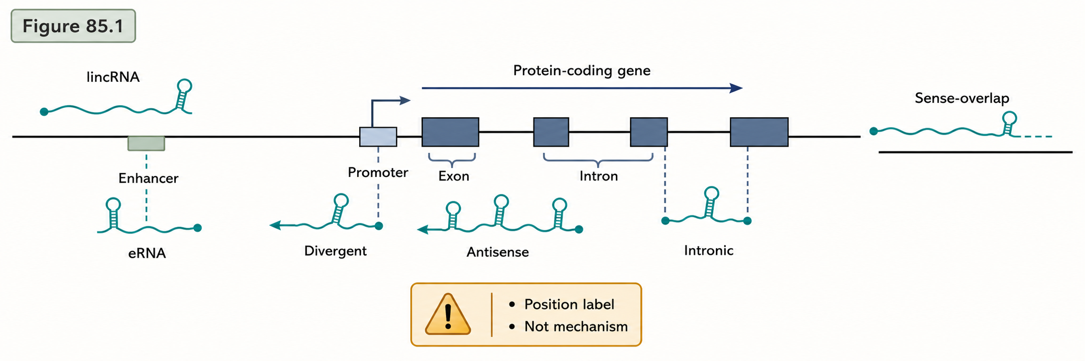
Figure 90.1. Genomic-position classes of lncRNAs. “Position-based lncRNA classes. The same RNA polymerase II transcript can be classified differently as neighboring gene models, isoforms, or coding-potential calls change.”
The instability of lncRNA classification follows from changes in both evidence and annotation boundaries. If a neighboring protein-coding gene gains a newly annotated alternative first exon, a previously intergenic lncRNA may become sense-overlapping. If long-read sequencing reveals that two short transcript models are parts of one longer isoform, separate lncRNA entries may collapse into one locus. If ribosome profiling and proteomics identify a functional micropeptide, a former lncRNA may be reclassified as a protein-coding or bifunctional transcript. If a database changes its coding-potential threshold, the same locus may move between “processed transcript,” “lncRNA,” “pseudogene transcript,” and “protein-coding candidate” categories.
This instability does not mean lncRNA biology is illusory. Protein-coding annotation also changes as transcript evidence improves. The difference is that protein-coding genes have a strong independent anchor: conserved protein sequence and open reading frame structure. Many lncRNAs lack an equivalent anchor, so their transcript models depend more strongly on sample coverage, sequencing depth, strand specificity, and computational assembly rules. A mature lncRNA annotation should therefore state what is known: the locus is transcribed; the transcript has a cap, splice junctions, or poly(A) site; the transcript accumulates in a specific compartment; the transcript lacks coding evidence under tested conditions; and the transcript has or lacks functional evidence.
Box 90.1. Classification Labels Are Not Mechanisms
A lncRNA label describes where a transcript model sits relative to other genome features; it does not say how the RNA works. An antisense lncRNA overlaps another gene on the opposite strand, but antisense position can reflect independent transcription, read-through, promoter collision, local chromatin state, or a mature RNA that base-pairs with the sense transcript. An intronic lncRNA may be an independent transcript, an unspliced host-gene fragment, or a stable intron-derived RNA. An enhancer RNA may be a signal of enhancer activity, a short-lived transcription byproduct, or in some cases part of a regulatory mechanism. When reading a lncRNA paper or catalog, translate class names into testable questions: What is the strand? Are the boundaries supported? Does the mature RNA accumulate? Is the phenotype separable from DNA elements and transcription through the locus?
90.2. Transcription, splicing, capping, polyadenylation, and localization
Most lncRNAs in mammalian annotations are produced by RNA polymerase II. RNA polymerase II transcription couples RNA synthesis to 5′ capping, splicing, 3′ end formation, and quality control through the carboxy-terminal domain of the polymerase and associated processing factors. The first physical product is not a finished lncRNA but a nascent transcript emerging from chromatin. As with pre-mRNA, the nascent 5′ end can receive a 7-methylguanosine cap. This cap protects the RNA from 5′ exonucleases and provides a binding platform for cap-associated complexes. Cap evidence is therefore a strong sign that a lncRNA is an independent transcription product rather than a random degradation fragment.
lncRNA splicing is diverse. Some lncRNAs contain multiple introns and splice junctions resembling mRNA architecture. Others are single-exon transcripts, inefficiently spliced transcripts, or transcripts with retained introns. Splicing can stabilize a lncRNA by recruiting exon-junction-associated factors and promoting proper processing. Conversely, weak splice sites can retain a transcript in the nucleus or expose it to nuclear decay. The imprinted Airn transcript in mouse is an instructive example of an atypical RNA polymerase II transcript that evades efficient splicing and remains nuclear rather than behaving like a typical exported mRNA. Such cases show that “RNA polymerase II transcript” does not automatically mean “mRNA-like itinerary.”
3′ end formation also separates lncRNA subclasses. Many lncRNAs are cleaved and polyadenylated by the canonical cleavage and polyadenylation machinery. Poly(A)-selected RNA sequencing therefore detects many lncRNAs efficiently. Other lncRNAs are nonpolyadenylated or processed by unusual mechanisms. MALAT1 and NEAT1 illustrate how specialized 3′ end processing can produce stable nuclear RNAs with maturation products distinct from conventional polyadenylated mRNA. Recent structural work on MALAT1 maturation and mascRNA biogenesis, and separate work on NEAT1 maturation and menRNA instability, reinforces the point that some lncRNA ends are generated by dedicated RNA structural and enzymatic contexts rather than by simple canonical polyadenylation.
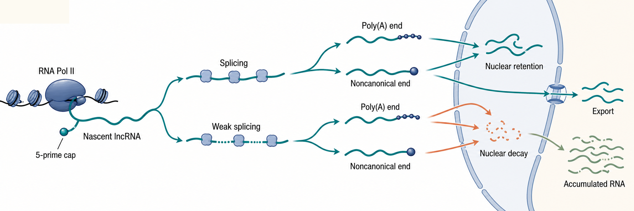
Figure 90.2. Processing itinerary of lncRNA transcripts. “Processing routes determine lncRNA accumulation. Capping, splice-site use, 3′ end formation, nuclear retention, export, and decay jointly determine which lncRNA molecules are detectable.”
Localization is both a consequence and a determinant of lncRNA biology. A lncRNA retained near its transcription site can act locally by recruiting proteins, altering chromatin-associated complexes, changing transcriptional interference, or organizing nuclear domains. A lncRNA enriched in nuclear speckles may influence RNA processing environments or reflect binding to speckle-associated factors. A lncRNA exported to the cytoplasm may interact with RNA-binding proteins, ribosomes, miRNAs, or decay machinery. Localization assays must distinguish chromatin-associated RNA, nucleoplasmic RNA, nuclear-body-enriched RNA, and cytoplasmic RNA. Fractionation can be contaminated by incompletely separated compartments, while imaging may miss low-copy transcripts or merge nascent transcription sites with mature RNA accumulations.
The processing itinerary creates several practical detection biases. Poly(A)-selected RNA sequencing misses nonpolyadenylated lncRNAs and undercounts unstable nuclear species. Ribosomal RNA depletion followed by total RNA sequencing captures more nonpolyadenylated and unspliced material but also increases background from pre-mRNAs, read-through transcripts, and partially degraded RNA. Short-read assembly can split or merge isoforms incorrectly, especially at repetitive loci. Long-read RNA sequencing improves isoform continuity but depends on RNA integrity, library chemistry, and read depth. For lncRNAs, method choice is not a minor technical detail; it can determine whether the transcript appears to exist.
Box 90.2. Processing Clues That Define a lncRNA Molecule
Treat a lncRNA annotation as a molecular itinerary rather than a name alone. Ask first whether the 5′ end is capped or otherwise defined; this separates independent transcription from fragments. Next ask whether introns are removed, retained, or absent; splicing can stabilize a transcript, but weak splice sites can promote nuclear retention or decay. Then ask how the 3′ end forms: canonical cleavage and polyadenylation, nonpolyadenylated maturation, or an uncertain assembly boundary. Finally ask where the RNA resides and how fast the RNA turns over. A polyadenylated, efficiently spliced, exported lncRNA will be captured by different assays and face different decay pathways from a chromatin-retained, weakly spliced, nonpolyadenylated transcript. Processing evidence is therefore not a decorative database field; it defines which RNA molecule exists in the cell.
90.3. Conservation at sequence, structure, synteny, and expression levels
Conservation is often invoked in lncRNA biology because functional DNA, RNA, and protein elements tend to be preserved by natural selection. For protein-coding genes, conservation is measured strongly through open reading frame preservation and amino acid sequence constraint. For lncRNAs, conservation must be parsed into several layers. Primary RNA sequence conservation means that homologous nucleotides remain alignable and constrained across species. This kind of conservation is present for some lncRNA regions but is generally weaker and patchier than for protein-coding exons. Low sequence conservation can reflect rapid turnover of nonfunctional transcription, but it can also reflect function carried by short motifs, RNA structure, transcriptional act, or locus position rather than by a long continuous sequence.
Structural conservation is harder to test. An RNA structure can be conserved even when many individual nucleotides change, provided compensatory substitutions preserve base pairing or higher-order architecture. The difficulty is that long RNAs can fold into many alternative structures, computational predictions are uncertain, and in vivo structure depends on proteins, transcription kinetics, modifications, and cellular conditions. A claimed conserved lncRNA structure is strongest when comparative covariation, biochemical probing, mutational rescue, and functional perturbation converge. A predicted hairpin alone is weak evidence because random long RNAs can contain many plausible local structures.
Syntenic conservation asks whether a transcript arises from a comparable genomic neighborhood across species. For example, a lncRNA near a developmental regulator may occupy a conserved interval even if its sequence is poorly alignable. Synteny can suggest that a locus has been preserved as part of a regulatory landscape. However, synteny alone is not proof that the RNA molecule is conserved as a functional product. The DNA regulatory element, promoter activity, transcription through the region, or local chromatin architecture may be the conserved feature. Expression conservation adds another layer: a transcript may be produced in comparable tissues, developmental stages, or cell states across species. Expression conservation is most persuasive when orthologous cell types are carefully matched and when detection methods have comparable sensitivity.
Table 90.1. Conservation evidence types for lncRNAs. Make conservation multidimensional and prevent collapse into sequence identity alone.
| Conservation layer | What is compared | Strongest evidence | Common false inference | Useful controls |
|---|---|---|---|---|
| Primary sequence | Alignable nucleotide blocks across orthologous loci or transcript exons. | Constraint above neutral flanks, preserved splice or end signals, and reproducible transcript boundaries. | Weak full-length identity proves nonfunction. | Neutral-region comparison, repeat masking, mappability checks, and coding/pseudogene exclusion. |
| Local motifs | Short RNA-binding, splice, poly(A), repeat-derived, or structural sequence elements. | Motif retention at comparable positions with matched expression, localization, or perturbation support. | A single motif match proves a conserved lncRNA mechanism. | Shuffled-sequence backgrounds, repeat-family controls, motif density tests, and orthogonal binding evidence. |
| RNA structure | Base-pairing patterns, local hairpins, higher-order folds, or structure-dependent processing elements. | Comparative covariation plus probing, mutational rescue, or structural data in relevant cells. | A predicted hairpin is enough to call conserved structure. | Random-folding nulls, covariation models, in vivo probing, and compensatory/disruptive mutants. |
| Synteny | Relative genomic neighborhood, nearby genes, and conserved regulatory interval. | Transcript initiation from an orthologous interval with boundary and promoter support in matched species. | Synteny proves the mature RNA product is conserved and causal. | Matched genome annotations, orthologous TSS and 3′ end evidence, and DNA-versus-RNA perturbation tests. |
| Promoter/chromatin context | Transcription start sites, enhancer/promoter marks, factor occupancy, and bidirectional transcription. | Conserved promoter activity or chromatin state in comparable cell types with RNA evidence. | Conserved chromatin marks mean the mature lncRNA is functional. | Nascent-versus-mature RNA assays, cell-type-matched epigenomes, and promoter-only control models. |
| Expression context | Tissue, developmental stage, stress state, disease state, or matched cell-type expression. | Orthologous cell types express the transcript with comparable timing, compartment, and processing state. | Absence from one catalog proves species absence. | Comparable library protocols, matched depth, single-cell or bulk validation, and positive-control loci. |
The most defensible conservation analysis therefore treats lncRNAs as composite loci. A lncRNA locus may show conserved promoter marks, syntenic position, a few conserved sequence blocks, tissue-specific expression, and one conserved RNA-binding motif without conserving the full transcript body. Another locus may show strong human-specific or primate-specific transcription driven by lineage-specific transposable elements. Such lineage specificity may still be biologically meaningful in a lineage, but it should not be confused with deep evolutionary conservation. Conversely, absence from a current catalog in mouse or zebrafish may reflect incomplete annotation rather than true absence.
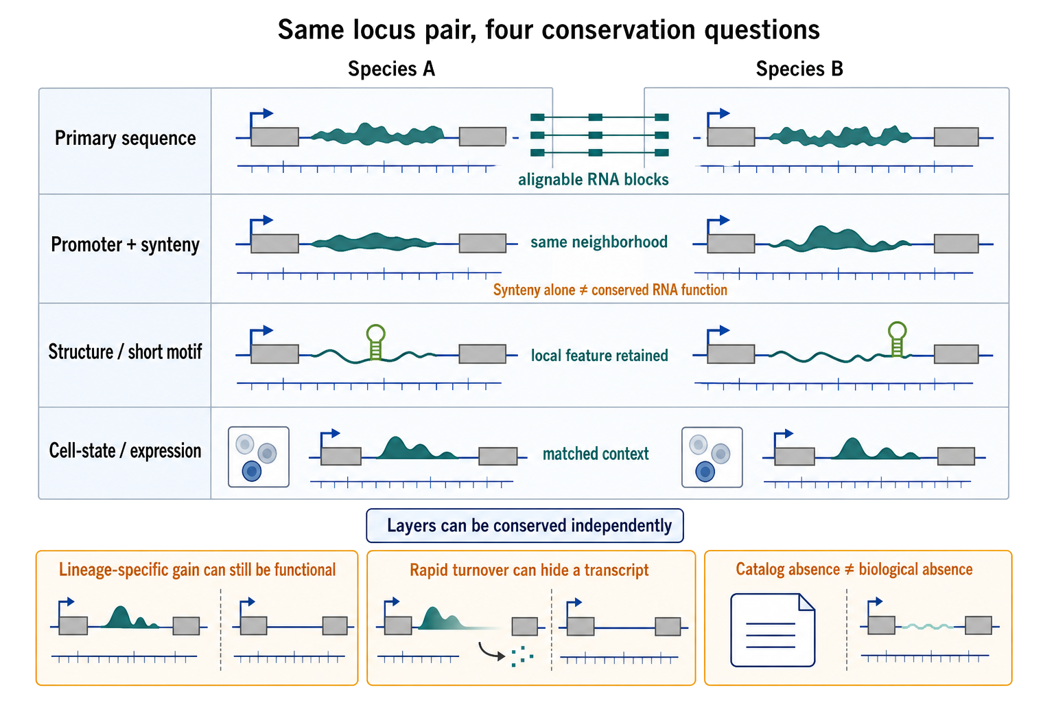
Figure 90.5. Four meanings of lncRNA conservation. “Four meanings of lncRNA conservation. The same locus pair can preserve alignable sequence, promoter and genomic neighborhood, a local structural or binding motif, or expression in a matched cell state independently. Lineage-specific transcripts can be functional without deep conservation, and rapid turnover or incomplete annotation can create false absence.”
90.4. Turnover, copy number, noise, and cell-type specificity
Many lncRNAs are expressed at lower steady-state abundance than typical mRNAs. Steady-state abundance reflects both synthesis and degradation. A low-copy lncRNA may be transcribed rarely, degraded quickly, retained at a single locus, expressed in only a small cell population, or diluted by averaging across heterogeneous tissue. These alternatives have different biological meanings. A transcript present at one or two molecules per nucleus can still have a local cis-regulatory role if the relevant target is the locus from which it is transcribed. The same copy number would be less plausible for a stoichiometric trans-acting role requiring contact with thousands of targets, unless the lncRNA is highly enriched in a specific subnuclear compartment or acts catalytically through associated proteins.
RNA decay pathways strongly shape lncRNA catalogs. Nuclear exosome activity, cap-binding surveillance, deadenylation, decapping, and 5′-to-3′ exonucleolysis can remove unstable lncRNAs before they accumulate. Promoter-proximal transcripts and enhancer RNAs are often short-lived, which makes them sensitive indicators of active regulatory regions but difficult to annotate as stable RNA genes. Some lncRNAs are stabilized by triple-helical structures, protein binding, unusual 3′ end formation, or nuclear-body incorporation. Thus, the difference between an annotated lncRNA and an undetected unstable transcript can be a difference in RNA half-life as much as in transcription rate.
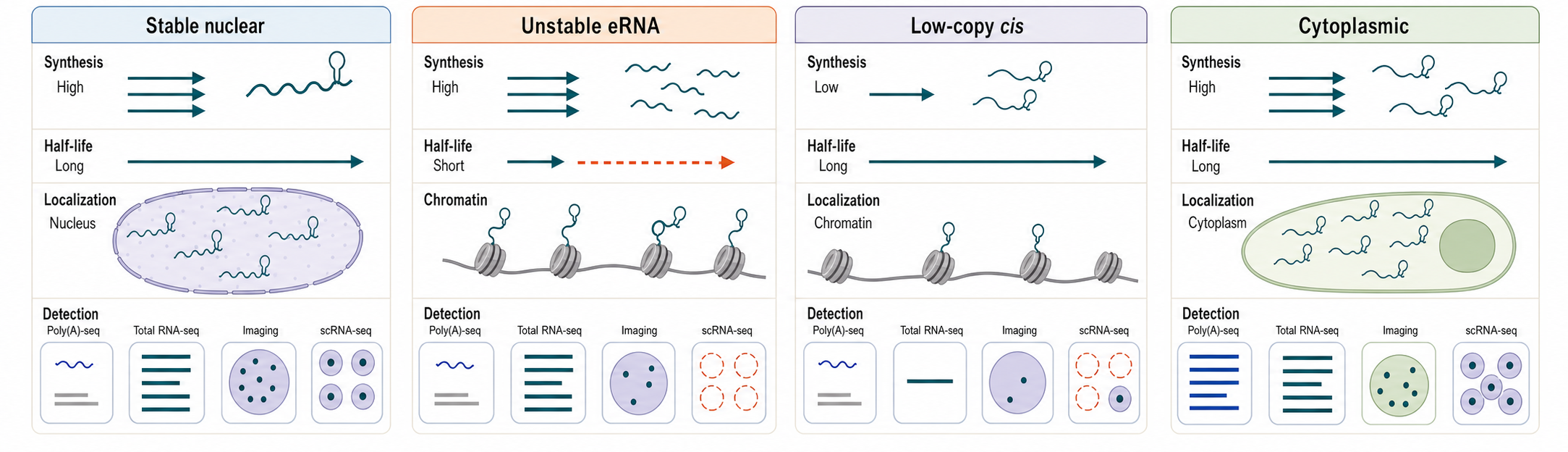
Figure 90.3. Abundance as synthesis minus degradation plus localization. “Low abundance is not a single biological state. A lncRNA may be rare because it is weakly transcribed, rapidly degraded, confined to a few cells, retained locally, or poorly captured by the assay.”
Cell-type specificity is a repeated theme in lncRNA studies. A lncRNA may be highly restricted to embryonic stem cells, neurons, immune cell subsets, germ cells, tumor states, or stress responses. Such specificity can make lncRNAs useful markers, but it also makes them vulnerable to sampling bias. A transcript absent from a bulk tissue sample may be present in a rare cell type. A transcript apparently specific to a disease sample may reflect altered cell composition rather than disease regulation within a cell type. Single-cell RNA sequencing helps resolve cellular sources but introduces dropout, sparse coverage, and 3′ or 5′ end bias. Many lncRNAs are long, low abundance, and incompletely captured by standard single-cell protocols, so absence from single-cell data should not be overinterpreted.
Noise is not merely a technical nuisance. Eukaryotic transcription is bursty, meaning promoters switch between active and inactive states. A weak promoter can produce intermittent transcripts that appear in only a fraction of cells. Some lncRNA expression patterns may reflect regulated bursts; others may reflect permissive low-level transcription from open chromatin. Distinguishing regulated low-copy expression from transcriptional noise requires replication, matched controls, orthogonal detection, and attention to chromatin context. A useful standard is to ask whether the transcript has reproducible boundaries, regulated abundance, coherent localization, and evidence that perturbing the RNA or its production changes a defined phenotype.
90.5. Annotation artifacts and evidence thresholds
lncRNA annotation is unusually artifact-prone because many candidate transcripts sit near the detection limit and lack protein-coding anchors. The first artifact class is incomplete transcript assembly. Short reads can create fragmented models when coverage is low, or fused models when read-through transcription crosses gene boundaries. Strand ambiguity can misassign reads from a coding gene to an antisense lncRNA if the library is not strongly strand-specific. Internal priming can create false poly(A) sites when oligo(dT) primers bind A-rich genomic sequence. Genomic DNA contamination can mimic intron-containing transcripts. Degraded RNA can create apparent short transcript fragments.
The second artifact class is biological byproduct mistaken for stable gene product. Pervasive transcription generates many unstable RNAs from promoters, enhancers, terminators, repeats, and open chromatin. Some byproducts are real RNA molecules, but not every real RNA molecule should be annotated as a stable lncRNA gene with implied function. Annotation should separate “transcription observed” from “mature transcript defined” and from “functional RNA product supported.” This separation is especially important for enhancer RNAs and promoter-upstream transcripts, where transcription may report regulatory activity without the RNA molecule being the effector.
The third artifact class is coding-potential misclassification. Some lncRNA transcripts contain small open reading frames. Ribosome profiling may show ribosome occupancy on these open reading frames, but ribosome occupancy alone does not prove production of a stable functional peptide. Conversely, absence of proteomic detection does not prove noncoding status, because small peptides can be unstable, cell-type-specific, or below mass spectrometry detection limits. Coding-potential decisions are strongest when open reading frame conservation, translation initiation evidence, ribosome phasing, peptide detection, and genetic separation of RNA and peptide functions are considered together.
Table 90.2. Annotation artifact checklist and controls. Give readers a practical diagnostic framework for evaluating lncRNA catalogs.
| Artifact or ambiguity | How it appears | Why lncRNAs are vulnerable | Control or evidence threshold |
|---|---|---|---|
| Read-through transcription | Coverage extends across terminators or joins a neighboring gene into a fused model. | Low abundance and weak boundaries make distal read-through look like an independent locus. | Independent 5′ and 3′ ends, long-read isoform support, termination evidence, and upstream-gene controls. |
| Strand ambiguity | Reads from a coding gene are assigned to an opposite-strand lncRNA model. | Antisense annotation depends directly on reliable strand assignment. | Strongly strand-specific libraries, strand-aware alignment, and strand-specific probes or primers. |
| Internal priming | Apparent poly(A) sites occur next to A-rich genomic sequence. | 3′ end evidence often anchors low-copy transcript models. | Downstream genomic-A filters, independent 3′ end methods, and non-oligo(dT) or direct RNA support. |
| Genomic DNA contamination | Intronic or unspliced signal persists without processing evidence. | Intronic and weakly spliced lncRNAs can resemble DNA-derived reads. | DNase and no-reverse-transcriptase controls, cap or splice evidence, and RNA-only enrichment. |
| Degraded RNA | Short fragments, truncated models, or strong 5′ or 3′ bias dominate the signal. | Low-copy transcripts are easily confused with stable degradation fragments. | RNA integrity metrics, replicate fragment patterns, cap evidence, and supported transcript ends. |
| pre-mRNA fragments | Host-gene introns or nascent RNA are assembled as independent intronic lncRNAs. | Intronic lncRNAs overlap genuine host-gene processing intermediates. | Independent promoter or poly(A) evidence, mature-RNA fractionation, and host-transcript splice controls. |
| Repeat multimapping | Reads collapse across repeats, paralogs, or repeat-derived exons. | Many lncRNAs contain transposable-element sequence and lineage-specific repeats. | Mappability filters, unique-k-mer evidence, long reads, repeat-aware assignment, and probe-specific imaging. |
| Coding-potential misclassification | Small ORFs or ribosome-profiling signal challenge the noncoding label. | lncRNA status is partly defined by absence of strong peptide evidence. | ORF conservation, initiation evidence, ribosome phasing, peptide detection, and RNA-versus-peptide tests. |
| Single-cell dropout | Sparse reads create false absence or exaggerated cell-type specificity. | Long, low-abundance lncRNAs are inefficiently captured and often end-biased. | Deep bulk or targeted validation, smFISH, matched cell-type comparisons, and detection-rate reporting. |
A practical evidence threshold for lncRNA annotation should include multiple tiers. Tier 1 is transcript existence: reproducible strand-specific reads, cap or 5′ end evidence, splice junctions when present, and 3′ end support. Tier 2 is transcript model quality: defined boundaries, isoform consistency, evidence across independent samples or technologies, and exclusion of read-through or contamination. Tier 3 is molecular behavior: abundance, half-life, localization, processing state, and cell-type specificity. Tier 4 is coding assessment: absence or presence of credible peptide-coding evidence. Tier 5 is functional evidence: perturbation, rescue, endogenous editing, allele-specific analysis, or separation of promoter, transcription, and RNA-product effects. This chapter is mainly about Tiers 1 to 4; Chapter 91 focuses on Tier 5.
Box 90.3. Evidence Language for lncRNA Annotation
Use cautious verbs that match the evidence. “Transcribed” means reads, nascent signal, or 5′ end data show RNA production from a locus. “Transcript model” means exon chains and ends have enough support to define a putative molecule. “Low coding potential” means available open reading frame, ribosome-profiling, conservation, and proteomic data do not support a conventional protein product; it is not proof that translation never occurs. “Conserved locus” means some feature such as synteny, promoter activity, motif content, structure, or expression context is preserved; it is not automatically conservation of the mature RNA product. “Functional lncRNA” should be reserved for cases where perturbation and rescue, endogenous editing, or separation-of-function experiments implicate the RNA molecule or its production in a defined phenotype.
Experimental Foundations and Evidence
Bulk RNA sequencing established that mammalian genomes produce many long transcripts outside protein-coding annotations. However, early catalogs were strongly influenced by library type. Poly(A)-selected libraries favored stable polyadenylated lncRNAs; total RNA libraries uncovered more nonpolyadenylated and immature transcripts; strand-specific protocols improved antisense assignment. Cap analysis of gene expression, RAMPAGE-like 5′ end mapping, and related methods helped distinguish genuine transcription start sites from assembled fragments. 3′ end sequencing helped map cleavage and polyadenylation sites, although internal priming controls remain necessary.
Long-read sequencing adds a different kind of evidence by connecting exons and ends across full-length molecules. For lncRNAs with multiple isoforms, repetitive sequence, or low coverage, long reads can reveal that short-read assemblies have over-split a locus or incorrectly connected distant exons. Yet long-read evidence is not automatically decisive. Reverse transcription artifacts, template switching, incomplete cDNA synthesis, RNA degradation, and insufficient depth can still distort models. Direct RNA sequencing avoids some cDNA artifacts but has its own error profile and requires enough intact RNA.
Imaging methods such as RNA fluorescence in situ hybridization provide molecule-level localization and copy-number estimates. Imaging can show whether a lncRNA accumulates at one or two chromosomal foci, fills a nuclear body, distributes through the nucleoplasm, or reaches the cytoplasm. This evidence is especially important for low-copy nuclear lncRNAs because bulk sequencing cannot distinguish a few local molecules from diffuse low-level accumulation. Imaging also exposes heterogeneity across cells. The limitation is probe specificity: repetitive regions, overlapping genes, and related paralogous sequences can create misleading signal unless probe design and controls are rigorous.
Metabolic labeling and transcriptional inhibition assays estimate RNA half-life. These methods show that many regulatory-region-associated transcripts turn over quickly, while a subset of lncRNAs is stable. Interpretation requires care because transcriptional inhibitors perturb cell physiology, and metabolic labeling efficiency can differ across nucleotide pools and cell states. Subcellular fractionation and chromatin-associated RNA sequencing can assign broad localization categories, but physical fraction purity and nascent RNA contamination must be checked.
Perturbation screens increasingly test lncRNA dependencies. CasRx and other RNA-targeting approaches can deplete transcripts without directly cutting DNA, while CRISPR interference can repress transcription initiation and CRISPR deletion can remove promoters, exons, splice sites, or regulatory DNA. Each perturbation has a different interpretation. RNA knockdown tests RNA-product contribution but can have off-target effects and incomplete depletion. Promoter repression tests transcriptional initiation but also removes promoter-associated DNA activity. Genomic deletion can remove enhancers or alter chromatin independently of the RNA. Strong functional annotation therefore requires matching the perturbation to the claim.
Biological Contexts Across Organisms and Cell States
Mammalian lncRNA catalogs are the richest because mammalian transcriptome projects sampled many tissues, developmental stages, disease states, and cell types. Mammalian genomes also contain many transposable elements that can donate promoters, splice sites, polyadenylation signals, and RNA-binding motifs to lncRNA loci. Repeat-derived sequence can make lncRNAs lineage-specific, rapidly evolving, and technically difficult to map. Some repeat-derived lncRNA sequence may be functional by recruiting repeat-binding proteins or forming RNA-DNA interactions, but repeats also increase multimapping and false assembly risk.
Plants have extensive noncoding transcription as well, including lncRNAs associated with flowering time, stress responses, chromatin regulation, and small-RNA pathways. Plant lncRNA annotation faces many of the same issues as mammalian annotation, but plant genomes differ in polyploidy history, transposon composition, developmental plasticity, and RNA-directed DNA methylation pathways. Fungal and invertebrate systems provide powerful genetics, yet lncRNA conservation across distant taxa can be limited and annotation density can vary by research community.
Developmental systems often reveal lncRNAs because stage-specific transcription creates sharp expression windows. Embryogenesis, germ-cell development, neural differentiation, immune activation, and stress responses are especially rich contexts. Cancer studies report many lncRNA changes, but cancer expression requires careful interpretation because tumors differ in cell composition, copy-number alterations, chromatin state, proliferation, hypoxia, and immune infiltration. A lncRNA associated with prognosis may be a cell-state marker rather than a direct driver. Clinical relevance therefore depends on whether the lncRNA is robustly detected, mechanistically linked, and independently validated.
Technology, Computational, Clinical, and Engineering Links
lncRNA annotation depends on computational decisions. Transcript assemblers infer exon chains from read coverage and splice junctions. Coding-potential tools combine open reading frame length, codon composition, conservation, sequence similarity, and sometimes machine-learning features. Comparative genomics tools evaluate sequence constraint and synteny. Single-cell analysis pipelines decide whether sparse lncRNA reads are retained, filtered, imputed, or ignored. These choices are not neutral. A stringent pipeline reduces false positives but may miss rare cell-type-specific lncRNAs; a permissive pipeline increases discovery but expands artifact burden.
Clinical and translational work uses lncRNAs as biomarkers, disease-associated loci, and possible therapeutic targets. A clinically useful lncRNA biomarker must be measurable reproducibly in the relevant sample type, whether tumor tissue, blood, exosomes, or another biospecimen. Low abundance and isoform ambiguity can make assay design difficult. Therapeutic targeting raises additional questions: where is the lncRNA located, which cells express it, whether the disease mechanism depends on the RNA product, and whether antisense oligonucleotides, small interfering RNAs, RNA-targeting CRISPR systems, or small molecules can reach the relevant compartment. Nuclear-retained lncRNAs may be suitable for RNase H-dependent antisense oligonucleotides, while cytoplasmic lncRNAs may be more accessible to RNA interference.
Engineering applications use lncRNA promoters, localization signals, repeat modules, and scaffold principles, but rational design remains early. The classification lessons from natural lncRNAs apply directly to engineering: a designed transcript must be processed, localized, stable enough, and noncoding or coding as intended. Without measuring these properties, a synthetic long RNA construct cannot be assumed to behave like a natural lncRNA simply because it lacks a long open reading frame.
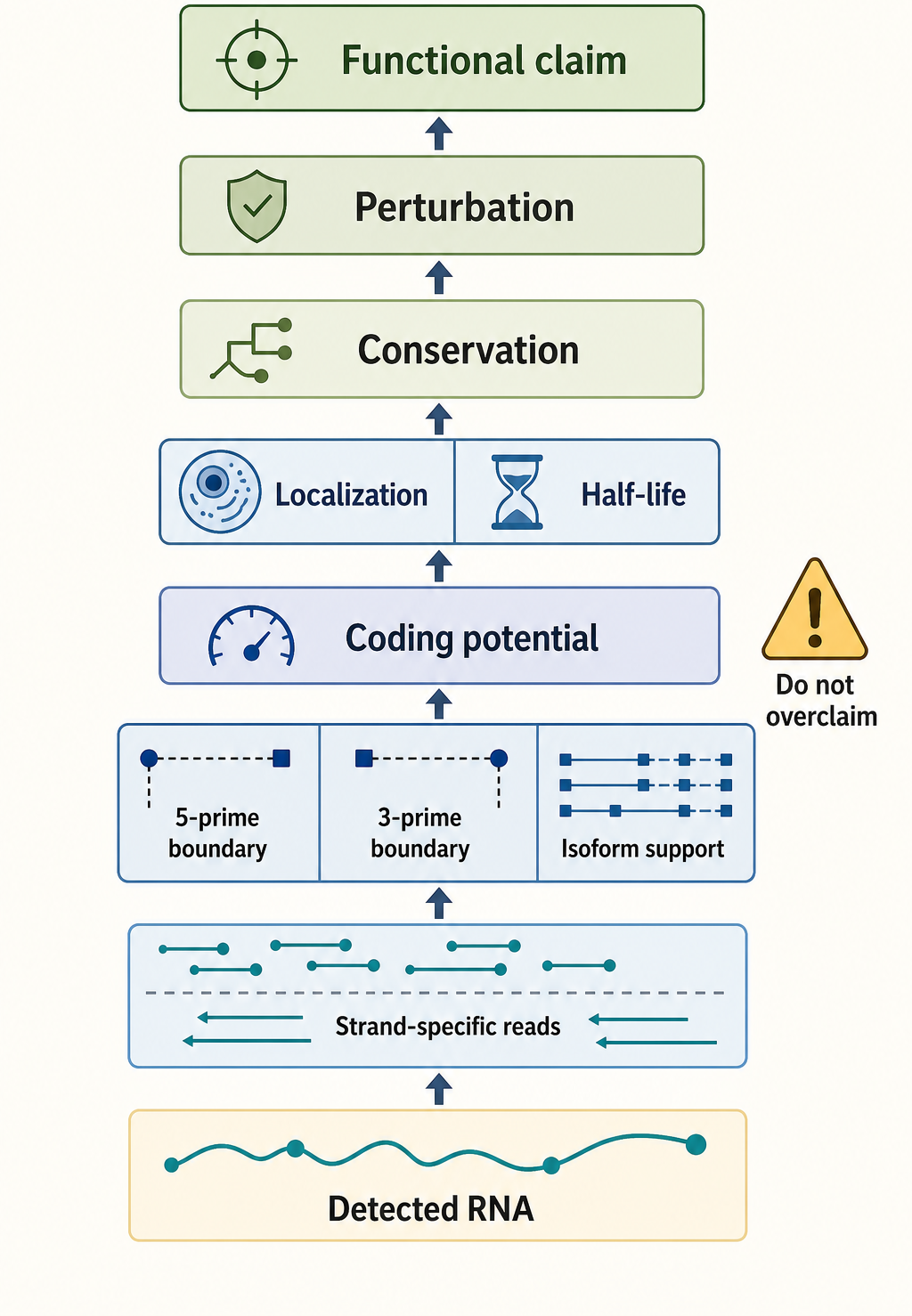
Figure 90.4. Evidence ladder for lncRNA annotation. “Evidence tiers in lncRNA annotation. A transcript model should not be promoted from detected RNA to functional RNA without boundary, coding, localization, and perturbation evidence appropriate to the claim.”
Recent Consensus
The current consensus is that lncRNAs are a real and important part of eukaryotic transcriptomes, but the category is heterogeneous and should not be treated as a unified functional mechanism. The review by Mattick and colleagues emphasizes definitions, functions, challenges, and recommendations, and it is the strongest local reference anchor for the broad framing of this chapter. The consensus also recognizes that annotation should be evidence-graded. A high-quality lncRNA model should have reproducible transcription evidence, strand assignment, boundary support, coding assessment, and ideally orthogonal validation of localization or processing.
There is also consensus that many lncRNA loci may act through mechanisms other than the mature RNA molecule. A phenotype from deleting a lncRNA locus may result from loss of a DNA enhancer, promoter competition, transcriptional interference, splicing-linked chromatin effects, or the RNA product. The proper interpretation depends on perturbation design. Finally, the field increasingly accepts that conservation can be partial and layered. Sequence conservation is valuable when present, but lncRNA conservation can also involve syntenic position, promoter logic, expression context, structural elements, or short motifs.
Open Questions, Controversies, Deprecated Models, and Common Misconceptions
Open questions:
- How many annotated lncRNAs have selected biological functions? The answer depends on the evidence threshold. If “function” means reproducible regulated transcription, many loci qualify. If “function” means an RNA product with a separable causal role at endogenous expression, the number is smaller and still being determined.
- How can researchers classify bifunctional transcripts that have both RNA-mediated activity and peptide-coding potential? These transcripts challenge the binary coding/noncoding division.
Common misconceptions:
- “Noncoding means nonfunctional.” That inference is wrong: rRNAs, tRNAs, snRNAs, snoRNAs, miRNAs, and many characterized lncRNAs are functional without coding for proteins. The opposite misconception is also common: because some noncoding RNAs are functional, any detected lncRNA must be functional. Detection is only the first evidence layer.
- “Poor sequence conservation proves nonfunction.” Poor conservation weakens some evolutionary arguments, but it does not exclude lineage-specific function, structural conservation, or locus-based mechanisms. A final misconception is that antisense position proves antisense regulation. Overlap creates a hypothesis; it does not establish mechanism.
Deprecated or weakened claims:
- Broad claims that most of the genome is functional merely because it is transcribed. Pervasive transcription is real, but transcriptional activity alone is insufficient to assign gene-like function to every transcript. A more durable model is evidence stratification: describe what has been observed, identify plausible mechanisms, and reserve strong functional language for cases with causal tests.