Chapter 109. Interferon-Stimulated RNA Pathways, Mitochondrial dsRNA, and Self-Nonself Discrimination
Scope Note
This chapter explains how interferon responses reshape cellular RNA metabolism, how interferon-stimulated genes restrict viral RNAs, how self double-stranded RNA is normally edited or hidden from cytosolic sensors, and how mitochondrial double-stranded RNA can become an inflammatory signal during organellar stress. The chapter links molecular mechanisms to infection models, interferonopathies, cancer immunology, cellular senescence, and systems-level immune profiling. Chapter 108 treats the core RNA sensors and proximal signaling adaptors; Chapter 109 focuses on the RNA environment created downstream of interferon and on the boundary between antiviral defense and autoinflammation.
Executive Summary
Interferons do not act only as soluble warning signals. They remodel the biochemical environment in which RNAs are synthesized, modified, translated, stored, sensed, and degraded. Type I and type III interferons induce hundreds of interferon-stimulated genes (ISGs), including sensors, nucleases, helicases, RNA-binding proteins, translation regulators, nucleotide-metabolism enzymes, and signaling adaptors. Together these proteins create an antiviral RNA environment: viral RNAs are more likely to be recognized, translation may be suppressed, replication complexes may be destabilized, and abnormal RNAs may be routed into decay or inflammatory signaling.
This antiviral state is powerful because many viruses expose RNA patterns that differ from ordinary host transcripts. Examples include uncapped 5′ triphosphate RNA, long double-stranded RNA (dsRNA), unusual base modifications, structured replication intermediates, or RNAs in compartments where host RNA is normally scarce. The same logic is dangerous because host cells also make endogenous dsRNA. Repetitive elements, convergent transcription, circular RNA structures, antisense transcripts, and mitochondrial bidirectional transcription can all generate RNA duplexes. Self-nonself discrimination is therefore not a single receptor property. It is a system-level outcome produced by compartmentalization, RNA processing, editing, modification, turnover, binding proteins, and receptor thresholds.
ADAR1 is central to this balance. The interferon-inducible ADAR1 p150 isoform edits adenosines to inosines in endogenous dsRNA, especially repetitive-element duplexes, and helps prevent inappropriate activation of MDA5 and PKR. Loss of ADAR1 editing can make self RNA resemble viral dsRNA, driving interferon production and severe autoinflammation. Primary studies now separate at least two protective functions: editing-dependent reduction of MDA5-stimulatory dsRNA and additional suppression of PKR activation, with context-dependent contributions from RNA-binding partners such as GGNBP2.
Mitochondrial dsRNA adds a second source of endogenous immunostimulatory RNA. Mammalian mitochondria transcribe both strands of a compact genome; incomplete processing, impaired degradation, altered RNA modification, or organellar damage can allow mitochondrial dsRNA to accumulate or escape into the cytosol. Recent work links cytosolic mitochondrial dsRNA to senescence-associated inflammation and to RNA modification-dependent control of mitochondrial RNA stability. The field is still clarifying which sensors dominate in each context, how mitochondrial RNA reaches the cytosol, and how organellar stress integrates with classic RIG-I-like receptor, PKR, OAS/RNase L, and cGAS-STING pathways.
Concept Inventory
- Interferon-stimulated gene (ISG): a gene whose expression increases after interferon signaling, usually through JAK-STAT-dependent transcriptional programs. ISGs include direct antiviral effectors, amplifiers of innate immune signaling, regulators that restrain inflammation, and genes whose antiviral role is indirect or context-dependent.
- Antiviral RNA environment: the interferon-induced cellular state in which RNA sensors, RNA-binding proteins, decay enzymes, translation inhibitors, and metabolic changes make viral RNA replication and expression less efficient. The term is a synthesis label, not a single pathway.
- Self dsRNA: endogenous double-stranded RNA produced by host transcription, RNA structure, repeat pairing, organellar transcription, or RNA processing defects. Self dsRNA is not harmless by definition; it becomes inflammatory when editing, sequestration, decay, or compartmental barriers fail.
- Nonself RNA: RNA interpreted by innate immune pathways as foreign or dangerous. Nonself status can reflect molecular features, such as 5′ triphosphate ends or long dsRNA, but it also depends on concentration, localization, modifications, associated proteins, and cellular state.
- RNA restriction factor: a host factor that reduces viral RNA replication, translation, stability, packaging, or spread. Some restriction factors directly bind viral RNA, whereas others change translation, nucleotide pools, RNA decay, membrane compartments, or immune signaling.
- ADAR1: an adenosine deaminase acting on RNA. ADAR1 edits adenosine to inosine in dsRNA. ADAR1 p110 is constitutive and mostly nuclear; ADAR1 p150 is interferon-inducible and shuttles between nucleus and cytoplasm. ADAR1 limits inappropriate recognition of endogenous dsRNA.
- MDA5: melanoma differentiation-associated protein 5, encoded by
IFIH1. MDA5 is a RIG-I-like receptor that senses long dsRNA and signals through MAVS to induce interferon and inflammatory genes. - Mitochondrial dsRNA: double-stranded RNA produced from mitochondrial transcripts, often involving complementary heavy-strand and light-strand RNAs. It can be degraded within mitochondria, retained in organelles, or, during stress, appear in the cytosol where it may activate innate immune pathways.
What to Know Before Reading This Chapter
Readers should understand the basic layout of innate RNA sensing from Chapter 108: RIG-I and MDA5 are cytosolic RNA helicase receptors; MAVS is the mitochondrial outer-membrane adaptor that links those receptors to interferon induction; PKR can inhibit translation after binding dsRNA; OAS enzymes synthesize 2′-5′ oligoadenylates that activate RNase L; and interferons induce a transcriptional antiviral state through JAK-STAT signaling. This chapter assumes that background but redefines each pathway where needed for self-nonself discrimination.
The running examples are viral replication intermediates, Alu-derived endogenous dsRNA, and mitochondrial dsRNA. Viral replication intermediates show why long dsRNA is useful as a danger signal. Alu-derived dsRNA shows why mammalian genomes create many self duplexes that must be edited or suppressed. Mitochondrial dsRNA shows why a host organelle can produce RNA with microbial-like features without infection.
109.1. Interferon induction and ISG-controlled RNA environments
Interferons are cytokines produced when cells detect infection, cellular damage, or inflammatory cues. Type I interferons, especially IFN-α and IFN-β, act on many cell types through the IFNAR receptor. Type III interferons, especially IFN-λ family members, signal through a more restricted receptor distribution and are prominent at epithelial barriers. Type II interferon, IFN-γ, has different immunological roles but can overlap with RNA-defense programs through macrophage activation, antigen presentation, and selected antiviral genes. The central point for RNA biology is that interferon signaling changes the fate of RNAs inside a cell.
The canonical pathway is conceptually simple. Interferon binds its receptor, receptor-associated Janus kinases phosphorylate STAT transcription factors, and STAT-containing complexes bind regulatory elements near interferon-stimulated genes. The resulting ISG program is broad. Some ISGs are receptors or receptor cofactors, such as RIG-I-like receptor components. Some are enzymes, such as OAS proteins, RNase L pathway components, ADAR1 p150, ISG15 conjugation machinery, and nucleotide-metabolism proteins. Some are RNA-binding or translation-control factors, such as IFIT proteins and PKR. Others change membranes, vesicle trafficking, antigen presentation, or cell death thresholds. The antiviral state is therefore not one molecular action but a coordinated shift in the probability that a viral RNA will be copied, translated, packaged, hidden, or destroyed.
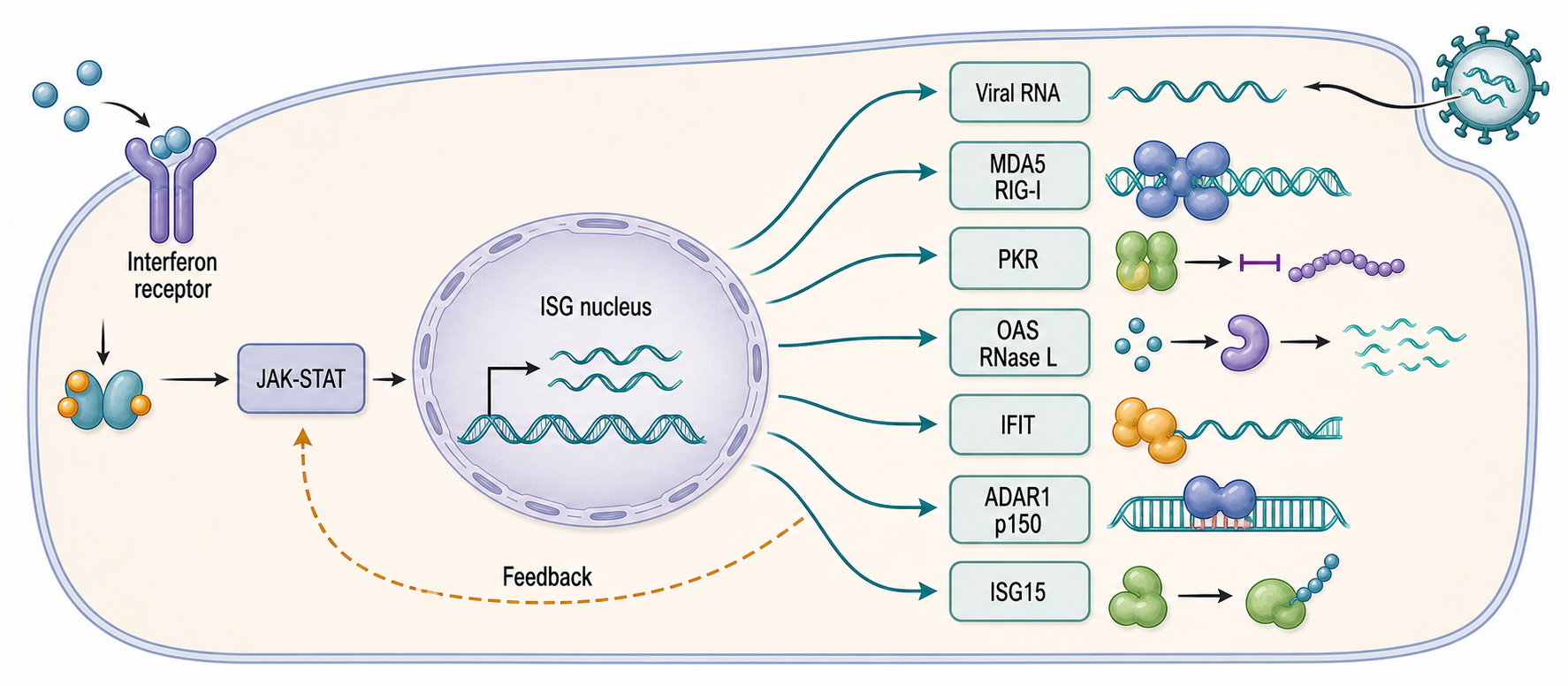
Figure 109.1. Interferon signaling remodels the RNA environment. Interferon does not create one antiviral mechanism; it raises and coordinates many RNA-centered barriers.
The phrase “ISG-controlled RNA environment” is useful because a viral RNA does not encounter one isolated sensor. A positive-sense RNA virus, for example, may expose replication intermediates to MDA5, produce capped or uncapped RNAs that compete with IFIT recognition, generate dsRNA that activates PKR or OAS, and rely on host translation machinery that has been rewired by interferon. A DNA virus may generate abundant nuclear or cytoplasmic viral transcripts and also express proteins that suppress ISG induction. Human cytomegalovirus U(L)26, for example, has been reported to attenuate IKKβ-mediated induction of ISG expression and protein ISGylation during infection. That example illustrates a general principle: viruses can antagonize not only the original sensor but also downstream RNA-state remodeling.
Interferon responses also have timing. Early after infection, basal sensors and preexisting restriction factors determine whether a cell detects viral RNA quickly. After interferon signaling begins, the cell expresses more sensors and effectors, raising sensitivity and broadening restriction. Later, negative regulators limit tissue damage. The same ISG can be protective in acute infection and pathogenic when chronically activated. This is why clinical interferon signatures are useful but not self-explanatory: they indicate pathway activation, not necessarily the initiating RNA species.
Box 109.1. Interferon Signature Is a Readout, Not a Diagnosis
An interferon signature is a pattern of interferon-stimulated gene mRNAs or proteins, not a molecular diagnosis. It means that a cell population has received interferon, activated overlapping STAT or IRF programs, or changed composition toward cells with high interferon tone. Before assigning viral infection, ask four questions. Which cell type carries the signal? Was interferon cytokine, receptor signaling, or only downstream gene expression measured? Is there direct evidence for viral RNA, viral protein, or infectious particles? Are endogenous triggers plausible, such as unedited repeat dsRNA, mitochondrial RNA release, RNA therapeutic exposure, DNA damage, or an inherited sensor variant? Strong interpretation links the signature to a candidate RNA ligand, a sensor branch, a time course, and a perturbation or rescue experiment.
109.2. RNA restriction factors and viral RNA decay
RNA restriction factors reduce viral replication by acting on RNA production, RNA translation, RNA stability, RNA localization, or the protein complexes that viral RNAs require. Some restriction factors are constitutively expressed and become more effective after interferon induction; others are strongly interferon-inducible. The category is functional rather than mechanistic. A restriction factor can be a nuclease, helicase, RNA-binding protein, translation factor antagonist, E3 ligase, membrane protein, metabolic enzyme, or signaling protein.
Viral RNA decay is one route to restriction. Host cells already maintain RNA quality-control pathways that distinguish ordinary mRNAs from aberrant transcripts. Nonsense-mediated decay, no-go decay, deadenylation-dependent decay, decapping, exosome-mediated decay, and endonucleolytic cleavage pathways all shape cellular transcriptomes. Viruses enter this environment with RNAs that may lack normal exon-junction architecture, carry unusual untranslated regions, form long structures, or be produced at abnormal locations and copy numbers. Upf1-mediated RNA decay pathways can target viral RNAs, although viruses differ in whether they are restricted by, evade, or exploit these pathways. KSHV studies show that nonsense-mediated RNA decay can target both cellular and viral RNAs and restrict viral gene expression in specific contexts.
Table 109.1. RNA restriction factors and RNA-centered antiviral mechanisms. RNA restriction factors recognize different viral RNA features or replication steps and can block translation, stability, copying, or packaging; antiviral association must be separated from direct RNA-centered mechanism.
| Factor or pathway | RNA feature/process | Primary effect | Example evidence | Caveat |
|---|---|---|---|---|
| IFIT cap discrimination | Uncapped RNA, 5′ triphosphate RNA, or incompletely methylated viral caps | Binds suspect 5′ ends and suppresses efficient viral RNA translation or replication | IFIT induction or loss changes cap-sensitive viral RNA expression in interferon-treated cells | Cap status is not the only determinant; many viruses cap, modify, or protein-shield their RNAs |
| PKR translation shutdown | Long or accessible dsRNA from viral replication, endogenous repeats, or structured host RNAs | Phosphorylates eIF2α and lowers global translation, limiting viral protein production | PKR activation follows dsRNA exposure and is restrained by ADAR1 p150 in self-dsRNA models | Translation loss can reflect stress or toxicity rather than selective viral RNA restriction |
| OAS/RNase L RNA cleavage | Cytosolic dsRNA that activates OAS enzymes and 2′-5′ oligoadenylate synthesis | Activates RNase L to cleave viral and host single-stranded RNAs | RNase L-dependent RNA cleavage and interferon amplification are observed after dsRNA-rich infection states | Cleavage is broad; antiviral specificity comes from activation context rather than exact RNA targeting |
| Upf1 and nonsense-mediated decay | Viral transcripts or RNA architectures accessible to host mRNA surveillance | Destabilizes susceptible viral RNAs and can restrict viral gene expression | Upf1-mediated decay reviews and KSHV NMD studies support viral RNA restriction | Effects are virus-specific; some viruses evade, exploit, or indirectly alter decay pathways |
| ADAR1 self-dsRNA editing | Endogenous repeat-derived or inverted-repeat dsRNA, especially in interferon-competent cells | A-to-I editing reduces MDA5 and PKR activation by self RNA | ADAR1 p150 loss activates MDA5 and PKR through separable mechanisms | ADAR1 can have isoform-specific and editing-independent effects; it is not simply a viral restriction factor |
| ISG15-linked modulation | Protein ISGylation of RNA-signaling or viral-replication environments | Tunes ISG induction, viral replication complexes, and antiviral protein function | HCMV U(L)26 attenuates IKKβ-linked ISG expression and protein ISGylation | ISGylation targets and outcomes differ by virus, cell type, and timing |
| Viral antagonists of RNA restriction | Viral RNA is shielded or host RNA-defense nodes are suppressed | Reduces sensing, RNA decay, translation shutdown, or downstream ISG amplification | DNA and RNA viruses encode factors that block ISG induction, RNA decay, or access to replication intermediates | Antagonist phenotypes are often pleiotropic and do not by themselves identify the initiating RNA ligand |
The OAS/RNase L pathway is a more explicitly antiviral RNA decay system. OAS enzymes bind dsRNA and synthesize 2′-5′ oligoadenylates, which activate RNase L. Activated RNase L cleaves single-stranded RNA, producing widespread RNA damage and sometimes secondary RNA fragments that amplify innate signaling. This can restrict viral replication by degrading viral and host RNAs, limiting translation, and pushing infected cells toward stress or death. The pathway is not precisely targeted to viral RNA; its antiviral specificity comes from dsRNA-dependent activation, compartmental context, and infection-induced thresholds.
Translation restriction can also behave like RNA restriction. PKR binds dsRNA and phosphorylates eIF2α, reducing cap-dependent translation. IFIT proteins can bind RNAs with atypical cap structures or exposed 5′ triphosphate features. ISGylation pathways can modify proteins that participate in RNA metabolism, thereby changing viral replication compartments or host translation. These mechanisms explain why an antiviral RNA environment often reduces viral protein output even before viral RNA abundance collapses.
Do not overgeneralize viral RNA decay as a universal host advantage. Some viruses rely on host decay factors, shield their RNAs in replication organelles, encode decapping or exonuclease antagonists, or tune viral RNA stability to avoid excessive sensor activation. Many experimental readouts also conflate RNA synthesis and decay. A lower viral RNA level after ISG induction may reflect impaired polymerase activity, reduced template availability, faster degradation, fewer infected cells, or selective death of infected cells. Rigorous interpretation requires time-resolved infection assays, normalization to infected-cell number, and, where possible, direct RNA half-life measurements.
109.3. ADAR1, MDA5, dsRNA editing, and autoinflammation
ADAR1, MDA5, dsRNA editing, and autoinflammation
ADAR1 is one of the clearest examples of a self-nonself discrimination factor. ADAR1 edits adenosine to inosine within dsRNA. Inosine is read by many enzymes and ribosomes as guanosine-like, but the most important innate-immunity effect is structural and informational: A-to-I editing can destabilize perfect dsRNA duplexes and mark endogenous duplexes as self-like. Mammalian genomes contain many repetitive elements, including Alu elements in primates, that can form intramolecular or intermolecular duplexes when transcribed in opposite orientations. Without editing or other safeguards, these endogenous duplexes can resemble viral dsRNA.
The ADAR1 locus produces distinct isoforms. ADAR1 p110 is constitutively expressed and primarily nuclear. ADAR1 p150 is interferon-inducible and contains an N-terminal Z-nucleic-acid-binding domain; it can act in the cytoplasm as well as the nucleus. The p150 isoform is especially important in interferon-stimulated cells because interferon increases both the abundance of dsRNA sensors and the need to prevent inappropriate sensing of host RNAs. A cell that induces MDA5, PKR, and OAS without increasing self-RNA safeguards would risk converting antiviral preparedness into self-directed inflammation.
The N-terminal Zα domain gives ADAR1 p150 a second, conformationally selective role. Z-RNA is a left-handed duplex conformation rather than a separate RNA sequence class, and ZBP1 is an innate sensor with Zα domains that can recognize Z-conformation nucleic acids. Genetic studies show that disrupting the ADAR1 Zα domain permits pathological ZBP1 activation. One mouse model produced severe type I interferon pathology independent of the canonical RIPK1-, RIPK3-, MLKL-, and caspase-8-dependent death routes. In murine fibroblasts, acute ADAR1 loss instead exposed an interferon-amplified ZBP1 branch in which the ZBP1 Zα domains and receptor-interacting protein homotypic interaction motif (RHIM) were required to recruit RIPK3 and MLKL and execute necroptotic or mixed apoptotic-necroptotic death. These systems establish that ZBP1 output is determined by genotype and cell context rather than by a universal consequence of ADAR1 failure.
The fibroblast study also separates catalytic editing from Z-conformation occupancy. Reconstitution with wild-type ADAR1 p150 suppressed an RNase-sensitive Z-RNA antibody signal, whereas a Zα-binding mutant did not; an editing-dead p150 mutant retained partial suppression, consistent with Zα-dependent binding or sequestration contributing alongside A-to-I editing. The strongest ligand evidence combined nuclease-controlled staining, Z-RNA immunoprecipitation, and Zα-dependent recovery of selected interferon-stimulated mRNA 3′ untranslated regions with ZBP1. Even that evidence is candidate- and model-specific: sequence propensity or antibody enrichment alone does not demonstrate cellular Z-RNA conformation, direct ADAR1 or ZBP1 occupancy, or a particular signaling output. Chapter 53 owns noncanonical conformer structure and detection; Chapter 108 owns receptor-level innate sensing.
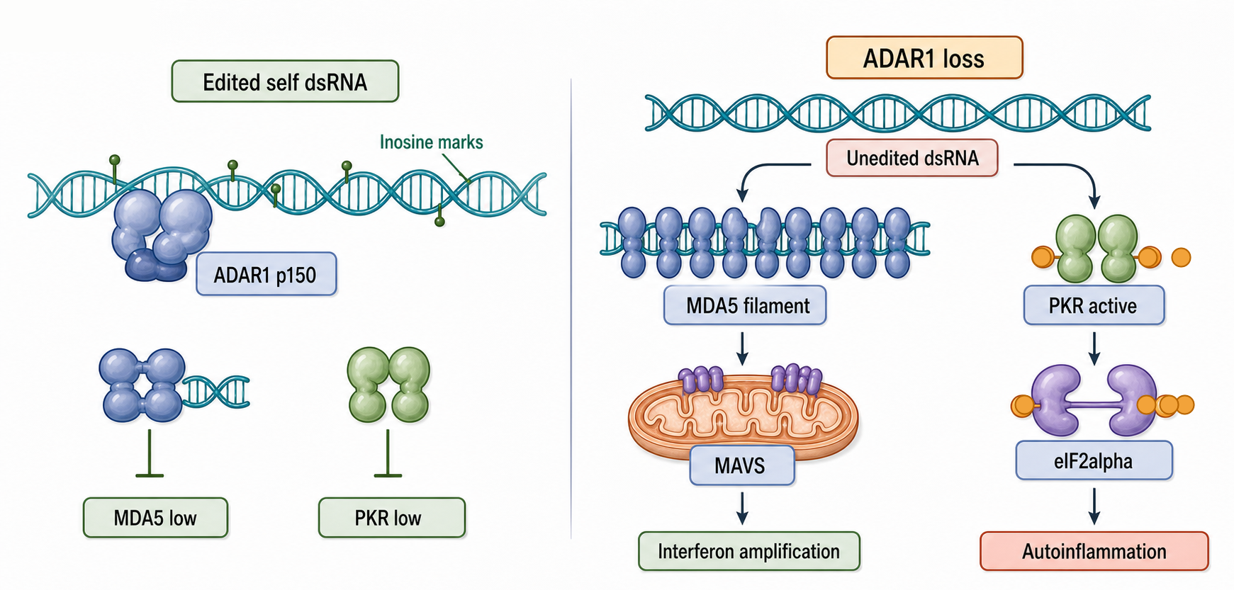
Figure 109.2. ADAR1 separates A-form dsRNA tolerance from ZBP1-dependent death. Self-nonself discrimination depends on RNA conformation, editing, occupancy, and signaling context; pathway dependence does not by itself identify a ligand.
MDA5 recognizes long A-form dsRNA and assembles signaling complexes that activate MAVS, whereas ZBP1 uses Zα domains to engage Z-conformation ligands and RHIM-dependent signaling to recruit RIPK3. The branches are distinguishable but can be coupled by interferon feedback. In ADAR1-deficient fibroblasts, MAVS or type I interferon receptor blockade prevented accumulation of both A-form and Z-form RNA signals, and added interferon accelerated their accumulation; ZBP1 mutations then separated ligand recognition and RIPK3 recruitment from the upstream interferon-amplification state. Thus pathway dependence does not equal ligand occupancy: MAVS dependence identifies an upstream signaling requirement, while ZBP1 immunoprecipitation or a Zα-dependent rescue tests a different proposition about which RNA engages the death sensor.
ADAR1 loss can increase MDA5 activation by leaving endogenous dsRNA unedited. This mechanism is strongly linked to autoinflammatory interferonopathies, including disorders in which variants in ADAR1 or MDA5 pathway components produce chronic interferon signatures. Recent work has refined the model. ADAR1 p150 prevents MDA5 and PKR activation through distinguishable mechanisms, meaning that ADAR1 is not merely a general dsRNA eraser. Another study identified GGNBP2 as a regulator of MDA5 sensing triggered by self dsRNA after loss of ADAR1 editing. These results matter because therapies that modulate ADAR1, MDA5, PKR, or ZBP1 may not have identical effects across tissues, RNA substrates, or disease genotypes.
Box 109.2. ADAR1 Tolerance Is Not Simple dsRNA Disposal
ADAR1 should not be treated as a nonspecific enzyme that makes all self dsRNA disappear. ADAR1 converts adenosine to inosine within duplex RNA, which can weaken perfect pairing, change sequence readout, alter protein binding, and make endogenous duplexes less favorable for MDA5 or PKR activation. Many edited RNAs remain present in the cell. The interferon-inducible p150 isoform also carries a Zα domain, and an editing-dead p150 protein can retain partial suppression of Z-conformation RNA in a murine fibroblast model, consistent with ligand binding or sequestration contributing alongside catalysis. Sensor outcome therefore depends on duplex conformation and length, editing density, RNA-binding proteins, localization, decay, and interferon state. In experiments, ask separately whether a phenotype follows catalytic activity, p150 versus p110 dosage, Zα-dependent recognition, direct RNA occupancy, or downstream MDA5-MAVS, PKR, or ZBP1-RIPK3 signaling.
Autoinflammation is not identical to autoimmunity. Autoinflammation refers to inappropriate activation of innate inflammatory pathways, often without antigen-specific adaptive immune targeting as the initiating event. In ADAR1-MDA5 disease mechanisms, the problem is that self RNA is interpreted as a viral-like molecular pattern. Adaptive immune features may follow, but the initiating logic is RNA surveillance. This distinction is clinically important because blocking cytokines, JAK-STAT signaling, MDA5 activation, or downstream inflammatory circuits may have different consequences from therapies aimed primarily at autoantibodies or lymphocyte specificity.
ADAR1 also intersects with cancer and RNA therapeutics. Many tumors experience interferon signaling, repeat-element derepression, or dsRNA stress. ADAR1 can protect cancer cells from dsRNA sensing, making ADAR1 inhibition attractive as an immuno-oncology strategy, but that same strategy risks inflammation in normal tissues. The therapeutic arm of the Zhang study is an important boundary case: the curaxin CBL0137 bypassed ADAR1 and induced predominantly Z-DNA, not Z-RNA, then promoted host-ZBP1-dependent necroptosis in tumor-microenvironment fibroblasts and potentiated anti-PD-1 treatment in syngeneic mouse melanoma models. Those results are proof of principle for exploiting a ZBP1-competent tumor microenvironment; they are not direct evidence that ADAR1 inhibition is clinically effective or that tumor regression was initiated by an RNA ligand. Detailed adaptive recruitment and checkpoint-therapy consequences hand off to Chapter 110. RNA editing technologies can also use ADAR biology to alter therapeutic RNAs or endogenous transcripts, but therapeutic editing must be judged separately from native ADAR1′s broad role in suppressing self dsRNA. A recent cancer-immunology review focused specifically on ADAR1 inhibition, tumor repeat dsRNA, and checkpoint response remains a final-reference-expansion item.
Other endogenous dsRNA sources and boundary cases
Endogenous dsRNA is not limited to Alu or mitochondrial transcripts. Convergent transcription can produce complementary RNAs. Structured long noncoding RNAs can contain paired regions. Circular RNAs can form intramolecular structures and, under some conditions, activate PKR if not properly modified or bound by proteins. Endogenous retroelements can generate dsRNA when derepressed by epigenetic therapy, cellular stress, or developmental state. EZH2 inhibition has been shown to activate a dsRNA-STING-interferon stress axis that potentiated PD-1 checkpoint blockade response in a prostate cancer model. That example is mechanistically complex because “viral mimicry” in cancer can involve repeat-derived RNAs, chromatin changes, DNA damage, cGAS-STING signaling, and interferon feedback.
The boundary between self and nonself is therefore graded. Perfectly foreign RNA can be ignored if hidden in a viral replication compartment. Self RNA can become inflammatory if it accumulates in the wrong compartment, becomes long enough, lacks editing, or appears during a state when sensor levels are high. RNA modifications can tune recognition but rarely supply an absolute passport. The same RNA molecule may be tolerated in the nucleus, degraded in mitochondria, and inflammatory in the cytosol.
Experimental Foundations and Evidence
Reading interferon-controlled RNA states
The simplest experiment is interferon treatment followed by infection or RNA profiling. Cells are treated with interferon, infected with a virus or transfected with RNA, and assayed for viral RNA, viral protein, host gene expression, cytokines, and cell viability. This design establishes that an interferon-induced state changes the outcome, but it does not identify the responsible ISG. CRISPR knockout or overexpression screens can identify candidate restriction factors, as illustrated by recent ISG screening work that found CCND3 as a restriction factor against high-pathogenic bandaviruses. Such screens need careful validation because cell proliferation, basal toxicity, and indirect immune signaling can appear as antiviral effects.
RNA-seq measures changes in RNA abundance, splicing, editing, repeat expression, and interferon signatures. It is powerful because it can capture host and viral transcripts in the same sample. Its main limitation is interpretive: abundance is not mechanism. A transcript can increase because transcription rises, decay slows, cell composition changes, or a subset of cells dominates the sample. Long-read and direct RNA sequencing can improve isoform and modification analysis, but recent reviews on direct RNA sequencing of viral and endogenous dsRNA-associated transcripts remain final-reference-expansion items for the most current methods.
dsRNA immunostaining, often using J2-like antibodies, can show accumulation and subcellular localization of dsRNA. However, antibody signal depends on fixation, duplex length, accessibility, and epitope context. A bright dsRNA signal does not automatically identify the RNA sequence, source, sensor, or inflammatory relevance. Combining dsRNA staining with mitochondrial markers, RNase treatments, subcellular fractionation, RNA sequencing, and genetic perturbation gives stronger evidence.
Editome profiling detects A-to-I editing because inosine is read as guanosine during reverse transcription. In RNA-seq data, editing can appear as A-to-G mismatches relative to the genome. Robust analysis must distinguish true editing from genetic variants, mapping artifacts in repeats, sequencing errors, and low-complexity alignments. ADAR1 biology is especially dependent on repeat-aware analysis because many important substrates are repetitive.
Evidence for a sensor pathway and evidence for ligand occupancy should be kept separate. Knockout, mutant-rescue, inhibitor, or receptor-blockade experiments can show that MAVS, interferon signaling, ZBP1, RIPK3, or MLKL is necessary for an outcome, but necessity does not identify the RNA bound by that factor. Antibody enrichment and colocalization narrow the candidate set but remain sensitive to epitope specificity, fixation, abundance, and indirect association. In the ADAR1-deficient fibroblast model, the most persuasive chain combined RNase-versus-DNase controls, Zα- and RHIM-mutant rescues, candidate-RNA recovery with ZBP1, and downstream RIPK3-MLKL activation. The chain supports selected ligands in that model without licensing a transcriptome-wide claim that every predicted or enriched sequence occupies ZBP1.
Infection models and viral antagonism
Infection models reveal the dynamic contest between viral RNA production and host RNA restriction. Acute infection can produce abundant pathogen-associated RNA patterns before interferon has induced maximal ISG expression. Chronic or persistent infection may instead reflect equilibrium: viral evasion, partial restriction, tissue-specific interferon tone, and immune-cell recruitment. DNA viruses, positive-strand RNA viruses, negative-strand RNA viruses, retroviruses, and segmented viruses expose different RNA intermediates and therefore stress different parts of the system.
Human cytomegalovirus and Kaposi sarcoma-associated herpesvirus show why DNA viruses belong in an RNA-centered innate immunity chapter. Although their genomes are DNA, their transcriptional programs produce viral RNAs that must be processed, exported, translated, and sensed or hidden. Viral proteins can suppress ISG induction or RNA decay, whereas host quality-control pathways can restrict viral transcripts. RNA viruses provide more obvious dsRNA replication intermediates, but many shelter replication in membrane-associated compartments, making accessibility as important as molecular pattern.
Animal models add tissue context and clinical relevance, but they also introduce species differences. Mouse interferon systems are invaluable for genetics and tissue pathology, yet ISG repertoires, ADAR substrates, repeat landscapes, and viral tropisms differ from humans. Human cell models capture human repeat biology and clinical variants more directly but may lack immune-cell interactions, tissue architecture, and chronic disease physiology. A strong evidence base usually triangulates cell culture, animal genetics, human genetic disease, and patient transcriptomics.
109.4. Mitochondrial dsRNA, organellar stress, and innate immune activation
Mitochondria are descendants of bacteria, but mitochondrial RNA immunogenicity is not explained by ancestry alone. Mammalian mitochondria have a compact circular genome transcribed from both strands. The heavy and light strands generate complementary RNAs that can form dsRNA when processing or degradation is incomplete. Mitochondrial RNAs also differ from typical nuclear mRNAs in processing, modification, polyadenylation, localization, and protein association. Under homeostatic conditions, mitochondrial RNA surveillance and organellar membranes limit cytosolic exposure. Under stress, that separation can fail.
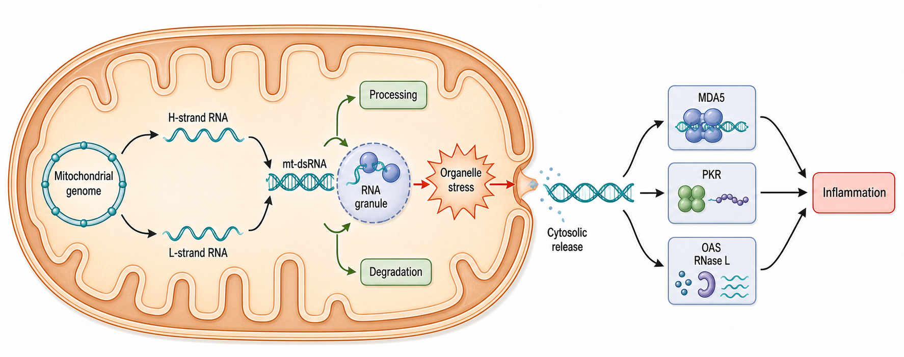
Figure 109.3. Mitochondrial dsRNA production, control, and cytosolic release. Mitochondrial dsRNA is a host-origin RNA that can become inflammatory when compartmental control fails.
Several mechanisms can increase mitochondrial dsRNA. First, imbalanced transcription from the two strands can produce excess complementary RNA. Second, defects in mitochondrial RNA processing or degradation can allow duplexes to persist. Third, altered mitochondrial RNA modifications may change stability or recognition. A 2024 Molecular Cell study reported that RNA 5-methylcytosine marks mitochondrial dsRNAs for degradation and cytosolic release, linking mitochondrial RNA modification to dsRNA fate. Fourth, organellar damage, permeability changes, mitophagy defects, or senescence-associated stress can allow mitochondrial RNA to reach cytosolic sensors. Recent work in senescent cells identified release of mitochondrial dsRNA into the cytosol as a driver of inflammatory phenotype.
The sensor pathway for mitochondrial dsRNA is context-dependent. Long cytosolic dsRNA can activate MDA5, PKR, or OAS/RNase L. Mitochondrial stress can also activate cGAS-STING through mitochondrial DNA or nuclear DNA damage, and some studies describe mixed RNA-DNA inflammatory axes. This creates a major interpretation problem: an interferon signature during mitochondrial stress does not prove that mitochondrial dsRNA is the initiating ligand. Strong evidence requires showing mitochondrial RNA accumulation or release, reducing the candidate mitochondrial RNA or its processing defect, and demonstrating loss of the inflammatory phenotype when the relevant RNA sensor pathway is disabled.
Box 109.3. Evidence Ladder for Mitochondrial dsRNA Claims
A convincing mitochondrial dsRNA claim usually climbs an evidence ladder. First, spatial evidence should show dsRNA signal near mitochondria and sensitivity to RNA-directed controls. Second, source evidence should identify complementary heavy-strand and light-strand mitochondrial transcripts while controlling for nuclear mitochondrial DNA segments, contamination, and mapping ambiguity. Third, a processing mechanism should connect mitochondrial RNA transcription, modification, degradation, or quality control to dsRNA accumulation before inflammatory output. Fourth, cytosolic exposure should be shown without relying only on damaged-cell readouts, because mitochondrial DNA release and cell death can produce overlapping inflammatory signatures. Fifth, causality requires rescue: reducing the candidate mitochondrial RNA or disabling the relevant RNA sensor branch should reduce the inflammatory phenotype while preserving comparable upstream stress where possible.
Mitochondrial dsRNA also teaches an important self-nonself lesson. The same cytosolic receptor machinery that detects viral replication can respond to host organellar transcripts when organelle quality control fails. The ligand is self by origin but danger-associated by location and abundance. This is why “self RNA” is too broad a category for innate immunity. The immune system monitors molecular patterns in compartments and physiological states, not legal ownership.
Mitochondrial RNA inflammation is relevant to aging, neurodegeneration, cancer, antiviral immunity, and inherited mitochondrial disease, but the strength of evidence varies by condition. A recent review summarizes mitochondrial RNA as an inflammatory contributor across disease contexts. For this chapter, the strongest local primary anchors are senescence-associated mitochondrial dsRNA release and modification-linked mitochondrial dsRNA control. Reviews and primary studies on PNPase, SUV3, mitochondrial degradosome activity, and mitochondrial RNA leakage mechanisms remain final-reference-expansion items.
109.5. Systems immunology, infection models, and clinical relevance
Biological contexts across organisms, cells, and disease states
Interferon-stimulated RNA pathways are strongest where the cost of viral replication is high and where epithelial or immune cells must respond rapidly. Barrier epithelia use type III interferons to create local antiviral states with less systemic inflammation than broad type I interferon responses. Plasmacytoid dendritic cells specialize in interferon production, whereas macrophages integrate interferon with phagocytosis, inflammasome activation, and tissue repair. Fibroblasts and epithelial cells often provide the tissue-intrinsic antiviral state that determines whether a virus spreads before immune-cell recruitment.
Cell type affects self-nonself discrimination because basal sensor levels, interferon responsiveness, RNA decay capacity, mitochondrial activity, repeat expression, and ADAR1 isoform abundance differ across tissues. Neurons, for example, are vulnerable to chronic inflammatory signaling and may rely heavily on RNA editing and mitochondrial quality control, whereas proliferating immune cells may tolerate stronger transient interferon activation. Tumors can exploit or suffer from these differences. A tumor with high repeat expression may be primed for dsRNA sensing, but the same tumor may upregulate ADAR1 or other suppressors to avoid inflammatory death.
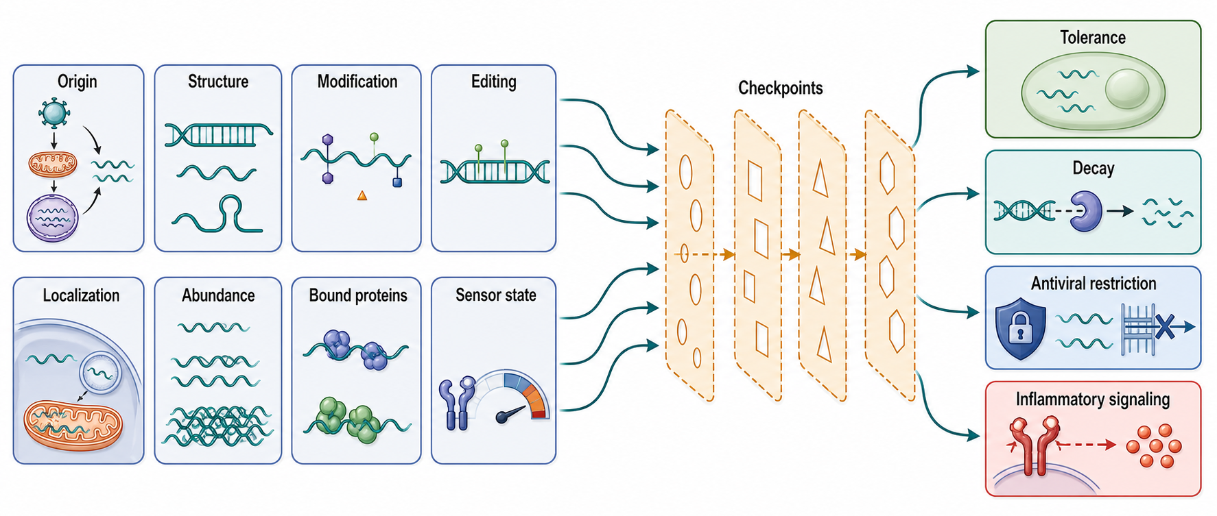
Figure 109.4. Self-nonself discrimination as a multilayer decision. RNA immune recognition is a systems decision, not a simple self-versus-foreign label.
Autoinflammatory interferonopathies provide natural experiments. Genetic defects that increase endogenous nucleic acid sensing often produce chronic interferon signatures, developmental injury, skin and neurological phenotypes, and variable systemic inflammation. ADAR1-related disease and gain-of-function MDA5 biology show that RNA editing and dsRNA sensing are not optional refinements; they are required to prevent self-RNA from activating antiviral pathways. Recent clinical reviews discuss emerging concepts and treatments in interferonopathies and monogenic lupus-like diseases, but disease-focused references for ADAR1-associated Aicardi-Goutieres syndrome, dyschromatosis symmetrica hereditaria, and IFIH1 gain-of-function phenotypes remain final-reference-expansion items.
Technology, computational, clinical, and engineering links
Systems immunology treats interferon-stimulated RNA pathways as network states rather than isolated genes. Bulk RNA-seq can quantify interferon signatures across tissues or patients. Single-cell RNA-seq can identify which cell types carry the signature. Perturb-seq and CRISPR screens can connect ISGs to viral restriction, self-dsRNA sensing, or inflammatory outputs. Proteomics and phosphoproteomics add signaling state. Cytokine measurements and clinical metadata connect molecular signatures to fever, tissue injury, treatment response, and prognosis. The broad systems-immunology argument is that human immune variation must be measured directly rather than inferred only from model organisms.
Table 109.2. Clinical and experimental contexts for interferon-stimulated RNA pathways. Infection, interferonopathy, tumor, therapeutic-RNA, and model-system contexts expose different candidate ligands and sensor-effector branches; common interferon readouts are insufficient to assign the initiating RNA pathway.
| Context | Candidate RNA ligand | Sensor or effector branch | Useful readouts | Major interpretation hazard |
|---|---|---|---|---|
| Acute viral infection | Viral replication intermediates, uncapped 5′ triphosphate RNA, or abundant viral transcripts | RIG-I or MDA5 through MAVS, plus PKR, OAS/RNase L, IFITs, and ISG amplification | Viral RNA kinetics, infectious titer, viral protein, IFNB or ISG transcripts, dsRNA staining | Lower viral RNA can reflect reduced synthesis, faster decay, fewer infected cells, or selective cell death |
| ADAR1-related autoinflammation | Unedited repeat-derived self dsRNA, including Alu-like duplexes in primates | MDA5-MAVS and PKR branches amplified by type I interferon signaling | A-to-I editome loss, interferon score, MDA5 or PKR activation, rescue by sensor-pathway perturbation | Tissue context, ADAR1 isoform, and editing-independent effects can change the dominant mechanism |
| ADAR1-Zα-ZBP1 fibroblast model | Interferon-amplified Z-conformation RNA candidates in selected ISG mRNA 3′ UTRs | ZBP1 Zα recognition and RHIM-dependent RIPK3-MLKL signaling | Nuclease-controlled Z-RNA signal, ZBP1-RNA recovery, Zα/RHIM mutant rescue, pMLKL, and viability | Z-RNA antibody enrichment and pathway dependence are not equivalent to transcript-specific occupancy; the evidence is from murine fibroblast systems |
| MDA5 gain-of-function disease | Endogenous dsRNA sensed at an abnormally low activation threshold | Hyperactive IFIH1/MDA5 signaling to MAVS and interferon genes | IFIH1 genotype, interferon signature, cytokine profile, and response to pathway inhibition | The ligand is often inferred from pathway activation rather than directly identified |
| Senescence-associated mitochondrial dsRNA | Cytosolic heavy-strand and light-strand mitochondrial RNA duplexes | MDA5, PKR, or OAS/RNase L, sometimes alongside DNA-stress pathways | Mitochondrial dsRNA staining, subcellular fractionation, mitochondrial RNA depletion, SASP cytokines | Mitochondrial DNA release, organelle damage, and cell death can mimic RNA-driven inflammation |
| Epigenetic cancer therapy and viral mimicry | Derepressed retroelement or repeat-derived dsRNA, with possible mitochondrial or DNA-stress inputs | RIG-I-like receptors, PKR, STING-linked interferon axes, and ADAR1 counter-regulation | Repeat RNA-seq, dsRNA staining, interferon signatures, checkpoint-response assays, ADAR1 perturbation | Viral mimicry can mix RNA sensing, chromatin stress, DNA damage, and changing immune-cell composition |
| RNA therapeutic delivery | In vitro transcription dsRNA byproducts, uncapped RNA, under-modified RNA, or formulation-associated RNA exposure | Endosomal TLR7/8, cytosolic RIG-I-like receptors, PKR, OAS/RNase L, and cytokine feedback | RNA purity, cap and modification assays, innate gene panels, cytokines, biodistribution, dose response | Immunogenicity may come from impurities, lipid or carrier toxicity, dose, tissue damage, or intended adjuvanticity |
Clinical interpretation requires restraint. An interferon signature in blood can reflect infection, inherited interferonopathy, cancer therapy, autoimmunity, tissue injury, or treatment with interferon or interferon-inducing drugs. It may be driven by immune-cell composition rather than uniform activation in every cell. Similarly, dsRNA signal in a tumor can indicate repeat derepression, viral infection, mitochondrial stress, RNA processing defects, or therapy-induced chromatin changes. A useful clinical model therefore links the RNA source, sensor pathway, cell type, cytokine output, and intervention.
Therapeutic engineering creates both opportunities and hazards. RNA therapeutics are designed to avoid unwanted innate sensing unless immune activation is the therapeutic goal. Chemical modification, purification to remove dsRNA byproducts, capping, poly(A) tail design, sequence optimization, and delivery route all influence recognition. In cancer, the goal may be reversed: exposing endogenous dsRNA, inhibiting ADAR1, or combining epigenetic therapy with checkpoint blockade may increase tumor immunogenicity. In interferonopathies, the goal is suppression of harmful sensing or downstream signaling. These opposing goals show why “immunostimulatory RNA” is not intrinsically good or bad; it is a context-dependent pharmacological property.
Computational work supports this field by predicting repeat-derived duplexes, identifying editing sites, quantifying viral and host RNA abundance, deconvolving cell states, and modeling ISG networks. Failure modes are common. Repeats are hard to map; short reads may collapse related elements; editing and genetic variation can be confused; mitochondrial reads can arise from nuclear mitochondrial DNA segments; and interferon signatures are correlated modules rather than direct measurements of a ligand. The best computational analyses state the uncertainty attached to each inferred RNA source.
Recent Consensus
The current consensus is that interferon responses create a multilayer RNA-defense state rather than a single antiviral pathway. Viral restriction can occur through sensing, translation inhibition, RNA decay, replication-complex disruption, metabolic changes, and cell-state shifts. RNA decay and translation control are central antiviral mechanisms, but they are often broad and can affect host RNAs as well as viral RNAs.
A second consensus is that endogenous dsRNA is abundant enough to require active tolerance mechanisms. ADAR1-mediated editing, RNA decay, nuclear retention, compartmentalization, RNA-binding proteins, and sensor thresholds all contribute. ADAR1 is a major self-nonself discrimination factor because it prevents MDA5 and PKR activation by endogenous dsRNA in interferon-competent cells. In selected murine models, ADAR1 p150 also restrains a Zα-dependent ZBP1 branch through both editing and ligand binding or sequestration, but whether interferon pathology or RIPK3-MLKL death dominates is context-dependent.
A third consensus is that mitochondrial dsRNA is a credible inflammatory ligand under specific stress conditions. It is not yet settled which leakage mechanisms, modifications, sensors, and disease contexts dominate. Recent primary work has made the field more concrete by tying mitochondrial dsRNA to senescence-associated inflammation and RNA modification-dependent control.
Open Questions, Controversies, Deprecated Models, and Common Misconceptions
Open questions:
- Which endogenous dsRNA species are most important in each disease? Repeat-derived nuclear RNAs, circular RNAs, mitochondrial dsRNAs, and viral RNAs can all activate overlapping sensors. Many studies measure pathway activation more confidently than ligand identity.
- How does ADAR1 suppress PKR separately from MDA5? Recent primary work supports distinct mechanisms, but substrate specificity, protein interactions, and tissue dependence remain active questions.
- Which endogenous transcripts form stable Z-RNA and directly occupy ZBP1 in vivo? Antibody enrichment, conformational prediction, genetic dependence, and candidate-RNA immunoprecipitation provide different levels of evidence and should not be collapsed into one claim.
- How does mitochondrial dsRNA reach the cytosol? Candidate routes include organellar permeability changes, mitophagy defects, RNA granule stress, vesicular trafficking, and cell damage. Current evidence is context-specific rather than universal.
Common misconceptions:
- “Interferon induction proves viral infection.” It does not. Endogenous dsRNA, mitochondrial stress, DNA damage, autoinflammatory mutations, and cancer therapy can all induce interferon signatures.
- “Self RNA is ignored and viral RNA is sensed.” The immune system responds to molecular pattern, localization, abundance, modification, binding partners, and cell state. Self RNAs can be inflammatory when these controls fail.
- “All dsRNA staining marks viral replication.” Antibody-based dsRNA signal can arise from viral RNA, endogenous repeats, mitochondrial RNA, structured RNAs, or fixation-dependent accessibility. Sequence-level and perturbation evidence are needed.
- “ZBP1-dependent pathology proves that every predicted Z-forming RNA is a ZBP1 ligand.” Genetic dependence maps the pathway, whereas direct occupancy requires RNA-protein evidence for specific molecules; neither sequence propensity nor antibody enrichment alone is sufficient.
Deprecated or weakened claims:
- Deprecated or incomplete model: ADAR1 simply destroys dsRNA danger signals. ADAR1 edits, remodels, and functionally marks dsRNA; it also has isoform-specific and editing-independent or sensor-specific effects that cannot be reduced to substrate removal.