Chapter 148. RNA Nanotechnology, Materials, Origami, Scaffolds, Condensate Engineering, and Molecular Machines
Scope Note
RNA nanotechnology treats RNA as a programmable material as well as an informational and regulatory molecule. This chapter covers synthetic RNA nanoparticles, RNA tiles, junctions, RNA origami, RNA scaffolds for biochemical organization and delivery, aptamer-functionalized materials, ligand-responsive hydrogels and surfaces, intracellular RNA materials, phase-separation-inspired engineering, molecular machines, strand-displacement systems, and responsive assemblies. The chapter emphasizes how sequence design, folding thermodynamics, tertiary motifs, chemical stability, immune sensing, manufacturing, and evidence standards determine whether an RNA structure is a useful material rather than only a predicted fold. The chapter owns material architecture and device behavior; the following therapeutic and delivery chapters own drug-modality selection, carrier pharmacology, biodistribution, manufacturing control, and clinical evidence.
Executive Summary
RNA nanotechnology uses RNA sequence, base pairing, tertiary motifs, chemical groups, and sometimes proteins or ligands to construct defined nanoscale objects. The field borrows principles from natural RNA folding, DNA nanotechnology, synthetic biology, and biomaterials, but RNA has its own advantages and liabilities. RNA can be transcribed, folded co-transcriptionally, displayed inside cells, selected as a ligand-binding aptamer, and coupled to ribozyme or regulatory functions. RNA is also polyanionic, nuclease-sensitive, conformationally heterogeneous, and visible to innate immune sensors when delivered to mammalian cells.
RNA nanoparticles are usually built from modular helices, loops, junctions, kissing-loop contacts, three-way junctions, multi-arm motifs, and sequence-programmed strand pairing. A design must specify not only the intended static drawing but the assembly path: which strands are made, when they meet, what off-pathway structures compete, what ions stabilize the final object, and how stoichiometry is controlled. RNA origami extends this logic by folding a long RNA strand, often with internal complementary segments and designed junctions, into a defined shape. The 2024 demonstration of folding molecular origami from ribosomal RNA supports the broader idea that long structured RNA can be repurposed as a programmable scaffold, but it also highlights the need to test folding experimentally rather than infer success from design alone [Shapiro_A_2024_rRNA_origami].
RNA scaffolds organize functional modules. A scaffold can display aptamers, recruit enzymes, cluster guide RNAs, present therapeutic cargos, position sensors, or concentrate reaction partners. The mechanistic promise is local concentration and geometry: a scaffold may increase the probability that two molecules meet or orient them in a productive order. The caveat is that scaffolding is not automatically beneficial. Flexible linkers, steric crowding, incomplete assembly, misfolding, degradation, immune activation, and cargo interference can erase or reverse the expected gain.
Aptamer materials exploit RNA sequences that bind ligands, proteins, small molecules, metabolites, or cell-surface receptors. An aptamer can act as an affinity element on a surface, a trigger in a hydrogel, a sensor in a fluorescent device, or a targeting ligand in a therapeutic assembly. Aptamer affinity measured in a purified buffer is not enough. Material performance depends on ligand concentration, competing molecules, serum proteins, nuclease exposure, surface orientation, mass transport, and whether binding produces a measurable mechanical, optical, electrical, or biological output.
Condensate engineering and intracellular RNA materials are a frontier area. Natural cells use RNA and RNA-binding proteins to build ribonucleoprotein granules with liquid-like, gel-like, or more solid material states. Synthetic systems try to program localization, reaction compartments, or regulatory hubs by tuning RNA valency, protein-binding motifs, repeat sequences, and phase-separation-promoting domains. The evidence bar is high because puncta are not automatically functional condensates, and phase separation can be confused with aggregation, overexpression, stress, or imaging artifacts.
RNA molecular machines and dynamic assemblies add time-dependent behavior. Strand displacement, ligand-induced conformational switching, ribozyme coupling, aptamer-triggered assembly, and toehold-mediated circuits can make structures that open, close, release cargo, report a signal, or change oligomeric state. Dynamic behavior requires kinetic control as well as thermodynamic design. Stability, immunogenicity, manufacturing, and design automation remain translational boundaries.
Concept Inventory
- RNA nanotechnology: the design and use of RNA molecules as programmable nanoscale structural, functional, or material components.
- RNA nanoparticle: a discrete nanoscale assembly containing RNA as a structural component, cargo, targeting ligand, regulator, or scaffold.
- RNA origami: an RNA design strategy in which one or more RNA strands fold into a prescribed shape through programmed helices, junctions, and tertiary contacts.
- RNA tile: a repeatable RNA unit with defined connection interfaces that can assemble into larger arrays or lattices.
- Junction motif: a branching RNA element, such as a three-way junction or multi-way junction, that organizes helices at controlled angles.
- Kissing-loop interaction: a tertiary contact between complementary loop sequences on two hairpins; useful for reversible or modular RNA-RNA assembly.
- RNA scaffold: an RNA structure used to position other molecules, such as enzymes, aptamers, therapeutic cargos, proteins, or guide RNAs.
- Aptamer material: a material or device whose function depends on ligand binding by an RNA aptamer.
- RNA hydrogel: a water-rich polymeric network in which RNA strands or RNA-containing modules contribute to crosslinking, responsiveness, recognition, or cargo release.
- Condensate engineering: the deliberate design of biomolecular assemblies with phase-separation-like material behavior, often by controlling multivalency and weak interactions.
- RNA molecular machine: an RNA-containing system that changes state, performs a mechanical or informational operation, or couples molecular recognition to movement, assembly, catalysis, or release.
- Strand displacement: a nucleic-acid reaction in which an incoming strand binds an exposed toehold and replaces another strand through branch migration.
What to Know Before Reading This Chapter
Readers should understand that RNA is a directional polymer with a negatively charged ribose-phosphate backbone, bases that can pair and stack, and a tendency to fold into secondary and tertiary structures. Chapters 3 and 4 introduce folding thermodynamics, ensembles, motifs, junctions, and tertiary architecture. Chapter 5 explains evidence standards for distinguishing mechanism, observation, inference, and artifact. Chapter 53 covers structural motifs; Chapter 58 covers natural RNA condensates; Chapter 65 covers inverse folding and programmable architecture; Chapters 108, 156, 157, 159, and 147 cover immune sensing, delivery, manufacturing, and synthetic biology. This chapter repeats the necessary background because a nanostructure is not established by a pleasing drawing. A valid RNA material must be designed, assembled, measured, challenged under relevant conditions, and connected to a function.
148.1. RNA Nanoparticles, Tiles, Junctions, and Origami
An RNA nanoparticle is a nanoscale assembly in which RNA contributes to shape, recognition, cargo display, or regulation. The word “particle” can describe a compact discrete object, a multivalent scaffold, a carrier, or an ordered assembly; it should not be used as proof that the structure is monodisperse or therapeutically deliverable. A rigorous description specifies the number of strands, approximate dimensions, stoichiometry, intended fold, assembly conditions, and evidence for the final state. A predicted 20-nanometer object, a smear on a gel, and a cryo-electron microscopy class average are different levels of evidence.
The simplest design vocabulary comes from RNA secondary structure. Watson-Crick helices act as stiff rods over short distances, hairpin loops cap helices or provide interaction surfaces, bulges and internal loops bend helices, and junctions connect multiple helical arms. A three-way junction is a branching element in which three helical segments meet; a four-way or higher-order junction can produce more complex topologies. Natural RNAs use such elements in ribosomal RNA, ribozymes, riboswitches, and viral RNAs. RNA nanotechnology turns those recurrent structural units into reusable engineering parts.
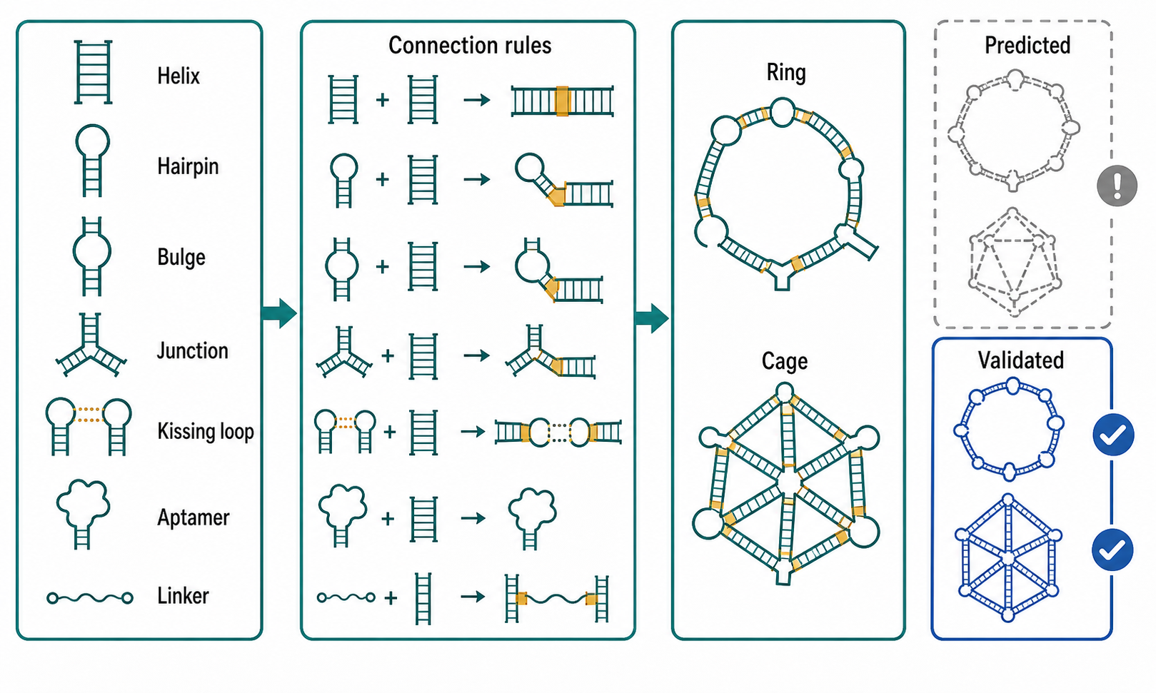
Figure 148.1. Motif Grammar of RNA Nanoparticle Design. Show how helices, hairpins, bulges, junctions, kissing loops, aptamers, and flexible linkers combine into particles, tiles, rings, and origami objects.
The important bridge from natural structure to engineering is modularity. A module is a part whose behavior can be reused with some predictability outside its original context. A kissing-loop interaction is a useful example. In a kissing loop, two hairpin loops with complementary exposed sequences base-pair with one another, often producing a reversible tertiary contact between separate domains or strands. Kissing loops can join particles, close rings, organize lattices, or create switchable assemblies. The boundary case is context dependence: loop sequence, loop length, helix orientation, magnesium concentration, flanking structure, and competing intramolecular pairing can determine whether the intended contact forms.
RNA tiles use repeatable interaction units to build larger objects. A tile can contain several helices and sticky ends or loop contacts that direct assembly with neighboring tiles. The designer specifies local connection rules, then relies on many tiles following the same rules to build a sheet, tube, cage, or array. This strategy is powerful because small units can create larger architectures. It is also artifact-prone because small off-stoichiometry errors, partially folded tiles, or unintended symmetry can produce heterogeneous aggregates that still look like “assembly” in low-resolution assays. Native gel shifts, atomic force microscopy, electron microscopy, size-exclusion chromatography, analytical ultracentrifugation, light scattering, and sequencing-based stoichiometry can give complementary evidence.
RNA origami is a more integrated strategy. In origami design, a long RNA strand, or sometimes a defined set of strands, is programmed to fold into a target architecture by forming many internal helices and junctions. DNA origami often uses a long scaffold strand and many short staple strands. RNA origami can instead exploit transcription: the RNA emerges from RNA polymerase and begins folding while still being synthesized. Co-transcriptional folding can help by allowing local domains to form in order, but it can also trap misfolded intermediates. Therefore RNA origami design must consider transcription direction, domain order, kinetic traps, magnesium dependence, and whether the final structure can refold after denaturation.
The verified chapter-local paper by Shapiro and colleagues reported folding molecular origami from ribosomal RNA, a striking example because ribosomal RNA is long, structured, and biologically familiar [Shapiro_A_2024_rRNA_origami]. The general lesson is not that any natural RNA can be reshaped at will. The lesson is that large RNAs can sometimes be treated as scaffolds for designed geometry when design, folding conditions, and structural assays are aligned. A reader should ask what parts of the original RNA sequence are retained, what constraints the original structure imposes, whether the new object folds in vitro or in cells, and how much of the population adopts the intended state.
Table 148.1. RNA Nanostructure Architecture Classes and Evidence Needs. Compare nanoparticles, tiles, junction-based hubs, rings, cages, origami, and arrays by design unit, assembly route, common evidence, and common artifacts.
| Architecture class | Core design parts | Assembly mode | Strongest evidence | Frequent overclaim |
|---|---|---|---|---|
| Discrete RNA nanoparticle | Helices, junctions, kissing loops, aptamers, cargo modules, and linkers arranged into a compact object | One or more strands anneal or fold into a defined stoichiometric particle under controlled ionic and thermal conditions | Concordant size, stoichiometry, purity, morphology, and activity measurements from orthogonal assays | Treating a gel shift or particle-size peak as proof of a uniform deliverable nanomedicine |
| RNA tile or tecton array | Repeatable units with sticky ends, loop-loop contacts, or programmed interfaces | Local connection rules propagate into sheets, tubes, cages, or lattices | Microscopy or scattering plus native assembly assays showing ordered repeat geometry and limited heterogeneity | Calling nonspecific higher-order aggregation a designed lattice |
| Junction-based hub | Three-way or multi-way junctions with helical arms, aptamers, cargo sites, or protein-binding motifs | Branched scaffold folds as a single RNA or assembles from defined strands | Mutational disruption and rescue of junction geometry paired with stoichiometry and functional output | Assuming a known junction motif keeps the same geometry in every sequence and ion context |
| Ring or polygon | Repeated edges, vertices, kissing-loop closures, and optional flexible hinges | Modular units close into finite cyclic assemblies rather than indefinite polymers | Native gels, microscopy, and distance reporters consistent with closed stoichiometric species | Mistaking linear concatemers, multimers, or off-stoichiometric products for closed rings |
| Cage or container | Junction vertices, helical edges, internal or surface cargo sites, and stabilizing tertiary contacts | Multistrand or single-strand assembly forms a hollow or shell-like object | Structural imaging plus cargo accessibility or encapsulation assays under solution conditions | Inferring encapsulation from cargo mixing without testing access, leakage, or release |
| RNA origami object | Long scaffold RNA, internal complementary segments, designed junctions, and tertiary contacts | Cotranscriptional or refolded pathway drives one strand or a small strand set into a prescribed shape | Structural probing or imaging showing intended domain organization and population homogeneity | Treating a predicted target drawing as evidence that the dominant RNA population folds correctly |
| Heterogeneous aggregate, failure state | Misfolded strands, partial tiles, exposed sticky regions, excess cargo, or nonspecific RNP contacts | Off-pathway association produces broad size distributions or poorly defined assemblies | Polydispersity, smear-like gels, inconsistent imaging, or loss of function after purification | Presenting aggregate-like material as a valid architecture class without mechanism-specific evidence |
Nanoparticle design proceeds in causal steps. First, the designer defines the function: shape display, cargo encapsulation, cell targeting, enzyme colocalization, sensor output, or dynamic response. Second, the designer chooses a topology, such as a ring, polygon, cage, rod, sheet, or branched hub. Third, the designer selects motifs that can implement the topology: helices for edges, junctions for vertices, kissing loops for contacts, aptamers for recognition, and flexible linkers where movement is useful. Fourth, the sequence is optimized to favor intended base pairing and disfavor competing folds. Fifth, the RNA is synthesized or transcribed, assembled under controlled ionic and thermal conditions, and purified. Sixth, the structure is measured and challenged under relevant conditions.
The evidence basis must match the claim. A native polyacrylamide gel can show a shifted species consistent with assembly, but it cannot prove a three-dimensional geometry. Atomic force microscopy can show surface-deposited shapes, but drying and surface interactions can distort flexible RNA. Cryo-electron microscopy can classify particles in vitreous ice, but small or heterogeneous RNA assemblies can be difficult to align. Chemical probing can show local pairing and accessibility, but it gives an ensemble-averaged or population-weighted view. Fluorescence resonance energy transfer can report distances or switching, but dye attachment can perturb folding. A strong structural claim usually integrates several methods.
Box 148.1. Assembly Evidence Is Not Architecture Evidence
An RNA nanostructure claim usually contains several smaller claims. Identity asks whether the expected RNA sequence, length, end chemistry, and modifications are present. Assembly asks whether the RNA forms a discrete species rather than a smear, concatemer, or aggregate. Architecture asks whether that species has the intended topology, dimensions, stoichiometry, and domain arrangement. Function asks whether the assembled object performs the proposed task under relevant conditions. A native gel shift can support assembly, but it does not establish architecture. A microscopy image can support shape, but it may not report solution behavior or population homogeneity. A functional signal can show activity, but it may come from a minority species or from free cargo. Strong claims connect at least two orthogonal structural assays with purification, stoichiometry, and a mechanism-specific functional test.
Do not overgeneralize from DNA nanotechnology. RNA and DNA share base-pairing logic, but RNA has a 2′ hydroxyl group, stronger propensity for A-form helices, richer tertiary motifs, different nuclease exposure, and different biological sensing. RNA can be genetically encoded or transcribed in situ, which is a major advantage for intracellular systems. RNA can also fold before a full sequence is available, which creates co-transcriptional pathways unavailable to annealed DNA constructs. A DNA-like drawing of crossed helices may hide these RNA-specific constraints.
Citation coverage is currently incomplete for this section. The local reference file contains the 2024 rRNA origami method paper and a 2014 synthetic nucleic acid technology review-method anchor [Chakraborty_S_2014_synthetic_nucleic_acid_technologies], but it lacks verified review coverage for RNA tectonics, classic RNA nanoparticle motifs, pRNA three-way junction systems, RNA tile arrays, and modern RNA origami software. Those gaps should be filled before final release.
148.2. RNA Scaffolds for Enzymes, Therapeutics, Delivery, and Spatial Organization
An RNA scaffold is an RNA structure that positions other molecules. The scaffold may be a designed hairpin array, a junction-based hub, a long structured RNA, a circular RNA, a guide RNA extension, an aptamer-bearing transcript, or a nanoparticle surface. The scaffold function is geometric and kinetic: it changes local concentration, orientation, timing, or compartmental access. A scaffold does not have to be rigid. In many biological contexts, a partly flexible scaffold is useful because it tethers partners while allowing them to search for productive orientations.
The prerequisite concept is effective concentration. If two enzymes act sequentially on a substrate, attaching them near one another can increase the probability that the intermediate encounters the second enzyme before diffusing away. In RNA systems, effective concentration can be created by repeated aptamer tags that bind protein fusions, by RNA motifs that bind endogenous proteins, by guide RNAs that recruit CRISPR-associated enzymes, or by RNA nanoparticles that display multiple copies of a ligand. The mechanism is not simply “more is better.” Too much clustering can sterically block active sites, sequester limiting proteins, or create nonproductive condensates.
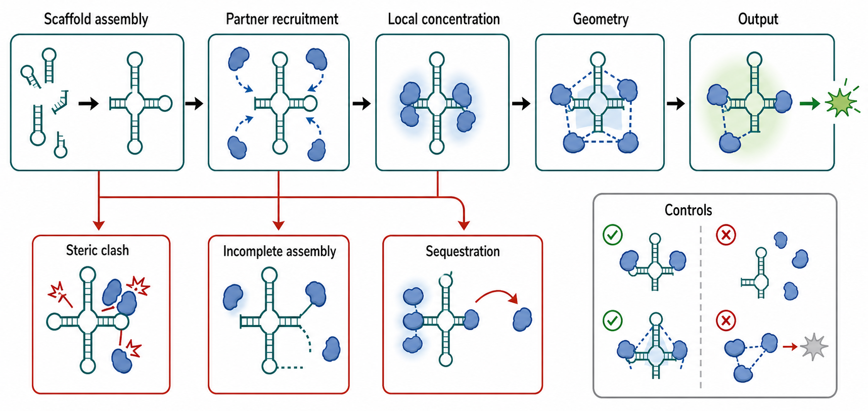
Figure 148.2. RNA Scaffold Logic from Tethering to Function. Trace the causal chain from scaffold expression or assembly to partner recruitment, local concentration, productive geometry, output, and failure modes.
Enzyme scaffolding provides a concrete example. Imagine an engineered transcript carrying two RNA hairpin aptamers, each recognized by a different RNA-binding protein fused to a metabolic enzyme. The RNA is transcribed in a cell, folds into a scaffold, recruits the two fusion proteins, and colocalizes them near a substrate-producing compartment. The intended outcome is increased flux through a pathway. Evidence for this design must separate at least four claims: the RNA is expressed and stable; the RNA recruits the proteins; the recruited proteins remain enzymatically active; and pathway flux changes because of scaffolded colocalization rather than expression differences or stress.
Box 148.2. Scaffolds Have an Optimum, Not a Maximum
Scaffold design is an optimization problem. A useful scaffold changes the rate-limiting step of a process: it may keep a substrate intermediate near the next enzyme, raise the local concentration of a weakly binding partner, or position a cargo where release is productive. Adding more binding sites does not guarantee improvement. Excess valency can trap partners in nonproductive orientations, bury active sites, titrate limiting proteins away from their normal targets, slow turnover by making complexes too stable, or nucleate stress-like aggregates. The relevant question is not “did the scaffold recruit the component?” but “did recruitment improve the intended mechanism without creating a larger cost?” Strong tests compare matched expression levels, binding-site mutants, spacing or linker variants, catalytic-dead partners, and direct output measurements. A scaffold that increases colocalization while lowering flux has failed as a functional design.
Therapeutic scaffolds use similar logic but face a harder environment. An RNA nanoparticle can display small interfering RNAs, microRNA mimics, aptamers, ribozymes, guide RNAs, immune agonists, proteins, peptides, or small-molecule cargos. A multivalent structure might combine a cell-targeting aptamer with a therapeutic RNA cargo, or display several copies of a receptor-binding motif to increase avidity. Avidity means that multiple weak interactions combine to produce stronger apparent binding to a surface. Avidity can improve targeting, but it can also reduce tissue penetration, increase uptake by clearance cells, or activate receptors unintentionally.
Delivery scaffolds must distinguish packaging from productive delivery. A carrier that protects RNA in serum is useful only if enough cargo reaches the correct intracellular compartment. For RNA interference cargos, the cargo must reach cytosolic Argonaute loading. For messenger RNA, the cargo must reach the cytosol and be translated. For guide RNAs, the guide must meet the relevant nuclease or editor. For aptamer drugs, the aptamer may need to remain extracellular or bind a cell-surface target. These compartment rules determine whether a scaffold is a delivery vehicle, a targeting ligand, a depot, or a misleading particle that accumulates but does not act.
Table 148.2. RNA Scaffold Use Cases and Required Controls. Link scaffold use cases to mechanism and controls.
| Use case | Recruited component | Expected output | Necessary controls | Translation boundary |
|---|---|---|---|---|
| Enzyme colocalization scaffold | RNA-binding protein fusions to sequential enzymes or regulatory domains | Increased pathway flux, faster intermediate handoff, or localized catalysis | Binding-site mutants, expression-matched controls, catalytic-dead controls, rescue of spacing or orientation, and direct product measurements | Crowding can block active sites, sequester limiting proteins, or create stress-associated aggregates |
| Therapeutic cargo display | siRNA, miRNA mimic, ribozyme, guide RNA, immune agonist, peptide, protein, or small-molecule payload | Multivalent presentation, protection, targeting, or combined recognition and action | Cargo loading, cargo accessibility, release, retained activity, serum stability, and carrier-only controls | Cargo accumulation is not productive delivery unless the payload reaches the correct intracellular compartment |
| Aptamer-targeted particle | Receptor-binding or ligand-binding aptamer displayed on an RNA particle or scaffold | Higher apparent binding, cell association, or receptor-triggered uptake | Aptamer mutation, receptor competition, target-negative cells, abundance-normalized uptake, and off-target binding tests | Avidity can reduce tissue penetration, increase clearance-cell uptake, or trigger unintended receptor signaling |
| Guide-RNA or CRISPR-effector scaffold | Cas nuclease, editor, transcriptional regulator, imaging tag, or repair-modulating domain | Target-localized editing, regulation, imaging, or recruitment of additional effectors | Guide-only control, effector-dead control, off-target profiling, guide abundance measurement, and target-site rescue | Added scaffold domains can alter nuclease specificity, RNA stability, or cell stress responses |
| Intracellular localization scaffold | Localization motifs, RNA-binding proteins, fluorescent tags, organelle-tethering modules, or endogenous partners | Spatial enrichment of RNA, protein, translation, sensing, or reaction output | Localization-motif mutants, orthogonal imaging, endogenous abundance checks, stress markers, and functional rescue | A bacterial, nuclear, cytosolic, or organelle design must satisfy different surveillance and compartment rules |
| Delivery depot or protective scaffold | Stabilizing motifs, chemical modifications, carrier interfaces, or release-responsive modules | Longer extracellular lifetime, controlled release, or protected transport to a target site | Nuclease challenge, release kinetics, intact-cargo assay, compartment-specific potency, and vehicle-only comparison | Serum protection does not establish cytosolic access, endosomal escape, or therapeutic potency |
Spatial organization can also be intracellular. Natural cells use RNA localization signals, RNA-binding proteins, cytoskeletal transport, membranes, and organelles to place transcripts and RNPs. Engineered RNA scaffolds borrow this logic by placing binding sites for localization proteins, adding aptamers that recruit fluorescent or functional proteins, or linking RNA devices to organelle-targeting systems. A scaffold designed for a bacterial cytoplasm does not automatically work in a mammalian nucleus, because compartment size, nuclease repertoire, RNA export, surveillance, and protein partners differ.
The evidence basis for scaffolding is often weaker than the visual model suggests. Colocalization microscopy can show overlapping fluorescent signals but not direct binding, stoichiometry, or function. Co-immunoprecipitation can show association but not geometry. Activity assays can show pathway change but not whether the scaffold caused it through proximity. Stronger evidence combines mutational controls that disrupt scaffold binding sites, rescue designs that restore geometry, orthogonal localization assays, endogenous abundance measurements, and direct functional output. Chapter 5′s distinction between observation and mechanism is especially important here.
Boundary cases include natural long noncoding RNAs, engineered guide RNAs, and hybrid RNP scaffolds. A natural lncRNA can scaffold chromatin proteins, but many lncRNA scaffold claims fail when deletion, rescue, localization, and abundance are tested rigorously. An engineered guide RNA can recruit a nuclease and additional effector domains, but it may also alter nuclease specificity or cell stress. A hybrid RNP scaffold may owe most of its material behavior to proteins rather than RNA. This chapter counts such systems as RNA scaffolds only when RNA sequence or structure is a designed determinant of assembly or function.
For translation, RNA scaffolds must compete with other platforms. Proteins, peptides, DNA, polymers, lipids, and inorganic nanoparticles can all organize cargos. RNA’s advantages are programmability, genetic encodability, aptamer selection, and direct interface with RNA biology. RNA’s disadvantages are nuclease sensitivity, immune visibility, polyanionic delivery barriers, and folding heterogeneity. The most plausible applications are those where RNA’s unique features matter: programmable interaction with RNA machinery, co-transcriptional intracellular formation, modular aptamer recognition, or simultaneous structural and regulatory function.
Chapter-local references support synthetic nucleic acid technologies at a broad level [Chakraborty_S_2014_synthetic_nucleic_acid_technologies] and rRNA-derived origami as a structural example [Shapiro_A_2024_rRNA_origami], but they do not verify specific enzyme-scaffold, therapeutic-scaffold, pRNA delivery, guide-RNA scaffold, or intracellular-localization examples. Those claims should receive targeted references before conversion into final claim-level teaching material.
148.3. Aptamer-Functionalized Materials and Ligand-Responsive Systems
An RNA aptamer is an RNA sequence that binds a target molecule through a folded three-dimensional structure. Targets can include small molecules, metabolites, proteins, ions, peptides, cell-surface receptors, or whole cells. Aptamers are often generated by systematic evolution of ligands by exponential enrichment, a selection process in which a large random library is enriched for binders through repeated binding, partitioning, amplification, and reselection. In materials, the aptamer is not only a binder. It is a recognition module coupled to a physical or chemical output.
Aptamer-functionalized materials include biosensor surfaces, beads, hydrogels, films, nanoparticles, microfluidic devices, and responsive coatings. The aptamer can capture a target, change conformation when bound, bring two material components together, release a blocked strand, alter fluorescence, change conductivity, open a pore, or trigger degradation. The mechanistic core is allostery at the material scale: a molecular binding event is converted into a macroscopic or device-readable change.
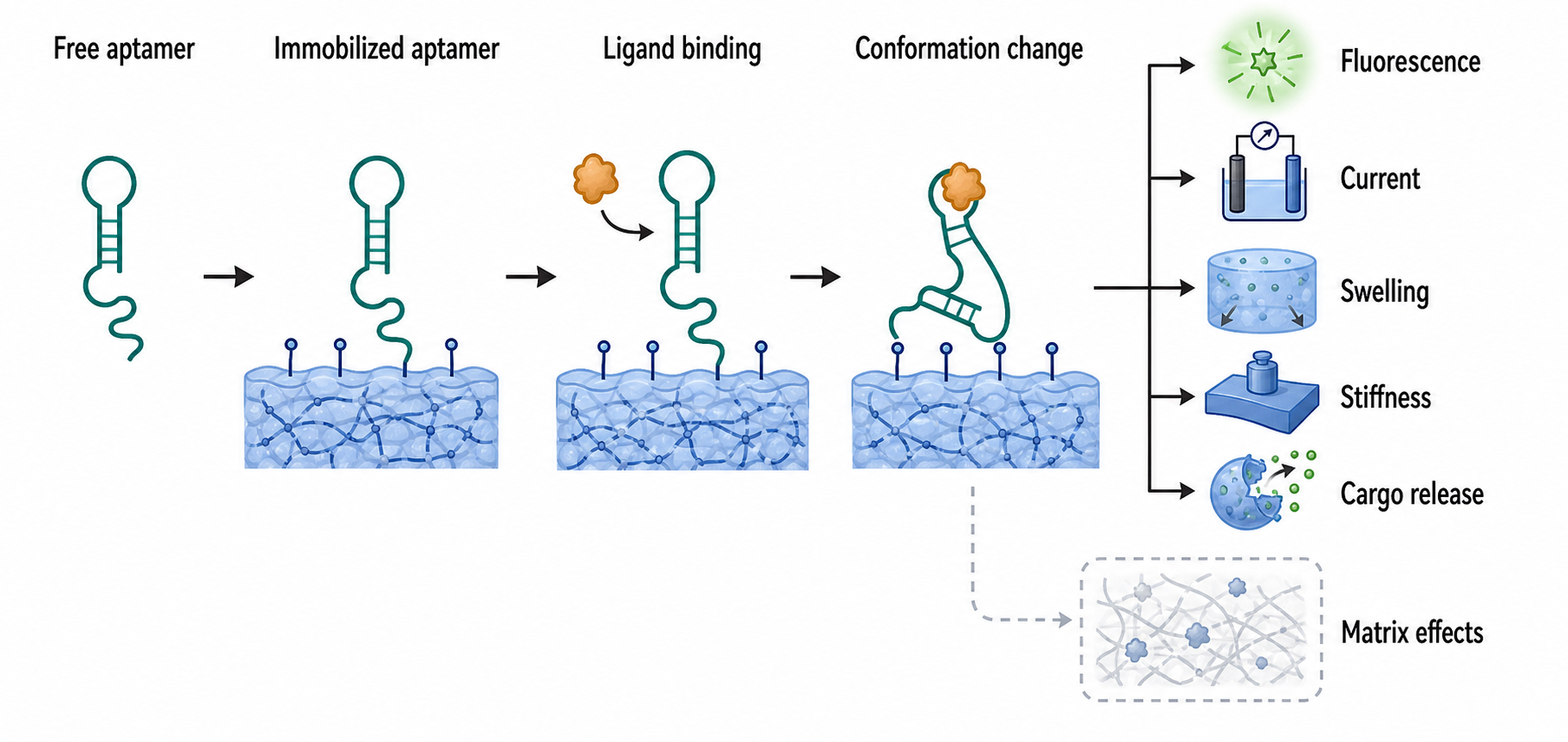
Figure 148.3. Aptamer Material Signal Transduction. Show how aptamer binding is converted into fluorescence, electrochemical current, swelling, stiffness, or cargo release.
A ligand-responsive hydrogel is a useful running example. A hydrogel is a water-rich network held together by crosslinks. If RNA aptamer-containing strands provide some of the crosslinks, ligand binding can stabilize or destabilize the network. In one design logic, a target ligand binds an aptamer and pulls a strand away from a complementary partner, weakening the network and releasing cargo. In another, ligand binding stabilizes a folded aptamer that acts as a crosslink, strengthening the gel. The output can be swelling, softening, fluorescence change, cargo release, or altered cell adhesion.
The first design step is choosing the recognition chemistry. An aptamer with nanomolar affinity in a low-salt buffer may perform poorly in serum because proteins, nucleases, divalent cations, pH, and competing molecules change the folding landscape. A small-molecule aptamer may be highly specific for a purified ligand but bind metabolites with similar structures in biological samples. A protein aptamer may lose affinity when immobilized because the surface blocks the binding face. Aptamer materials therefore require affinity, specificity, and function tests under the material’s actual operating conditions.
Surface immobilization creates special boundary cases. RNA attached to a gold surface, polymer, bead, electrode, or nanoparticle is no longer a freely diffusing molecule. Linker length, surface density, orientation, steric crowding, electrostatic repulsion, and drying can change folding and target access. High aptamer density may increase total binding capacity but reduce per-aptamer activity. Low density may preserve folding but reduce signal. A carefully designed material often uses spacer sequences, passivating molecules, and density optimization rather than simply maximizing aptamer loading.
Aptamer biosensors must couple binding to a transduction mechanism. Fluorescent sensors may use dye-labeled aptamers, fluorogenic aptamers that activate a small-molecule dye, or strand-displacement reporters. Electrochemical sensors may place an aptamer on an electrode so ligand-induced motion changes electron transfer. Mechanical sensors may change stiffness or swelling. Optical surfaces may change refractive index or plasmonic response. Each output has different artifacts. Fluorescence can be affected by photobleaching or sample autofluorescence. Electrochemical signals can drift because of fouling. Hydrogel swelling can reflect salt or pH rather than ligand.
Table 148.3. Aptamer Material Evidence Ladder. Separate aptamer selection, purified binding, material integration, complex-sample performance, and biological application.
| Evidence level | What it shows | What it cannot show | Required next test |
|---|---|---|---|
| Aptamer selection or enrichment | A sequence population was enriched for binding under the selection conditions | True affinity, structural mechanism, specificity in complex samples, or material compatibility | Purify candidate aptamers and measure binding, specificity, and folding dependence |
| Purified-buffer binding | Dissociation, competition, or conformational response can be measured in a controlled solution | Performance after immobilization, nuclease exposure, serum proteins, fouling, or competing metabolites | Test binding under the intended salt, pH, temperature, matrix, and competitor conditions |
| Structural or mutational validation | The aptamer fold or key nucleotides contribute to target recognition | Material-level signal, surface accessibility, or biological specificity | Incorporate fold-preserving and fold-disrupting variants into the material design |
| Material integration | The aptamer remains attached to a surface, gel, particle, film, or electrode and can encounter target | That binding is efficiently converted into fluorescence, current, swelling, stiffness, or release | Quantify orientation, density, linker effects, background signal, and target-dependent output |
| Device transduction | Target binding produces a measurable material or reporter change | Specificity in realistic samples, reversibility, durability, or biological utility | Challenge the device with near-neighbor ligands, matrix controls, repeated cycles, and time courses |
| Complex-sample performance | The material responds in serum, lysate, environmental sample, tissue fluid, or mixed-cell context | Mechanism of response, long-term stability, pharmacology, or clinical usefulness | Separate degradation, nonspecific adsorption, fouling, and matrix effects from aptamer-dependent recognition |
| Biological or therapeutic application | Signal, capture, release, targeting, or potency occurs in cells, tissue, animals, or a validated use model | General safety, tissue specificity, manufacturability, or regulatory readiness | Measure dose response, biodistribution or localization, immune activation, toxicity, and batch reproducibility |
Therapeutic aptamer materials add pharmacological constraints. An aptamer displayed on an RNA nanoparticle might target a receptor and deliver a cargo. A hydrogel might release an RNA drug in response to a metabolite or inflammatory cue. A surface might capture an extracellular RNA-binding protein or viral particle. These concepts are attractive because aptamers are programmable and can be selected for many targets. However, aptamers often require chemical stabilization, and their in vivo residence time, renal clearance, protein binding, and immune sensing must be tested. An aptamer that works as an analytical reagent is not automatically a drug ligand.
The evidence basis for aptamer materials is layered. A selection paper establishes a binding sequence only under selection conditions. A biochemical paper may measure dissociation constants, specificity, and structural dependence. A material paper must then show that the aptamer remains folded and responsive after incorporation. A biological application must show that the signal or release event occurs in the intended sample or organism and is not explained by degradation, nonspecific adsorption, or matrix effects. This layering is why aptamer-material claims should state the evidence level explicitly.
Do not overgeneralize the term “specific.” Specificity can mean discrimination among purified ligands, selectivity in a serum sample, cell-type targeting, or absence of clinical off-target effects. These are not equivalent. Similarly, “responsive” can mean rapid reversible switching, slow irreversible release, equilibrium swelling, or degradation-triggered collapse. A reader should ask what input was applied, what output was measured, how reversible the response was, and whether competing inputs were tested.
The local references do not provide high-confidence aptamer-material review coverage. The broad synthetic nucleic acid technology reference can support general statements about nucleic-acid predictability and design [Chakraborty_S_2014_synthetic_nucleic_acid_technologies], but final release needs targeted citations for RNA aptamer selection, fluorogenic aptamers, aptamer hydrogels, aptamer biosensors, immobilization artifacts, and therapeutic aptamer materials.
148.4. RNA Condensate Engineering and Intracellular Materials
RNA condensate engineering is the deliberate design of RNA-containing assemblies that concentrate molecules in space and often show phase-separation-like behavior. A biomolecular condensate is a compartment formed by many weak interactions rather than by a surrounding lipid membrane. Natural examples include nucleoli, stress granules, processing bodies, germ granules, and many nuclear bodies. These structures contain RNA, RNA-binding proteins, enzymes, and regulatory factors. Chapter 58 treats natural RNA condensates in detail; this section focuses on how engineers try to build or redirect such material states.
The basic physical concept is multivalency. A molecule is multivalent when it carries multiple interaction sites. An RNA with repeated protein-binding hairpins, many G-quadruplex-prone regions, repetitive low-complexity sequences, or many complementary patches can interact with multiple partners. A protein with intrinsically disordered regions or multiple RNA-binding domains can do the same. When multivalent interactions cross a threshold, molecules can demix from the surrounding solution into a dense phase. The dense phase can behave like a liquid, viscoelastic gel, aging solid, or heterogeneous assembly depending on interaction strength, valency, concentration, ATP-dependent remodeling, and cellular environment.
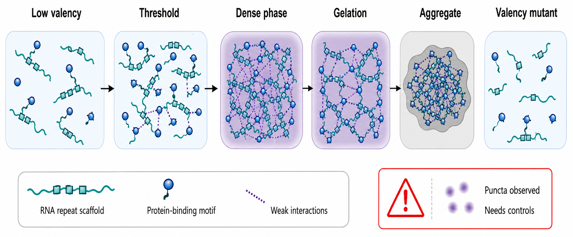
Figure 148.4. Valency-Controlled RNA Condensate Engineering. Explain how RNA repeat number, protein-binding motifs, weak interactions, and concentration thresholds produce scaffolded assemblies, condensate-like phases, gels, or aggregates.
Engineered intracellular RNA materials use several design routes. One route expresses an RNA bearing repeated binding sites for a designed RNA-binding protein fused to an enzyme, fluorophore, or regulatory domain. Another route uses base-pairing among repeated RNA modules to build a mesh. A third route combines RNA aptamers with proteins containing phase-separation-promoting domains. A fourth route uses guide RNAs to localize effectors to DNA, nascent RNA, or chromatin while also increasing local valency. In all cases, the intended function may be localization, reaction acceleration, sequestration, sensing, memory, or controlled release.
A concrete example is an engineered RNA hub that recruits an enzyme cascade. The RNA contains repeated binding motifs for two protein fusions. When expressed, the RNA and proteins form puncta. If those puncta increase product formation, the designer might claim that the RNA creates a synthetic metabolic compartment. A rigorous interpretation requires controls: puncta should depend on the RNA motifs; enzyme abundance should be comparable; catalytically inactive controls should separate localization from enzymatic activity; dissolution or mutation of the hub should reduce product; and cell stress markers should be checked. Without those controls, puncta may be overexpression aggregates.
Condensate engineering differs from ordinary scaffolding by emphasizing collective material behavior. A small scaffold may bring two molecules together without forming a separate phase. A condensate-like material involves many molecules and an emergent dense state. The distinction matters because dense phases have properties that affect reaction rates, diffusion, selectivity, and aging. A condensate can enrich a substrate but also exclude a cofactor. It can accelerate one reaction and inhibit another by slowing diffusion. It can buffer concentration or create hysteresis. Therefore material characterization is part of mechanism, not an aesthetic supplement.
Evidence for phase separation is commonly overclaimed. Spherical puncta, fusion events, fluorescence recovery after photobleaching, concentration thresholds, and sensitivity to salt or aliphatic alcohols can support a condensate interpretation, but none is decisive alone. Some aggregates are round. Some solid assemblies exchange slowly. Some perturbations disrupt many weak interactions nonspecifically. Stronger evidence combines live-cell dynamics, concentration dependence, reversibility, mutational tuning of valency, in vitro reconstitution, biophysical measurement of material properties, and functional rescue. Chapter 5′s artifact-control framework is essential for this literature.
Box 148.3. Puncta Are Observations, Not Condensate Mechanisms
Fluorescent puncta are a starting observation, not a mechanism. A punctum can be a liquid-like condensate, a gel, a solid aggregate, a membrane-associated cluster, an overexpression artifact, or a stress response. A condensate-engineering claim should show that puncta depend on the designed RNA features, appear over a concentration or valency threshold, change predictably when interaction sites are removed or restored, and retain the intended function. Material-state evidence strengthens the claim: fusion behavior, fluorescence recovery after photobleaching, exchange rates, dissolution by targeted perturbation, in vitro reconstitution with defined components, and measurements of diffusion or viscoelasticity can all help. No single assay is decisive. The most important control is functional: if the RNA material is proposed to accelerate a reaction, sequester a protein, or localize an editor, the output should track the engineered material state rather than only the presence of bright spots.
RNA-specific issues include length, sequence composition, structure, modification, and surveillance. Long RNAs can provide many binding sites, but they are also substrates for nuclear retention, export, decay, editing, and immune sensing. Repetitive RNAs can nucleate pathological RNP assemblies, as seen in repeat-expansion disease contexts, and engineered repeats may mimic some hazards. Structured RNAs may resist nucleases but also activate sensors or sequester proteins. Modified nucleotides can tune stability and immune recognition but may alter condensate behavior. Intracellular RNA materials must therefore be evaluated as cell-biological perturbations, not inert building blocks.
Condensate-inspired design can also be extracellular or cell-free. RNA hydrogels, coacervates, and RNP droplets can be used as reaction compartments, delivery depots, or sensing environments. Such systems are easier to characterize than living-cell condensates because composition and concentration can be controlled. However, cell-free success may not translate to cells, where molecular crowding, active remodeling, nucleases, and surveillance pathways differ. Conversely, a system that forms only under overexpressed cellular conditions may fail in a purified assay because it requires endogenous partners.
Therapeutic translation is speculative but important. Designed condensates might concentrate RNA editors at a target transcript, sequester toxic RNA-binding proteins, localize mRNA translation, or build synthetic organelles for cell therapies. Each application faces safety questions. Persistent condensates could trap essential proteins, alter stress responses, interfere with RNA decay, or seed pathological aggregation. Reversible, dose-controlled, and degradable designs are likely safer than irreversible intracellular materials. Claims about therapeutic condensates should therefore be labeled as frontier engineering unless supported by animal or clinical evidence.
The chapter-local references do not provide a condensate-engineering anchor. This draft treats the section as expert synthesis that needs citations for natural RNA granule material properties, engineered phase separation, RNA repeat toxicity, intracellular synthetic organelles, and assay standards. The absence of a local review anchor is a major Curation-deferred source need for this chapter.
148.5. Molecular Machines, Dynamic Assemblies, Strand-Displacement Systems, and Responsive Nanostructures
An RNA molecular machine is an RNA-containing system that changes state in a programmed way and uses that change to perform a task. The task may be mechanical movement, cargo release, signal generation, catalytic activation, assembly, disassembly, localization, or computation. The phrase “machine” should be used carefully. Thermal motion drives molecular systems, and most RNA machines operate by shifting ensembles and kinetic pathways rather than by acting like macroscopic gears. A valid machine claim identifies input, state change, output, energy source or thermodynamic bias, and reset behavior.
Natural RNAs already show machine-like behavior. Riboswitches bind metabolites and change gene expression platforms. Ribozymes fold active sites and catalyze chemical reactions. Spliceosomal small nuclear RNAs participate in rearranging RNP machines. The ribosome uses rRNA as a core structural and catalytic framework for translation. Synthetic RNA machines borrow these principles but usually simplify them: an aptamer domain senses input, a helix or strand-displacement domain transmits the signal, and an output module reports or acts.
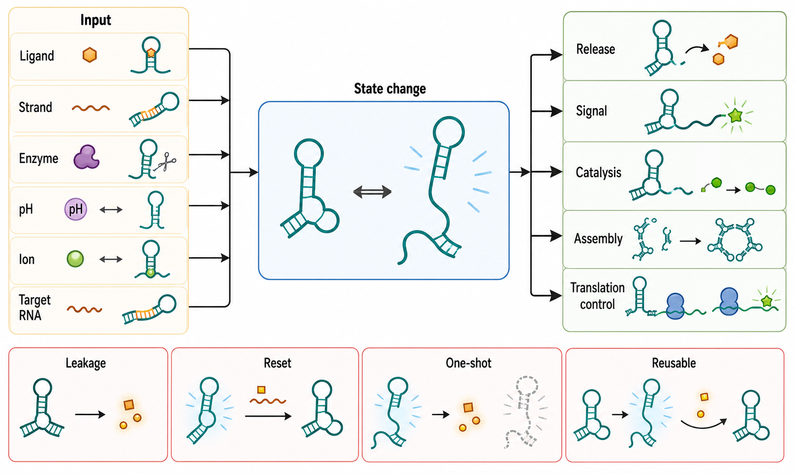
Figure 148.5. Dynamic RNA Machine Input-State-Output Map. Map inputs such as ligand, strand, enzyme, pH, ion, or target RNA to conformational state changes and outputs such as release, signal, catalysis, assembly, or translation control.
Strand displacement is one of the clearest dynamic mechanisms. A strand-displacement system contains a double-stranded or partially paired nucleic-acid complex with an exposed single-stranded toehold. An incoming strand binds the toehold and then replaces an incumbent strand through branch migration. In RNA systems, strand displacement can trigger fluorescence, release a cargo, expose an aptamer, activate translation, or assemble a larger structure. The designer controls kinetics by changing toehold length, sequence, temperature, salt, secondary structure, and competing off-target interactions.
RNA strand-displacement design differs from DNA strand displacement because RNA folds strongly and may interact with proteins and nucleases. A toehold drawn as unpaired may be hidden in an alternative hairpin. An output strand may be degraded before reporting. A branch migration domain may be slowed by stable secondary structure. In cells, RNA-binding proteins can block or stabilize intermediates. Therefore RNA circuits often require chemical probing, kinetic assays, and cellular controls in addition to sequence design.
Ligand-responsive nanostructures couple aptamer binding to assembly or motion. A small molecule can stabilize an aptamer fold that brings two helices together. A protein can bridge aptamer-decorated particles. A metabolite can expose a cleavage site or hide a degradation element. A receptor can cluster multivalent RNA particles at a cell surface. The design principle is modular signal transduction: recognition changes structure, and structure changes function. The challenge is leakage. If the output occurs without input, the device has poor dynamic range. If the input stabilizes a nonfunctional off-pathway structure, the device may bind ligand but fail to respond.
Ribozyme-coupled devices provide another example. A self-cleaving ribozyme can be placed in an RNA scaffold so that ligand binding either promotes or inhibits cleavage. Cleavage can release a cargo, destabilize a transcript, or change a material network. The mechanism requires both aptamer communication and catalytic chemistry. Mutating the ribozyme active site should remove cleavage without removing binding. Mutating the aptamer should remove ligand dependence. Time-course assays should distinguish folding-limited activation from true ligand control.
Molecular machines can also operate through assembly state. An RNA ring might open when a strand invades one edge. A cage might release an RNA cargo after encountering a disease-associated microRNA. A nanoparticle might expose a cell-binding aptamer only after an environmental trigger. These designs are attractive for precision delivery and diagnostics, but they require realistic input concentrations. A device that responds to micromolar trigger RNA in buffer may be irrelevant if the cellular trigger is present at a few copies per cell or buried in RNPs.
Table 148.4. Dynamic RNA Device Design Variables. Summarize design variables that control dynamic device behavior.
| Variable | Affects thermodynamics | Affects kinetics | Common artifact | Useful assay |
|---|---|---|---|---|
| Toehold length and exposure | Sets initial binding stability for the trigger strand or RNA input | Longer or more accessible toeholds usually accelerate initiation | Designed toehold is hidden in an alternative hairpin or bound by proteins | Time-resolved fluorescence, chemical probing, and toehold-mutant comparison |
| Branch-migration domain | Determines final duplex stability and strand-exchange favorability | Stable secondary structure can slow or stall displacement | Endpoint product forms only after nonphysiologic heating or excess trigger | Stopped-flow or gel time course across trigger concentrations and temperatures |
| Aptamer-output coupling | Ligand binding stabilizes or destabilizes the output conformation | Coupling strength controls switching rate and response lag | Ligand binds but does not propagate a functional conformational change | Ligand titration with FRET, probing, or reporter output normalized to RNA abundance |
| Ribozyme communication module | Ligand or strand state biases active-site formation or cleavage suppression | Folding order can make cleavage activation time-dependent | Cleavage changes RNA abundance rather than controlled device state | Active-site mutant, aptamer mutant, and cleavage time course |
| Trigger abundance and compartment | Determines whether the intended input can outcompete background states | Low-copy or RNP-buried triggers may make reactions too slow in cells | Buffer response requires trigger concentrations never reached in the intended system | Cellular dose-response paired with direct trigger and device-state measurements |
| Ion, pH, temperature, and crowding conditions | Stabilize helices, tertiary contacts, aptamer folds, and off-pathway states | Environmental shifts can change folding and strand-exchange rates | Device appears specific because the assay buffer suppresses competing folds | Matrix-matched assays and condition sweeps with structural readouts |
| Chemical modification or nuclease protection | Can stabilize intended structures or disrupt motif and aptamer energetics | Protection may slow activation, cleavage, or release | Improved signal reflects slower degradation rather than better switching | Intact-RNA quantification alongside activity and switching assays |
| Reporter and output module | Changes apparent free energy of the observed state through dye, cargo, or fusion effects | Reporter maturation, photophysics, or cargo release can dominate timing | Fluorescence increase is misread as structural opening | Orthogonal reporter-free structural assay and output-module swap |
| Reset and reversibility design | Sets whether the device has a reversible equilibrium or one-shot product bias | Reset strand, enzyme turnover, or degradation controls reuse rate | Single-turnover release is described as a reusable machine | Repeated input-removal cycles, washout tests, and mass-balance analysis |
Evidence for dynamic behavior requires time-resolved measurements. Endpoint gels can show that a final product forms, but they do not show the path, rate, leakage, reversibility, or intermediate traps. Fluorescence time courses, stopped-flow experiments, single-molecule FRET, chemical probing across time, microscopy of assembly, and computational kinetic modeling can help. For a device intended to work in cells, reporter output should be compared with direct measurements of RNA abundance and state. Otherwise increased fluorescence could reflect stabilization, transcriptional induction, or altered degradation rather than machine operation.
Design automation is especially useful for dynamic systems because humans cannot easily reason through all competing folds and kinetic traps. Thermodynamic models can predict secondary structures and ensemble probabilities. Inverse-folding algorithms can search for sequences that favor a target structure. Kinetic simulators can estimate folding pathways or strand-displacement rates. Machine-learning tools can learn from prior designs. These tools are helpful filters, not final evidence. They depend on training data, parameter assumptions, ionic conditions, pseudoknot handling, and the absence of proteins or crowding unless explicitly modeled.
Several overgeneralizations are common in this area. A switch is not a sensor unless input concentration is relevant and output is measurable above background. A molecular machine is not necessarily reusable; many nucleic-acid devices are single-turnover. A responsive nanostructure is not automatically smart delivery; it must encounter the trigger in the right compartment. A computationally predicted conformational change is not proof of motion. A fluorescence increase is not proof of structural opening unless controls exclude abundance and environment effects.
The chapter-local references are insufficient for final citation of strand-displacement systems, RNA switches, ribozyme devices, dynamic nanostructures, and RNA computation. The 2014 synthetic nucleic acid technology reference supports broad discussion of predictive nucleic-acid design [Chakraborty_S_2014_synthetic_nucleic_acid_technologies], but targeted primary and review citations are needed for the specific device classes.
148.6. Stability, Immunogenicity, Manufacturing, Design Automation, and Translation Barriers
The most important translational question for RNA materials is not whether an RNA can be drawn, folded, or imaged once. The question is whether a reproducible product can be manufactured, stored, delivered, and made to function in the intended environment with acceptable safety. This section connects molecular design to product reality. It overlaps with Chapter 159 on RNA synthesis and release testing, Chapter 156 on lipid nanoparticles, Chapter 157 on delivery systems, Chapter 108 on innate immune sensing, and Chapter 163 on responsible translation.
RNA stability has several meanings. Chemical stability refers to resistance to backbone cleavage, hydrolysis, oxidation, and chemical modification. Enzymatic stability refers to resistance to ribonucleases in serum, cells, tissues, or environmental samples. Structural stability refers to maintaining the intended fold or assembly state. Functional stability refers to preserving the desired output, such as binding, release, catalysis, or delivery. A design can be chemically intact but structurally rearranged; structurally intact but biologically inactive; or active in buffer but rapidly degraded in serum.
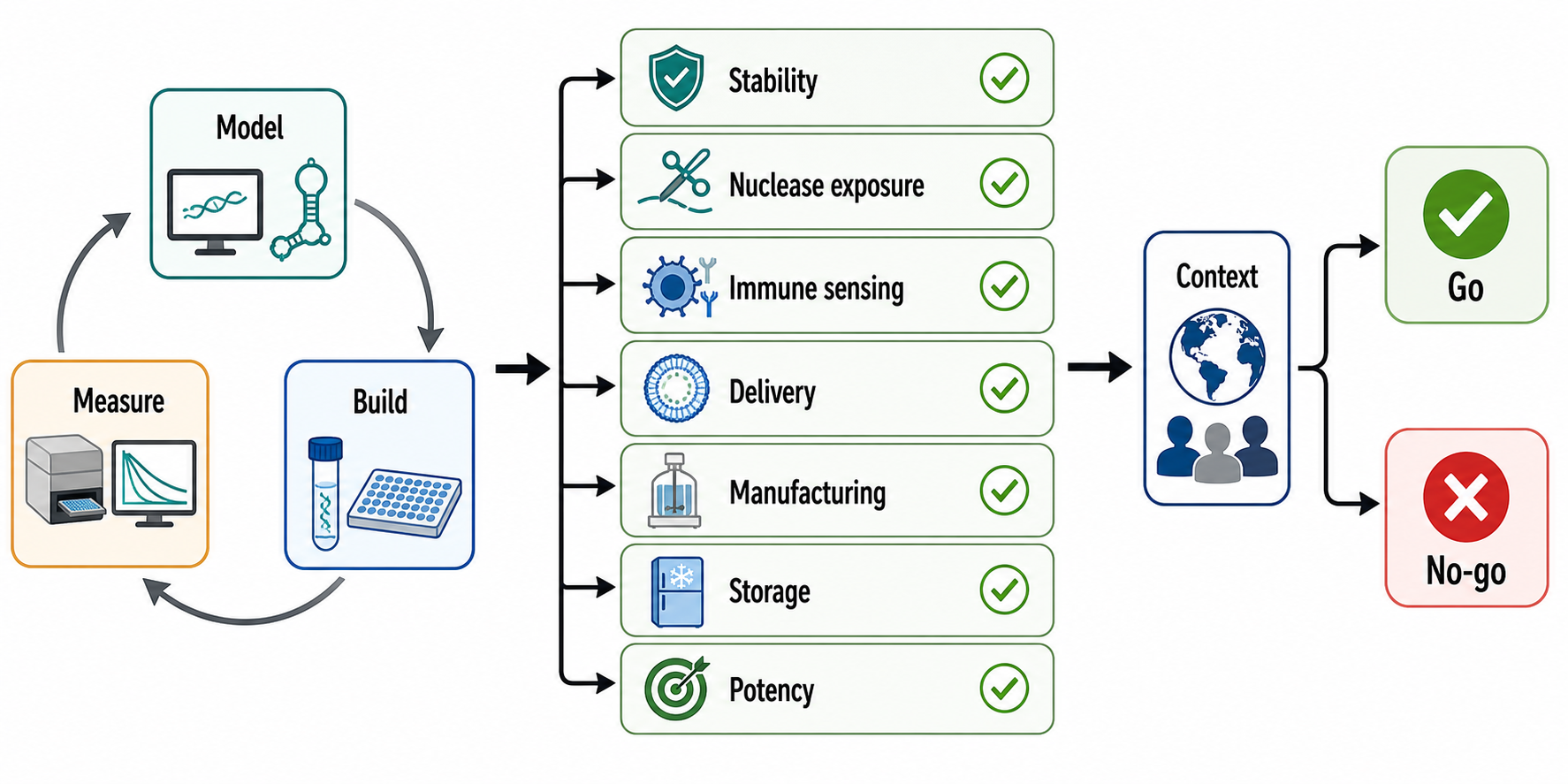
Figure 148.6. Translational Stress Test for RNA Materials. Present a checklist-style workflow for challenging an RNA material across chemical stability, nuclease exposure, immune sensing, delivery, manufacturing, storage, and functional potency.
RNA’s 2′ hydroxyl contributes to folding and tertiary chemistry but also makes the backbone vulnerable to base-catalyzed transesterification. Divalent metal ions can stabilize tertiary structure but may also promote cleavage under some conditions. Chemical modifications such as 2′ O-methyl, 2′ fluoro, locked nucleic acid, phosphorothioate linkages, terminal caps, circularization, and modified nucleobases can increase stability, but they can also alter folding, aptamer binding, immune sensing, protein interactions, and manufacturing analytics. A modification pattern validated for an siRNA duplex may not preserve an RNA origami junction or aptamer hydrogel.
Immunogenicity is a central boundary for mammalian applications. Innate immune receptors can detect double-stranded RNA, 5′ triphosphorylated RNA, uncapped RNA, long RNA, uridine-rich RNA, or RNA delivered to endosomes and cytosol. Toll-like receptors, RIG-I-like receptors, protein kinase R, OAS/RNase L pathways, and related systems are treated in Chapter 108. For RNA nanomaterials, immunogenicity depends on sequence, length, structure, modifications, delivery vehicle, impurities, dose, route, cell type, and whether the product accumulates in immune cells. Immune activation can be a desired vaccine or adjuvant effect, but it is a liability for most scaffolds, sensors, and chronic materials.
Manufacturing starts with how the RNA is made. Short RNAs can be chemically synthesized with high control over modifications but may become difficult or costly as length and structural complexity increase. Longer RNAs can be produced by in vitro transcription, but products may include abortive transcripts, double-stranded RNA byproducts, incorrect ends, sequence variants, template carryover, proteins, salts, and endotoxin if workflows are not controlled. Genetically encoded intracellular RNAs avoid ex vivo purification but introduce expression variability, host-cell processing, localization, surveillance, and biosafety concerns. Each route has different release tests.
Analytical control for RNA materials is harder than for a simple oligonucleotide. A product specification may need identity, length distribution, sequence integrity, modification stoichiometry, residual template, residual protein, endotoxin, residual solvents, ion content, folding state, particle size, polydispersity, morphology, cargo loading, ligand display, activity, sterility, and stability after storage. For a dynamic device, the specification may also include switching range, leakage, response time, and reversibility. For a therapeutic delivery product, potency must be measured in a biologically relevant assay rather than inferred from particle formation alone.
Design automation is advancing but uneven. Useful computational tasks include secondary-structure prediction, inverse folding, motif selection, junction geometry, codon or transcription-order planning, off-target complementarity screening, immune-motif filtering, aptamer integration, and kinetic simulation. Automated pipelines can generate candidates and detect obvious conflicts. They cannot yet guarantee that a large RNA nanostructure will fold as designed in serum or inside a cell. The strongest design workflow is iterative: model, build, measure, update the model, redesign, and retest.
The 2014 synthetic nucleic acid technology paper in the local references emphasizes the predictive power and limits of synthetic nucleic acid approaches [Chakraborty_S_2014_synthetic_nucleic_acid_technologies]. That perspective remains important. Predictability is not absolute; it is domain-specific. Short duplexes may be predictable by nearest-neighbor models. Large RNA assemblies may require empirical motif libraries and structural validation. Intracellular materials may require cell-specific models of abundance, protein binding, degradation, and immune sensing. A design that is rational at one level can fail at another.
Translational barriers can be grouped into six classes. The first is folding heterogeneity: the intended structure competes with misfolded or partial structures. The second is biological instability: nucleases, RNA surveillance, and dilution reduce product lifetime. The third is delivery: the material must reach the correct tissue, cell, and compartment. The fourth is immunogenicity and toxicity: the product or vehicle may trigger innate immunity, complement, stress, or off-target biology. The fifth is manufacturability: product quality must be reproducible at scale. The sixth is value: the RNA material must outperform simpler alternatives enough to justify complexity.
Regulatory and clinical translation require careful claims. A biosensor material may face diagnostic-device validation rather than drug regulation. A therapeutic scaffold may be regulated as an RNA drug, biologic, combination product, or advanced therapy depending on composition and use. A cell-therapy material expressed inside engineered cells has different risks from an injected nanoparticle. Environmental or agricultural uses raise persistence, spread, and ecological questions. Chapter 163 treats governance more fully, but designers should consider use context early because it determines acceptable risk.
Consensus in the field is cautious. RNA is a powerful programmable material because it can combine sequence-specific recognition, folding, ligand binding, catalysis, and genetic encodability. The most convincing demonstrations are those that connect design to orthogonal structural evidence and a function measured under relevant conditions. The least convincing claims rely on schematic cartoons, single endpoint gels, uncontrolled puncta, or buffer-only binding data presented as translational readiness. The open questions are whether RNA nanostructures can be manufactured at scale with sufficient homogeneity, whether intracellular RNA materials can be made safe and controllable, whether aptamer materials can remain selective in complex samples, and whether design automation can generalize beyond narrow training regimes.
Common misconceptions are worth making explicit. First, “RNA is programmable” does not mean every designed sequence folds correctly. Second, “self-assembly” does not mean error-free assembly. Third, “biocompatible” does not mean immunologically silent. Fourth, “nanoscale” does not mean deliverable. Fifth, “aptamer-targeted” does not mean tissue-specific in vivo. Sixth, “phase-separated” does not mean functional. Seventh, “machine” does not mean powered, reusable, or precise. These cautions are not pessimism; they are the standards that let RNA nanotechnology mature from elegant demonstrations into robust tools.
The largest Curation-deferred source need for this chapter is citation coverage. The local bibliography explicitly lacks a high-confidence review anchor and contains only two verified papers, both useful but insufficient for a field this broad. Before finalization, the chapter needs curated reviews and primary papers for RNA nanoparticles, pRNA and three-way junction architectures, RNA tiles, RNA origami software, RNA aptamer materials, RNA hydrogels, RNA condensate engineering, strand-displacement circuits, RNA device kinetics, immunogenicity of RNA materials, and manufacturing standards for complex RNA assemblies.
Recent Consensus
RNA nanotechnology is most mature as a design and discovery field, with strong mechanistic grounding in RNA folding, motif modularity, aptamer recognition, and nucleic-acid self-assembly. Translation is uneven. Cell-free structures and sensors are easier to validate than in vivo materials. Therapeutic and intracellular applications require delivery, immune, stability, and manufacturing evidence that goes beyond structural assembly. The safest consensus statement is that RNA provides a uniquely programmable material platform, but each application must earn its claim through context-specific characterization.
Open Questions, Controversies, Deprecated Models, and Common Misconceptions
Open questions:
- How can co-transcriptional folding pathways be predicted for large origami?
- How can RNA nanostructure manufacturing be scaled without losing homogeneity?
- How can RNA materials be stabilized without destroying function?
- How can intracellular materials be designed to remain reversible and nonpathogenic?
- How can computational design tools be benchmarked across independent laboratories?
Controversies:
- Should some engineered puncta be called condensates?
- Can aptamer targeting provide enough in vivo specificity for complex therapeutics?
- How much of RNA nanotechnology should be modeled after DNA nanotechnology versus natural RNP assembly?
Deprecated or weakened claims:
- A gel shift should not be treated as proof of a designed nanoparticle.
- Fluorescence puncta should not be treated as proof of functional phase separation.
- Buffer affinity should not be treated as proof of biological targeting.