Chapter 108. Innate Immune Sensing of RNA: TLRs, RIG-I-Like Receptors, PKR, OAS/RNase L, and TRIM25-Linked Signaling
Scope Note
This chapter explains how animal innate immune systems interpret RNA as a sign of infection, cellular damage, or therapeutic intervention. The focus is on RNA-sensing Toll-like receptors in endosomes, cytosolic RIG-I-like receptors, double-stranded RNA-responsive effector pathways such as PKR and OAS/RNase L, and TRIM25-linked regulatory circuits that connect RNA recognition to interferon, inflammation, translational arrest, RNA cleavage, and cell death. The chapter treats self versus nonself discrimination as an active, compartment-dependent process rather than as a single molecular barcode.
Executive Summary
Innate RNA sensing depends on three linked ideas. First, RNA molecules carry molecular patterns: base-paired regions, single-stranded uridine-rich or guanosine/uridine-rich sequences, 5′ triphosphate or diphosphate ends, missing cap structures, unusual length distributions, and modification patterns. Second, receptors interpret those patterns only within specific compartments. Host ribosomal RNA and messenger RNA are abundant, so the immune system cannot simply respond to every RNA molecule. Endosomal Toll-like receptors survey material delivered by uptake, phagocytosis, or autophagy; RIG-I-like receptors survey the cytosol; PKR and OAS proteins respond to cytosolic double-stranded RNA; RNase L, stress granules, mitochondria, and inflammasome adaptors shape the downstream outcome. Third, the same pathways can protect against viruses, drive autoinflammatory disease when self RNA is mislocalized or improperly edited, and be deliberately engaged or avoided in RNA therapeutics.
TLR3 recognizes double-stranded RNA in acidified endosomes and signals through TRIF. TLR7 and TLR8 recognize degradation products of single-stranded RNA and signal through MyD88, with cell-type-specific outputs. TLR13, present in mice but not humans, recognizes a conserved bacterial 23S ribosomal RNA sequence and illustrates how RNA sequence can be interpreted as a microbial pattern. Cytosolic RIG-I preferentially recognizes short duplex RNA with 5′ triphosphate or diphosphate ends, whereas MDA5 assembles on long double-stranded RNA and detects many picornavirus-like or replication-derived RNAs. LGP2 lacks signaling CARD domains and modulates RIG-I-like receptor activation. Activated RIG-I and MDA5 signal through MAVS on mitochondria and related membranes to induce type I and type III interferons and inflammatory genes. TRIM25-dependent ubiquitin signaling, phosphorylation, dephosphorylation, ATP-dependent proofreading, and condensate-like assembly tune the threshold.
PKR binds double-stranded RNA, dimerizes, phosphorylates eIF2alpha, and reduces translation while promoting stress responses. OAS enzymes synthesize 2′,5′-linked oligoadenylates after binding double-stranded RNA; these second messengers activate RNase L, which cleaves RNA and amplifies antiviral signaling. Inflammasome connections arise through RNA virus replication, ion flux, mitochondrial stress, RNase L-generated fragments, and crosstalk between interferon and IL-1-family cytokine pathways. RNA modifications such as N1-methylpseudouridine, 2′-O-methylation, adenosine-to-inosine editing, and tRNA/rRNA modifications can reduce inappropriate sensing, although the effect depends on receptor, sequence, structure, dose, purity, and delivery route. Viruses evade sensing by capping, hiding, editing, binding, degrading, sequestering, or modifying RNA and by targeting receptor adaptors. The field is mechanistically mature in core pathways but unsettled in many context-specific questions, especially how endogenous RNA species, therapeutic nanoparticles, and chronic inflammatory states intersect.
Concept Inventory
- Innate RNA sensing: the set of receptor and effector pathways that detect RNA features associated with infection, stress, or mislocalization. A pathogen-associated molecular pattern is a molecular feature enriched in microbes or viruses, such as a 5′ triphosphate RNA end. A damage-associated molecular pattern is an endogenous molecule that becomes stimulatory because it appears in the wrong compartment, wrong concentration, or wrong biochemical state. Self versus nonself discrimination is therefore probabilistic and contextual. Mammalian cells tolerate enormous amounts of self RNA because most self RNA is capped, processed, modified, protein-bound, compartmentalized, rapidly degraded when misplaced, or shielded from receptors.
- Endosomal RNA-sensing Toll-like receptors: type I membrane receptors whose ligand-binding domains face the endosomal lumen. TLR3 primarily recognizes double-stranded RNA, TLR7 and TLR8 recognize single-stranded RNA degradation products, and TLR13-like systems recognize bacterial ribosomal RNA in species that encode them. RIG-I-like receptors are DExD/H-box ATPases in the cytosol. RIG-I and MDA5 have N-terminal CARD domains that transmit signals to MAVS, whereas LGP2 lacks CARD domains and is regulatory. PKR and OAS/RNase L are not only sensors but also antiviral effectors because they directly alter translation or RNA integrity after recognizing double-stranded RNA. TRIM25 is an E3 ubiquitin ligase and signaling organizer best known for promoting RIG-I activation, although its role is regulated by viral antagonists, host cofactors, and cell state.
What to Know Before Reading This Chapter
RNA molecules differ by strand polarity, terminal chemistry, base modification, folding, length, protein occupancy, and cellular compartment. A 5′ cap is a host mRNA mark added during nuclear transcription and maturation; many RNA virus replication products instead expose triphosphate or diphosphate 5′ ends. Double-stranded RNA is rare as a long naked molecule in healthy mammalian cytosol, but it appears during viral replication, from inverted repeats, from mitochondrial transcription, and from synthetic duplex delivery. Endosomes are acidic vesicles that receive extracellular and phagocytosed material, so endosomal receptors can sample microbial or damaged-cell RNA without being exposed continuously to all cytosolic RNA. Interferons are cytokines that induce hundreds of antiviral genes; inflammasomes are cytosolic complexes that activate inflammatory caspases and IL-1-family cytokines. These pathways cooperate, but they are not interchangeable.
108.1. Self versus nonself RNA features and compartmentalization
The simplest description of innate RNA sensing says that host cells detect viral RNA. That statement is useful but incomplete. Cells do not have a universal label that says “viral.” Instead, receptors respond to a set of molecular and spatial features that are statistically enriched during infection. Important features include long double-stranded RNA, blunt-ended or base-paired 5′ triphosphorylated RNA, uncapped or improperly capped RNA, single-stranded RNA fragments in acidified endosomes, unusual nucleotide composition, missing 2′-O-methylation at the cap-adjacent nucleotide, and RNA that appears in a compartment where the receptor can see it. Recent reviews emphasize this proofreading model rather than a rigid self/nonself binary (Uehata and Takeuchi 2020, PMID: 32708595; Solotchi and Patel 2024, PMID: 38884803).
Compartmentalization is as important as chemistry. Nuclear mRNA precursors, ribosomal RNA, transfer RNA, mitochondrial transcripts, and noncoding RNAs all contain structures that could stimulate receptors if they were presented in the wrong context. Healthy cells reduce this risk by capping mRNA, editing some double-stranded regions, decorating abundant RNAs with modifications, binding RNA with proteins, maintaining organelle membranes, and degrading leaked nucleic acids. Endosomal receptors are normally separated from cytosolic RNA; cytosolic RIG-I-like receptors are normally separated from endosomal degradation products. Loss of this separation converts self RNA into a damage signal. Examples include mitochondrial double-stranded RNA release, defective RNA editing by ADAR1, abnormal ribonuclease activity, immune-complex delivery of self RNA to endosomes, and damaged-cell uptake by plasmacytoid dendritic cells. Chapter 109 treats interferon-stimulated restriction factors and mitochondrial double-stranded RNA in greater depth.
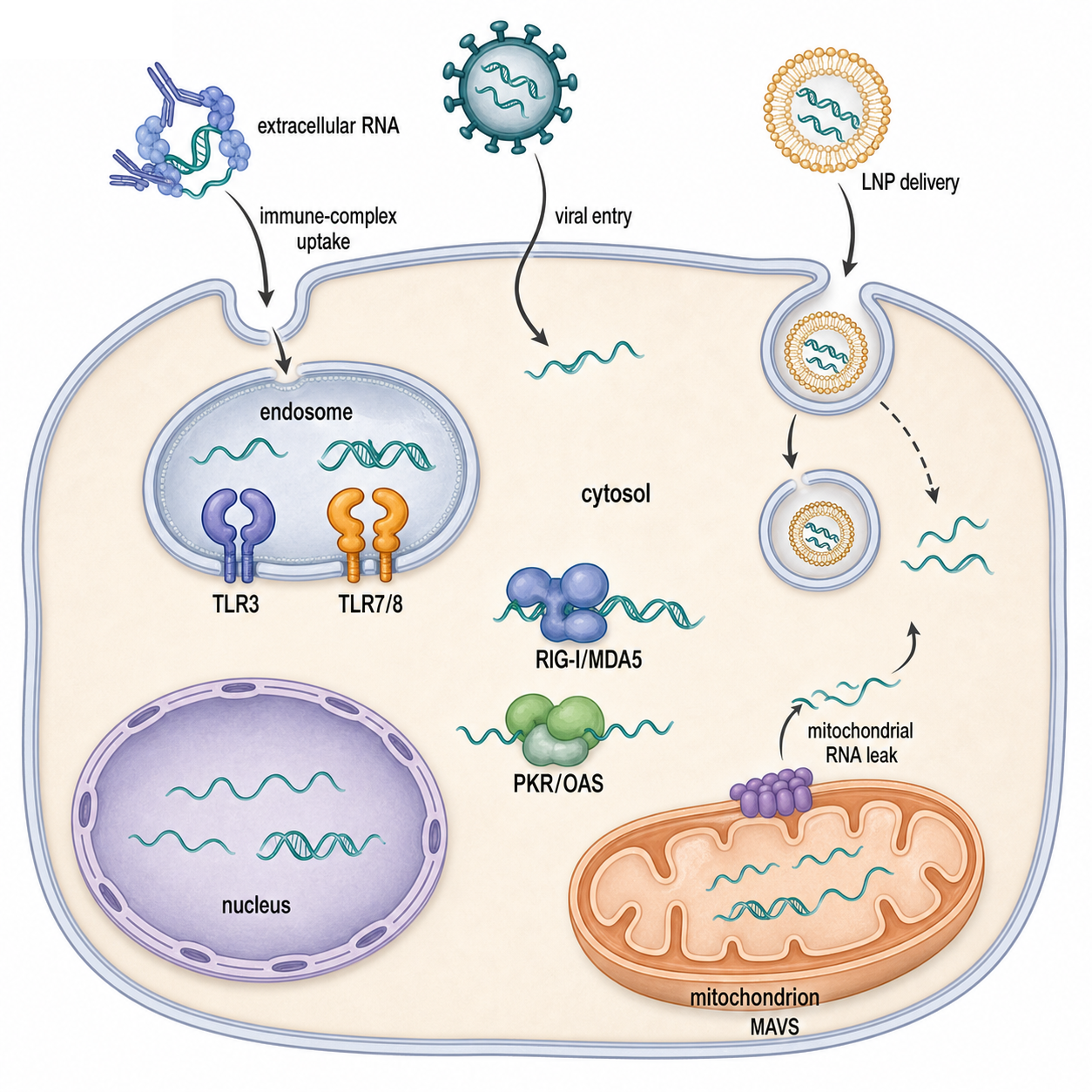
Figure 108.1. Compartmental logic of self versus nonself RNA sensing. RNA immunogenicity depends on location as well as chemistry: endosomal uptake, cytosolic viral RNA, mitochondrial leakage, or therapeutic delivery exposes different receptors to ligands normally segregated from them.
Self RNA is not invisible. It is actively managed. Host messenger RNA has a 5′ cap, a poly(A) tail, exon junction and RNA-binding proteins, coding-region constraints, and a lifetime controlled by decay pathways. Host transfer RNA and ribosomal RNA are heavily modified and usually assembled into ribonucleoprotein particles. Mitochondrial RNA is transcribed and processed inside an organelle, but mitochondrial stress can expose double-stranded RNA to cytosolic or endosomal sensors. Because each safeguard is partial, immune tolerance depends on layered thresholds. A receptor may bind weakly but fail to signal; a ligand may signal only after endosomal proteolysis, ATP-dependent receptor proofreading, ubiquitin-dependent adaptor assembly, or interferon priming.
Alternative RNA conformations add another layer to this discrimination problem. Z-RNA is a transient, left-handed double-helical conformation that sequence, torsional stress, protein binding, and the surrounding ribonucleoprotein environment can stabilize. Z-DNA-binding protein 1 (ZBP1) contains Zα domains that recognize Z-conformation nucleic acids and can couple recognition to receptor-interacting protein kinase 3 (RIPK3)-linked inflammatory cell-death pathways. A sequence capable of adopting Z-RNA is not evidence that ZBP1 occupies that RNA in a living cell: ligand assignment requires conformationally informative structural or binding evidence together with ZBP1- and Zα-domain-dependent perturbations. Chapter 53 owns the structural principles and evidence for noncanonical RNA conformers; this chapter owns their consequences for innate sensing.
Box 108.1. Self RNA Is Actively Tolerated, Not Chemically Invisible
Self RNA is not automatically harmless because it lacks stimulatory chemistry. Many host RNAs contain local duplexes, uridine-rich stretches, exposed phosphates during processing, or degradation fragments that resemble features used by innate sensors. Tolerance comes from several safeguards acting together: nuclear and organellar compartmentalization, 5′ capping, 2′-O-methylation and other modifications, RNA-binding proteins, rapid decay of misplaced RNA, and receptor thresholds that require productive signaling complexes.
The key teaching point is that innate receptors read presentation, not ancestry. A host transcript can become immunostimulatory if immune complexes deliver it to endosomal TLR7 or TLR8, if mitochondrial stress exposes dsRNA, if ADAR1 editing fails to dampen endogenous duplexes, or if synthetic delivery puts an otherwise normal sequence into the wrong compartment. Conversely, a viral RNA feature may be weakly sensed if it is capped, shielded, modified, sequestered in replication organelles, or present below the activation threshold.
This threshold logic explains why dose, delivery route, and formulation matter for therapeutic RNA. A purified, capped, modified mRNA inside a lipid nanoparticle can encode a protein without strong innate immune activation in one context, yet a contaminated preparation containing double-stranded RNA byproducts can strongly induce interferon. A short interfering RNA can be tolerated after chemical modification and careful sequence design, yet a similar duplex can activate TLR7, TLR8, RIG-I, or PKR if it reaches the wrong compartment or contains stimulatory motifs. The practical lesson is not that one modification or one purification step makes RNA “self,” but that many molecular and cellular filters jointly set the sensing threshold.
108.2. Endosomal TLR3, TLR7, TLR8, and TLR13-like sensing systems
Endosomal Toll-like receptors detect nucleic acids that arrive in the endolysosomal pathway after viral entry, phagocytosis, receptor-mediated uptake, immune-complex internalization, autophagy, or nanoparticle trafficking. These receptors are synthesized in the endoplasmic reticulum, trafficked to endosomes with accessory proteins, and proteolytically processed before full activity. Acidic pH helps expose and generate ligand fragments. This design reduces accidental sensing of extracellular or cytosolic host RNA and places receptor activation near adaptors that initiate interferon and inflammatory signaling.
TLR3 is the main endosomal double-stranded RNA receptor. It recognizes the sugar-phosphate backbone and geometry of double-stranded RNA rather than a precise sequence, dimerizes on RNA, and signals through the adaptor TRIF. TRIF activates TBK1 and IKK-family pathways that induce interferon regulatory factors and NF-kappaB-dependent genes. TLR3 can respond to viral replication intermediates, synthetic poly(I:C), and in some disease contexts endogenous double-stranded RNA. The local bibliography includes recent structural and regulatory reviews plus primary studies on self RNA and mitochondrial double-stranded RNA in inflammatory settings (Chen et al. 2021, PMID: 34414698; Shimizu 2024, PMID: 39168045; Zhang et al. 2025, PMID: 40915996; Milcarek et al. 2026, PMID: 42048448). Canonical early TLR3 discovery and TRIF pathway primary papers remain final-provenance expansion items before finalizing the reference layer.
TLR7 and TLR8 sense single-stranded RNA degradation products. They are not simple receptors for intact viral genomes. Structural and biochemical work shows that TLR7 and TLR8 can bind nucleosides and short oligoribonucleotides in cooperative ligand-binding sites, which explains why endosomal digestion and nucleotide composition influence activation. Human TLR7 is especially important in plasmacytoid dendritic cells, where it drives strong type I interferon production. Human TLR8 is prominent in monocytes, macrophages, and conventional dendritic cells and often produces inflammatory cytokines as well as interferon-linked outputs. Mouse-human differences are substantial: mouse TLR8 has historically appeared less responsive in many assays, and mouse TLR7 biology cannot be mapped one-to-one onto human TLR7 and TLR8. These species differences matter when interpreting vaccine adjuvants, autoimmune phenotypes, and RNA drug immunostimulation.
Table 108.1. Endosomal RNA-sensing TLRs and their ligand logic. Endosomal TLR3, TLR7, TLR8, and TLR13-like systems differ in RNA features, adaptors, expressing cells, and species distribution; experimental agonists do not erase receptor- and organism-specific ligand boundaries.
| Receptor | Main RNA feature | Adaptor | Prominent cell types | Species notes | Common experimental ligand | Caveat |
|---|---|---|---|---|---|---|
| TLR3 | Endosomal double-stranded RNA geometry and sugar-phosphate backbone. | TRIF. | Dendritic, epithelial, fibroblast, and stromal contexts vary by tissue. | Present in humans and mice, but expression and disease phenotypes are cell-type dependent. | Poly(I:C), viral dsRNA preparations, or defined dsRNA delivered to endosomes. | Poly(I:C) can also activate cytosolic MDA5 if delivery bypasses endosomal restriction, so TLR3 assignment needs compartment and receptor controls. |
| TLR7 | Endosomal single-stranded RNA degradation products, especially uridine-rich or guanosine/uridine-rich features with nucleoside and oligoribonucleotide contributions. | MyD88. | Plasmacytoid dendritic cells, B cells, and selected myeloid contexts. | Human and mouse TLR7 are both important, but dosage, sex-linked expression, and cell distribution affect interpretation. | GU-rich ssRNA, viral ssRNA, immune-complexed self RNA, or synthetic ssRNA delivered to acidified endosomes. | Intact RNA uptake is not enough; endosomal digestion, modifications, dose, and delivery vehicle determine whether signaling crosses threshold. |
| TLR8 | Endosomal single-stranded RNA degradation products, commonly interpreted through uridine plus short oligoribonucleotide binding logic. | MyD88. | Human monocytes, macrophages, and conventional dendritic cells are prominent settings. | Human TLR8 is functionally prominent; mouse TLR8 behavior differs and should not be mapped directly onto human TLR8. | GU-rich ssRNA fragments, synthetic ssRNA, or RNA-derived degradation products in endosomal assays. | TLR8 often skews toward inflammatory cytokines as well as interferon-linked outputs, and mouse models can underrepresent human TLR8 biology. |
| TLR13-like systems | Conserved bacterial 23S rRNA sequence features rather than broad dsRNA or ssRNA degradation-product sensing. | MyD88. | Mouse and selected mammalian myeloid or dendritic-cell contexts. | Mice encode TLR13; humans do not encode a functional TLR13 ortholog. | Bacterial 23S rRNA fragments or oligoribonucleotides containing the recognized conserved sequence. | TLR13 is useful for teaching sequence-specific RNA surveillance, but it should not be used as a human receptor model. |
TLR13 is not present in humans, but it is pedagogically useful because it shows a different RNA-sensing principle. Mouse TLR13 recognizes a conserved sequence in bacterial 23S ribosomal RNA, including the region affected by some macrolide resistance mutations. This is sequence-specific RNA surveillance rather than broad dsRNA or degradation-product sensing. TLR13-like biology also warns against assuming that “RNA sensing” has the same receptor inventory in all mammals. Verified TLR13 discovery and bacterial 23S rRNA ligand papers remain final-provenance expansion items; the current references.md has adequate chapter anchors but lacks the canonical primary TLR13 records.
Endosomal RNA sensing has two recurrent caveats. First, uptake is not evidence of receptor activation. RNA can enter endosomes without engaging TLRs, and TLR activation can reflect contaminants, immune complexes, delivery vehicles, or cell death. Second, endosomal TLR activation does not prove viral replication. Inactivated virus, extracellular vesicles, damaged cells, or synthetic RNA can deliver RNA ligands. Good experiments therefore combine receptor genetics, ligand purification, nuclease sensitivity, endosomal acidification controls, cell-type comparisons, and downstream pathway readouts.
108.3. RIG-I, MDA5, LGP2, 5’ triphosphate RNA, and dsRNA recognition
RIG-I-like receptors patrol the cytosol for RNA features that are difficult for healthy mammalian cells to expose persistently. RIG-I contains two N-terminal CARD domains, a helicase core, and a C-terminal domain that binds RNA ends. In the inactive state the CARD domains are restrained. Binding to short double-stranded RNA with a 5′ triphosphate or diphosphate end, together with ATP-dependent conformational cycling, releases the CARD domains for signaling. The strongest ligands often combine end chemistry with duplex structure and appropriate length. This is why a naked 5′ triphosphate is not the whole signal: nearby base pairing, terminal geometry, protein occupancy, and competing host modifications all influence activation (Rehwinkel and Gack 2020, PMID: 32203325; Solotchi and Patel 2024, PMID: 38884803).
MDA5 detects RNA differently. It forms cooperative filaments along long double-stranded RNA, a property suited to viral replication intermediates that extend far beyond the length of typical local hairpins. MDA5 is essential for sensing many picornaviruses and other viruses that generate long dsRNA structures, whereas RIG-I is central for many negative-strand RNA viruses and 5′ triphosphate-bearing products. The classic genetic distinction between RIG-I and MDA5 virus specificity was established by Kato et al. (2006, PMID: 16625202), although later work has shown overlap and context dependence. LGP2 binds RNA and shares helicase-related architecture but lacks CARD domains. It can promote or inhibit RIG-I-like receptor signaling depending on ligand, concentration, cell type, and timing, partly by influencing RNA availability and MDA5 filament behavior.
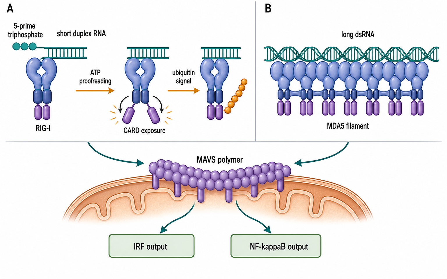
Figure 108.2. RIG-I-like receptor proofreading and MAVS activation. RIG-I recognizes end-bearing short duplexes and exposes its signaling domains after ATP-dependent activation, whereas MDA5 assembles on long double-stranded RNA; both pathways converge on MAVS to activate IRF and NF-κB outputs.
Activated RIG-I or MDA5 engages MAVS, an adaptor anchored on mitochondria, peroxisomes, and mitochondria-associated membranes. MAVS can assemble prion-like signaling polymers that recruit TRAF proteins, TBK1, IKKepsilon, and canonical IKK complexes. The outputs include IRF3 and IRF7 activation, NF-kappaB activation, type I and type III interferon expression, chemokines, and many interferon-stimulated genes. Because MAVS is linked to mitochondrial membranes, RNA sensing is physically connected to organelle stress, apoptosis regulators, metabolic state, and inflammasome-adjacent signals. Recent primary work on MAVS aggregation control and RIG-I condensation illustrates that receptor signaling is spatially organized rather than a simple linear chain (Li et al. 2023, PMID: 37563140; Wang et al. 2025, PMID: 40229436).
TRIM25-linked signaling is a central regulatory example. TRIM25 is an E3 ubiquitin ligase reported to catalyze K63-linked ubiquitination events that promote RIG-I CARD activation and downstream signaling. The broader consensus is that ubiquitin scaffolds, phosphorylation state, dephosphorylation, RNA binding, and host cofactors together regulate RIG-I, while viruses often target TRIM25 or related ubiquitin machinery. Some details remain debated because experimental systems differ in receptor abundance, ligand type, cell type, and viral antagonist expression. For a chapter-level model, TRIM25 should be treated as a signaling organizer and threshold regulator, not as a single on/off switch. Onomoto et al. (2021, PMID: 33462384) review host and viral regulation, and Chang et al. (2023, PMID: 37978243) provides a recent example of receptor turnover through RNF125-linked degradation.
The evidence base for RIG-I-like receptor biology is strong but method-sensitive. Transfected synthetic RNA can bypass natural entry routes. Overexpressed receptors can signal from weak ligands. Viral infection produces many RNA species at once: genomes, antigenomes, defective interfering RNAs, replication intermediates, host shutoff products, and stress-induced endogenous RNAs. Rigorous ligand assignment usually needs receptor knockouts, MAVS dependency, phosphatase or ubiquitin controls, RNA immunoprecipitation, nuclease and phosphatase treatment, infection-stage sampling, and rescue with defined RNA ligands. The field increasingly treats ligand recognition as kinetic proofreading, in which receptors sample many RNAs but signal strongly only from ligands that sustain productive conformations.
108.4. PKR, OAS/RNase L, TRIM25-linked signaling, and inflammasome connections
PKR, encoded by EIF2AK2, is a double-stranded RNA-activated kinase. It contains N-terminal dsRNA-binding motifs and a C-terminal kinase domain. Binding to dsRNA promotes dimerization and autophosphorylation, enabling PKR to phosphorylate eIF2alpha. Phosphorylated eIF2alpha reduces general translation initiation because it inhibits recycling of eIF2-GDP to eIF2-GTP. In infected cells, this can limit viral protein synthesis and shift the cell toward stress granule formation, integrated stress response signaling, apoptosis, or inflammatory outputs. PKR is therefore both a sensor and an effector: the RNA ligand directly changes protein synthesis capacity.
PKR specificity is not absolute. Long perfect dsRNA is a potent activator, but structured cellular RNAs, viral decoys, short duplexes at high concentration, and protein-bound RNAs can alter activation. Many viruses encode PKR antagonists: dsRNA-binding proteins that hide ligands, pseudo-substrate proteins that block kinase action, RNAs that act as decoys, and factors that manipulate stress granules. Cesaro and Michiels (2021, PMID: 34759908) review viral inhibition strategies, and Gao et al. (2022, PMID: 35858300) provides a recent example linking viral evasion to stress granule reprogramming. The main boundary case is that PKR activation may indicate dsRNA stress but does not identify the RNA source without additional mapping.
OAS/RNase L is a parallel dsRNA-triggered effector pathway. OAS proteins bind double-stranded RNA and synthesize 2′,5′-linked oligoadenylates from ATP. These unusual oligoadenylates bind and activate RNase L, an endoribonuclease that cleaves viral and host RNA. RNase L activation can suppress viral replication by degrading RNA templates and can amplify innate signaling by producing RNA fragments that stimulate RIG-I-like receptors or by altering cellular stress pathways. Human OAS genetics is clinically important; inborn errors and polymorphisms can affect susceptibility to severe viral inflammatory disease, including SARS-CoV-2-related multisystem inflammatory syndrome in children in one cited study (Lee et al. 2023, PMID: 36538032).
Table 108.2. Cytosolic dsRNA-responsive effector pathways. PKR, OAS-RNase L, RIG-I-like receptors, and inflammasome-linked pathways respond to overlapping RNA contexts but execute distinct biochemical actions and pathologies; shared downstream inflammation does not identify the proximal sensor.
| Pathway | Proximal ligand or trigger | Direct biochemical action | Antiviral benefit | Pathology risk | Key caveat |
|---|---|---|---|---|---|
| RIG-I-MAVS | Short blunt or base-paired RNA with 5′ triphosphate or diphosphate ends, plus suitable terminal geometry. | ATP-dependent RIG-I activation exposes CARD domains that engage MAVS polymers and IRF/NF-kappaB signaling. | Induces type I and type III interferons, chemokines, and interferon-stimulated antiviral programs. | Excessive activation can drive systemic cytokines, tissue inflammation, or responses to mislocalized self RNA. | End chemistry alone is insufficient; duplex context, phosphorylation, ubiquitin regulation, protein occupancy, and delivery route shape signaling. |
| MDA5-MAVS | Long cytosolic double-stranded RNA, often from viral replication intermediates. | Cooperative MDA5 filament assembly signals through MAVS to activate IRF and NF-kappaB outputs. | Detects many long-dsRNA-producing infections and sustains antiviral interferon programs. | Chronic or misdirected sensing of endogenous, repeat-derived, or mitochondrial dsRNA can contribute to interferon-driven disease. | The RIG-I/MDA5 split is a useful starting model, but receptor usage overlaps and is modified by cell type, virus, ligand length, and LGP2. |
| PKR-eIF2alpha | Cytosolic double-stranded RNA, with strong activation by long or abundant duplexes. | PKR dimerizes, autophosphorylates, and phosphorylates eIF2alpha to reduce translation initiation. | Limits viral protein synthesis and couples RNA detection to stress-response programs. | Broad translation arrest, stress granules, apoptosis, or inflammatory stress can damage tissue when activation is prolonged. | PKR activation reports dsRNA stress but does not identify the RNA source without ligand mapping and viral-antagonist controls. |
| OAS/RNase L | Double-stranded RNA bound by OAS proteins. | OAS enzymes synthesize 2′,5′ oligoadenylates that activate RNase L-mediated RNA cleavage. | Degrades viral RNA templates and can amplify antiviral signaling through secondary RNA fragments. | Host RNA cleavage can produce immunostimulatory fragments, apoptosis, or tissue pathology in some contexts. | RNase L cleavage can be both restrictive and inflammatory; OAS repertoires and RNase L consequences differ across species and tissues. |
| Inflammasome-linked crosstalk | RNA virus-induced mitochondrial stress, ion flux, membrane damage, interferon priming, PKR stress, or RNase L-generated fragments. | Inflammasome complexes activate caspase-1, IL-1-family cytokine maturation, and inflammatory cell-death programs. | Recruits inflammatory defense and can remove infected cells when coordinated with antiviral signaling. | IL-1-family cytokines and pyroptotic injury can worsen tissue inflammation. | A positive inflammasome readout during RNA virus infection usually does not prove direct RNA binding by the inflammasome sensor. |
RNase L biology also illustrates why RNA degradation is immunologically ambiguous. Cleavage can remove viral RNA, but cleavage fragments can become immunostimulatory. RNase L can promote apoptosis or inflammatory signaling in some contexts and tissue pathology in others. A recent hair follicle regeneration study links RNase L to altered innate signaling in a nonclassical tissue context (Kirby et al. 2025, PMID: 39903537), but this should not be overgeneralized to all RNase L biology. Core OAS discovery, 2-5A biochemistry, and RNase L structural activation references remain final-provenance expansion items.
Inflammasome connections arise because RNA virus infection perturbs many cytosolic systems at once. Inflammasomes such as NLRP3 are not primarily sequence-specific RNA receptors in the same way as RIG-I or TLR7. They respond to cellular disturbance: ion flux, mitochondrial dysfunction, membrane damage, reactive oxygen species, viral proteins, and altered organelle homeostasis. RNA sensing can feed into this through interferon priming, MAVS-mitochondrial coupling, PKR-linked stress responses, RNase L-mediated RNA cleavage, and cell death pathways. Reviews on RNA respiratory viruses emphasize both antiviral protection and viral manipulation of inflammasomes (Rodrigues and Zamboni 2025, PMID: 39891396). A cautious phrasing is important: inflammasome activation during RNA virus infection often depends on RNA-sensing pathways, but a positive inflammasome readout alone does not prove direct RNA recognition.
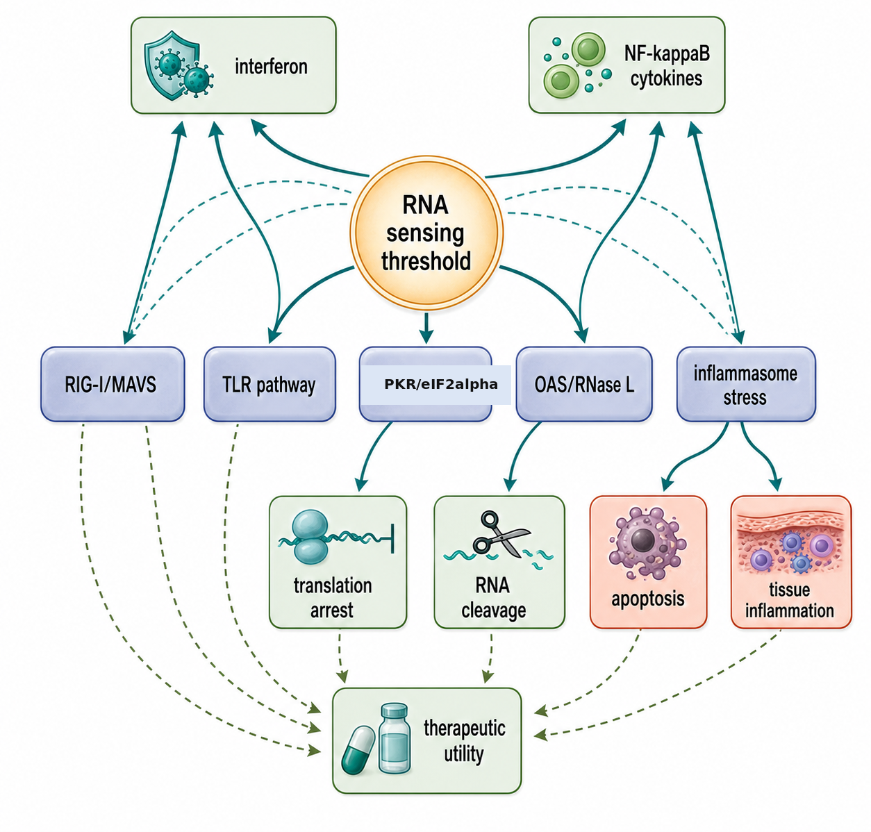
Figure 108.3. Effector outcomes downstream of RNA sensing. Crossing an RNA-sensing threshold can produce antiviral restriction, cytokines, translational arrest, RNA cleavage, inflammasome-linked stress, apoptosis, or necroptosis; terminal outcomes and the ADAR1-Z-RNA-ZBP1 branch remain context-dependent and require pathway-specific evidence.
The downstream outputs of these pathways are deliberately redundant. Interferons induce antiviral states in infected and neighboring cells. NF-kappaB-dependent cytokines recruit and activate immune cells. Translational arrest conserves resources and suppresses viral protein production. RNA cleavage removes templates and creates secondary signals. Cell death can prevent virion production but may also damage tissue. This redundancy is adaptive during rapidly replicating infections, yet it is dangerous when triggered chronically by self RNA. The same molecular logic underlies autoinflammation, interferonopathies, autoimmune nucleic-acid sensing, and adverse reactions to some RNA therapeutics.
ZBP1 illustrates why recognition, pathway engagement, and biological outcome must be separated. The Zα domains provide conformational recognition, whereas receptor-interacting homotypic motifs couple ZBP1 to RIPK3 and context-dependent apoptosis, necroptosis, or inflammatory signaling. In interferon-treated mouse embryonic fibroblasts, Zhang et al. found that acute ADAR1 loss enabled Zα- and RHIM-dependent ZBP1 activation, association with RIPK3 and MLKL, MLKL phosphorylation, and a mixture of apoptosis and necroptosis. The same study’s melanoma experiments tested a different intervention: the curaxin CBL0137 induced Z-DNA in host tumor-microenvironment fibroblasts, and combination treatment with anti-PD-1 depended on host ZBP1 in B16-F10 and YUMMER1.7 mouse tumors. Those experiments did not test ADAR1 loss in tumor cells, did not assign the therapeutic effect to Z-RNA, and do not establish efficacy in human cancer. In another mouse genetic setting, Jiao et al. found fatal ZBP1-amplified interferon pathology that did not require RIPK3 or MLKL, demonstrating that ZBP1 dependence does not imply terminal necroptosis in every tissue or genotype (Jiao et al. 2022, PMID: 35859176; Zhang et al. 2022, PMID: 35614224).
The ligand evidence in the ADAR1-deficient fibroblast system is stronger than sequence prediction alone but still has a defined resolution. RNase-sensitive Z22-antibody staining and RNA pulldown, Zα-dependent ZBP1 association with selected interferon-stimulated mRNA 3′ UTRs, ZBP1 proximity to the Z22 signal, and downstream Zα- and RHIM-domain perturbations, RIPK3 inhibition, MLKL phosphorylation, and cell-death genetics support a causal RNA-ligand-to-cell-death pathway. Antibody enrichment, proximity, and targeted pulldown nevertheless do not provide nucleotide-resolved in-cell structures or establish that every predicted Z-forming transcript is occupied by ZBP1. Chapter 109 develops ADAR1-mediated self-RNA protection.
Experimental Foundations and Evidence
The strongest evidence for innate RNA-sensing pathways comes from convergent methods. Genetic loss-of-function experiments identify required receptors and adaptors. Biochemical reconstitution defines ligand binding, ATPase activity, enzymatic output, or kinase activation. Structural biology shows how RNA contacts receptor domains and how receptors dimerize or assemble filaments. Infection studies test whether the pathway matters during viral replication. Therapeutic and synthetic RNA experiments show how sequence, chemistry, purification, and delivery change sensing. No single method is sufficient because each has artifacts.
Synthetic ligands are powerful but easily overinterpreted. Poly(I:C) is useful for double-stranded RNA responses but does not mimic all viral dsRNA. In vitro-transcribed 5′ triphosphate RNA can activate RIG-I, but transcription reactions may contain abortive products, dsRNA contaminants, or sequence mixtures. Lipid-based transfection can deliver RNA to compartments that natural infection would not reach. Reporter assays can exaggerate weak activation if receptors or adaptors are overexpressed. Conversely, primary cells may fail to respond if they lack the relevant receptor, lack endosomal processing, or are not interferon-primed. Good evidence therefore asks whether a ligand is necessary, sufficient, chemically defined, and presented through a physiologically plausible route.
Box 108.2. Evidence Ladder for Assigning an RNA Sensor
An RNA-sensing assignment is strongest when several evidence layers point to the same receptor. First, the RNA ligand should be chemically and physically defined: length, strandedness, end chemistry, modifications, contaminants, and delivery route. Second, receptor dependence should be shown with loss-of-function genetics or carefully controlled inhibition, ideally with rescue. Third, pathway specificity should match the adaptor and output: TRIF for TLR3, MyD88 for TLR7 or TLR8, MAVS for RIG-I-like receptors, eIF2alpha phosphorylation for PKR, and 2′,5′ oligoadenylate or RNase L activity for OAS/RNase L.
A lower tier of evidence is still useful but should be phrased cautiously. Reporter activation after RNA transfection, interferon induction during infection, or loss of response in one receptor knockout can show pathway involvement, but these results do not prove direct binding to the suspected RNA. Strong claims need compartment controls, nuclease or phosphatase treatments, dose-response behavior, primary-cell validation, and checks for viral antagonists or delivery artifacts.
Viral infection experiments are biologically rich but chemically complex. A single RNA virus can produce genomic RNA, antigenomic RNA, defective interfering genomes, copy-back RNAs, capped or cap-snatched RNAs, dsRNA replication intermediates, subgenomic RNAs, and host RNAs released by stress. Viral proteins also block signaling, alter organelles, and change RNA metabolism. This is why receptor specificity is best established by combining viral genetics with RNA purification, time-resolved sampling, receptor knockout, pathway rescue, and defined ligand add-back. The influenza A virus literature is a good example: RIG-I sensing depends on viral RNA features, nuclear and cytoplasmic trafficking, viral antagonists, and cell context rather than a single static ligand description (Liu and Zhou 2019, PMID: 30760567).
108.5. RNA modifications, viral evasion, autoimmunity, and therapeutics
Biological contexts across cells and organisms
Cell type strongly shapes RNA-sensing output. Plasmacytoid dendritic cells specialize in TLR7- and TLR9-driven type I interferon production. Monocytes and macrophages often integrate TLR8, inflammasome, and cytokine outputs. Epithelial cells rely heavily on cytosolic RIG-I-like receptors because they are common initial targets of respiratory and enteric RNA viruses. Neurons, hepatocytes, fibroblasts, and tumor cells can all sense RNA, but their thresholds and consequences differ. An identical RNA ligand can therefore be an interferon inducer, an inflammatory cytokine trigger, a translational arrest signal, a weak adjuvant, or a toxic stressor depending on the cell.
Species differences are not details. Mice encode TLR13 and humans do not. Human TLR8 is more functionally prominent than mouse TLR8 in many contexts. OAS gene repertoires differ across mammals. Viral antagonists are often host-specific because they target particular receptor surfaces or adaptor interactions. This matters for preclinical RNA therapeutics and vaccines. A mouse result showing strong or weak innate sensing may reflect the mouse receptor inventory, dose, route, and formulation rather than a general human rule.
Autoimmunity and autoinflammation show what happens when the system misclassifies endogenous RNA. Self RNA can reach endosomes through immune complexes, apoptotic debris, extracellular vesicles, or tissue damage. Cytosolic dsRNA can accumulate from repetitive elements, mitochondrial transcription, defective RNA editing, or impaired RNA decay. Persistent interferon signaling can then maintain a feed-forward loop: interferon induces more sensors, more sensors lower the activation threshold, and additional endogenous RNA species become stimulatory. The clinical categories include systemic lupus erythematosus-like nucleic-acid autoimmunity, monogenic interferonopathies, and tissue-specific inflammatory disorders. The mechanisms vary, so “RNA sensing causes autoimmunity” is too broad; the useful claim is that mislocalized or improperly processed RNA can activate defined innate pathways that contribute to particular autoimmune phenotypes.
Technology, computational, clinical, and engineering links
RNA therapeutics sit directly on top of innate sensing biology. For mRNA vaccines and protein-replacement mRNA, developers often try to preserve enough local inflammation for immunogenicity when vaccination is intended, or reduce inflammation when protein expression is the goal. Control points include cap structure, poly(A) tail, untranslated regions, codon composition, nucleoside modifications such as N1-methylpseudouridine, removal of dsRNA contaminants by chromatography, sequence avoidance of endosomal TLR motifs, dose, route, and delivery vehicle. Lipid nanoparticles are not passive packages: they determine which cells take up RNA, how much RNA remains in endosomes, how much escapes to cytosol, and how strongly innate pathways are triggered. Chatterjee et al. (2024, PMID: 38437552) review endosomal escape as a bottleneck for LNP therapeutics. Canonical Kariko-Weissman modified nucleoside and mRNA vaccine innate-sensing references remain final-provenance expansion items.
Box 108.3. Therapeutic RNA Tuning Is Context-Specific
Therapeutic RNA design does not have one immune goal. A vaccine may benefit from controlled local innate activation because interferon and inflammatory cytokines help recruit and instruct adaptive immunity. A protein-replacement mRNA, in contrast, often needs low innate activation because interferon can reduce translation, shorten RNA persistence, and cause systemic adverse effects. A RIG-I or TLR agonist deliberately moves in the opposite direction by making receptor activation the drug mechanism.
Three questions keep the design logic grounded. Which cells receive the RNA? Which compartment sees the RNA before degradation or escape? Which readout defines success: antigen expression, protein replacement, tumor inflammation, antiviral state, or safety? Cap chemistry, N1-methylpseudouridine or other nucleoside changes, dsRNA-contaminant removal, motif avoidance, dose, route, and lipid nanoparticle composition all tune the answer. None of these features alone guarantees immune silence or immune activation.
Synthetic agonists deliberately activate RNA sensors. RIG-I agonist RNAs can be designed to carry 5′ triphosphate ends and short duplex structures, aiming to induce interferon and tumor cell death or antiviral states. TLR7 and TLR8 agonists are used or explored as vaccine adjuvants and cancer immunotherapy agents. Poly(I:C) and derivatives activate TLR3 and MDA5-linked pathways, although toxicity and formulation constrain use. The cited hepatocellular carcinoma study of synthetic RIG-I agonist RNA is one applied example (Ulloa et al. 2025, PMID: 39945619). In these settings, efficacy and toxicity are inseparable from receptor distribution, tumor cell competence, immune-cell recruitment, and systemic cytokine risk.
Viral evasion strategies are diverse because RNA sensing has multiple choke points. Viruses can cap or mimic host mRNA, hide dsRNA inside replication organelles, encode dsRNA-binding proteins, cleave or relocalize adaptors, inhibit TRIM25 or other ubiquitin regulators, block PKR activation, degrade signaling molecules, manipulate stress granules, methylate RNA caps to avoid IFIT and other restriction factors, or produce decoy RNAs. Some viral proteins target more than one pathway. Some evasion mechanisms reduce sensing but create vulnerabilities, such as dependence on a viral capping enzyme or a host lipid compartment. These vulnerabilities are attractive drug targets, but viral diversity makes broad generalization risky.
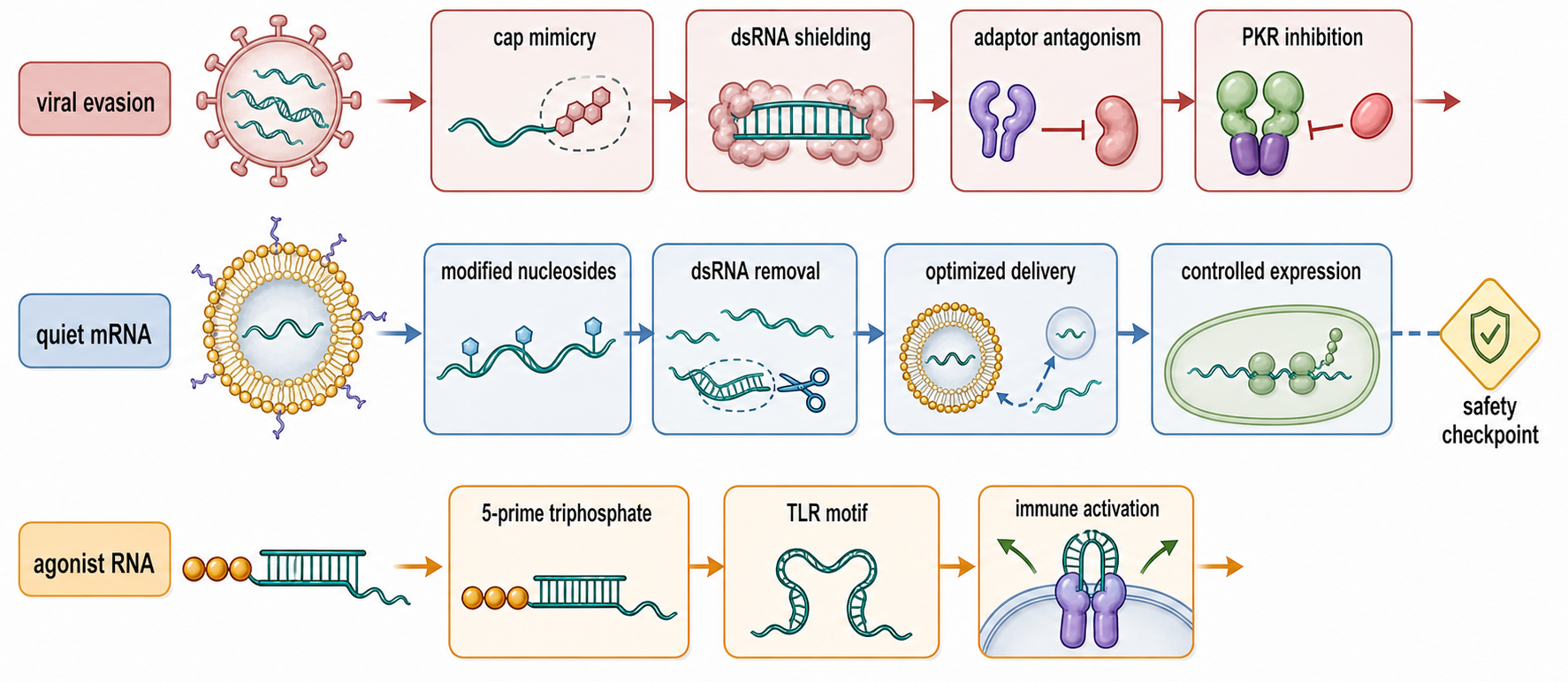
Figure 108.4. Tuning innate sensing in viral evasion and RNA therapeutics. Viruses evade RNA sensing by hiding ligands or blocking signaling, whereas RNA therapeutics are engineered either to minimize innate activation through capping, nucleoside modification, purification, and delivery or to trigger it deliberately with agonist RNA features.
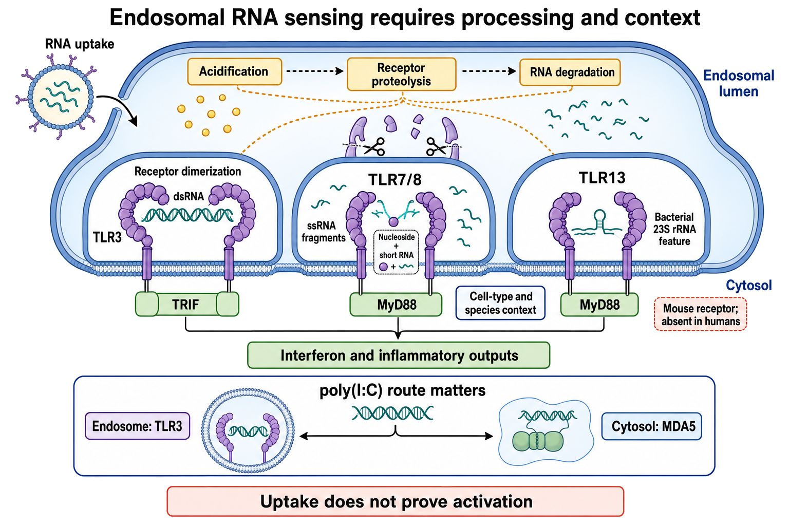
Figure 108.5. Endosomal RNA processing and adaptor-specific TLR activation. Endosomal uptake does not prove receptor activation; ligand processing, receptor maturation, delivery compartment, receptor inventory, and cell type determine which RNA-sensing pathway signals.
Computational design increasingly uses innate-sensing rules, but the rules are incomplete. Algorithms can flag long duplex potential, uridine-rich motifs, CpG or UpA content, 5′ end structures, predicted dsRNA contaminants, and modification patterns. They can optimize codon usage, reduce innate stimulation, or design agonist RNAs. However, receptor activation depends on folding in the delivered molecule, impurities from synthesis, protein binding, nanoparticle composition, cell type, and kinetics of endosomal escape. A sequence-only model cannot fully predict innate activation without manufacturing and delivery information. This is a recurring theme for RNA engineering: the same molecule is both an information polymer and a physical ligand.
Recent Consensus
The current consensus is that innate RNA sensing is layered, redundant, and context-dependent. TLR3, TLR7, TLR8, RIG-I, MDA5, PKR, and OAS proteins have well-supported ligand preferences, but biological responses are determined by ligand chemistry, compartment, receptor expression, cell state, and antagonism. RIG-I and MDA5 are not simply “short RNA” and “long RNA” sensors, although that distinction is a useful starting point. Endosomal TLR7 and TLR8 do not merely bind any single-stranded RNA; they respond strongly to degradation products and nucleotide features generated in endosomes. PKR and OAS/RNase L are not downstream decorations of interferon biology; they are direct dsRNA-responsive effectors that can determine whether translation continues and whether RNA remains intact.
Another consensus point is that endogenous RNA can be immunostimulatory without being foreign. The decisive issue is presentation. Self RNA delivered to endosomes in immune complexes, mitochondrial RNA released during stress, Alu-derived duplexes insufficiently edited by ADAR1, or RNA fragments generated by abnormal nuclease activity can all participate in inflammation. This does not erase the pathogen-sensing function of the system; it shows why tolerance mechanisms, RNA metabolism, and compartment barriers are part of innate immunity.
Open Questions, Controversies, Deprecated Models, and Common Misconceptions
Open questions:
- Which endogenous RNA species drive inflammatory diseases?
- How do the relative contributions of TLRs, RIG-I-like receptors, PKR, OAS/RNase L, cGAS-linked stress pathways, and inflammasomes shift between cell types and disease stages?
- How should TRIM25-linked RIG-I activation be represented when ubiquitin ligases, unanchored ubiquitin chains, phosphorylation events, and viral antagonists complicate simple diagrams?
- How does the immunological impact of RNA modifications depend on receptor and context?
- When does N1-methylpseudouridine reduce innate responses without making modified RNA universally nonimmunogenic?
Common misconceptions:
- “Noncoding RNA means immune ligand.” Most noncoding RNA is normal self RNA, and immune stimulation depends on chemistry, structure, localization, modification, dose, and receptor context.
- “Double-stranded RNA.” Is not a single biological entity; length, end structure, sequence, editing, protein binding, and compartment matter.
- “Interferon induction is not always beneficial.” It can restrict viruses, enhance vaccine responses, reduce therapeutic protein expression, or drive pathology.
- “A receptor knockout phenotype proves direct binding to the suspected RNA.” Knockout phenotypes can reflect indirect signaling, altered cell state, redundancy, or ligand misassignment; direct binding requires biochemical or structural evidence.
- “Lack of response in one cell line proves safety in primary immune cells or in vivo.” Innate sensing and toxicity are cell-type-, dose-, route-, and context-dependent.